Embed Size (px)
Citation preview
201
Acta Neurologica Taiwanica Vol 15 No 3 September 2006
From the Section of Neurology, Tungs’ Taichung MetroharborHospital, Taichung, Taiwan.Received January 20, 2006. Revised February 8, 2006.Accepted April 12, 2006.
Reprint requests and correspondence to: Huai-Hua Yeh, MD.No. 699, Sec. 1, Chung-Chi Road, Wu-Chi, Taichung, Taiwan.E-mail: [email protected]
INTRODUCTION
Stroke of uncertain origin in a patient always posesa challenge to and a burden on every neurologic practi-tioner to uncover the underlying cause by means of athorough and concise workup. Cardiac myxoma is anuncommon cause of stroke. However, it is the mostcommon benign cardiac tumor, found more frequentlyin young adults with stroke or transient ischemic attack(1 in 250) than in older patients (1 in 750)(1). Clinical
suspicion and prudent interventions are required forearly diagnosis, which is the key to reducing the mor-bidity and mortality. However, non-specific systemicsymptoms and minor embolic phenomena may be over-looked in the absence of any history of cardiac prob-lems. In this situation, cardiac evaluation may not beperformed or ignored, and the diagnosis of this rare con-dition may be delayed until the onset of a more signifi-cant embolic disease, such as stroke, with functionalimpairment. We present a patient with atrial myxoma
Young Stroke, Cardiac Myxoma, and Multiple Emboli: A Case Report and Literature Review
Huai-Hua Yeh, Chi-Chiang Yang, Wai-Fai Tung, Hsin-Fan Wang, and Jai-Nien Tung
Abstract- Cardiac myxoma is a source of emboli to the vascular tree, especially to the central nervous sys-tem. Although it is rare, its early recognition is particularly important because of its unique clinical featuresof subsequently leading to intracerebral or subarachnoid hemorrhage, even brain metastases, and its poten-tial for surgical cure. Missing the diagnosis may lead to devastating results, including stroke, even suddendeath. A 40-year-old male with no other conventional vascular risk factors such as hypertension, diabetes orhyperlipidemia presented with right hemiplegia, global aphasia, vomiting, and fever. Infarction over the leftmiddle cerebral artery was disclosed on magnetic resonance imaging study, and echocardiogram showed ahuge mass, about 5cm in size, on the mitral valve which was histopathologically proved to be a cardiacmyxoma. He also presented with multiple emboli to the kidneys and the left eye. There is uncertainty aboutthe role of anticoagulation. The treatment of choice remains surgical excision of the cardiac myxoma whichmay lead to normalization of serum interleukin-6 levels and resolution of constitutional symptoms, and theintracranial aneurysms may regress and resolve.
Key Words: Cardiac myxoma, Stroke, Cardiac emboli
Acta Neurol Taiwan 2006;15:201-205
202
Acta Neurologica Taiwanica Vol 15 No 3 September 2006
and acute cerebral infarction over the left middle cere-bral arterial territory who underwent immediate surgicalintervention with fair functional recovery.
CASE REPORT
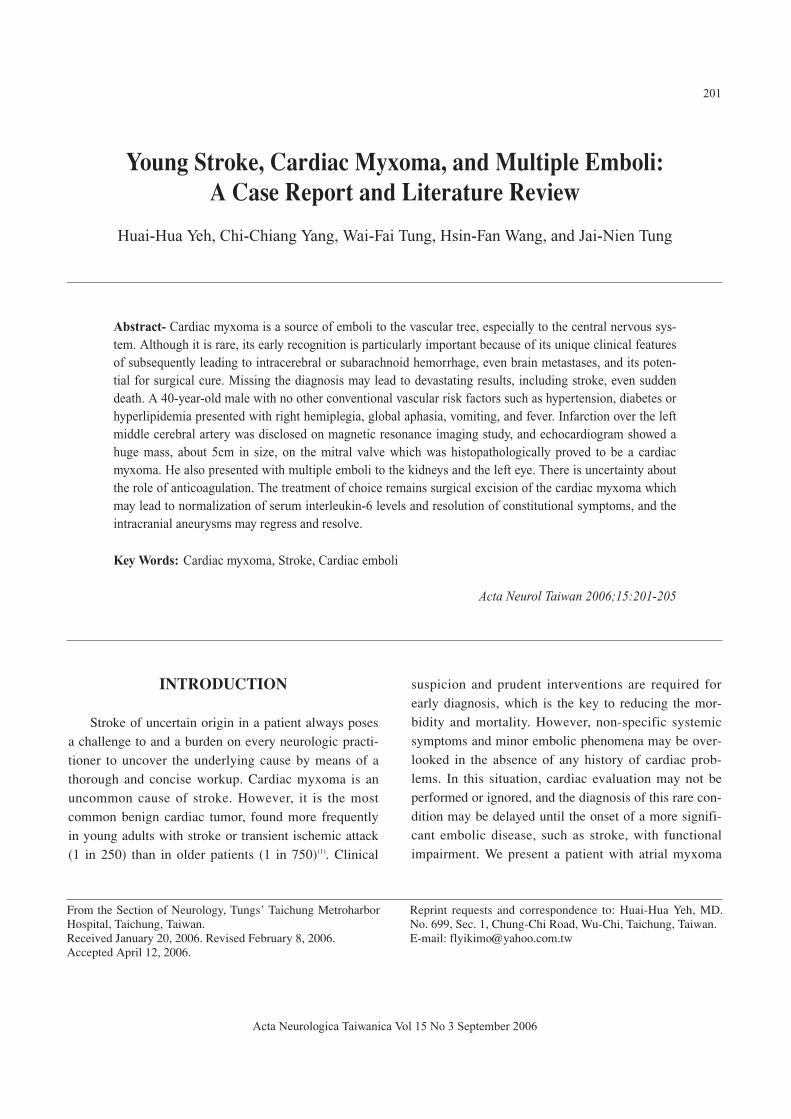
A 40-year-old married male without any systemicillness before, including hypertension, diabetes mellitus,hyperlipidemia and smoking, cardiopulmonary distressor syncope, had an episode of minor stroke with one-week admission to a medical center about 6-7 years ago.No traceable risk factors were noted at that time. Therewas no regular medications taken and no follow-up afterdischarge. He developed rapid onset of right-sided weak-ness and aphasia, and fell to the ground on the day ofadmission. Several episodes of vomiting, high-gradefever of above 39 ˚C along with profused sweating werenoted later. He was transferred to our hospital and neuro-logical examination showed an awake but lethargicpatient with Glasgow Coma Scale (GCS) of 10(E3V1M6). Right hemianopia, global aphasia, righthemiplegia with muscle power grading at 2/5, and rightBabinski sign were present. NIH Stroke Scale (NIHSS)score was 26 and Barthel index (BI) was 0 on admission.Physical examination showed low-grade fever with bodytemperature of 37.8 ˚C, coffee-ground substance fromthe nasogastric tube, a deep-colored urine, smooth respi-ration, clear breathing sounds, regular heart beats and nomurmurs. Laboratory data revealed peripheral leukocy-tosis (13,300/mm3) but no anemia (Hemoglobin 14.6gm/dL), elevated C-reactive protein (CRP) (5.7 mg/dl,normal <0.8), and microhematuria. Chest X-ray showedno evidence of active lung lesions, electrocardiogramwas normal, and sonogram of abdomen did not revealany lesions involving the genitourinary tracts, includingthe kidneys. Carotid duplex and transcranial duplexstudy disclosed stenotic flow at bilateral anterior andmiddle cerebral arteries, and vertebrobasilar arteries.Noncontrasted brain computed tomography (CT)showed multiple hypodense lesions over the left frontaland temporoparietal lobes, the anterior limb of left inter-nal capsule and the caudate nucleus. Magnetic resonanceimaging (MRI) of the brain disclosed hypointensities on
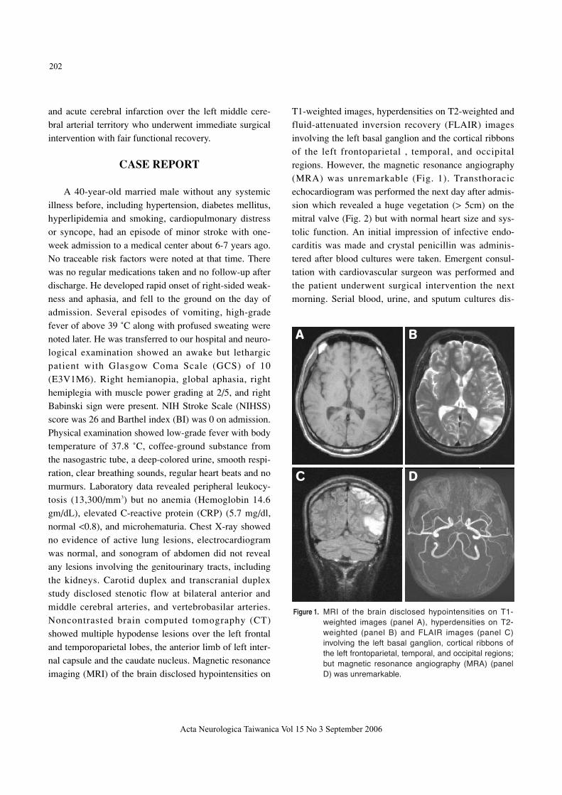
T1-weighted images, hyperdensities on T2-weighted andfluid-attenuated inversion recovery (FLAIR) imagesinvolving the left basal ganglion and the cortical ribbonsof the left frontoparietal , temporal, and occipitalregions. However, the magnetic resonance angiography(MRA) was unremarkable (Fig. 1). Transthoracicechocardiogram was performed the next day after admis-sion which revealed a huge vegetation (> 5cm) on themitral valve (Fig. 2) but with normal heart size and sys-tolic function. An initial impression of infective endo-carditis was made and crystal penicillin was adminis-tered after blood cultures were taken. Emergent consul-tation with cardiovascular surgeon was performed andthe patient underwent surgical intervention the nextmorning. Serial blood, urine, and sputum cultures dis-
Figure 1. MRI of the brain disclosed hypointensities on T1-weighted images (panel A), hyperdensities on T2-weighted (panel B) and FLAIR images (panel C)involving the left basal ganglion, cortical ribbons ofthe left frontoparietal, temporal, and occipital regions;but magnetic resonance angiography (MRA) (panelD) was unremarkable.
A B
C D
203
Acta Neurologica Taiwanica Vol 15 No 3 September 2006
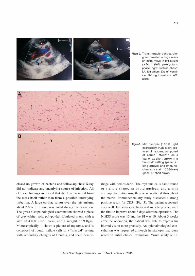
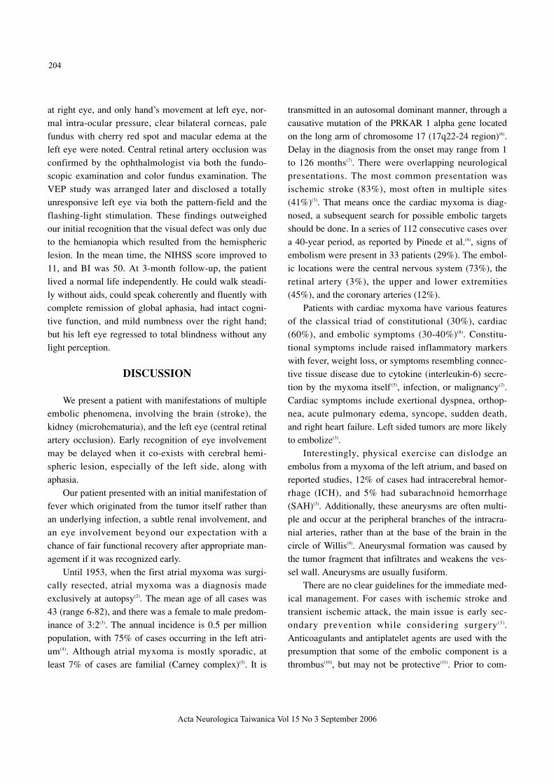
closed no growth of bacteria and follow-up chest X-raydid not indicate any underlying source of infection. Allof these findings indicated that the fever resulted fromthe mass itself rather than from a possible underlyinginfection. A large cardiac tumor over the left atrium,about 7 5cm in size, was noted during the operation.The gross histopathological examination showed a pieceof grey-white, soft, polypoidal, lobulated mass, with asize of 4.0 3.0 1.5cm, and a weight of 9.0gm.Microscopically, it shows a picture of myxoma, and iscomposed of round, stellate cells in a “mucoid” settingwith secondary changes of fibrosis, and focal hemor-
rhage with hemosiderin. The myxoma cells had a roundor stellate shape, an ovoid nucleus, and a pinkeosinophilic cytoplasm; they were scattered throughoutthe matrix. Immunochemistry study disclosed a strongpositive result for CD34 (Fig. 3). The patient recoveredvery well. His sensory aphasia and muscle powers werethe first to improve about 3 days after the operation. TheNIHSS score was 15 and the BI was 10. About 3 weeksafter the operation, the patient was able to express hisblurred vision more precisely. An ophthalmological con-sultation was requested although hemianopia had beennoted on initial clinical evaluation. Visual acuity of 1.0
Figure 3. Microscopic (100 lightmicroscopy, H&E stain) pic-tures of myxoma, composedof round, stellate cells(panel a.: short arrow) in a“mucoid” setting (panel a.:long arrow); and immuno-chemistry stain (CD34+++)(panel b.: short arrow).
A
B
Figure 2. Transthoracic echocardio-gram revealed a huge masson mitral valve in left atrium(> 5cm) (left: presystolicphase. right: systolic phase:LA: left atrium. LV: left ventri-cle. RV: right ventricle. AO:aorta)
204
Acta Neurologica Taiwanica Vol 15 No 3 September 2006
at right eye, and only hand’s movement at left eye, nor-mal intra-ocular pressure, clear bilateral corneas, palefundus with cherry red spot and macular edema at theleft eye were noted. Central retinal artery occlusion wasconfirmed by the ophthalmologist via both the fundo-scopic examination and color fundus examination. TheVEP study was arranged later and disclosed a totallyunresponsive left eye via both the pattern-field and theflashing-light stimulation. These findings outweighedour initial recognition that the visual defect was only dueto the hemianopia which resulted from the hemisphericlesion. In the mean time, the NIHSS score improved to11, and BI was 50. At 3-month follow-up, the patientlived a normal life independently. He could walk steadi-ly without aids, could speak coherently and fluently withcomplete remission of global aphasia, had intact cogni-tive function, and mild numbness over the right hand;but his left eye regressed to total blindness without anylight perception.
DISCUSSION
We present a patient with manifestations of multipleembolic phenomena, involving the brain (stroke), thekidney (microhematuria), and the left eye (central retinalartery occlusion). Early recognition of eye involvementmay be delayed when it co-exists with cerebral hemi-spheric lesion, especially of the left side, along withaphasia.
Our patient presented with an initial manifestation offever which originated from the tumor itself rather thanan underlying infection, a subtle renal involvement, andan eye involvement beyond our expectation with achance of fair functional recovery after appropriate man-agement if it was recognized early.
Until 1953, when the first atrial myxoma was surgi-cally resected, atrial myxoma was a diagnosis madeexclusively at autopsy(2). The mean age of all cases was43 (range 6-82), and there was a female to male predom-inance of 3:2(3). The annual incidence is 0.5 per millionpopulation, with 75% of cases occurring in the left atri-um(4). Although atrial myxoma is mostly sporadic, atleast 7% of cases are familial (Carney complex)(5). It is
transmitted in an autosomal dominant manner, through acausative mutation of the PRKAR 1 alpha gene locatedon the long arm of chromosome 17 (17q22-24 region)(6).Delay in the diagnosis from the onset may range from 1to 126 months(7). There were overlapping neurologicalpresentations. The most common presentation wasischemic stroke (83%), most often in multiple sites(41%)(3). That means once the cardiac myxoma is diag-nosed, a subsequent search for possible embolic targetsshould be done. In a series of 112 consecutive cases overa 40-year period, as reported by Pinede et al.(4), signs ofembolism were present in 33 patients (29%). The embol-ic locations were the central nervous system (73%), theretinal artery (3%), the upper and lower extremities(45%), and the coronary arteries (12%).
Patients with cardiac myxoma have various featuresof the classical triad of constitutional (30%), cardiac(60%), and embolic symptoms (30-40%)(8). Constitu-tional symptoms include raised inflammatory markerswith fever, weight loss, or symptoms resembling connec-tive tissue disease due to cytokine (interleukin-6) secre-tion by the myxoma itself (5), infection, or malignancy(2).Cardiac symptoms include exertional dyspnea, orthop-nea, acute pulmonary edema, syncope, sudden death,and right heart failure. Left sided tumors are more likelyto embolize(3).
Interestingly, physical exercise can dislodge anembolus from a myxoma of the left atrium, and based onreported studies, 12% of cases had intracerebral hemor-rhage (ICH), and 5% had subarachnoid hemorrhage(SAH)(3). Additionally, these aneurysms are often multi-ple and occur at the peripheral branches of the intracra-nial arteries, rather than at the base of the brain in thecircle of Willis(9). Aneurysmal formation was caused bythe tumor fragment that infiltrates and weakens the ves-sel wall. Aneurysms are usually fusiform.
There are no clear guidelines for the immediate med-ical management. For cases with ischemic stroke andtransient ischemic attack, the main issue is early sec-ondary prevention while considering surgery(3).Anticoagulants and antiplatelet agents are used with thepresumption that some of the embolic component is athrombus(10), but may not be protective(11). Prior to com-
205
Acta Neurologica Taiwanica Vol 15 No 3 September 2006
mencing therapy, hemorrhage needs to be excluded withCT or MRI, the latter preferably with MRA to helpexclude large aneurysm formation associated with tumorembolization(3).
The removal of the myxoma in a patient with recentstroke poses a difficult management problem. The con-cern has been that cardiopulmonary bypass and antico-agulation may exacerbate the neurological injury.Timing of surgery is still controversial, and needs to beclarified as more experience is accrued(3). Resection ofcardiac myxoma may lead to normalization of seruminterleukin-6 levels and resolution of constitutionalsymptoms(12), and the intracranial aneurysms may beregressed and resolved(13).
The recurrence rate is low (5%), but long-term andserial echocardiography is advisable especially foryoung patients(4).
CONCLUSION
The presence of embolic phenomena, especially inyoung patients with neurological symptoms, shouldprompt early neuroimaging and echocardiography, evenin the absence of EKG or auscultation abnormalities.Here we emphasize the importance of MRA of the brain,VEP study and the early and routine consultation withophthalmologist once the cardiac myxoma is proved.MRA of the brain is preferred for the exclusion of co-existing aneurysmal formation. Ophthalmological evalu-ation and VEP study can help in the early recognition ofarterial occlusion of the ophthalmic or central retinalartery even when the patient does not complain of or isunaware of the visual problems.
REFERENCES
1. Hart RG, Albers GW, Koudstaal PJ. Cardioembolic stroke.
In: Ginsberg MD, Bogousslavsky J, eds. Cerebrobascular
Disease: Pathophysiology, Diagnosis and Management.
London: Blackwell Science, 1998:1392-429.
2. Reynen K. Cardiac myxomas. N Engl J Med 1995;333:
1610-7.
3. Ekinci EI, Donnan GA. Neurological manifestations of car-
diac myxoma: a review of the literature and report of cases.
Intern Med J 2004;34:243-9.
4. Pinede L, Duhaut P, Loire R. Clinical presentation of left
atrial cardiac myxoma. A series of 112 consecutive cases.
Medicine (Baltimore) 2001;80:159-72.
5. McCarthy PM, Piehler JM, Schaff HV, et al. The signifi-
cance of multiple, recurrent and “complex”cardiac myxo-
mas. J Thorac Cardiovasc Surg 1986;91:389-96.
6. Kirschner LS, Carney JA, Pack SD, et al. Mutations of the
gene encoding the protein kinase type I-alpha regulatory
subunit in patients with the Carney complex. Nat Genet
2000;26:89-92.
7. MacGowan SW, Sidhu P, Aherne T, et al. Atrial myxoma:
national incidence, diagnosis, and surgical management. Ir
J Med Sci 1993;162:223-6.
8. Campbell K. Early diagnosis of an atrial myxoma with cen-
tral retinal artery occlusion. Ann Ophthalmol 1974;6:1207-
8, 1210-1.
9. Gonsalves CG, Nidecker AC. Cerebral aneurysms and car-
diac myxoma. J Can Assoc Radiol 1979;30:127-8.
10. Gee GT, Bazan C 3rd, Jinkins JR. Imaging of cerebral
infarction caused by atrial myxoma. Neuroradiology 1994;
36:271-2.
11. Knepper LE, Biller J, Adams HP Jr, et al. Neurologic mani-
festations of atrial myxoma. A 12-year experience and
review. Stroke 1988;19:1435-40.
12. Mendoza CE, Rosado MF, Bernal L. The role of inter-
leukin-6 in cases of cardiac myxoma. Clinical features,
immunologic abnormalities, and a possible role in recur-
rence. Text Heart Inst J 2001;28:3-7.
13. Damasio H, Seabra-Gomes R, da Silva JP, et al. Multiple
cerebral aneurysms and cardiac myxoma. Arch Neurol
1975;32:269-70.