Embed Size (px)
Citation preview

This is an author produced version of a paper published in
Theriogenology.
This paper has not been peer-reviewed and does not include the journal
pagination.
Citation for the published paper:
R. Gonzalez & Y.C.B Sjunnesson. (2013) Effect of blood plasma collected
after adrenocorticotropic hormone administration during the preovulatory
period in the sow on oocyte in vitro maturation. Theriogenology. Volume: 80,
Number: 6, pp 673-683.
http://dx.doi.org/10.1016/j.theriogenology.2013.06.017.
Access to the published version may require journal subscription.
Published with permission from: Elsevier.
Standard set statement from the publisher:
“NOTICE: this is the author’s version of a work that was accepted for publication in
<Theriogenology>. Changes resulting from the publishing process, such as peer
review, editing, corrections, structural formatting, and other quality control
mechanisms may not be reflected in this document. Changes may have been made to
this work since it was submitted for publication. A definitive version was
subsequently published in THERIOGENOLOGY, [VOL#80 ISSUE#6 (2013-07-
23)] DOI#10.1016/j.theriogenology.2013.06.017¨
Epsilon Open Archive http://epsilon.slu.se

1
Effect of blood plasma collected after adrenocorticotropic hormone
administration during the preovulatory period in the sow on oocyte
in vitro maturation
Authors: R. González and, Y.C.B. Sjunnesson
Department of Clinical Sciences, Swedish University of Agricultural Sciences (SLU),
Uppsala, Sweden
Address: Corresponding author (*)
Raquel González (*)
Division of Reproduction,
Department of Clinical Sciences,
Faculty of Veterinary Medicine and Animal Science,
Swedish University of Agricultural Sciences (SLU),
SE-750 07 Uppsala, Sweden
Tel. +46-18-671162; Fax: +46-18-673545
E-mail address: [email protected]; [email protected]
Ylva C. B. Sjunnesson (née Brandt)
Division of Reproduction,
Department of Clinical Sciences,
Faculty of Veterinary Medicine and Animal Science,
Swedish University of Agricultural Sciences (SLU),
SE-750 07 Uppsala, Sweden

2
Tel. +46-18-672174; Fax: +46-18-673545
E-mail address: [email protected]
Abstract
Reproduction may be affected by stressful events changing the female endocrine or
metabolic profile. An altered environment during oocyte development could influence the
delicate process of oocyte maturation. Here, the effect of simulated stress by media
supplementation with blood plasma from sows after adrenocorticotropic hormone (ACTH)
administration during the preovulatory period was assessed. Oocytes were matured for 46 h
in the presence of plasma from ACTH-treated sows, or plasma from NaCl-treated control
sows, or medium without plasma (BSA group). The plasma used had been collected at 36 h
and 12 h (± 2 h) before ovulation (for the first 24 h + last 22 h of maturation, respectively).
Subsequent fertilisation and embryo development were evaluated. Actin cytoskeleton and
mitochondrial patterns were studied by confocal microscopy both in the oocytes and the
resulting blastocysts. Nuclear maturation did not differ between treatments. Subtle
differences were observed in the actin microfilaments in oocytes; however, mitochondrial
patterns were associated with the treatment (P<0.001). These differences in mitochondrial
patterns were not reflected by in vitro outcomes, which were similar in all groups. In
conclusion, an altered hormonal environment provided by a brief exposure to plasma from
ACTH-treated sows during in vitro oocyte maturation, could induce alterations in actin
cytoskeleton and mitochondrial patterns in oocytes. However, these changes might not
hamper the subsequent in vitro embryo development.
[217 words]
Key words: confocal microscopy / stress / porcine / actin cytoskeleton / mitochondria

3
1. Introduction
In livestock, stress is responsible for suboptimal reproductive efficiency, which has
important production and economic consequences [1]. Chronic stress is a contributing factor
of reproductive failure in wild animals maintained in captivity [2] and stress of management
may also disrupt the outcomes of assisted reproduction techniques in wild species [3, 4].
Moreover, in humans, infertility has been linked with maternal psychosocial stress [5, 6].
Recently it was shown that stress also reduced the probability of conception during the fertile
window in women [7]. The changes in reproductive performance observed in stressful
situations are mainly attributed to alterations in the signalling pathway at the hypothalamus-
pituitary axis level [8]. Glucocorticoids (GCs) released in stressful events can interfere with
the secretion of gonadotrophins, but they can also affect gonadal function directly at the level
of the ovary, modulating steroidogenesis or gametogenesis [8]. Therefore, the changes that
GCs can cause in the hormonal profile may have an impact on the growing population of
follicles or on those follicles close to ovulation. An altered follicular milieu could, in turn, result
in the release of an oocyte with a compromised developmental competence [9, 10].
However, several studies addressing the direct effects of GCs on oocyte in vitro maturation
in mammals have shown conflicting results [11-14]. Previous in vivo studies in the pig model
have shown that stress simulated by the administration of adrenocorticotropic hormone
(ACTH) every 2 to 4 h during the preovulatory period, from the beginning of oestrus (~48h
before ovulation) up to ~12h after ovulation, caused differences in the hormonal profile of
treated animals and alterations in signs of sexual display [15, 16]. Furthermore, after artificial
insemination, fewer embryos were recovered in the group of ACTH-treated sows compared
to controls [17]. However, it was not possible to identify the specific step, from the preantral
follicle to the early embryo stage, at which the oocytes/embryos in the simulated stress-group
were lost [17]. Therefore, a possible impact of ACTH administration during the maturation of
the oocyte has not been evaluated yet.

4
Mitochondria are energy-converting organelles which generate ATP by oxidative
phosphorylation [18] and distribute it to areas of high energy requirement. This is crucial for
key events such as oocyte maturation, fertilisation and embryo development [19, 20]. A
relocation of mitochondria occurs as the oocyte matures, as shown in different species [20-
23]; and a disproportional inheritance of mitochondria during early cleavage can result in
arrested development [24]. Therefore, mitochondrial distribution is a reliable marker of
cytoplasmic oocyte maturation [25] and embryo quality [24].
The characteristics of the cytoskeleton are also useful markers to determine oocyte
and embryo quality. Actin is a part of the structural cell skeleton and most development
events, such as polar body release, nuclear migration and embryo cleavage, are dependent
on normal actin distribution [25, 26]. The polymerization of non-filamentous actin into
microfilaments (F-actin) is important for both porcine meiosis and mitosis (i.e., for oocyte
maturation and early embryo development) [27]. In addition, actin cytoskeleton integrity was
previously related with embryo quality [28].
In this study, in an attempt to evaluate whether porcine oocyte maturation may be
affected by an altered hormonal profile, blood plasma collected from ACTH-administered
sows (simulated stress condition) during the preovulatory period was added at the equivalent
points of in vitro oocyte maturation. Thus, the specific aims were to assess (i) the effect of an
altered environment caused by plasma supplementation from ACTH-treated sows during in
vitro maturation (IVM) of pig oocytes and (ii) their subsequent ability to undergo fertilisation
and embryo development. Mitochondrial distribution and actin cytoskeleton in both oocytes
and blastocysts were investigated as markers of cytoplasmic maturation and blastocyst
quality, together with in vitro outcomes, and compared between the treated group and
controls.

5
2. Materials and methods
Unless otherwise indicated, all the reagents used in this study were purchased from
Sigma-Aldrich, Stockholm, Sweden.
2.1. Animals and plasma samples
No additional in vivo experiments were performed to collect blood plasma. The plasma
samples used in the present study were collected through jugular vein catheters from sows
subjected to ACTH administration or control animals from a previous experiment [16]. Briefly,
from the onset of standing oestrus, the sows were administered with synthetic ACTH (5
µg/kg bodyweight) or 0.9% NaCl every 4h, from the beginning of oestrus (~ 48 h before
ovulation) until ~ 12 h after ovulation. Ovulation was monitored by transrectal
ultrasonography. Plasma from representative sows with similar oestrous cycles was selected
(determined by the LH surge, 17ß-estradiol levels and their time to ovulation) (n = 3; in each
group) and pooled for culture supplementation during IVM (see below) to minimize individual
variability. The mean plasma concentrations of cortisol and reproductive hormones are
shown (Table 1). Cortisol and progesterone levels were significantly higher in the ACTH
group than in the control in samples taken 45 min after the injection (Table 1) [16]. Boar
husbandry was carried out as indicated [29]. The Ethics Committee for Experimentation with
Animals, Uppsala, Sweden, approved the experimental design in advance.

6
2.2. Oocyte collection and in vitro maturation
Ovaries from prepubertal gilts were transported in 0.9% (wt/vol) sodium chloride
solution at 32 to 34°C within 3 to 4 hours after slaughter. Cumulus oocyte complexes (COCs)
were collected from 3 to 6 mm follicles by manual aspiration using Hepes-buffered TCM-199
(M-2520) supplemented with 2.2 g/L NaHCO3 (S-5761), 1 mM L-glutamine (G-8540), 0.5
mg/mL polyvinyl alcohol (PVA, P-8136), 20 IU/mL heparin (H-3149) and 50 µg/ mL
gentamicin (G-1264). The COCs were washed three times in the same collection medium but
without heparin, and after selection, they were rinsed once in maturation medium. The
medium used for maturation was TCM-199 (M-2154) supplemented with 50 ng/mL epidermal
growth factor (E-4127), 5 µL/mL insulin-transferrin-selenite (ITS, I-3146), 10 IU/mL PMSG
and 5 IU/mL hCG (Suigonan, Intervet Scandinavia, Skovlunde, Denmark), 1 mM L-glutamine,
1 mM sodium pyruvate (P-3662), 100 µM 2ß-mercapto-ethanol (M-7522), and 50 µg/mL
gentamicin. Groups of 40-55 COCs were cultured in 500 µL of maturation medium into 4-well
culture dishes (Nunclon, Nalgene, Nunc International, Roskilde, Denmark) for the first 24 h
(with all the supplements) and for an additional period of 22 h in maturation medium without

7
hormones and without ITS, at 38.5°C under 5% CO2 in air. Pig oocytes were exposed to 10%
pooled plasma from ACTH-treated sows (n = 3; ACTH group), non-treated control sows (n =
3; no ACTH group). An additional control group without plasma was included, where the
medium was supplemented with 1mg/mL BSA (A-3311) (BSA group) during in vitro
maturation. The plasma added to the IVM medium for the first 24 h was collected around 36
± 2 h before ovulation (to cover the period between 48-24h before ovulation) and the plasma
added for the last 22 h of culture was recovered around 12 ± 2 h before ovulation
(representing the period between 24-0h before ovulation). Different endpoints were
evaluated after the maturation period: a) nuclear maturation: oocytes were stained to monitor
progression to metaphase II; b) cytoplasmic maturation: by means of cytoskeleton
morphology and actively respiring mitochondria staining in oocytes; c) ability of exposed
oocytes to be fertilised and progress up to the blastocyst stage; and d) quality of the
resulting blastocysts, their cytoskeletal integrity and actively respiring mitochondria. After
maturation, some oocytes were stained with Hoechst 33342 (B-2261; 10 µg/mL) to assess
their chromatin status or used for confocal laser scanning microscopy (CLSM) (6
independent replicates). An additional group of immature oocytes was included for CLSM
analysis (3 replicates) to compare actin and mitochondrial patterns with mature oocytes after
in vitro maturation. Degenerated oocytes were excluded from confocal analysis. For
subsequent in vitro fertilisation ability and embryo development (see below) after oocyte
exposure to treatments, a total of 12 replicates were carried out (Table 2).
2.3. Semen collection and sperm preparation
Fresh semen was collected on several occasions from one boar using the gloved-hand
method. The semen was extended (1:1, v/v) in Beltsville thawing solution (BTS, 005974-
ZG863, IMV Technologies, L’Aigle Cedex, France), and was then washed using non-
capacitating medium consisting of the following: 113.1 mM NaCl (S-5886), 4.78 mM KCl (P-

8
5405), 5.56 mM D-Glucose (G-6152), 1.19 mM KH2PO4 (P-5655), 1.19 mM MgSO4•7H2O (M-
1880), 21.58 mM sodium lactate (L-7900), 50 µg/mL gentamicin and 5 µg mL phenol red (P-
5530). Briefly, 0.5 mL of semen was transferred to a 15 mL tube containing 4 mL of non-
capacitating medium at room temperature and centrifuged at 300 X g for 5 min. Afterwards,
the supernatant was discarded and the spermatozoa were washed again under the same
conditions. The pellet was gently resuspended in IVF medium (see below), and the sperm
suspension was kept at 38.5°C under 5% CO2 in air for 30 min before being added to
fertilisation wells.
2.4. In vitro fertilisation
After 46 h of maturation, oocytes were partially denuded by gently pipetting in IVF
medium and rinsed twice before transferring them to a 4-well culture dish containing a final
volume of 500 µL IVF medium. The IVF medium composition was 90 mM NaCl, 12 mM KCl,
0.5 mM NaH2PO4 (S-5011), 25 mM NaHCO3, 0.5 mM MgSO4, 2 mM sodium pyruvate, 8 mM
CaCl2•2H2O (C-7902), 6 mM lactic acid (L-7900), 6 mg/ml BSA, 1.9 mM caffeine (C-0750),
50 µg/mL gentamicin and 5 µg/mL phenol red. The final sperm concentration used was 5 x
105 cells/mL. Gametes were co-incubated for 24 h at 38.5°C under 5% CO2 in air.
2.5. In vitro culture
After gamete co-incubation, presumptive zygotes were transferred to 1.5 mL tubes
containing 750 µL NCSU-23 medium [30] supplemented with 10 mM hepes (H-6147), 50
µg/mL insulin (I-6634) and 25 µg/mL gentamicin to remove the remaining cumulus cells and
spermatozoa using a vortex (1 min). Presumptive zygotes were then washed three times in
hepes-NCSU-23 and rinsed once in porcine culture medium (PZM) [31] supplemented with
2.78 mM myo-inositol (I-7508) and 5 µg/mL phenol red before placing them in 500 µL PZM.
After 24 hours in PZM, which corresponded with 48 h post-insemination (hpi), cleavage rate
9
was assessed in Hepes-NCSU-23 and the non-cleaved oocytes were removed. Embryos
were transferred to fresh PZM, covered with 300 µL mineral oil (M-8410), and were kept in
culture up to day 7. Embryo culture was carried out in a humidified atmosphere of 5% O2, 5%
CO2 and 90% N2. Cleavage rates were calculated as the percentage of embryos found on
day 2 of culture over the total number of presumptive zygotes in culture. Blastocyst rate was
calculated as the percentage of blastocysts found on day 7 of culture over the total number
of embryos on day 2. Blastocyst quality was scored under a stereomicroscope as grade 1
(excellent to good) to 4 (dead or degenerating) according to the embryo grading scheme
from the International Embryo Transfer Society [32]. The cytoskeleton integrity and the
distribution of actively respiring mitochondria were also evaluated in the resulting blastocysts
by confocal microscopy (see below).
2.6. Simultaneous staining of mitochondria, nuclear chromatin and actin microfilaments
For mitochondria visualization, the cell-permeant MitoTracker Orange CMTMRos probe
(M-7510, Invitrogen, Stockholm, Sweden) was used, which stains mitochondria in live cells
and its accumulation is dependent upon membrane potential. It is readily sequestered only
by actively respiring organelles dependent upon their oxidative activity. Oocytes and
blastocysts were incubated in 500 µL IVM or PZM medium, respectively, with 200 mM
MitoTracker Orange at 38.5°C under 5% CO2 in air for 30 min. Afterwards, oocytes and
embryos were rinsed three times in their corresponding medium and were fixed overnight at
4°C with 2% (w/v) paraformaldehyde solution in PBS supplemented with 0.1% (w/v) PVA.
After fixation, samples were washed five times in PBS with 0.1% PVA and permeabilized in
0.1% (v/v) Triton X (X100, CAS 9002931) in PBS with PVA for 10 min at room temperature.
Once permeabilized, the samples were incubated with Alexa Fluor® 488 Phalloidin
(Invitrogen, A-12379) for F-actin labelling for 1 hour at room temperature. They were washed
four times (15 min per wash) in PBS with PVA at room temperature, followed by staining with

10
Hoechst 33342 (2.5 µg/mL), to evaluate the maturation status of the oocyte or to perform
nuclei counts on the blastocysts. Finally, the samples were rinsed three times in PBS with
PVA before mounting them on diagnostic slides (Menzel gläser, Thermo Scientific,
Braunschweig, Germany). For mounting, the samples were moved in 2 µL of PBS and were
placed in 2 µL of Vectashield (Vector laboratories, Burlingame, USA), covered with a
coverslip, sealed with nail polish and stored refrigerated at 4°C in the dark until confocal laser
scanning microscopy was performed.
2.7. Assessment of staining by confocal laser scanning microscopy (CLSM)
Hoechst staining was evaluated under UV-fluorescence at 410 nm. Alexa Fluor 488-
Phalloidin and MitoTracker Orange CMTMRos were excited by argon and helium/neon lasers
at 488 nm and 633 nm, respectively. Samples were analysed using a confocal laser
scanning microscope (LSM 510, Carl Zeiss AB, Jena, Germany). The ZEN 2008 Light
Edition Software (Carl Zeiss) was used for image analysis (http://www.zeiss.com). All
pictures were analyzed together, retrospectively, at the end of the experiment to avoid
changes in criteria classification throughout time. Imaging scoring was performed blind to the
scorer. Two different individuals analyzed the pictures independently in duplicate for each
staining.
2.8. Classification of actin staining
Cortical microfilaments were classified as: a) Complete: an evenly and sharply stained
band of actin appearing immediately beneath the plasma membrane; b) Thin: the cortex
showed a complete band of actin staining, but the density was notably lower than in the
previous category and sometimes very weakly stained; c) Discontinuous: the cortical staining
was interrupted and not evenly distributed throughout the periphery of the oocyte; d) Partial:
when only a hemisphere of actin staining was observed beneath the oolema and; e) Absent:

11
no cortical microfilaments were observed. The cytoplasmic microfilaments stained
moderately and were classified in the following categories: a) Even: when a homogeneous
mesh of actin staining was observed in the ooplasm; b) Not even: the actin pattern staining
was heterogeneous throughout the cytoplasm and, c) Absence: no staining was observed. In
addition, the presence of cumulus cells projections through the zona pellucida was identified
by actin staining and classified in different patterns: a) Absence: no transzonal projections of
the cumulus cells were observed; b) Intermediate presence: the presence of transzonal
projections was evident, ranging from a few projections to a more abundant presence, but
the stain was weak and the density of the projections was low; c) Complete presence: there
was an intensely- stained network of cumulus cell processes within the whole area of the
zona (Fig. 1A-F). These criteria were adapted from previous studies [33-35].

12
Figure 1. Representative images illustrating the different patterns of actin microfilaments (MF)
and actively respiring mitochondria in mature and immature pig oocytes. Actin microfilaments are
stained in green, actively respiring mitochondria in orange and chromatin in blue. (A-C, I-K) Oocytes at
metaphase II (MII) and (D-H, L) at germinal vesicle (GV) stage. (A) Oocyte showing complete cortical MF, but
cytoplasmic MF are not evenly distributed. (B) Mature oocyte showing decreased density of cortical and
cytoplasmic MF. The MII spindle and the polar body are stained in blue (arrow). (c) Cortical and cytoplasmic MF
are evenly distributed. The polar body (arrow) also shows strong actin staining and some of the cumulus cell
processes within the zona pellucida (z) are evident. (D) Immature oocyte showing complete formation of cortical
MF, but the density is lower than in matured oocytes. Transzonal cumulus cell projections are distinct within the
zona (z). There is no apparent formation of cytoplasmic MF. (E) Oocyte showing only a partial formation of
cortical microfilaments. (F) Immature oocyte in which cortical and cytoplasmic microfilaments are not formed yet.
Only the cumulus cell processes within the zona pellucida (z) are evident. (G) Immature oocyte showing a
peripheral distribution of actively respiring mitochondria. Some mitochondria are located surrounding the GV
(arrow). (H) Oocyte in GV displaying a peripheral distribution of actively respiring mitochondria. Actin staining is
also represented. Note the low density and discontinuity of cortical MF and the cumulus cell processes within the
zona (z). (I) Metaphase II oocyte indicating a peripheral and polarized mitochondria distribution on the
hemisphere where the spindle and the PB were located (not evident is this view). (J) Oocyte with a semi-
peripheral mitochondrial type after maturation. The cortex is lacking active mitochondria staining. A more intense
band of actively respiring and aggregated mitochondria is evident in the subcortical area. (K) Matured oocyte with
central mitochondrial pattern: the cortex does not present actively respiring mitochondria and they are located
towards the central area of the ooplasm. For better identification of the cytoplasm borders, the cytoplasm is
encircled in white. (L) Immature oocyte with mitochondria diffused evenly in the cytoplasm. All images are oocyte
equatorial sections obtained by CLSM. Bar = 40 µm.
Blastocysts were classified into grades I to III according to the integrity of their actin
cytoskeleton, as previously described [28, 36, 37]. Grade I was characterized by distinct actin
staining in the blastomeres borders with an abundant and even microfilament complement in
the cytoplasm (Fig. 3A-D). Grade II was typified by gross maintenance of the cell outline, but
with large areas where the cell borders were indistinct or stained only patchily. In addition,
small clumps of actin could be seen within the cytoplasm (Fig. 3E). In grade III, the integrity
of the cells was lost and the blastocysts had large areas where the cells borders were not
13
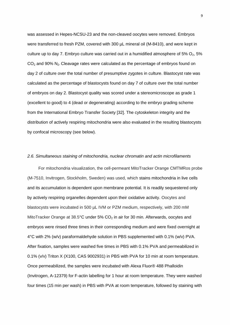
distinct and microfilaments were mainly visible as clumps of actin in the cytoplasm (Fig. 3F).
To classify the blastocyst into the different categories, 30 consecutive Z-stack planes were
taken from each embryo. The different slices were analysed individually and after merging all
scanned image layers to obtain a three-dimensional image.
Figure 3. Representative images illustrating the different patterns of actin microfilaments (MF)
and actively respiring mitochondria in pig blastocysts. Actin MF are depicted in green and actively
respiring mitochondria in orange. Three-dimensional picture showing actin and mitochondria dual staining in a
grade I (both for actin and mitochondria) blastocyst: (A) tilted view or (B) front view. (C) The same embryo as in
A-B, but only the central slice of the Z-scan is shown. Actin cytoskeleton integrity was classified as (D) grade I,

14
(E) grade II or (F) grade III. Mitochondrial pattern was categorized as (G) grade I, (H) grade II and (I) grade III.
See the text for further details. White boxes are indicating the position of the embryo when tilted. Pictures without
white boxes are showing the top view. Bar = 50 µm.
2.9. Classification of mitochondria staining
Oocytes were classified according to one of the following mitochondrial patterns, as
defined previously [21, 23, 38]: a) Peripheral: actively respiring mitochondria were observed
in the cortical region of the oocyte; b) Semi-peripheral: actively respiring mitochondria were
more evident in the subcortical area whereas the cortex of the oocyte was lacking actively
respiring mitochondria; c) Polarized: actively respiring mitochondria were located mainly in
one of the hemispheres of the oocyte. This pattern showed different variations, including
polarization opposite to, or corresponding to the same side as, the meiotic spindle and the
polar body. Oocytes showing a simultaneous peripheral and polarized distribution were
included in this category; d) Central: mitochondria were localized in the centre of the
cytoplasm and the periphery of the oocyte was lacking mitochondria labelling; d) Diffused:
when the mitochondria were homogeneously distributed throughout the cytoplasm (Fig. 1G-
L).
In blastocysts, the distribution of actively respiring mitochondria was scored from grade
I to III. Grade I blastocysts had an even distribution of mitochondria where the stain was
distinctly present in almost all the cells. Grade II blastocysts were characterized by an
intermediate pattern, where the staining was not evenly distributed in the whole embryo;
some of the blastomeres presented large clumps of staining while others lacked actively
respiring mitochondria. In grade III blastocysts, the distribution of actively respiring
mitochondria was very heterogeneous and 50-75% of the embryo did not present distinct
points of staining (Fig. 3G-I). Z-stack (30 slices) were analysed as previously described for
mitochondrial classification.

15
2.10. Statistical analysis
Data expressed in percentages (maturation, cleavage and blastocyst rates) were
analysed using one-way ANOVA. Before the analysis, normal distribution of data and
residuals were tested using Shapiro-Wilk’s test. Homogeneity of variances was evaluated
using Levene’s test. If needed, variables were arcsine-transformed to satisfy parametric
assumptions. When significant differences were found, comparison between treatments was
made using the post hoc Tukey test. When variables did not meet the assumptions of the
parametric test (blastocyst cell number and blastocyst quality), Kruskal-Wallis ANOVA was
used, followed by Mann-Whitney U test to detect differences between experimental groups.
The actin and mitochondrial patterns of oocytes and blastocysts were analysed as
categorical data using correspondence analysis for data visualization [39]. As a
complementary test to the correspondence analysis, Cramér’s V coefficient, together with
Pearson chi-squared test, were calculated to measure the strength association or
dependency between categorical variables [40]. Results are presented as mean ± S.E.M,
unless otherwise stated. Probability values of less than 0.05 were considered significant.
Statistical analyses were performed using Statistica (Statsoft, Inc. 2001. STATISTICA, data
analysis software system, version 6; www.statsoft.com).

16
3. Results
3.1 In vitro maturation, fertilisation and embryo culture
In order to evaluate the effect of simulated stress on nuclear maturation, 716 oocytes
were used. Some of them were used for confocal laser scanning microscopy (CLSM)
assessment as indicated below. No significant differences between groups were observed in
the proportion of oocytes that reached the metaphase II (MII) after 46 h of in vitro maturation
(Table 2). For subsequent fertilisation and embryo culture, 1,333 oocytes were used. The
cleavage rate, (calculated on day 2 of culture) and blastocyst rate (on day 7 of culture) did
not differ between the experimental groups (Table 2).
Blastocyst quality assessed by the number of cells after 7 d of in vitro culture was similar
between groups (Table 3). When blastocyst quality was scored from 1 (best quality) to 4
(worst quality), no significant differences between groups were detected if all blastocysts
were included in the statistical analysis. When only expanded blastocysts (and onwards)
were included in the analysis, the blastocysts obtained in the ACTH group had significantly
lower quality compared to the control groups (Kruskal-Wallis test = 13.1; P<0.01) (Table 3).

17
3.2. Confocal laser scanning microscopy analysis in oocytes
To assess actin cytoskeleton morphology (Figure 1), 165 oocytes were used. There
were no significant differences in the cortex microfilaments in mature oocytes after treatment
(ACTH, no ACTH or BSA groups) during IVM. Around 60% of oocytes in all groups showed a
complete pattern of cortex microfilaments. In contrast, only 20% of immature oocytes (non-
cultured) presented evenly and sharply stained cortex microfilaments (P<0.001).
Cytoplasmic microfilaments differed between maturation status and treatments
(Pearson chi-square: 94.7, P<0.001; Cramér’s coefficient: 0.54). Briefly, in most of the
immature oocytes (84%), cytoplasmic microfilaments were absent and only 6% of oocytes
had an even distribution of microfilaments through the cytoplasm. Among mature oocytes in
the BSA group, 52% presented an uneven pattern and 45% had an even pattern of
microfilaments. However, in the plasma groups, lower numbers of oocytes presented an
even distribution pattern (19% in the ACTH group and 11% in the no ACTH group).
Regarding the transzonal cumulus cell projections, the percentage of mature oocytes
without projections was observed in 41% (control BSA), 64% (control no ACTH) and 56%
(ACTH group). The proportion of oocytes examined after maturation with a complete

18
presence of transzonal projections was 0-3.5% in all treated groups. No differences were
detected between treatments (P>0.05). In comparison, 98.5% of immature oocytes
presented either a complete network (36%) or various degrees of transzonal cumulus
projections (62.5%) (Pearson chi-square: 71.4; P<0.001; Cramér’s coefficient: 0.46).
Mitochondrial staining (Figure 1) was assessed using 160 oocytes. There was an effect
of the treatment during oocyte maturation on the mitochondria distribution (Pearson chi-
square: 16.3; P<0.05; Cramér’s coefficient: 0.29). When immature oocytes were compared to
matured oocytes, there were also differences in mitochondria distribution (Pearson chi-
square: 77; P<0.001; Cramér’s coefficient: 0.40). In immature oocytes, actively respiring
mitochondria were mainly observed in the periphery of the oocyte. In the BSA and ACTH
group, the predominant pattern was an evenly diffused distribution of mitochondria, whereas
a higher proportion of oocytes displaying a polarized pattern was found in the no ACTH
group (Figure 2).

19
Figure 2. Distribution of mitochondrial patterns in oocytes after in vitro maturation under
simulated stress conditions analysed by confocal laser scanning microscopy. Oocytes were
exposed to plasma collected from ACTH stress-simulated sows (ACTH group; n = 32) or belonged to different
control groups: plasma collected from non-treated ACTH females (no ACTH group; n = 38) or without plasma,
supplemented with BSA (n = 28). An additional group of immature (non-matured) oocytes (n = 62), stained after
collection was included.
3.3. Confocal laser scanning microscopy analysis in blastocysts
Confocal image analysis of blastocysts (Figure 3) showed that neither the actin
cytoskeleton integrity nor the mitochondrial activity pattern was affected by the treatment
exposure during IVM (Table 4).

20
4. Discussion
To tackle a possible impact of an altered hormonal environment at key events such as
oocyte maturation, we have used pooled blood plasma obtained from sows subjected to
ACTH administration before ovulation [16]. A similar approach was previously used [41] to
study the effect of induced nutritrional hyperlipidaemia on in vitro bovine embryos;
supplementing the embryo culture medium with blood plasma collected from cows under
different dietary treatments. In the present study, plasma collection in relation to ovulation
was known, so that the plasma recovered in vivo could be added at the corresponding time
point of in vitro culture. In addition, levels of cortisol and reproductive hormones were known.
Progesterone and cortisol levels were significantly higher after ACTH administration [16]. Our
results indicated that, under such conditions, the exposure to plasma from ACTH-
administered sows had limited impact on in vitro oocyte maturation and the subsequent
ability of the oocytes to progress up to the blastocyst stage.
Glucocorticoids stimulate the production of progesterone by granulosa cells through the
enzymes that regulate ovarian steroidogenesis, resulting in the combined effect of enhanced
biosynthesis and decreased metabolism of progesterone (reviewed by [8]). The
administration of ACTH raises both circulating and intrafollicular levels of cortisol [42], but the
net decline of cortisol from peak concentration in plasma after ACTH administration was

21
shorter in comparison to that of the decline in follicular fluid [42]. The administration of ACTH
during estrous in the sow changed the plasma levels of progesterone and possibly 17ß-
estradiol, LH and inhibin α and shortened the duration of signs of standing estrous [16].
Since follicular fluids were not available, it was not possible to determine the levels of cortisol
and other steroids in the follicular fluid after ACTH-administration. Thus, in the present study,
plasma collected at the cortisol and progesterone peaks were selected to guarantee that
plasma composition differed between the ACTH-exposed groups and controls in the assayed
hormones. Blood plasma (or serum) and follicular fluid composition are similar, but contain
different concentrations of hormones [43]. Nonetheless, some compounds are correlated
between plasma and follicular fluid samples and variations in peripheral blood composition
will be reflected in the follicular fluid [44] influencing oocyte competence. We have
supplemented the in vitro maturation medium with 10% of blood plasma since it is the
standard proportion used for culture media. Therefore in our work, the effect of an altered
environment provided by blood plasma supplementation on oocyte maturation might have
been underestimated in comparison to the environment within the follicle under ACTH
exposure.
Nuclear maturation was not impaired after the exposure of oocytes to simulated stress
during IVM. In agreement with other studies using GCs (cortisol, dexamethasone or
corticosterone) to assess the effect of stress on the oocyte, maturation was not affected by
the exposure to GCs in the mouse [11, 13] or sheep [12]. In addition, in the pig, a minimal
inhibitory concentration of dexamethasone (1 µg/mL ~ 2.5 µM) was needed to cause a
negative effect on the progression of oocytes up to metaphase II, but the subsequent
fertilisation ability of the oocytes was not affected [14]. This minimal inhibitory concentration
of GCs is out of the physiological range, even for the levels observed after a stressful event.
Cortisol is bound with high affinity to the cortisol-binding protein and also circulates bound to
other proteins [43, 45]. Around 2-5% of total endogenous cortisol corresponds to the free
unbound, and thus biologically active, fraction [43, 45]. By contrast, dexamethasone, a

22
synthetic GC with similar structure to cortisol, has 25-50 times the glucocorticoid potency of
cortisol and circulates mainly in the unbound form [46]. Regardless of the greater
glucocorticoid potency of dexamethasone in comparison to the natural glucocorticoid,
extremely high levels were needed to observe an effect on oocyte maturation in different
species [12-14]. Therefore, it appears unlikely that a brief exposure to high levels of cortisol
in vivo damage the oocyte directly. A recent study has evaluated the effect of maternal
restraint stress on oocyte competence both in vivo and in vitro [47]. Their data suggests that
restraint stress impaired oocyte competence acting through the hypothalamic-pituitary-
adrenal-gonadal axes, whereas cortisol may have affected ovulation and oocyte competence
by an indirect effect on the ovary rather than at the oocyte level [47]. In target tissues, cortisol
can be either inactivated to cortisone or regenerated from its inactive metabolite by the
bidirectional system of the 11B-hydroxisteroid dehydrogenase (HSD11B) enzymes [45].
Recently, it has been demonstrated that primarily the HSD11B enzyme system acts within
porcine oocytes, converting cortisol to cortisone, and this enzymatic inactivation increases
during oocyte maturation [48]. This HSD11B activity may also have limited the potentially
harmful effect of physiological levels of cortisol in the oocyte in the present work. The
maturation of the follicle is likely influenced by the change in estradiol:progesterone ratios
within the follicle that eventually affected oocyte maturation [32]. Follicles undergoing atresia
are characterized by high progesterone and low estradiol concentrations in the follicular fluid
and, therefore, oocytes exposed to high levels of progesterone have lower developmental
competence [32]. Nonetheless, in the present work, the high concentrations of progesterone
provided by the plasma from ACTH-treated animals seemed not to limit oocyte maturation
and subsequent development in comparison to their untreated counterparts. In our system,
epidermal growth factor (EGF), insulin-transferrin-selenite (ITS) and 2-ßmercaptoethanol
were added. 2-ßmercaptoethanol improves cytoplasmic maturation, having a beneficial effect
on subsequent embryo development [49]. Both ITS and EGF also improve oocyte maturation

23
in the pig [50, 51]. Therefore, the presence of these compounds may have facilitated the
ability of oocytes to overcome a possible negative effect of the altered environment.
The characteristics of actin microfilaments observed both in immature (at germinal
vesicle stage) and mature (MII) oocytes were similar to those previously described in porcine
oocytes [34, 35]. Minor differences were observed by CLSM in actin microfilament patterns in
mature oocytes after treatment. The control group with BSA presented a greater proportion
of oocytes (10-15%) with remnants of transzonal cumulus cell projections compared with the
plasma groups. This difference may be ascribed to the source of proteins, favouring cumulus
expansion in the presence of plasma.
A relocation of mitochondria occurs throughout oocyte maturation [21, 38, 52]. In the
pig, at the germinal vesicle stage, the majority of oocytes displayed a peripheral distribution
of mitochondria, whereas at the end of IVM, mitochondria had migrated to the inner regions
of the cytoplasm, a characteristic of high quality oocytes being to display a diffused
homogeneous distribution of mitochondria in the cytoplasm [21, 53]. A polarization of
mitochondria has been observed in MII oocytes in other animal models [22, 23], as well as in
humans [54]. We have also observed polarization of mitochondria to a different extent in
matured oocytes after treatment, but not in any of the immature ones. In the present study, a
statistically significant effect of treatment on the mitochondria was found. Our results suggest
that there was an influence of the maturation status of the oocyte, as well as a possible
influence of the environment in which the oocytes are maturing on the distribution of
mitochondria. In agreement with this finding, the mitochondrial distribution under in vitro
conditions differed from the in vivo situation [55]; relocation of mitochondria was altered by
the exposure of porcine oocytes to polychlorinated biphenyls during maturation [53]. Using
multiphoton laser scanning microscopy, it was found that the pronuclear accumulation of
mitochondria may be positively correlated with development to the blastocyst stage in rhesus
monkey embryos [56]. In our study, the different mitochondrial distribution pattern in the
various treatment groups after IVM was not reflected by cleavage or blastocyst rates. The

24
oocytes were only exposed to altered hormonal levels during in vitro maturation and,
therefore, the exposure time might have not been enough to detect differences at the
blastocyst level. Whether the different spatial pattern of mitochondrial distribution in oocytes
after maturation is related to their subsequent ability to generate blastocysts warrants future
research.
Differences in blastocyst quality were observed only when expanded blastocysts (or
blastocysts at later developmental stages ) were included in the analysis. However, actin
cytoskeleton integrity and the general mitochondrial pattern of blastocysts were not affected
by the treatment. Unfortunately, it was not possible to compare the quality score given using
a stereomicroscope and the assessment by CLSM for each blastocyst, since the blastocysts
were not followed individually during the staining procedure. Therefore, from our data, a
detrimental effect on cytoplasmic maturation cannot be completely excluded. Whether the
slight differences in blastocyst quality and maximum number of blastocyst cells reached in
the treatments may be reflected in pregnancy rates and live birth rates after embryo transfer
requires investigation.
In conclusion, the present work revealed that an altered hormonal environment during
in vitro oocyte maturation, provided by a brief exposure to plasma collected from simulated-
stressed sows by ACTH administration during the preovulatory period, could induce
alterations in actin cytoskeleton and mitochondrial patterns in oocytes. However, these
changes might not hamper the subsequent in vitro embryo development.
Acknowledgements
The authors would like to thank Ickholmen abattoir for providing the ovaries and
Monica Nilsson for her help collecting the ovaries. We are grateful to Celina Abraham for her
initial laboratory assistance and to Jane Morrell, Margareta Wallgren and Amanda Pimenta
for their help with the boar semen collection. Morten Veljstedt and Anne Dorthe Roed at the
Department of Basic Animal and Veterinary Sciences, Faculty of Life Sciences, University of

25
Copenhagen, Denmark are gratefully acknowledged for their help with the pig IVF protocols.
We are also thankful to Jane Morrell for linguistic review of this manuscript. Supported by
The Swedish Research Council Formas (grant numbers 221-2007-374 and 221-2010-549).

26
References
[1] Dobson H, Tebble JE, Smith RF, Ward WR. Is stress really all that important?
Theriogenology. 2001;55:65-73.
[2] Swaisgood RR, Owen MA, Czekala NM, Mauroo N, Hawk K, Tang JCL. Evaluating
stress and well-being in the giant panda: a system for monitoring. In: Wildt DE, Zhang A,
Zhang H, Janssen DL, Ellis S, editors. Giant pandas Biology, veterinary medicine and
management: Cambridge University Press; 2006. p. 299-314.
[3] Dorn CG. Application of reproductive technologies in North American bison (Bison
bison). Theriogenology. 1995;43:13-20.
[4] González R, Berlinguer F, Espeso G, Ariu F, del Olmo A, Garde JJ, et al. Use of a
neuroleptic in assisted reproduction of the critically endangered Mohor gazelle (Gazella
dama mhorr). Theriogenology. 2008;70:909-22.
[5] Campagne DM. Should fertilization treatment start with reducing stress? Hum Reprod.
2006;21:1651-8.
[6] Cwikel J, Gidron Y, Sheiner E. Psychological interactions with infertility among women.
Eur J Obstet Gynecol Reprod Biol. 2004;117:126-31.
[7] Louis GM, Lum KJ, Sundaram R, Chen Z, Kim S, Lynch CD, et al. Stress reduces
conception probabilities across the fertile window: evidence in support of relaxation.
Fertil Steril. 2011;95:2184-9.
[8] Whirledge S, Cidlowski JA. Glucocorticoids, stress and fertility. Minerva Endocrinol.
2010;35:109-25.
[9] Leroy JL, Opsomer G, Van Soom A, Goovaerts IG, Bols PE. Reduced fertility in high-
yielding dairy cows: are the oocyte and embryo in danger? Part I. The importance of
negative energy balance and altered corpus luteum function to the reduction of oocyte
and embryo quality in high-yielding dairy cows. Reproduction in Domestic Animals.
2008;43:612-22.

27
[10] Webb R, Garnsworthy PC, Gong JG, Armstrong DG. Control of follicular growth: local
interactions and nutritional influences. J Anim Sci. 2004;83 (Suppl. E): E63–E74.
[11] Andersen CY. Effect of glucocorticoids on spontaneous and follicle-stimulating
hormone induced oocyte maturation in mouse oocytes during culture. J Steroid Biochem
Mol Biol. 2003;85:423-7.
[12] Gonzalez R, Ruiz-Leon Y, Gomendio M, Roldan ER. The effect of glucocorticoids on
ERK-1/2 phosphorylation during maturation of lamb oocytes and their subsequent
fertilization and cleavage ability in vitro. Reprod Toxicol. 2010;29:198-205.
[13] Gonzalez R, Ruiz-Leon Y, Gomendio M, Roldan ER. The effect of glucocorticoids on
mouse oocyte in vitro maturation and subsequent fertilization and embryo development.
Toxicol In Vitro. 2010;24:108-15.
[14] Yang JG, Chen WY, Li PS. Effects of glucocorticoids on maturation of pig oocytes and
their subsequent fertilizing capacity in vitro. Biol Reprod. 1999;60:929-36.
[15] Brandt Y, Einarsson S, Ljung A, Lundeheim N, Rodríguez-Martínez H, Madej A.
Effects of continuous elevated cortisol concentrations during oestrus on concentrations
and patterns of progesterone, oestradiol and LH in the sow. Anim Reprod Sci.
2009;110:172-85.
[16] Brandt Y, Lundeheim N, Madej A, Rodriguez-Martinez H, Einarsson S. Effects of
ACTH injections during estrus on concentrations and patterns of progesterone, estradiol,
LH, and inhibin alpha and time of ovulation in the sow. Domest Anim Endocrinol.
2007;32:122-37.
[17] Brandt Y, Madej A, Rodriguez-Martinez H, Einarsson S. Effects of exogenous ACTH
during oestrus on early embryo development and oviductal transport in the sow. Reprod
Domest Anim. 2007;42:118-25.
[18] Alberts B, Johnson A, Lewis J, Raff M, Roberts K, Walter P. Molecular biology of the
cell. Fourth edition ed: Garland Science.; 2002.
28
[19] Van Blerkom J. Mitochondria in human oogenesis and preimplantation embryogenesis:
engines of metabolism, ionic regulation and developmental competence. Reproduction.
2004;128:269-80.
[20] Wang LY, Wang DH, Zou XY, Xu CM. Mitochondrial functions on oocytes and
preimplantation embryos. J Zhejiang Univ Sci B. 2009;10:483-92.
[21] Brevini TA, Vassena R, Francisci C, Gandolfi F. Role of adenosine triphosphate, active
mitochondria, and microtubules in the acquisition of developmental competence of
parthenogenetically activated pig oocytes. Biol Reprod. 2005;72:1218-23.
[22] Nishi Y, Takeshita T, Sato K, Araki T. Change of the mitochondrial distribution in
mouse ooplasm during in vitro maturation. J Nippon Med Sch. 2003;70:408-15.
[23] Velilla E, Rodriguez-Gonzalez E, Vidal F, Izquierdo D, Paramio MT. Mitochondrial
organization in prepubertal goat oocytes during in vitro maturation and fertilization. Mol
Reprod Dev. 2006;73:617-26.
[24] Van Blerkom J, Davis P, Alexander S. Differential mitochondrial distribution in human
pronuclear embryos leads to disproportionate inheritance between blastomeres:
relationship to microtubular organization, ATP content and competence. Hum Reprod.
2000;15:2621-33.
[25] Ferreira EM, Vireque AA, Adona PR, Meirelles FV, Ferriani RA, Navarro PA.
Cytoplasmic maturation of bovine oocytes: structural and biochemical modifications and
acquisition of developmental competence. Theriogenology. 2009;71:836-48.
[26] Sun QY, Schatten H. Regulation of dynamic events by microfilaments during oocyte
maturation and fertilization. Reproduction. 2006;131:193-205.
[27] Wang WH, Abeydeera LR, Prather RS, Day BN. Actin filament distribution in blocked
and developing pig embryos. Zygote. 2000;8:353-8.
[28] Zijlstra C, Kidson A, Schoevers EJ, Daemen AJJM, Tharasanit T, Kuijk EW, et al.
Blastocyst morphology, actin cytoskeleton quality and chromosome content are
correlated with embryo quality in the pig. Theriogenology. 2008;70:923-35.

29
[29] Blomqvist G, Persson M, Wallgren M, Wallgren P, Morrell JM. Removal of virus from
boar semen spiked with porcine circovirus type 2. Anim Reprod Sci. 2011;126:108-14.
[30] Petters RM, Wells KD. Culture of pig embryos. J Reprod Fertil Suppl. 1993;48:61-73.
[31] Yoshioka K, Suzuki C, Tanaka A, Anas IM, Iwamura S. Birth of piglets derived from
porcine zygotes cultured in a chemically defined medium. Biol Reprod. 2002;66:112-9.
[32] Gordon I. Laboratory production of cattle embryos. Second Edition. Biotecnology in
Agriculture Series No. 27 United Kingdom: CABI Publishing; 2003.
[33] Albarracin JL, Morato R, Izquierdo D, Mogas T. Effects of roscovitine on the nuclear
and cytoskeletal components of calf oocytes and their subsequent development.
Theriogenology. 2005;64:1740-55.
[34] Suzuki H, Jeong BS, Yang X. Dynamic changes of cumulus-oocyte cell communication
during in vitro maturation of porcine oocytes. Biol Reprod. 2000;63:723-9.
[35] Suzuki H, Saito Y, Kagawa N, Yang X. In vitro fertilization and polyspermy in the pig:
factors affecting fertilization rates and cytoskeletal reorganization of the oocyte. Microsc
Res Tech. 2003;61:327-34.
[36] Skidmore JA, Schoevers E, Stout TA. Effect of different methods of cryopreservation
on the cytoskeletal integrity of dromedary camel (Camelus dromedarius) embryos. Anim
Reprod Sci. 2009;113:196-204.
[37] Tharasanit T, Colenbrander B, Stout TA. Effect of cryopreservation on the cellular
integrity of equine embryos. Reproduction. 2005;129:789-98.
[38] Liu S, Li Y, Gao X, Yan JH, Chen ZJ. Changes in the distribution of mitochondria
before and after in vitro maturation of human oocytes and the effect of in vitro maturation
on mitochondria distribution. Fertil Steril. 2010;93:1550-5.
[39] Beh E. Simple Correspondence Analysis: A Bibliographic Review. Int Stat Rev.
2004;72:257-84.
[40] Guisande-González C, Barreiro-Felpeto A, Maneiro-Estraviz I, Riveiro-Alarcón I,
Vergara-Castaño AR, Vaamonde-Liste A. Tratamiento de datos. Madrid. Spain.
Ediciones Díaz de Santos; 2006.

30
[41] Leroy JLMR, Van Hoeck L, Clemente M, Rizos D, Gutierrez-Adan A, Van Soom A, et
al. The effect of nutritionally induced hyperlipidaemia on in vitro bovine embryo quality.
Hum Reprod. 2010;25:768-78.
[42] Montgomery A, Viveiros M, Cummings E, Liptrap R. Rate of decline in cortisol
concentrations in ovarian follicles following ACTH treatment in the sow. Can J Vet Res.
1997;61:309-11.
[43] Andersen CY. Possible new mechanism of cortisol action in female reproductive
organs: physiological implications of the free hormone hypothesis. J Endocrinol.
2002;173:211-7.
[44] Leroy JL, Vanholder T, Delanghe JR, Opsomer G, Van Soom A, Bols PE, et al.
Metabolite and ionic composition of follicular fluid from different-sized follicles and their
relationship to serum concentrations in dairy cows. Anim Reprod Sci. 2004;80:201-11.
[45] Goodman HM. Basic Medical Endocrinology. Third Edition. San Diego, USA:
Academic Press; 2003.
[46] Magiakou MA, Chrousos GP. Corticosteroid therapy, non-endocrine disease and
corticosteroid withdrawal. In: Bardwin C, editor. Current therapy in endocrinology and
metabolism. 5th ed ed. Philadelphia: Mosby Yearbook; 1994. p. 120-4.
[47] Zhang SY, Wang JZ, Li JJ, Wei DL, Sui HS, Zhang ZH, et al. Maternal restraint stress
diminishes the developmental potential of oocytes. Biol Reprod. 2011;84:672–81.
[48] Webb RJ, Sunak N, Wren L, Michael AE. Inactivation of glucocorticoids by 11beta-
hydroxysteroid dehydrogenase enzymes increases during the meiotic maturation of
porcine oocytes. Reproduction. 2008;136:725-32.
[49] Abeydeera LR, Wang WH, Cantley TC, Prather RS, Day BN. Presence of beta-
mercaptoethanol can increase the glutathione content of pig oocytes matured in vitro
and the rate of blastocyst development after in vitro fertilization. Theriogenology.
1998;50:747-56.
[50] Hu J, Ma X, Bao JC, Li W, Cheng D, Gao Z, et al. Insulin-transferrin-selenium (ITS)
improves maturation of porcine oocytes in vitro. Zygote. 2011;19:191-7.

31
[51] Uhm SJ, Gupta MK, Yang JH, Chung HJ, Min TS, Lee HT. Epidermal growth factor
can be used in lieu of follicle-stimulating hormone for nuclear maturation of porcine
oocytes in vitro. Theriogenology. 2010;73:1024-36.
[52] Sun QY, Wu GM, Lai L, Park KW, Cabot R, Cheong HT, et al. Translocation of active
mitochondria during pig oocyte maturation, fertilization and early embryo development in
vitro. Reproduction. 2001;122:155-63.
[53] Brevini TA, Vassena R, Paffoni A, Francisci C, Fascio U, Gandolfi F. Exposure of pig
oocytes to PCBs during in vitro maturation: effects on developmental competence,
cytoplasmic remodelling and communications with cumulus cells. Eur J Histochem.
2004;48:347-56.
[54] Dell'Aquila ME, Ambruosi B, De Santis T, Cho YS. Mitochondrial distribution and
activity in human mature oocytes: gonadotropin-releasing hormone agonist versus
antagonist for pituitary down-regulation. Fertil Steril. 2009;91:249-55.
[55] Egerszegi I, Alm H, Ratky J, Heleil B, Brussow KP, Torner H. Meiotic progression,
mitochondrial features and fertilisation characteristics of porcine oocytes with different
G6PDH activities. Reprod Fertil Dev. 2010;22:830-8.
[56] Squirrell JM, Schramm RD, Paprocki AM, Wokosin DL, Bavister BD. Imaging
mitochondrial organization in living primate oocytes and embryos using multiphoton
microscopy. Microsc Microanal. 2003;9:190-201

32