Embed Size (px)
Citation preview
Z:\Manuscripts MJT\manuscript new lipid\manuscript revisedbb 72707 (3).doc 1
TITLE: ABCG1 is Deficient in Alveolar Macrophages of GM-
CSF Knock-Out Mice and Patients with Pulmonary
Alveolar Proteinosis
AUTHORS: Mary Jane Thomassen1, Barbara P. Barna1, Achut G.
Malur2, Tracey L. Bonfield3, Carol F. Farver3,4, Anagha
Malur1, Heidi Dalrymple1, Mani S. Kavuru1 and Maria
Febbraio5
INSTITUTION: 1Program in Lung Cell Biology and Translational
Research, Division of Pulmonary and Critical Care
Medicine and 2Department of Microbiology and
immunology, East Carolina University, Greenville, NC
and 3Departments of Pulmonary, Allergy and Critical
Care Medicine; 4Anatomic Pathology and 5Cell
Biology, The Cleveland Clinic Foundation, Cleveland,
OH
CORRESPONDING AUTHOR: Dr. Mary Jane Thomassen
Division of Pulmonary and Critical Care Medicine East Carolina University Brody School of Medicine
3E-149 Brody Medical Sciences Building Greenville, NC 27834
(252) 744-1117, FAX (252) 744-2583 email: [email protected]
Running Title: ABCG1 Deficiency in Alveolar Macrophages
by guest, on Novem
ber 21, 2018w
ww
.jlr.orgD
ownloaded from
Z:\Manuscripts MJT\manuscript new lipid\manuscript revisedbb 72707 (3).doc 2
ABSTRACT
Patients with pulmonary alveolar proteinosis (PAP) display impaired surfactant
clearance, foamy, lipid-filled alveolar macrophages, and elevated cholesterol metabolites
within the lung. Neutralizing autoantibodies to granulocyte-macrophage colony
stimulating factor (GM-CSF) are also present, resulting in virtual GM-CSF deficiency. We
investigated ABCG1 and ABCA1 expression in alveolar macrophages of PAP patients
and GM-CSF knock-out (KO) mice which exhibit PAP-like pulmonary pathology and
elevated pulmonary cholesterol. Alveolar macrophages from both sources displayed a
striking similarity in transporter gene dysregulation consisting of deficient ABCG1
accompanied by highly elevated ABCA1. Peroxisome proliferator-activated receptor
gamma (PPARγ), a known regulator of both transporters, was deficient as previously
reported. In contrast, the liver-X-receptor (LXR)α, which also upregulates both
transporters, was highly elevated. GM-CSF treatment increased ABCG1 expression in
macrophages in vitro and in PAP patients in vivo. Overexpression of PPARγ by
lentivirus-PPARγ transduction of primary alveolar macrophages, or activation by
rosiglitazone also elevated ABCG1 expression. Results suggest that ABCG1 deficiency
in PAP and GM-CSF KO alveolar macrophages is due to absence of a GM-CSF-
mediated PPARγ pathway. These findings document the existence of ABCG1 deficiency
in human lung disease and highlight a critical role for ABCG1 in surfactant homeostasis.
Key Words: Alveolar macrophage, LXRα, PPARγ, ABCA1, ABCG1, foam cells.
by guest, on Novem
ber 21, 2018w
ww
.jlr.orgD
ownloaded from
Z:\Manuscripts MJT\manuscript new lipid\manuscript revisedbb 72707 (3).doc 3
INTRODUCTION
Pulmonary alveolar proteinosis (PAP) is a rare autoimmune disease, in which
surfactant clearance is defective and granulocyte-macrophage colony stimulating factor
(GM-CSF) is deficient due to neutralizing autoantibodies (1). In patients with PAP, the
lungs are filled with excess lipoproteinaceous material, and alveolar macrophages are
engorged with lipid resulting in a foamy appearance (2). We have shown that alveolar
macrophages from PAP patients are also severely deficient in peroxisome proliferator-
activated receptor gamma (PPARγ), a key regulator of lipid metabolism (3). This
deficiency is correctable by GM-CSF therapy (3). Subsequently we reported that
cholesterol and the cholesterol metabolites, cholestenoic acid and 27-hydroxycholesterol
(27-OH), are highly elevated in PAP bronchoalveolar lavage fluid (4). Interestingly, a
PAP-like pulmonary histopathology is displayed by GM-CSF KO mice (5;6). Exogenous
or local over-expression of GM-CSF reverses this pathology (7;8). Pulmonary cholesterol
is also elevated in the GM-CSF KO mouse lung (9), and GM-CSF KO macrophages are
deficient in PPARγ (10;11).
Surfactant, which is produced by type II alveolar pneumocytes, is composed of
90% lipid, 10% protein, and a small amount of carbohydrate. Approximately 80-90% of
surfactant lipid is phospholipid; the other lipids, in decreasing order, are cholesterol,
triacylglycerol and free fatty acids (12). The processes mediating and regulating
surfactant clearance, recycling, and catabolism are not completely defined (13). Two
primary surfactant catabolic pathways have been described (reviewed in (14)). The first
pathway is a recycling process requiring type II pneumocytes. The second pathway is a
clearance pathway in which surfactant is phagocytosed and degraded by alveolar
by guest, on Novem
ber 21, 2018w
ww
.jlr.orgD
ownloaded from
Z:\Manuscripts MJT\manuscript new lipid\manuscript revisedbb 72707 (3).doc 4
macrophages. Although a small amount of surfactant is degraded by type II
pneumocytes, recent findings emphasize the critical role of the alveolar macrophage in
surfactant catabolism (15).
The ATP-binding cassette (ABC) transporters, ABCA1 and ABCG1, are members
of a group of transmembrane proteins that transport a wide variety of substrates across
membranes (16-19). Macrophages are a rich source of both ABCA1 and ABCG1, which
can be regulated by a number of pathways, including PPARγ activation and activation of
LXRα via uptake of cholesterol or oxysterols (20-22). Cholesterol is esterified by
macrophages and accumulated cholesterol esters are stored within the cell giving it a
foamy appearance (reviewed in (18)). ABCA1 and ABCG1 play a critical role in
mediating cellular efflux of both cholesterol and phospholipids to lipoproteins (18).
Recent findings have implicated these transporters in surfactant homeostasis (16;23;24).
ABCA1 KO mice exhibit morphologic abnormalities in the lungs including a massive
accumulation of lipid laden macrophages and type II pneumocytes (24). Similarly, lungs
from ABCG1 KO mice show abnormal accumulation of lipid deposits in both alveolar
macrophages and type 2 pneumocytes (16).
Although the phenotype of GM-CSF KO mice appears to resemble that of PAP,
the status of genes involved in alveolar macrophage lipid efflux has not been investigated
in either case. We hypothesized that the ABC transporters might be dysregulated in PAP
and in GM-CSF KO mice. Results indicate that alveolar macrophages from PAP patients
and GM-CSF KO mice display a striking similarity of transporter dysregulation
characterized by deficient ABCG1 and elevated ABCA1.
by guest, on Novem
ber 21, 2018w
ww
.jlr.orgD
ownloaded from
Z:\Manuscripts MJT\manuscript new lipid\manuscript revisedbb 72707 (3).doc 5
MATERIALS AND METHODS
Study Population. This protocol was approved by the Institutional Review Board and
written informed consent was obtained from all subjects. Healthy control individuals had no
history of lung disease and were not on medication (n=6). The diagnosis of idiopathic PAP
was established by histopathological examination of material from open lung or
transbronchial biopsies showing the characteristic filling of the alveoli with eosinophilic
amorphous material with preserved lung architecture and absence of inflammation, and
exclusion of secondary etiologies by negative lung cultures or occupational history (25). All
PAP patients were symptomatic with dyspnea, were hypoxemic on room air and had typical
alveolar infiltrates on radiographs. Six PAP patients participated in a prospective clinical trial
of recombinant human GM-CSF (Leukine; Berlex, Seattle, WA) as previously described (26).
Treatment consisted of 250 mcg/d by subcutaneous administration, with increased dosage
every 2 wk and maximum daily dosage of 18 mcg/kg/d by 8 wk. Patients were evaluated at
baseline before initiation of GM-CSF therapy and after 6 months of therapy. Clinical
improvement or response to therapy was defined a priori as ≥ 10 mm Hg improvement in
PaO2 from baseline.
Cell collection. Alveolar macrophages were derived from bronchoalveolar lavage (BAL)
obtained by fiberoptic bronchoscopy as previously described (27). Differential cell counts
were obtained from cytospins stained with a modified Wright’s stain. Differential cell counts
from BALs indicated that 93 ± 3% of PAP and 98 ± 0.3% of healthy control cells were
alveolar macrophages. Mean viability of lavage cells was greater than 95% as determined
by trypan blue dye exclusion.
by guest, on Novem
ber 21, 2018w
ww
.jlr.orgD
ownloaded from
Z:\Manuscripts MJT\manuscript new lipid\manuscript revisedbb 72707 (3).doc 6
Mice. Animal studies were conducted in conformity with Public Health Service (PHS) Policy
on humane care and use of laboratory animals and were approved by the institutional animal
care committee. The GM-CSF KO mice were generated by Dr. Glenn Dranoff (5). The mice
have been backcrossed eight generations to C57Bl/6. C57Bl/6 wild type mice were obtained
from Jackson Laboratory (Bar Harbor, ME) for controls. Bronchoalveolar lavage and
peritoneal cells were obtained from 8-12 week-old GM-CSF KO mice and age and gender
matched wild-type C57Bl/6 controls. For BAL cell harvest, mice received ketamine (80
mg/kg) and xylazine (10 mg/kg) intraperitoneally (IP). The thoracic cavity was opened and
the lungs were exposed. After cannulating the trachea a tube was inserted and
bronchoalveolar lavage was carried out with warmed (37oC) PBS in 1ml aliquots. To obtain
elicited peritoneal macrophages, mice were injected intraperitoneally with 0.5 ml of sterile
4% Brewer’s Thioglycollate medium (Sigma). Four days later, mice were sacrificed and cells
recovered by lavage with 5 ml of sterile PBS using a 23 gauge needle. BAL and peritoneal
cell differentials from all animals used in experiments revealed > 90% macrophages. For all
experiments 3 sets of pooled BAL or peritoneal cells from 3-5 mice were used. Cytospins of
BAL or peritoneal cells were stained with Oil Red O to detect intracellular neutral lipids, and
counterstained with Gill’s hematoxylin stain.
Lentivirus Plasmid and Transduction. Primary human alveolar macrophages were
transduced with a self inactivating lentivirus expression vector that was previously utilized
in the generation of a stable cell line expressing the human parainfluenza virus type 3 C
protein (28). cDNA corresponding to the human PPARγ was cloned into the multiple
cloning sites downstream of a CMV promoter using standard techniques as described
(28). The recombinant lenti-viral plasmid thus obtained was then transfected into 293FT
by guest, on Novem
ber 21, 2018w
ww
.jlr.orgD
ownloaded from
Z:\Manuscripts MJT\manuscript new lipid\manuscript revisedbb 72707 (3).doc 7
cells along with plasmids encoding the gag, pol and rev genes and a plasmid possessing
the vesicular stomatitis glycoprotein (G) using Lipofectamine 2000. At 48 hrs post
transfection, cell culture supernatant containing the Lentivirus-PPARγ (Lenti-PPARγ) was
purified by centrifugation at 27,000 rpm at 40C for 3.5 hrs. The Lenti-PPARγ virus pellet
was resuspended in TE and aliquots of 100µl were stored at -800C. The concentration of
Lenti-PPARγ virus was determined by a p24 ELISA (CELL BIOLABS, San Diego, CA). A
lentivirus expressing the enhanced Green Fluorescent Protein (Lenti-eGFP) was
obtained using a similar protocol and utilized as a control in experiments for the
determination of transduction efficiency. The percentage efficiency of in vitro
transduction as determined by examination of Lenti-eGFP incorporation under
fluorescent microscopy was 87 ± 1 (n=3) in human alveolar macrophages.
RNA Purification and Analysis. Total RNA was extracted from human and mouse cells by
RNAeasy protocol (Qiagen, Valencia, CA). Expression of mRNA was determined by real
time RT-PCR using the ABI Prism 7000 Detection System (TaqMan; Applied Biosystems,
Foster City, CA.) according to the manufacturer’s instructions. RNA specimens were
analyzed in duplicate using primer sets for mouse or human PPARγ, LXRα, ABCA1 and
ABCG1 (ABI). Threshold cycle (CT) values for genes of interest were normalized to a
housekeeping gene [glyceraldehyde 3 phosphate dehydrogenase, (GAPDH)] and used to
calculate the relative quantity of mRNA expression in PAP or GM-CSF KO samples
compared to healthy human or wild type murine controls. Data were expressed as a fold
change in mRNA expression relative to control values (29).
by guest, on Novem
ber 21, 2018w
ww
.jlr.orgD
ownloaded from
Z:\Manuscripts MJT\manuscript new lipid\manuscript revisedbb 72707 (3).doc 8
Immunoblotting. Cultured human alveolar macrophages or freshly obtained BAL cells from
5 animals/group of GM-CSF KO and wild-type C57Bl/6 mice were washed in phosphate-
buffered saline (PBS) and lysed in a modified NP-40 lysis buffer as previously described
(30). An equivalent amount of protein (25 µg) from each sample group was analyzed by
7.5% SDS gel electrophoresis and transferred onto nitrocellulose membranes. Primary
antibodies to ABCG1 (Santa Cruz, E-20) were diluted 1:500 and β-actin (Chemicon) were
diluted 1:5000 before incubation with membranes. Proteins were visualized by
chemiluminescence.
Statistics. Data were analyzed by student’s t-test using Prism software (GraphPad, Inc.,
San Diego, CA.). Significance was defined as p ≤ 0.05.
by guest, on Novem
ber 21, 2018w
ww
.jlr.orgD
ownloaded from
Z:\Manuscripts MJT\manuscript new lipid\manuscript revisedbb 72707 (3).doc 9
RESULTS
ABC transporter gene expression is dysregulated in PAP and GM-CSF KO alveolar
macrophages. Endogenous mRNA expression of lipid transporter genes was
determined in uncultured preparations of bronchoalveolar lavage (BAL)-derived cells
which consisted of >90% alveolar macrophages (see Methods section). ABCG1 mRNA
level was significantly (p= 0.03) decreased in PAP alveolar macrophages as compared to
healthy controls (Figure 1A), and in GM-CSF KO alveolar macrophages (p=0.02)
compared to wild-type mice (Figure 1B). In contrast, ABCA1 mRNA level was elevated in
both cases (p=0.03 PAP patients; p=0.004 GM-CSF KO mice) (see Figure 1A and B).
Because transporter expression may be governed by both LXRα (17;31) and PPARγ (32),
we investigated the status of these regulatory genes in alveolar macrophages. LXRα
mRNA level was elevated in both PAP patients (p=0.005) and GM-CSF KO mice
(p=0.0006), while PPARγ mRNA was decreased (p=0.04 PAP patients; p=0.0002 GM-
CSF KO mice) as reported previously (3;10) (Figure 1A and B). Apolipoprotein E
(APOE), another gene subject to LXR regulation was also investigated and found to be
highly increased in alveolar macrophages from both PAP patients (7.1±1.9 fold increase,
p=0.03) and GM-CSF KO mice (618±1.4, p<0.0001) as compared to controls.
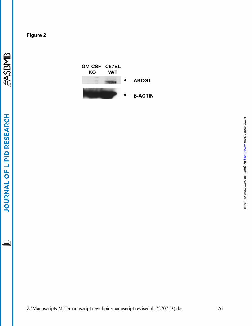
ABCG1 protein expression is reduced in GM-CSF KO alveolar macrophages.
Immunoblotting revealed no detectable ABCG1 protein in BAL cell lysates from GM-CSF
KO mice (Figure 2). Protein bands of approximately 110 kda corresponding to ABCG1
were visible however in BAL cell lysates from wild type C57Bl/6 mice (Figure 2).
by guest, on Novem
ber 21, 2018w
ww
.jlr.orgD
ownloaded from
Z:\Manuscripts MJT\manuscript new lipid\manuscript revisedbb 72707 (3).doc 10
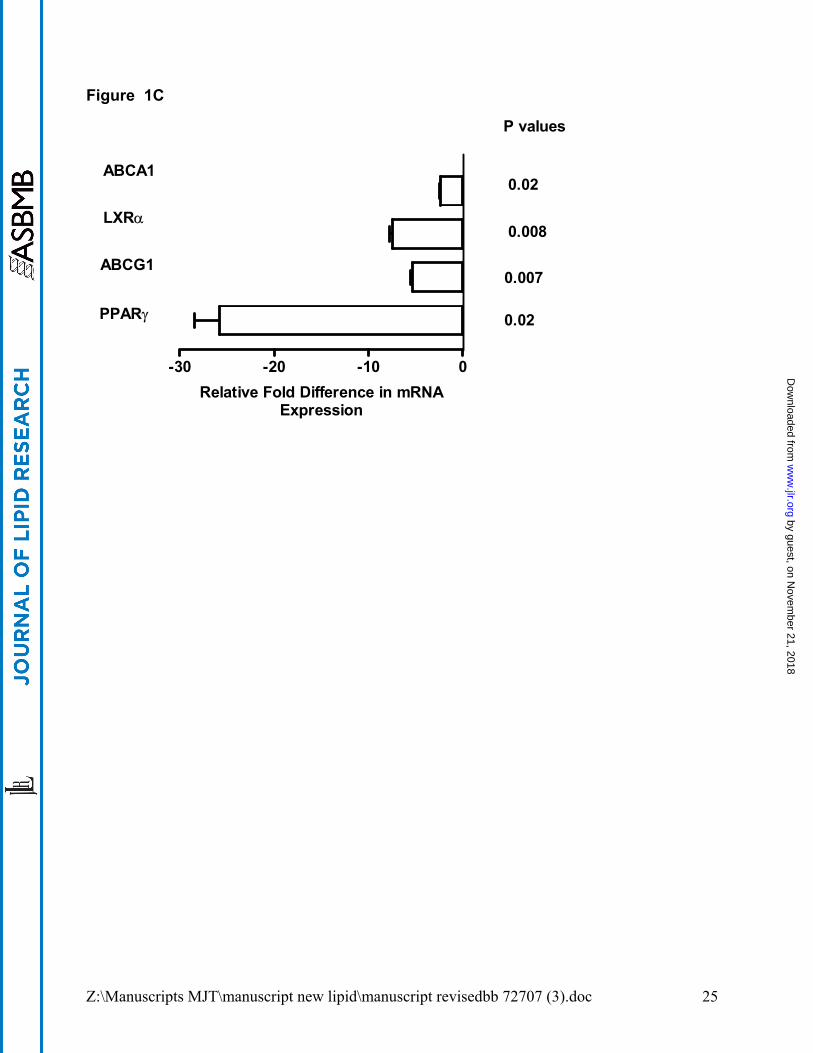
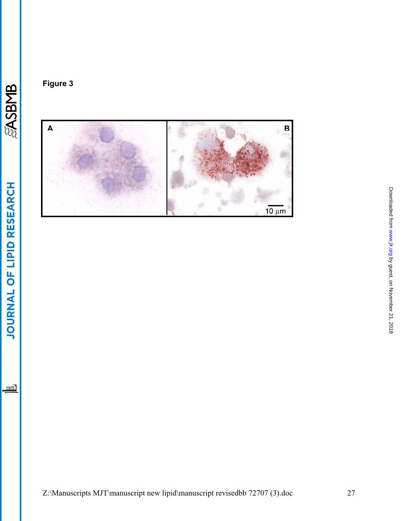
GM-CSF KO and PAP alveolar macrophages are morphologically similar. Oil Red O
staining highlighted the foamy cytopathology of GM-CSF KO alveolar macrophages
which strongly resembled that previously reported in PAP (33). Compared to wild type
C57Bl/6 mice (Figure 3A), GM-CSF KO alveolar macrophages were filled with
cytoplasmic deposits of Oil Red O-positive (neutral) lipids and large lipid vacuoles (Figure
3B). The majority (97.6 ± 1.0%) of wild type alveolar macrophages (n=5) were Oil Red O
negative, with only 2.4 ± 1.0% positive cells. In contrast, 92.4 ± 4.3% of GM-CSF KO
macrophages were Oil Red O positive (n=5, p=0.001). Oil Red O staining was
unremarkable in GM-CSF KO peritoneal macrophages which resembled those from wild-
type mice (data not shown) although PPARγ and ABCA1 gene expression were
decreased (Figure 1C) as noted by Ditiakovski et al (11). ABCG1 was also decreased in
peritoneal macrophages but in contrast to alveolar macrophages, LXRα was not elevated
(Figure 1C). Furthermore, as expected since LXR was not elevated, APOE expression
was also not elevated in peritoneal cells.
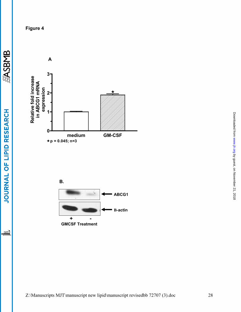
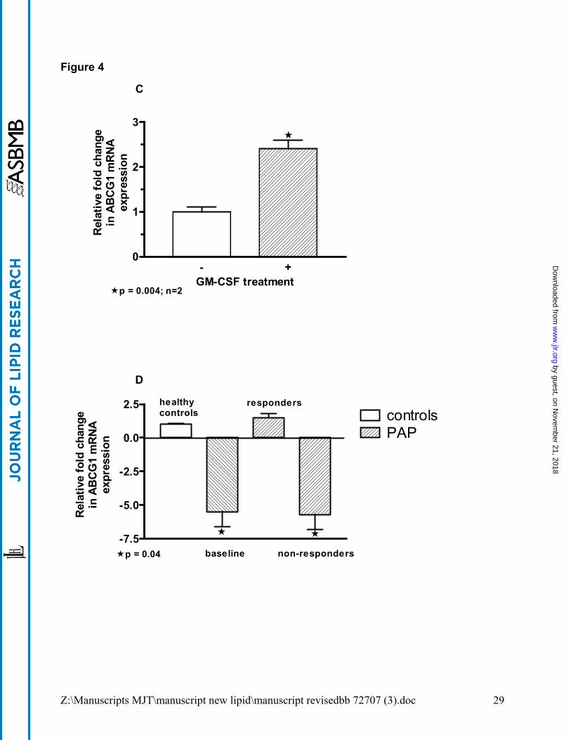
GM-CSF treatment upregulates ABCG1 expression. To determine whether GM-CSF
modified ABCG1 levels, human and murine macrophages were treated with GM-CSF (100
ng/ml) in vitro. GM-CSF exposure significantly increased ABCG1 mRNA (p=0.04, n=3) and
protein expression in alveolar macrophages from healthy controls compared to untreated
macrophages (Figure 4A and B). Alveolar macrophages from a PAP patient also responded
to GM-CSF culture with a 3.1-fold increase in ABCG1 mRNA level as compared to culture
media (data not shown). Similarly, GM-CSF KO macrophages cultured in the presence of
GM-CSF displayed elevated ABCG1 mRNA (p=0.004) (see Figure 4C). ABCG1 mRNA
expression was also examined in BAL cells from PAP patients (n=6) who had received GM-
by guest, on Novem
ber 21, 2018w
ww
.jlr.orgD
ownloaded from
Z:\Manuscripts MJT\manuscript new lipid\manuscript revisedbb 72707 (3).doc 11
CSF therapy in vivo (Figure 4D). ABCG1 mRNA levels were significantly deficient (p=0.04)
before therapy (baseline) compared to healthy controls (n=6), and remained so in clinical
non-responders (n=4). In two clinically responsive PAP patients post-GM-CSF therapy
however, ABCG1 mRNA levels were restored to control range (Figure 4D).
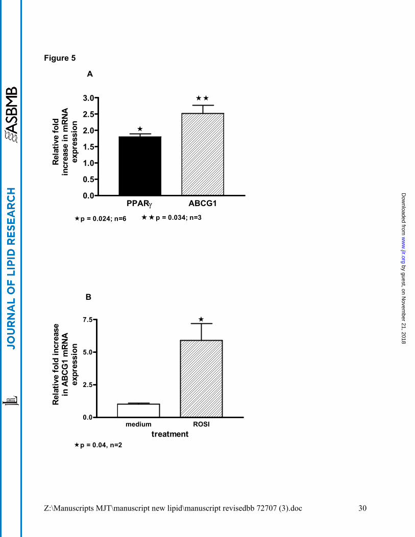
PPARγ overexpression or activation increases ABCG1 expression. Although PPARγ
pathways are cited as regulating ABCG1 in experimental models, no data are available for
primary human alveolar macrophages. A lentivirus-PPARγ vector (LPPARγ) and as control,
a lentivirus-eGFP (LeGFP) vector were used to transduce primary human alveolar
macrophages in vitro. Compared to LeGFP, LPPARγ significantly increased ABCG1 mRNA
(p=0.03, n=3) and as anticipated, PPARγ mRNA was also increased (p=0.02) (figure 5A). As
an additional control PPARα mRNA was also evaluated and lenti-PPARγ transduction had no
effect on PPARα expression. PPARγ and ABCG1 mRNA levels were not affected in LeGFP
transduced alveolar macrophages compared to untreated macrophages (data not shown).
Ligand activation of PPARγ via Rosiglitazone (10µM) treatment of human alveolar
macrophages in vitro also produced brisk upregulation in ABCG1 mRNA levels (Figure 5B).
by guest, on Novem
ber 21, 2018w
ww
.jlr.orgD
ownloaded from
Z:\Manuscripts MJT\manuscript new lipid\manuscript revisedbb 72707 (3).doc 12
DISCUSSION
This report demonstrates similar phenotypes and profiles of lipid transport genes
in alveolar macrophages from PAP patients and GM-CSF KO mice, thus supporting the
role of GM-CSF in the etiology of this disease and identifying potential downstream target
genes. Alveolar macrophages from both PAP patients and GM-CSF KO mice present a
foamy appearance and contain excess Oil Red O positive neutral lipids. Peritoneal
macrophages from GM-CSF KO mice, in contrast, do not exhibit lipid accumulation,
suggesting that a portion of the etiology of the disease is specific to the demands of the
macrophage environment. We previously reported decreased expression of PPARγ in
PAP alveolar macrophages (3), and now show this to be a characteristic of both alveolar
and peritoneal macrophages of GM-CSF KO mice. Ditiakovski et al also reported similar
findings in GM-CSF KO peritoneal macrophages (11). Such data indicate that PPARγ is
downsteam of GM-CSF.
In both PAP patients and GM-CSF KO mice, LXRα expression was increased in
alveolar macrophages. This seems counter to the hypothesis put forth by Ricote et. al.
(34), which suggested that LXRα is regulated by PPAR/RXR heterodimers. Accordingly,
one would expect a down regulation of LXRα in GM-CSF KO alveolar macrophages
where PPARγ is absent. In fact, such down regulation in LXRα mRNA level is observed
in peritoneal macrophages. Our previous report indicated elevated cholesterol and
oxysterol levels in PAP lung (4). Thus, the increased LXRα expression found in alveolar
macrophages in the current study is consistent with the localized increase in
cholesterol/cholesterol metabolites that would directly influence LXRα expression (20).
Baldan et al (16), however, also reported increased LXRα and ABCA1 in alveolar
by guest, on Novem
ber 21, 2018w
ww
.jlr.orgD
ownloaded from
Z:\Manuscripts MJT\manuscript new lipid\manuscript revisedbb 72707 (3).doc 13
macrophages of ABCG1 KO mice. Interestingly, these ABCG1 KO mice also accumulate
lipid in the lung.
A novel observation in this study is the demonstration of an ABCG1 transporter
deficiency in human pulmonary disease. Previously, ABCA1 deficiency was recognized
in Tangier’s disease (35). The accumulation of surfactant lipid despite elevated ABCA1
and LXRα in PAP and GM-CSF KO alveolar macrophages suggests that expression of
these genes is not sufficient to maintain surfactant homeostasis and prevent foam cell
formation. Such findings may indirectly implicate ABCG1 as the primary transporter for
efflux of accumulated surfactant phospholipid. Alternatively, the dysregulation of these
(and potentially other) genes may result in aberrant compartmentalization of surfactant
such that it is unavailable for processing or efflux. Our data further suggest that ABCA1
expression may be regulated by RXR/LXR dimers, as suggested by Ricote et al (34), and
in the case of LXRα altered as a consequence of lipid accumulation. ABCG1 may also
be regulated by RXR/LXR heterodimers (34), but we found decreased ABCG1
expression in both peritoneal and alveolar macrophages. Recent studies have shown
that PPARγ activation may induce ABCG1 expression in an LXR independent manner
(32), and a deficiency of this pathway may be responsible for the decreased ABCG1 we
observed in PAP and GM-CSF KO mice.
The extremely large increase in APOE mRNA expression in GM-CSF KO mice
compared to human alveolar macrophages is likely to be related to species-specific
differences in lipoprotein metabolism. Mice lack cholesterol ester transfer protein (CETP)
and therefore there is a difference in the manner by which cholesterol is transferred
between lipoproteins and dispersed (36;37). Mice may be more dependent upon liver-
mediated mechanisms for cholesterol excretion than are humans and the exaggerated
by guest, on Novem
ber 21, 2018w
ww
.jlr.orgD
ownloaded from
Z:\Manuscripts MJT\manuscript new lipid\manuscript revisedbb 72707 (3).doc 14
increase in APOE found in GM-CSF KO mice may reflect the importance of a murine
salvage pathway.
In summary, results of the current studies support the hypothesis that GM-CSF
promotes surfactant catabolism by upregulating ABCG1 via PPARγ pathways. First, the
deficiency of alveolar macrophage ABCG1 reported in both PAP patients and GM-CSF
KO mice parallels the deficiency of PPARγ. Second, GM-CSF increased ABCG1
expression in both in vitro experiments and in a clinical trial of recombinant GM-CSF
therapy in PAP patients. In the latter case, PAP patients who demonstrated an excellent
clinical response with almost complete resolution of disease also displayed upregulated
ABCG1 mRNA levels in BAL cells, whereas ABCG1 did not increase in BAL cells of
clinically unresponsive patients. Such findings with ABCG1 parallel those we reported
previously showing PPARγ restoration in clinically responsive PAP patients receiving
recombinant GM-CSF [3]. Finally, the increase in alveolar macrophage ABCG1
expression after either lentivirus vector-mediated PPARγ overexpression or ligand
activation of PPARγ demonstrates the functional link between PPARγ and ABCG1 in
alveolar macrophages.
ACKNOWLEDGEMENTS: This work was funded by NIH HL67676, North Carolina
Biotechnology Center grant #FRG-1013 and the generous support of Regina Taussig.
We thank Irina Polyakova for her expert assistance with the animals.
by guest, on Novem
ber 21, 2018w
ww
.jlr.orgD
ownloaded from
Z:\Manuscripts MJT\manuscript new lipid\manuscript revisedbb 72707 (3).doc 15
Reference List
1. Kitamura,T., Tanaka,N., Watanabe,J., Uchida,K., Kanegasaki,S., Yamada,Y., and
Nakata,K. 1999. Idiopathic pulmonary alveolar proteinosis as an autoimmune
disease with neutralizing antibody against granulocyte/macrophage colony-
stimulating factor. J. Exp. Med. 190:875-880.
2. Seymour,J.F., and Presneill,J.J. 2002. Pulmonary alveolar proteinosis (Progress in
the first 44 years). Am. J. Respir. Crit. Care Med. 166:215-235.
3. Bonfield,T.L., Farver,C.F., Barna,B.P., Malur,A., Abraham,S., Raychaudhuri,B.,
Kavuru,M.S., and Thomassen,M.J. 2003. Peroxisome proliferator-activated
receptor-gamma is deficient in alveolar macrophages from patients with alveolar
proteinosis. Am. J Respir Cell Mol. Biol. 29:677-682.
4. Meaney,S., Bonfield,T.L., Hansson,M., Babiker,A., Kavuru,M.S., and
Thomassen,M.J. 2004. Serum cholestenoic acid as a potential marker of pulmonary
cholesterol homeostasis: increased levels in patients with pulmonary alveolar
proteinosis. J Lipid Res. 45:2354-2360.
5. Dranoff,G., Crawford,A.D., Sadelain,M., Ream,B., Rashid,A., Bronson,R.T.,
Dickersin,G.R., Bachurski,C.J., Mark,E.L., Whitsett,J.A. et al 1994. Involvement of
granulocyte-macrophage colony-stimulating factor in pulmonary homeostasis.
Science 264:713-716.
6. Stanley,E., Lieschke,G.J., Grail,D., Metcalf,D., Hodgson,G., Gall,J.A.M.,
Maher,D.W., Cebon,J., Sinickas,V., and Dunn,A.R. 1994. Granulocyte/macrophage
colony-stimulating factor-deficient mice show no major perturbation of
by guest, on Novem
ber 21, 2018w
ww
.jlr.orgD
ownloaded from
Z:\Manuscripts MJT\manuscript new lipid\manuscript revisedbb 72707 (3).doc 16
hematopoiesis but develop a characteristic pulmonary pathology. Proc. Natl. Acad.
Sci. USA 91:5592-5596.
7. Huffman,J.A., Hull,W.M., Dranoff,G., Mulligan,R.C., and Whitsett,J.A. 1996.
Pulmonary epithelial cell expression of GM-CSF corrects the alveolar proteinosis in
GM-CSF-deficient mice. J. Clin. Invest. 97:649-655.
8. Reed,J.A., Ikegami,M., Cianciolo,E.R., Lu,W., Cho,P.S., Hull,W., Jobe,A.H., and
Whitsett,J.A. 1999. Aerosolized GM-CSF ameliorates pulmonary alveolar
proteinosis in GM-CSF-deficient mice. Am. J. Physiol. 276:L556-L563.
9. Abe,A., Hiraoka,M., Wild,S., Wilcoxen,S.E., Paine,R., III, and Shayman,J.A. 2004.
Lysosomal phospholipase A2 is selectively expressed in alveolar macrophages. J.
Biol. Chem. 279:42605-42611.
10. Malur,A., Febbraio,M., Swaisgood,C.M., Bonfield,T.L., Farver,C., Barna,B.P.,
Kavuru,M.S., and Thomassen,M.J. 2006. Contributions of GM-CSF deficiency and
surfactant excess to pulmonary lipid gene function. Proc Am Thorac Soc 3:A189.
11. Ditiatkovski,M., Toh,B.H., and Bobik,A. 2006. GM-CSF deficiency reduces
macrophage PPAR-gamma expression and aggravates atherosclerosis in ApoE-
deficient mice. Arterioscler. Thromb. Vasc. Biol 26:2337-2344.
12. Akino,T. 1992. Lipid components of the surfactant system. In Pulmonary Surfactant.
B.Robertson, van Golde,L.M.G., and Batenburg,J.J., editors. Elsevier. Amsterdam.
19-31.
by guest, on Novem
ber 21, 2018w
ww
.jlr.orgD
ownloaded from
Z:\Manuscripts MJT\manuscript new lipid\manuscript revisedbb 72707 (3).doc 17
13. Trapnell,B.C., and Whitsett,J.A. 2002. GM-CSF regulates pulmonary surfactant
homeostasis and alveolar macrophage-mediated innate host defense. Annu. Rev.
Physiol. 64:775-802.
14. Hawgood,S., and Poulain,F.R. 2001. The pulmonary collectins and surfactant
metabolism. Ann. Rev. Physiol. 63:495-519.
15. Trapnell,B.C., Whitsett,J.A., and Nakata,K. 2003. Pulmonary alveolar proteinosis. N.
Engl. J. Med. 349:2527-2539.
16. Baldan,A., Tarr,P., Vales,C.S., Frank,J., Shimotake,T.K., Hawgood,S., and
Edwards,P.A. 2006. Deletion of the Transmembrane Transporter ABCG1 Results in
Progressive Pulmonary Lipidosis. J. Biol. Chem. 281:29401-29410.
17. Schmitz,G., and Langmann,T. 2005. Transcriptional regulatory networks in lipid
metabolism control ABCA1 expression. Biochimica et Biophysica Acta (BBA) -
Molecular and Cell Biology of Lipids 1735:1-19.
18. Cavelier,C., Lorenzi,I., Rohrer,L., and von Eckardstein,A. 2006. Lipid efflux by the
ATP-binding cassette transporters ABCA1 and ABCG1. Biochimica et Biophysica
Acta (BBA) - Molecular and Cell Biology of Lipids 1761:655-666.
19. Kennedy,M.A., Barrera,G.C., Nakamura,K., Baldan,A., Tarr,P., Fishbein,M.C.,
Frank,J., Francone,O.L., and Edwards,P.A. 2005. ABCG1 has a critical role in
mediating cholesterol efflux to HDL and preventing cellular lipid accumulation. Cell
Metabolism 1:121-131.
by guest, on Novem
ber 21, 2018w
ww
.jlr.orgD
ownloaded from
Z:\Manuscripts MJT\manuscript new lipid\manuscript revisedbb 72707 (3).doc 18
20. Fu,X., Menke,J.G., Chen,Y., Zhou,G., MacNaul,K.L., Wright,S.D., Sparrow,C.P., and
Lund,E.G. 2001. 27-hydroxycholesterol is an endogenous ligand for liver X receptor
in cholesterol-loaded cells. J. Biol. Chem. 276:38378-38387.
21. Zelcer,N., and Tontonoz,P. 2006. Liver X receptors as integrators of metabolic and
inflammatory signaling. J. Clin. Invest. 116:607-614.
22. Chawla,A., Boisvert,W.A., Laffitte,B.A., Barak,Y., Liao,D., Nagy,L., and
Edwards,P.A. 2001. A PPAR gamma-LXR-ABCA1 pathway in macrophages is
involved in cholesterol efflux and atherogenesis. Mol. Cell 7:161-171.
23. Out,R., Hoekstra,M., Hildebrand,R.B., Kruit,J.K., Meurs,I., Li,Z., Kuipers,F., Van
Berkel,T.J., and Van,E.M. 2006. Macrophage ABCG1 deletion disrupts lipid
homeostasis in alveolar macrophages and moderately influences atherosclerotic
lesion development in LDL receptor-deficient mice. Arterioscler. Thromb. Vasc. Biol
26:2295-2300.
24. Bates,S.R., Tao,J.Q., Collins,H.L., Francone,O.L., and Rothblat,G.H. 2005.
Pulmonary abnormalities due to ABCA1 deficiency in mice. Am J Physiol Lung Cell
Mol Physiol 289:L980-L989.
25. Shah,P.L., Hansell,D., Lawson,P.R., Reid,K.B.M., and Morgan,C. 2000. Pulmonary
alveolar proteinosis: clinical aspects and current concepts on pathogenesis. Thorax
55:67-77.
26. Kavuru,M.S., Sullivan,E.J., Piccin,R., Thomassen,M.J., and Stoller,J.K. 2000.
Exogenous granulocyte-macrophage colony-stimulating factor administration for
pulmonary alveolar proteinosis. Am. J. Respir. Crit. Care Med. 161:1143-1148.
by guest, on Novem
ber 21, 2018w
ww
.jlr.orgD
ownloaded from
Z:\Manuscripts MJT\manuscript new lipid\manuscript revisedbb 72707 (3).doc 19
27. Thomassen,M.J., Buhrow,L.T., Connors,M.J., Kaneko,F.T., Erzurum,S.C., and
Kavuru,M.S. 1997. Nitric oxide inhibits inflammatory cytokine production by human
alveolar macrophages. Am. J. Respir. Cell Mol. Biol. 17:279-283.
28. Malur,A.G., Chattopadhyay,S., Maitra,R.K., and Banerjee,A.K. 2005. Inhibition of
STAT 1 phosphorylation by human parainfluenza virus type 3 C protein. J. Virol.
79:7877-7882.
29. Livak,K.J., and Schmittgen,T.D. 2001. Analysis of relative gene expression data
using real-time quantitative PCR and the 2(-Delta Delta C(T)) Method. Methods
25:402-408.
30. Barna,B.P., Mattera,R., Jacobs,B.S., Drazba,J., Estes,M.E., Prayson,R.A., and
Barnett,G.H. 2001. Epidermal growth factor regulates astrocyte expression of the
interleukin-4 receptor via a MAPK-independent pathway. Cell. Immunol. 208:18-24.
31. Sabol,S.L., Brewer,H.B., Jr., and Santamarina-Fojo,S. 2005. The human ABCG1
gene: identification of LXR response elements that modulate expression in
macrophages and liver. J. Lipid Res. 46:2151-2167.
32. Li,A.C., Binder,C.J., Gutierrez,A., Brown,K.K., Plotkin,C.R., Pattison,J.W.,
Valledor,A.F., Davis,R.A., Wilson,T.M., Witztum,J.L. et al 2004. Differential inhibition
of macrohage foam-cell formation and atherosclerosis in mice by PPARalpha,
beta/delta, and gamma. J. Clin. Invest 114:1564-1576.
33. Iyonaga,K., Suga,M., Yamamoto,T., Ichiyasu,H., Miyakawa,M., and Ando,M. 1999.
Elevated bronchoalveolar concentrations of MCP-1 in patients with pulmonary
alveolar proteinosis. Eur. Respir. J. 14:383-389.
by guest, on Novem
ber 21, 2018w
ww
.jlr.orgD
ownloaded from
Z:\Manuscripts MJT\manuscript new lipid\manuscript revisedbb 72707 (3).doc 20
34. Ricote,M., Valledor,A.F., and Glass,C.K. 2004. Decoding transcriptional programs
regulated by PPARs and LXRs in the macrophage: effects on lipid homeostasis,
inflammation, and atherosclerosis. Arterioscler. Thromb Vasc. Biol. 24:230-239.
35. Oram,J.F., Lawn,R.M., Garvin,M.R., and Wade,D.P. 2000. ABCA1 is the cAMP-
inducible apolipoprotein receptor that mediates cholesterol secretion from
macrophages. J. Biol. Chem. 275:34508-34511.
36. Hogarth,C.A., Roy,A., and Ebert,D.L. 2003. Genomic evidence for the absence of a
functional cholesteryl ester transfer protein gene in mice and rats. Comparative
Biochemistry and Physiology Part B: Biochemistry and Molecular Biology 135:219-
229.
37. Masson,D., Staels,B., Gautier,T., Desrumaux,C., Athias,A., Le Guern,N.,
Schneider,M., Zak,Z., Dumont,L., Deckert,V. et al 2004. Cholesteryl ester transfer
protein modulates the effect of liver X receptor agonists on cholesterol transport and
excretion in the mouse. J. Lipid Res. 45:543-550.
by guest, on Novem
ber 21, 2018w
ww
.jlr.orgD
ownloaded from
Z:\Manuscripts MJT\manuscript new lipid\manuscript revisedbb 72707 (3).doc 21
FIGURE LEGENDS
Figure 1. Dysregulation of lipid regulatory genes in macrophages of PAP patients
and GM-CSF KO mice. Expression of mRNA was determined by quantitative PCR
relative to controls. Data shown are from: (A) freshly obtained, uncultured human
alveolar macrophages; (B), murine alveolar macrophages; and (C) murine peritoneal
macrophages. Human macrophage data were compiled from groups of 3 PAP patients
and 3 healthy controls; murine data were obtained from 3 pooled sets of macrophages
(3-5 animals each) from GM-CSF KO and wild-type C57Bl/6 mice.
Figure 2. ABCG1 is deficient in GM-CSF KO alveolar macrophages. Lysates (25 ug
protein) from freshly obtained, uncultured GM-CSF KO and wild-type C57BL/6 alveolar
macrophages (5 animals/group) were analyzed for ABCG1 and β-actin expression by
immunoblot.
Figure 3. GM-CSF KO alveolar macrophages are filled with neutral lipids. Cytospin
preparations of uncultured BAL cells from C57Bl/6 wild type (A) and GM-CSF KO (B)
mice were stained with Oil Red O.
Figure 4. GM-CSF increases ABCG1 expression in human and murine
macrophages. Expression of ABCG1 was determined by quantitative PCR or
immunoblot compared to controls. For in vitro studies, macrophages were cultured with
or without GM-CSF (100 ng/ml) for 24-48h. (A). ABCG1 mRNA expression in cultured
by guest, on Novem
ber 21, 2018w
ww
.jlr.orgD
ownloaded from
Z:\Manuscripts MJT\manuscript new lipid\manuscript revisedbb 72707 (3).doc 22
human alveolar macrophages from healthy controls (n=3). (B). Immunoblot of lysate
from cultured human alveolar macrophages showing increased ABCG1 protein. (C).
ABCG1 mRNA expression in cultured GM-CSF KO macrophages. (D). ABCG1 mRNA
expression in freshly obtained bronchoalveolar lavage cells from PAP patients (n=6)
before (baseline) and after 6 months of recombinant GM-CSF therapy compared to
healthy controls (n=6). ABCG1 levels in BAL cells from clinical non-responders (n=4)
remained significantly below controls (p = 0.04) whereas in clinical responders (n=2),
levels were restored to control range.
Figure 5. PPARγ overexpression or activation increases ABCG1 expression in
primary alveolar macrophages. Human alveolar macrophages were: (A) transduced
with Lenti-enhanced Green Fluorescent Protein (LeGFP) (control) or lentivirus-PPARγ
(LPPARγ) vectors followed by 48h culture; or (B) cultured with or without Rosiglitazone
(10µM) for 24h. Expression of mRNA expression was quantified by PCR.
by guest, on Novem
ber 21, 2018w
ww
.jlr.orgD
ownloaded from
Z:\Manuscripts MJT\manuscript new lipid\manuscript revisedbb 72707 (3).doc 23
Figure 1A
-20 -10 0 10 20
-ABCA1
LXRα
ABCG1
PPARγ
p values
0.03
0.04
0.03
0.005
Relative Fold Difference in mRNAExpression
by guest, on Novem
ber 21, 2018w
ww
.jlr.orgD
ownloaded from
Z:\Manuscripts MJT\manuscript new lipid\manuscript revisedbb 72707 (3).doc 24
Figure 1B
-20 -10 0 10 20
-
ABCA1
LXRα
ABCG1
PPARγ
P values
0.004
0.0006
0.02
0.0002
Relative Fold Difference in mRNAExpression
by guest, on Novem
ber 21, 2018w
ww
.jlr.orgD
ownloaded from
Z:\Manuscripts MJT\manuscript new lipid\manuscript revisedbb 72707 (3).doc 25
Figure 1C
-30 -20 -10 0
ABCA1
LXRα
ABCG1
PPARγ
P values
0.02
0.008
0.007
0.02
Relative Fold Difference in mRNAExpression
by guest, on Novem
ber 21, 2018w
ww
.jlr.orgD
ownloaded from
Z:\Manuscripts MJT\manuscript new lipid\manuscript revisedbb 72707 (3).doc 26
Figure 2
GM-CSF C57BL KO W/T
ABCG1
β-ACTIN
by guest, on Novem
ber 21, 2018w
ww
.jlr.orgD
ownloaded from
Z:\Manuscripts MJT\manuscript new lipid\manuscript revisedbb 72707 (3).doc 27
Figure 3
BA
by guest, on Novem
ber 21, 2018w
ww
.jlr.orgD
ownloaded from
Z:\Manuscripts MJT\manuscript new lipid\manuscript revisedbb 72707 (3).doc 28
Figure 4
medium GM-CSF0
1
2
3
p = 0.045; n=3
A
Rel
ativ
e fo
ld in
crea
sein
AB
CG1
mR
NAex
pres
sion
GMCSF Treatment
ABCG1
Β-actin
+ -
B.
by guest, on Novem
ber 21, 2018w
ww
.jlr.orgD
ownloaded from
Z:\Manuscripts MJT\manuscript new lipid\manuscript revisedbb 72707 (3).doc 29
Figure 4
-7.5
-5.0
-2.5
0.0
2.5 respondershealthycontrols
p = 0.04 baseline non-responders
D
controls PAP
Rel
ativ
e fo
ld c
hang
ein
AB
CG1
mR
NAex
pres
sion
- +0
1
2
3
p = 0.004; n=2
C
GM-CSF treatment
Rel
ativ
e fo
ld c
hang
ein
ABC
G1
mR
NAex
pres
sion
by guest, on Novem
ber 21, 2018w
ww
.jlr.orgD
ownloaded from
Z:\Manuscripts MJT\manuscript new lipid\manuscript revisedbb 72707 (3).doc 30
Figure 5
PPARγ ABCG10.0
0.5
1.0
1.5
2.0
2.5
3.0
A
p = 0.024; n=6 p = 0.034; n=3
Rela
tive
fold
incr
ease
in m
RNA
expr
essi
on
medium ROSI0.0
2.5
5.0
7.5
B
p = 0.04, n=2treatment
Rel
ativ
e fo
ld in
crea
sein
AB
CG1
mR
NAex
pres
sion
by guest, on Novem
ber 21, 2018w
ww
.jlr.orgD
ownloaded from


![Acrich MJT 5050 Series - Seoul SemiconductorSpecification]SAW0L60A_R2.0.pdf · Product Data Sheet SAW0L60A - Acrich MJT 5050 Rev2.0, Aug 29, 2017 1 Product Brief Description Key Applications](https://img.pdfslide.net/doc/110x75/5ae2665f7f8b9a097a8cec6f/acrich-mjt-5050-series-seoul-specificationsaw0l60ar20pdf-product-data-sheet.jpg)