Embed Size (px)
Citation preview

z.onepro Ultrasound System Specification Sheet

K90139C | August 2014 | z.onepro Ultrasound System Specifications i
Table of Contents
SpecificationsZONE Sonography™ Technology (ZST) . . . . . . . . . . . . . . . . . . . . . 2Architecture . . . . . . . . . . . . . . . . . . . . . . . . . . . . . . . . . . . . . . . . . . . . . . 2Applications . . . . . . . . . . . . . . . . . . . . . . . . . . . . . . . . . . . . . . . . . . . . . . 2Site Requirements . . . . . . . . . . . . . . . . . . . . . . . . . . . . . . . . . . . . . . . . 2System Warranty . . . . . . . . . . . . . . . . . . . . . . . . . . . . . . . . . . . . . . . . . . 2Dimensions . . . . . . . . . . . . . . . . . . . . . . . . . . . . . . . . . . . . . . . . . . . . . . . 3System Design . . . . . . . . . . . . . . . . . . . . . . . . . . . . . . . . . . . . . . . . . . . . 3Display . . . . . . . . . . . . . . . . . . . . . . . . . . . . . . . . . . . . . . . . . . . . . . . . . . . 3User Interface . . . . . . . . . . . . . . . . . . . . . . . . . . . . . . . . . . . . . . . . . . . . . 4
TransducersC4-1 Curved Array. . . . . . . . . . . . . . . . . . . . . . . . . . . . . . . . . . . . . . . . . 5C6-2 Curved Array. . . . . . . . . . . . . . . . . . . . . . . . . . . . . . . . . . . . . . . . . 5C9-3 Curved Array. . . . . . . . . . . . . . . . . . . . . . . . . . . . . . . . . . . . . . . . . 6C10-3 Curved Array . . . . . . . . . . . . . . . . . . . . . . . . . . . . . . . . . . . . . . . 6E9-4 Endocavity . . . . . . . . . . . . . . . . . . . . . . . . . . . . . . . . . . . . . . . . . . . 7L8-3 Linear Array . . . . . . . . . . . . . . . . . . . . . . . . . . . . . . . . . . . . . . . . . . 7L10-5 Linear Array . . . . . . . . . . . . . . . . . . . . . . . . . . . . . . . . . . . . . . . . . 8L14-5sp (Special Procedures) Linear Array . . . . . . . . . . . . . . . . . . 8L14-5w (Wide Field-of-View) Linear Array . . . . . . . . . . . . . . . . . . 9P4-1c Phased Array. . . . . . . . . . . . . . . . . . . . . . . . . . . . . . . . . . . . . . . . 9A2CW . . . . . . . . . . . . . . . . . . . . . . . . . . . . . . . . . . . . . . . . . . . . . . . . . . .10A5CW . . . . . . . . . . . . . . . . . . . . . . . . . . . . . . . . . . . . . . . . . . . . . . . . . . .10P8-3TEE Phased Array . . . . . . . . . . . . . . . . . . . . . . . . . . . . . . . . . . . .10Transducer Performance Data . . . . . . . . . . . . . . . . . . . . . . . . . . . .11
Imaging ModesB-Mode . . . . . . . . . . . . . . . . . . . . . . . . . . . . . . . . . . . . . . . . . . . . . . . . . .12M-Mode . . . . . . . . . . . . . . . . . . . . . . . . . . . . . . . . . . . . . . . . . . . . . . . . .12Color and Power Doppler . . . . . . . . . . . . . . . . . . . . . . . . . . . . . . . .13Pulsed Wave Doppler . . . . . . . . . . . . . . . . . . . . . . . . . . . . . . . . . . . .13Continuous Wave Doppler . . . . . . . . . . . . . . . . . . . . . . . . . . . . . . .14Dual Imaging . . . . . . . . . . . . . . . . . . . . . . . . . . . . . . . . . . . . . . . . . . . .14Simultaneous Dual Imaging . . . . . . . . . . . . . . . . . . . . . . . . . . . . . .14Imaging Mode Combinations . . . . . . . . . . . . . . . . . . . . . . . . . . . .14Imaging Formats . . . . . . . . . . . . . . . . . . . . . . . . . . . . . . . . . . . . . . . . .15Contrast Enhanced Ultrasound . . . . . . . . . . . . . . . . . . . . . . . . . . .15Auto-Opt with ZST . . . . . . . . . . . . . . . . . . . . . . . . . . . . . . . . . . . . . .15Acoustic Zoom . . . . . . . . . . . . . . . . . . . . . . . . . . . . . . . . . . . . . . . . . .15Display Zoom . . . . . . . . . . . . . . . . . . . . . . . . . . . . . . . . . . . . . . . . . . . .15
SpecificationsImage Display . . . . . . . . . . . . . . . . . . . . . . . . . . . . . . . . . . . . . . . . . . .16Cine Memory . . . . . . . . . . . . . . . . . . . . . . . . . . . . . . . . . . . . . . . . . . . .16
Exam Management & Presets . . . . . . . . . . . . . . . . . . . . . . . . . . . . .17Image Management . . . . . . . . . . . . . . . . . . . . . . . . . . . . . . . . . . . . .17Connectivity . . . . . . . . . . . . . . . . . . . . . . . . . . . . . . . . . . . . . . . . . . . . .18Optional Peripherals . . . . . . . . . . . . . . . . . . . . . . . . . . . . . . . . . . . . .18User Editable Worksheets Option . . . . . . . . . . . . . . . . . . . . . . . . .18
Measurements & AnalysisAuto-Dop Trace (Automatic Doppler Measurements) . . . . . .19General Capabilities . . . . . . . . . . . . . . . . . . . . . . . . . . . . . . . . . . . . . .19Generic B-Mode Measurements / Calculations . . . . . . . . . . . .19Generic M-Mode Measurements/Calculations . . . . . . . . . . . . .19Generic PW Measurements/Calculations . . . . . . . . . . . . . . . . . .19OB Measurements/Calculations . . . . . . . . . . . . . . . . . . . . . . . . . .20Abdominal Measurements/Calculations . . . . . . . . . . . . . . . . . .21Pediatric Hip Measurement . . . . . . . . . . . . . . . . . . . . . . . . . . . . . . .22B-Mode (Fetal Heart) . . . . . . . . . . . . . . . . . . . . . . . . . . . . . . . . . . . . .22PW-Doppler (Fetal Heart) . . . . . . . . . . . . . . . . . . . . . . . . . . . . . . . . .22M-Mode (Fetal Heart) . . . . . . . . . . . . . . . . . . . . . . . . . . . . . . . . . . . .23GYN Measurements / Calculations . . . . . . . . . . . . . . . . . . . . . . . .23Vascular Measurements / Calculations . . . . . . . . . . . . . . . . . . . .23Carotid . . . . . . . . . . . . . . . . . . . . . . . . . . . . . . . . . . . . . . . . . . . . . . . . . .23Upper Extremity Arterial Calc (Right/Left)Stenosis, Diameter, PW Doppler, Report Page . . . . . . . . . . . . .23Lower Extremity Arterial Calc (Right/Left)Stenosis, Diameter, PW Doppler, Report Page . . . . . . . . . . . . .24Lower Extremity Venous MeasurementsDiameter, Checklists, Report Page . . . . . . . . . . . . . . . . . . . . . . . .24Upper Extremity Venous MeasurementsDiameter, Checklists, Report Page . . . . . . . . . . . . . . . . . . . . . . . .25
SpecificationsAnnotation Package . . . . . . . . . . . . . . . . . . . . . . . . . . . . . . . . . . . . .26Safety and Regulatory . . . . . . . . . . . . . . . . . . . . . . . . . . . . . . . . . . . .26
Specifications
K90139C | August 2014 | z.onepro Ultrasound System Specifications2
ZONE Sonography™ Technology (ZST)ZONE Sonography Technology is an entirely new approach to ultrasound image acquisition and processing. Conventional systems acquire acoustic data line-by-line and focus it with a beamformer using only a small fraction of the actual information contained in the echo data set. ZONE Sonography Technology has the ability to utilize all of the information contained in the returning echo data set and as such can cover the field of view in much fewer transmit / receive cycles. While it might be intuitive that simultaneously collecting data from these larger regions would be more efficient, it is understandably less intuitive that fewer acquisitions could result in improved image quality. ZONE Sonography Technology enables this performance advantage by retrospectively analyzing these complete echo data sets to synthesize a continuous transmit focus at every image point.
With ZONE Sonography Technology, some of the image quality improvements include:
1. Focused image across the full field of viewa. Dynamic transmit & receive focus
(Every pixel in the frame is in focus)i. No need for transmit focal zone control and
resultant frame rate tradeoffsb. Enhanced image resolution, uniformity, contrast, and
penetration
2. Faster acoustic acquisitiona. Temporal accuracy (reduced motion blur)b. Acoustic time available to interleave modes without
performance compromise
3. Patient specific imaginga. Compensating for physiological sound speed
variations in patients
4. Novel Techniquesa. Compound contrast imagingb. Flexible image formats (phased array imaging on
curved transducers, linear on curved, etc.)
Architecturen > 100,000 dynamic channels per frame
n Frame Rate: > 60 frames per second
n Total System Dynamic Range 250 dB
n Boot-up time: < 30 seconds
Languages Supported:
n English n German n Swedish
n French n Italian n Russian
n Spanish
Applicationsn Abdominal
n Abdominal Vascular
n Anesthesia
n Breast
n Contrast Enhanced Ultrasound* (CEUS)
n Echocardiography *
n TEE
n Emergency Medicine (FAST Exam, Central Lines, Peripheral Venous Access)
n Endocavity (Endovaginal, Endorectal)
n Prostate
n Gynecologic (including Endovaginal)
n Intraoperative (Vascular/Superficial)
n Interventional (Guided Needle Procedures)
n Musculoskeletal (MSK)
n Neonatal/Pediatric Abdomen, Echocardiography, Head, Hip
n Obstetrics (all trimesters)
n Ocular
n Superficial Parts
n Testicular/Scrotum
n Thyroid
n Transcranial Imaging with Doppler
n Vascular (Extracranial, Peripheral, Deep)
Site Requirementsn 100-240VAC, 50-60Hz
n 180W (616 BTU/hr) with no peripherals
n 470W (1608 BTU/hr) with peripherals
n Ambient air temperature of 0 – 35˚ Celsius
n Ambient relative humidity of up to 80%, non-condensing
n Ingress Protection Rating: IP20
System Warrantyn 1st year warranty includes parts for normal wear and
failure, and labor
n 5 year warranty includes parts for normal wear and failure
* Feature is only available for sale in specific countries. Please contact your local representative for availability.
K90139C | August 2014 | z.onepro Ultrasound System Specifications 3
Specifications
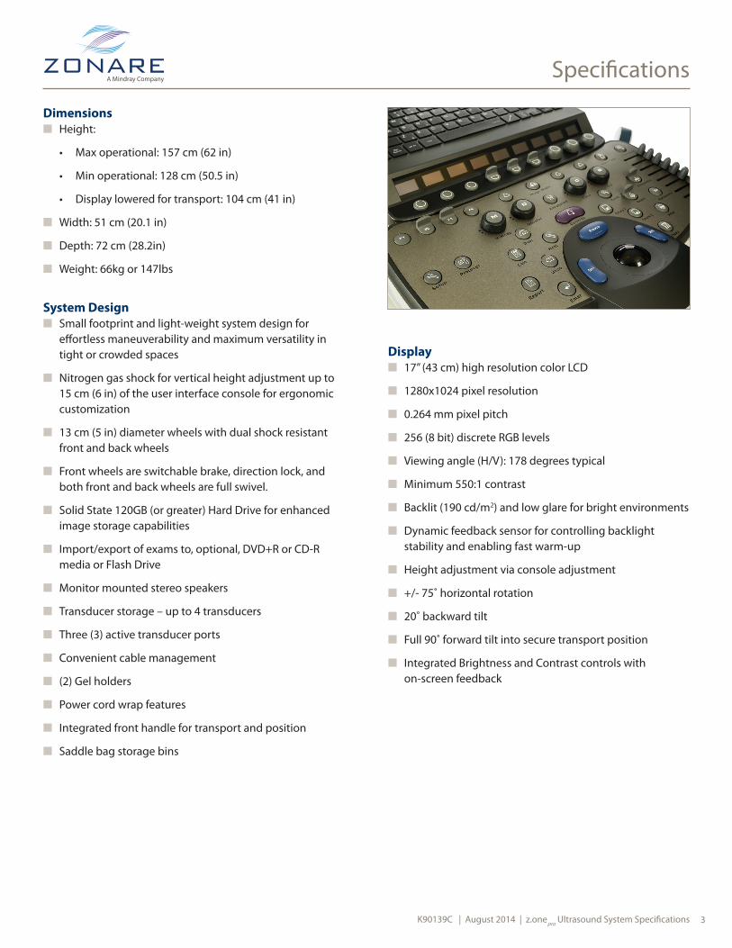
Dimensionsn Height:
• Max operational: 157 cm (62 in)
• Min operational: 128 cm (50.5 in)
• Display lowered for transport: 104 cm (41 in)
n Width: 51 cm (20.1 in)
n Depth: 72 cm (28.2in)
n Weight: 66kg or 147lbs
System Designn Small footprint and light-weight system design for
effortless maneuverability and maximum versatility in tight or crowded spaces
n Nitrogen gas shock for vertical height adjustment up to 15 cm (6 in) of the user interface console for ergonomic customization
n 13 cm (5 in) diameter wheels with dual shock resistant front and back wheels
n Front wheels are switchable brake, direction lock, and both front and back wheels are full swivel.
n Solid State 120GB (or greater) Hard Drive for enhanced image storage capabilities
n Import/export of exams to, optional, DVD+R or CD-R media or Flash Drive
n Monitor mounted stereo speakers
n Transducer storage – up to 4 transducers
n Three (3) active transducer ports
n Convenient cable management
n (2) Gel holders
n Power cord wrap features
n Integrated front handle for transport and position
n Saddle bag storage bins
Displayn 17” (43 cm) high resolution color LCD
n 1280x1024 pixel resolution
n 0.264 mm pixel pitch
n 256 (8 bit) discrete RGB levels
n Viewing angle (H/V): 178 degrees typical
n Minimum 550:1 contrast
n Backlit (190 cd/m2) and low glare for bright environments
n Dynamic feedback sensor for controlling backlight stability and enabling fast warm-up
n Height adjustment via console adjustment
n +/- 75˚ horizontal rotation
n 20˚ backward tilt
n Full 90˚ forward tilt into secure transport position
n Integrated Brightness and Contrast controls with on-screen feedback

Specifications
K90139C | August 2014 | z.onepro Ultrasound System Specifications4
User Interfacen Streamlined keyboard layout for best user ergonomics
n Optional special procedures UI with fewer hard keys *
n Home base design for easy access to major modes
n Full size, backlit QWERTY keyboard with non-English accents and characters
n OLEDs display customized controls for selected imaging modes provides for a less cluttered keyboard
n Context-sensitive backlit keys
n (8) DGC slide potentiometers with 45 mm travel and +/- 20dB Gain
n (4) User-programmable Function keys
• Unassigned • Full Image Display• Archive • Hide Pt. Bar• Auto trace • Image Width• Body Patterns • Lt/Rt Invert• B-Mode • Microphone• B-Steer • Power Dop• Bx Guide • Presets• Compounding • Print• Contrast • Protocol• Cursor • Record• Custom Preset • Remove Data Fields• d-PDI • Review• Display Format • Shutdown• Dual • Stpwatch Phse/Reset• ECG • Stpwatch Start/Stop• Elastography • Simul Dual• Ext. Sync • Transducer• Freeze • Up/Down Invert
n (3) User-programmable Mode keys
• Unassigned • d-PDI• 3D • Elastography• Auto trace • Presets• Aux CW • Power Doppler• Contrast • TDI• CW • Transducer
n Context-sensitive onscreen menu
n 38mm diameter trackball
* Option is only available for sale in specific countries. Please contact your local representative for availability.
K90139C | August 2014 | z.onepro Ultrasound System Specifications 5
Transducers
New transducer technology, wide bandwidth imaging, and multiple frequency imaging with an expanded range of frequencies including Compound Harmonics. These features provide:
n Increased sensitivity and resolution n More clinical information and expanded applications
The transducers are lightweight and ergonomically designed to offer easier imaging access, increased operator comfort, and greater overall clinical impact across all patient types.
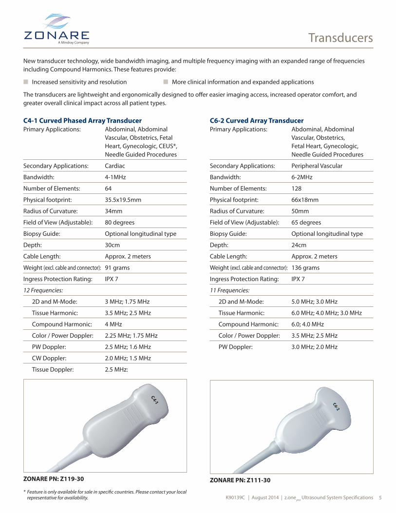
C4-1 Curved Phased Array TransducerPrimary Applications: Abdominal, Abdominal
Vascular, Obstetrics, Fetal Heart, Gynecologic, CEUS*, Needle Guided Procedures
Secondary Applications: Cardiac
Bandwidth: 4-1MHz
Number of Elements: 64
Physical footprint: 35.5x19.5mm
Radius of Curvature: 34mm
Field of View (Adjustable): 80 degrees
Biopsy Guide: Optional longitudinal type
Depth: 30cm
Cable Length: Approx. 2 meters
Weight (excl. cable and connector): 91 grams
Ingress Protection Rating: IPX 7
12 Frequencies:
2D and M-Mode: 3 MHz; 1.75 MHz
Tissue Harmonic: 3.5 MHz; 2.5 MHz
Compound Harmonic: 4 MHz
Color / Power Doppler: 2.25 MHz; 1.75 MHz
PW Doppler: 2.5 MHz; 1.6 MHz
CW Doppler: 2.0 MHz; 1.5 MHz
Tissue Doppler: 2.5 MHz:
ZONARE PN: Z119-30 ZONARE PN: Z111-30
C6-2 Curved Array TransducerPrimary Applications: Abdominal, Abdominal
Vascular, Obstetrics, Fetal Heart, Gynecologic, Needle Guided Procedures
Secondary Applications: Peripheral Vascular
Bandwidth: 6-2MHz
Number of Elements: 128
Physical footprint: 66x18mm
Radius of Curvature: 50mm
Field of View (Adjustable): 65 degrees
Biopsy Guide: Optional longitudinal type
Depth: 24cm
Cable Length: Approx. 2 meters
Weight (excl. cable and connector): 136 grams
Ingress Protection Rating: IPX 7
11 Frequencies:
2D and M-Mode: 5.0 MHz; 3.0 MHz
Tissue Harmonic: 6.0 MHz; 4.0 MHz; 3.0 MHz
Compound Harmonic: 6.0; 4.0 MHz
Color / Power Doppler: 3.5 MHz; 2.5 MHz
PW Doppler: 3.0 MHz; 2.0 MHz
* Feature is only available for sale in specific countries. Please contact your local representative for availability.

K90139C | August 2014 | z.onepro Ultrasound System Specifications6
Transducers
C9-3 Curved Array TransducerPrimary Applications: Abdominal, Abdominal
Vascular, OB, Pediatric/Small Adult Abdomen
Secondary Applications: Peripheral Vascular
Bandwidth: 9-3MHz
Number of Elements: 128
Physical footprint: 46x14mm
Radius of Curvature: 33mm
Field of View (Adjustable): 67 degrees
Biopsy Guide: Optional longitudinal type
Depth: 18cm
Cable Length: Approx. 2 meters
Weight (excl. cable and connector): 91 grams
Ingress Protection Rating: IPX 7
13 Frequencies:
2D and M-Mode: 7.0 MHz; 5.5 MHz; 3.5 MHz
Tissue Harmonic: 8.0 MHz; 7.0 MHz; 5.0 MHz
Compound Harmonic: 8.0 MHz; 6.0 MHz
Compound: 7.0 MHz
Color / Power Doppler: 5.0 MHz; 3.5 MHz
PW Doppler: 5.0 MHz; 3.1 MHz
ZONARE PN: Z109-30
C10-3 Curved Phased Array TransducerPrimary Applications: Neonatal Heart, Neonatal
Head, Neonatal Abdominal, Pediatric Echo, Pediatric Abdominal, Ocular
Secondary Applications: Peripheral Vascular
Bandwidth: 10-3 MHz
Number of Elements: 64
Physical footprint: ~16 mm
Radius of Curvature: 16 mm
Field of View: 80 degrees
Biopsy Guide: None available
Virtual Apex Array: N/A
Depth: 14 cm
Cable Length: Approx. 2 meters
Weight (excl. cable and connector): 45 grams
Ingress Protection Rating: IPX 7
18 Frequencies:
2D and M-Mode: 7.5 MHz; 6.0 MHz; 4.0 MHz
Tissue Harmonic: 9.0 MHz; 7.0 MHz
Compound Harmonic: 9.0 MHz; 7.0 MHz
Color / Power Doppler: 6.2 MHz; 5.2 MHz; 4.5 MHz; 3.5 MHz
PW Doppler: 5.0 MHz; 3.5 MHz; 3.2MHz
CW Doppler: 6.0 MHz; 3.6 MHz; 3.2MHz
Tissue Doppler: 7.0 MHz
ZONARE PN: Z124-30

K90139C | August 2014 | z.onepro Ultrasound System Specifications 7
Transducers
E9-4 Endocavity TransducerPrimary Applications: Endovaginal including
First Trimester Obstetrics, Gyn (uterus, ovaries)
Secondary Applications: Endorectal including Prostate, Rectal Wall Needle Guided Procedures
Bandwidth: 9-4 MHz
Number of Elements: 128
Physical footprint: 23x20mm
Radius of Curvature: 12mm
Field of View: 135 degrees
Biopsy Guide: 1. Optional disposable 2. Optional re-useable
Virtual Apex Array: N/A
Depth: 14cm
Cable Length: Approx. 2 meters
Weight (excl. cable and connector): 159 grams
Ingress Protection Rating: IPX 7
9 Frequencies:
2D and M-Mode: 8.0 MHz; 5.0 MHz; 4.0 MHz
Tissue Harmonic: 7.5 MHz; 6.5 MHz
Compound Harmonic: 7.0 MHz
Compound: 7.0 MHz
Color Doppler: 4.5 MHz
PW Doppler: 4.5 MHz
ZONARE PN: Z103-30 ZONARE PN: Z106-30
L8-3 Linear Array TransducerPrimary Applications: Peripheral Vascular,
Needle Guided Procedures
Secondary Applications: Pediatric Hips, Technically Difficult Small Parts
Bandwidth: 8-3 MHz
Number of Elements: 128
Physical footprint: 48x11mm
Field of View (Adjustable): 38mm
Biopsy Guide: Optional transverse type
Virtual Apex Array: Wider Field of View
Depth: 10cm
Cable Length: Approx. 2 meters
Weight (excl. cable and connector): 113 grams
Ingress Protection Rating: IPX 7
12 Frequencies:
2D and M-Mode: 7.0 MHz; 5.5 MHz; 4.0 MHz
Tissue Harmonic: 7.5 MHz; 6.0 MHz
Compound Harmonic: 7.0 MHz
Spatial Harmonics: 7.0 MHz
Compound Spatial Harmonics: 7.0 MHz
Color / Power Doppler: 5.5 MHz; 4.0 MHz
PW Doppler: 4.6 MHz; 3.5 MHz
K90139C | August 2014 | z.onepro Ultrasound System Specifications8
Transducers
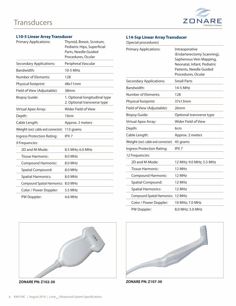
L10-5 Linear Array TransducerPrimary Applications: Thyroid, Breast, Scrotum,
Pediatric Hips, Superficial Parts, Needle Guided Procedures, Ocular
Secondary Applications: Peripheral Vascular
Bandwidth: 10-5 MHz
Number of Elements: 128
Physical footprint: 48x11mm
Field of View (Adjustable): 38mm
Biopsy Guide: 1. Optional longitudinal type 2. Optional transverse type
Virtual Apex Array: Wider Field of View
Depth: 10cm
Cable Length: Approx. 2 meters
Weight (excl. cable and connector): 113 grams
Ingress Protection Rating: IPX 7
9 Frequencies:
2D and M-Mode: 8.5 MHz; 6.0 MHz
Tissue Harmonic: 8.0 MHz
Compound Harmonic: 8.0 MHz
Spatial Compound: 8.0 MHz
Spatial Harmonics: 8.0 MHz
Compound Spatial Harmonics: 8.0 MHz
Color / Power Doppler: 5.5 MHz
PW Doppler: 4.6 MHz
ZONARE PN: Z102-30
L14-5sp Linear Array Transducer (Special procedures)
Primary Applications: Intraoperative (Endarterectomy Scanning), Saphenous Vein Mapping, Neonatal, Infant, Pediatric Patients, Needle Guided Procedures, Ocular
Secondary Applications: Small Parts
Bandwidth: 14-5 MHz
Number of Elements: 128
Physical footprint: 37x13mm
Field of View (Adjustable): 26mm
Biopsy Guide: Optional transverse type
Virtual Apex Array: Wider Field of View
Depth: 6cm
Cable Length: Approx. 2 meters
Weight (excl. cable and connector): 45 grams
Ingress Protection Rating: IPX 7
12 Frequencies:
2D and M-Mode: 12 MHz; 9.0 MHz; 5.5 MHz
Tissue Harmonic: 12 MHz
Compound Harmonic: 12 MHz
Spatial Compound: 12 MHz
Spatial Harmonics: 12 MHz
Compound Spatial Harmonics: 12 MHz
Color / Power Doppler: 10 MHz; 7.0 MHz
PW Doppler: 8.0 MHz; 5.0 MHz
ZONARE PN: Z107-30

K90139C | August 2014 | z.onepro Ultrasound System Specifications 9
Transducers
L14-5w Linear Array Transducer(Wide field-of-view)
Primary Applications: Small Parts including Breast, Thyroid, Testes, Superficial Anatomy, Needle Guided Procedures, Ocular
Secondary Applications: Superficial Vascular
Bandwidth: 14-5 MHz
Number of Elements: 192
Physical footprint: 62x10mm
Field of View (Adjustable): 55mm
Biopsy Guide: 1. Optional longitudinal type 2. Optional transverse type
Virtual Apex Array: Wider Field of View
Depth: 10cm
Cable Length: Approx. 2 meters
Weight (excl. cable and connector): 91 grams
Ingress Protection Rating: IPX 7
12 Frequencies:
2D and M-Mode: 12 MHz; 7.0 MHz
Tissue Harmonic: 12 MHz
Compound Harmonic: 12 MHz
Spatial Compound: 12 MHz
Spatial Harmonics: 12 MHz
Compound Spatial Harmonics: 12 MHz
Color / Power Doppler: 9.0MHz; 7.0MHz; 5.5 MHz
PW Doppler: 7.0 MHz; 5.0 MHz
ZONARE PN: Z110-30 ZONARE PN: Z108-30
P4-1c Phased Array TransducerPrimary Applications: Cardiac, Transcranial Imaging/
Doppler, Trauma (FAST Exams), Deep Abdominal, Abdominal Vascular, Renal, Aorta
Secondary Applications: Technically Difficult: Obstetrics, Fetal Heart
Bandwidth: 4-1 MHz
Number of Elements: 64
Physical footprint: 27x20mm
Field of View (Adjustable): 84 degrees
Biopsy Guide: N/A
Virtual Apex Array: N/A
Depth: 30cm
Cable Length: Approx. 2 meters
Weight (excl. cable and connector): 91 grams
Ingress Protection Rating: IPX 7
20 Frequencies:
2D and M-Mode: 4.0 MHz; 3.0 MHz; 2.5 MHz; 2.0 MHz
Tissue Harmonic: 5.0 MHz; 4.0 MHz; 3.5 MHz; 3.0 MHz
Compound Harmonic: 4.0 MHz
Compound: 3.0 MHz
Color / Power Doppler: 3.0 MHz; 2.5 MHz; 2.0 MHz
PW Doppler: 3.2 MHz; 2.0 MHz; 1.8 MHz
CW Doppler: 3.0 MHz; 2.4 MHz; 2.0 MHz
Tissue Doppler: 3.5 MHz

K90139C | August 2014 | z.onepro Ultrasound System Specifications10
Transducers
ZONARE PN: Z116-00
ZONARE PN: Z118-00
A2CW Transducer *Primary Applications: Adult, Adolescent
Echocardiography
Secondary Applications: N/A
Center Frequency: 2.0 MHz
Bandwidth: > 5%
Number of Elements: 2
Physical footprint: 17mm diameter
Note: Requires Echocardiography option (CW and ECG) **
A5CW Transducer *Primary Applications: Peripheral Vascular
Secondary Applications: N/A
Center Frequency: 5.0 MHz
Bandwidth: > 10%
Number of Elements: 2
Physical footprint: 11mm diameter
Note: Requires Echocardiography option (CW and ECG) **
* Transducer is only available for sale in specific countries. Please contact your local representative for availability.
P8-3TEE Phased Array TransducerPrimary Applications: Adult Transesophageal
Echocardiography
Secondary Applications: N/A
Bandwidth: 8-3 MHz
Number of Elements: 64
Physical footprint: 14x10mm
Field of View (Adjustable): 90 degrees
Biopsy Guide: N/A
Depth: 18cm
Cable Length: Approx. 2 meters
Ingress Protection Rating: IPX 7
8 Frequencies:
2D and M-Mode: 7MHz, 6MHz, 5MHz
Color / Power Doppler: 3.6MHz, 2.8MHz
PW Doppler: 3.1 MHz; 2.8MHz
CW Doppler: 3.0 MHz
Tissue Doppler: 3.5 MHz
Note: Requires Echocardiography option **
ZONARE PN: Z115-00
** Feature is only available for sale in specific countries. Please contact your local representative for availability.

K90139C | August 2014 | z.onepro Ultrasound System Specifications 11
Transducers
Transducer Performance Data at -6dB
Transducer Axial Resolution Lateral Resolution Elevation Focus (mm) (mm) (mm)
C4-1 0.6 1.6 80
C6-2 0.4 0.9 70
C9-3 0.3 0.7 45
C10-3 0.2 0.8 35
E9-4 0.3 0.8 35
L8-3 0.2 0.5 25
L10-5 0.2 0.4 18
L14-5sp 0.2* 0.3 12.5
L14-5w 0.1 0.3 20
P4-1c 0.4 2.4 75
P8-3TEE 0.3 1.5 50

K90139C | August 2014 | z.onepro Ultrasound System Specifications 12
Imaging Modes
B-Mode
Live Imaging Controls
n Auto-Opt with ZST – Adjust Gain/DGC and Sound Speed Correction
n Depth – up to 30cm (see transducers)
n Frequency change
n Tissue Harmonic Imaging
n Acoustic Zoom
n PW Doppler Cursor
n Acoustic Output
n Dual Imaging
n B-Steer
Live & Retrospective Imaging Controls
n Auto-Opt with ZST – Gain/DGC
n 2D Gain/DGC
n Display Zoom (pan/zoom on frozen image up to 4X mag)
n Grayscale Map
n B Mode Tints
n Dynamic Range
n Persistence
n Edge Enhancement/Smoothing
n Up/Down Invert
n Left/Right Invert
M-Mode
Live Imaging Controls
n Depth
n Frequency
n Tissue Harmonic Imaging
n Cursor Position
n Acoustic Zoom
n Display Zoom (Zoom on Frozen Image)
n Acoustic Output
Live & Retrospective Imaging Controls
n M Gain/DGC
n Sweep Speed
n Strip Tints
n Display Format (Two B reference image sizes and full screen strip)
n M Map
n M Dynamic Range
n Persistence
n Edge Enhancement/Smoothing
n Up/Down Invert
n Left/Right Invert
K90139C | August 2014 | z.onepro Ultrasound System Specifications13
Imaging Modes
Color and Power Doppler
Live Imaging Controls
n ROI Position/Size
n ROI Size – up to full screen
n Velocity Scale
n Wall Filter
n Flash Suppression
n Steering Angle (linear transducers only)
n Acoustic Zoom
n Acoustic Output
n Dual Imaging
Live & Retrospective Imaging Controls
n Color Gain
n Display Zoom (Zoom on Frozen Image)
n Color Map
n Edge Enhancement/Smoothing
n Baseline Shift
n Invert
n Persistence
Pulsed Wave DopplerLive Imaging Controls
n Triplex Mode (simultaneous live 2D, Color, PW Doppler strip)
n Update (quick switch between live 2D or Color and live PW Doppler strip)
n TDI (Tissue Doppler Imaging)
n Cursor Position
n Gate Size 1-15 mm
n Steering Angle in 1 degree increments
n Velocity Scale, Baseline, Filter
n Acoustic Power
n Sweep Speed
Live & Retrospective Imaging Controls
n PW Gain
n Angle Correct – manual 0 to +/-76 degrees
n Angle Correct – ‘Quick 60’
n Baseline Shift
n Wall Filter
n Invert
n Strip Tints
n Display Format (1/2; 1/3; full screen strip)
n Sweep Speed (5 speeds)
n PW Map
n PW Dynamic Range
n Audio Volume
K90139C | August 2014 | z.onepro Ultrasound System Specifications 14
Imaging Modes
Continuous Wave Doppler *
Live Imaging Controls
n Update
n Cursor Position
n Velocity Scale, Baseline Filter
n Acoustic Power
n Sweep Speed (5 speeds)
Live & Retrospective Imaging Controls
n CW Gain
n Dynamic Range
n CW Map
n Velocity Scale
n Baseline Shift
n Wall Filter
n Focal Depth Position
n Display Format (1/2; 1/3; full screen strip)
n Acoustic Power
n Sweep Speed (5 speeds)
Dual Imagingn Available for all imaging transducers
n Displays two images side-by-side
n Ability to display two frozen, one active/one frozen, or two active images
n Allows Left/Right switching of the active side of the display, while automatically freezing the other side
n Archiving while in dual image will store both images
n All measurements and calculations can be performed across combined dual images
n Annotations and body markers
Simultaneous Dual Imagingn Available for all imaging transducers
n Displays two images (same or different modes), side-by-side in real-time
n Simultaneous display of Color Velocity & Power Doppler Imaging
n Allows Left/Right switching of the active side of the display: active side allows optimization, mode changes, while both sides are live simultaneously
n Retrospective processing adjustments affect both images at the same time
n Real-time dual image adjustments
n Dual image archiving will store both images in single frame or loop format
Imaging Mode Combinationsn B+CD/PD
n B+M
n B+PW (real-time duplex)
n B+CD/PD+PW (real-time Triplex)
n B+CW (update mode)
n B+CD+CW (update mode)
* Feature is only available for sale in specific countries. Please contact your local representative for availability.
K90139C | August 2014 | z.onepro Ultrasound System Specifications15
Imaging Modes
Imaging Formatsn Convex
n Linear
n Phased
n Micro-Convex
n Virtual Apex Array
n Curved Phased
n Image Width – user selectable width and positioning
Auto-Opt with ZST n Available on all imaging transducers
n Instantly equalizes tissue gain during live or frozen/Cine images
n Automatically measures the various sound speeds within the body habitus and compensates to determine the most accurate
Acoustic Zoomn Available in all imaging modes
Display Zoomn Available for all imaging transducers
n From 1.25X to 4X magnification on frozen image, with panning capabilities
n Precise measurement of small structures made easier
Contrast Enhanced Ultrasound* (CEUS)Contrast Imaging is a harmonic detection mode specifically designed to enhance signals from ultrasound contrast agents similar to 2D (B-Mode), a suite of additional optimization controls are provided to further enhance contrast agent imaging performance.
Available as option on
• C4-1
Display Formats
n Single Image display
n Simultaneous Dual Image display
n Non-Simultaneous Dual Image display
Stopwatch Display
n Elapsed time: The duration of time that passes after the stopwatch is started, usually the time elapsed from the injection of the contrast agent.
n Phase time: The duration of time between the start and stop of a phase. Four phase timers are supported.
n Cine elapsed time: This is the elapsed and phase time for the displayed frame/strip within the Cine memory.
Contrast Imaging User Controls
n Frame rate
n Power
n 2D Gain/DGC
n Contrast Gain
n Depth
n Frequency
n Display Zoom (pan/zoom on frozen image up to 4X mag)
n Dynamic Range
n Edge Enhancement/Smoothing
n Persistence
n Maps
n L/R Invert
n U/D Invert
n Auto-Opt with ZST – Gain/DGC
n Tints
n Compounding
n Harmonics
* CEUS is only available for sale in specific countries. Please contact your local representative for availability.
K90139C | August 2014 | z.onepro Ultrasound System Specifications 16
Specifications
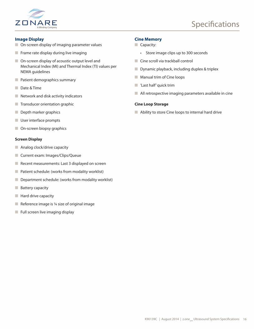
Image Displayn On-screen display of imaging parameter values
n Frame rate display during live imaging
n On-screen display of acoustic output level and Mechanical Index (MI) and Thermal Index (TI) values per NEMA guidelines
n Patient demographics summary
n Date & Time
n Network and disk activity indicators
n Transducer orientation graphic
n Depth marker graphics
n User interface prompts
n On-screen biopsy graphics
Screen Display
n Analog clock/drive capacity
n Current exam: Images/Clips/Queue
n Recent measurements: Last 3 displayed on screen
n Patient schedule: (works from modality worklist)
n Department schedule: (works from modality worklist)
n Battery capacity
n Hard drive capacity
n Reference image is ¼ size of original image
n Full screen live imaging display
Cine Memoryn Capacity:
• Store image clips up to 300 seconds
n Cine scroll via trackball control
n Dynamic playback, including duplex & triplex
n Manual trim of Cine loops
n ‘Last half’ quick trim
n All retrospective imaging parameters available in cine
Cine Loop Storage
n Ability to store Cine loops to internal hard drive
Specifications
K90139C | August 2014 | z.onepro Ultrasound System Specifications17
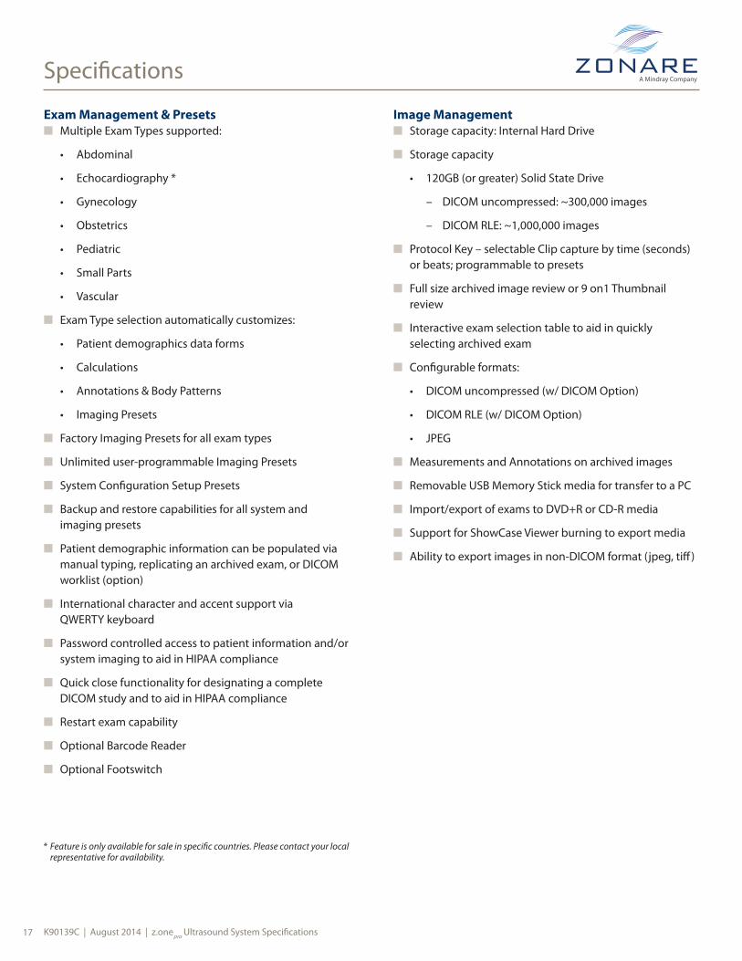
Exam Management & Presetsn Multiple Exam Types supported:
• Abdominal
• Echocardiography *
• Gynecology
• Obstetrics
• Pediatric
• Small Parts
• Vascular
n Exam Type selection automatically customizes:
• Patient demographics data forms
• Calculations
• Annotations & Body Patterns
• Imaging Presets
n Factory Imaging Presets for all exam types
n Unlimited user-programmable Imaging Presets
n System Configuration Setup Presets
n Backup and restore capabilities for all system and imaging presets
n Patient demographic information can be populated via manual typing, replicating an archived exam, or DICOM worklist (option)
n International character and accent support via QWERTY keyboard
n Password controlled access to patient information and/or system imaging to aid in HIPAA compliance
n Quick close functionality for designating a complete DICOM study and to aid in HIPAA compliance
n Restart exam capability
n Optional Barcode Reader
n Optional Footswitch
Image Managementn Storage capacity: Internal Hard Drive
n Storage capacity
• 120GB (or greater) Solid State Drive
– DICOM uncompressed: ~300,000 images
– DICOM RLE: ~1,000,000 images
n Protocol Key – selectable Clip capture by time (seconds) or beats; programmable to presets
n Full size archived image review or 9 on1 Thumbnail review
n Interactive exam selection table to aid in quickly selecting archived exam
n Configurable formats:
• DICOM uncompressed (w/ DICOM Option)
• DICOM RLE (w/ DICOM Option)
• JPEG
n Measurements and Annotations on archived images
n Removable USB Memory Stick media for transfer to a PC
n Import/export of exams to DVD+R or CD-R media
n Support for ShowCase Viewer burning to export media
n Ability to export images in non-DICOM format (jpeg, tiff)
* Feature is only available for sale in specific countries. Please contact your local representative for availability.

K90139C | August 2014 | z.onepro Ultrasound System Specifications 18
Specifications
Connectivity
Inputs & Outputs
n HDMI
n USB 4 ports
n SATA connection
n Ethernet (10/100 Base T)
n Wireless capable via optional bridge
DICOM Option
n Verify Service Class
n Print Service Class
n Store Service Class
n Basic Modality Worklist Query
n In Progress
n Queue for mobile exams with autosend feature upon network connection
n Exam push to DICOM from onboard archive
n Variable compression settings
n Configuration Setup
n Structured Reporting – Vascular
n Structured Reporting – Cardiac
n Structured Reporting – OB / GYN
DICOM Removable Media
n USB Memory Stick
n DVD+R or CD-R media, optional
Optional Peripheralsn Sony UP-D711MD B/W Thermal Printer
• Printer and Mounting Kit (Bracket): ZONARE PN: Z417-00
• Mounting Kit only (Bracket): ZONARE PN: Z418-00
n Datalogic Barcode Reader
• Reader: ZONARE PN: Z373-00
User Editable Worksheets OptionOffline created worksheets can be imported into the system. There is no capability to modify the worksheet template once uploaded onto system. Data and worksheet template is exported in XML format.

K90139C | August 2014 | z.onepro Ultrasound System Specifications19
Measurements & Analysis
Auto-Dop Trace (Automatic Doppler Measurements)n Automatic tracing of spectral Doppler waveform
n Derived from heart cycle or time
n Independent of PW gain
n Integrates data into Calc Package
n Requires Auto-Opt with ZST option
n Available measurements: RI, PI, Accel, S/D, HR, AT, TAMX, TAMN
General Capabilitiesn Interactive worksheets for reviewing and editing
in-process results
n Formatted reports for Obstetrics, Gynecology, Echocardiography and Vascular
n Multiple measurements can be used to derive final results (average, last, max)
n Dynamic display of measurement results
n Reposition caliper
n Configurable, customizable calc menus
n Export data feature to third party software
n Export serial data
Generic B-Mode Measurements / Calculationsn Distance: up to 4 diameters
n Circumference/Area (Ellipse or Trace)
n Volume (3 distance)
n % Stenosis (Area or Diameter)
n Depth (from transducer face)
Generic M-Mode Measurements/Calculationsn M distance
n Heart Rate (bpm – beats per minute)
n AV Plane
Generic PW Measurements/Calculations n Velocity
n Velocity Pairs
n RI (Resistive Index)
n PI (Pulsatility Index)
n Acceleration / Slope
n S/D (Systolic / Diastolic ratio)
n A:B (Generic velocity ratio)
n HR (Heart Rate)
n AT (Acceleration Time)
n TAMX (Time Average Max)
n TAMN (Time Average Mean)
n Volume Flow
K90139C | August 2014 | z.onepro Ultrasound System Specifications 20
Measurements & Analysis
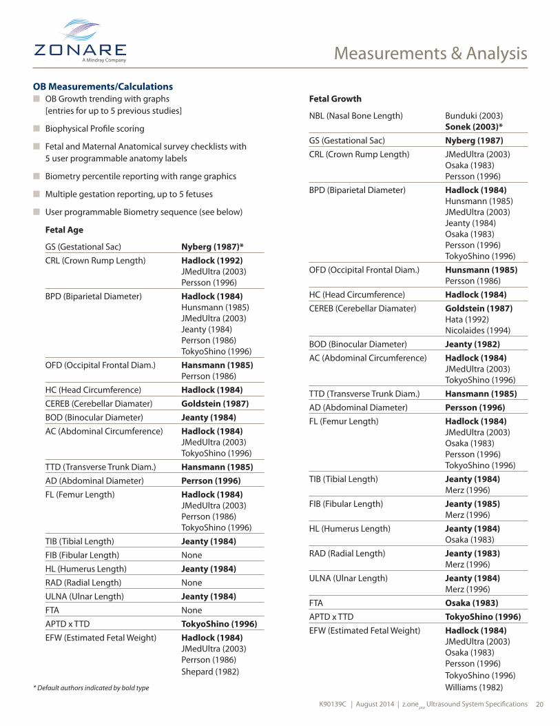
OB Measurements/Calculationsn OB Growth trending with graphs
[entries for up to 5 previous studies]
n Biophysical Profile scoring
n Fetal and Maternal Anatomical survey checklists with 5 user programmable anatomy labels
n Biometry percentile reporting with range graphics
n Multiple gestation reporting, up to 5 fetuses
n User programmable Biometry sequence (see below)
Fetal Age
GS (Gestational Sac) Nyberg (1987)*CRL (Crown Rump Length) Hadlock (1992) JMedUltra (2003) Persson (1996)BPD (Biparietal Diameter) Hadlock (1984) Hunsmann (1985) JMedUltra (2003) Jeanty (1984) Perrson (1986) TokyoShino (1996)OFD (Occipital Frontal Diam.) Hansmann (1985) Perrson (1986)HC (Head Circumference) Hadlock (1984)CEREB (Cerebellar Diamater) Goldstein (1987)BOD (Binocular Diameter) Jeanty (1984)AC (Abdominal Circumference) Hadlock (1984) JMedUltra (2003) TokyoShino (1996)TTD (Transverse Trunk Diam.) Hansmann (1985)AD (Abdominal Diameter) Perrson (1996)FL (Femur Length) Hadlock (1984) JMedUltra (2003) Perrson (1986) TokyoShino (1996)TIB (Tibial Length) Jeanty (1984)FIB (Fibular Length) NoneHL (Humerus Length) Jeanty (1984)RAD (Radial Length) NoneULNA (Ulnar Length) Jeanty (1984)FTA NoneAPTD x TTD TokyoShino (1996)EFW (Estimated Fetal Weight) Hadlock (1984) JMedUltra (2003) Perrson (1986) Shepard (1982)
Fetal Growth
NBL (Nasal Bone Length) Bunduki (2003) Sonek (2003)*GS (Gestational Sac) Nyberg (1987)CRL (Crown Rump Length) JMedUltra (2003) Osaka (1983) Persson (1996)BPD (Biparietal Diameter) Hadlock (1984) Hunsmann (1985) JMedUltra (2003) Jeanty (1984) Osaka (1983) Persson (1996) TokyoShino (1996)OFD (Occipital Frontal Diam.) Hunsmann (1985) Persson (1986)HC (Head Circumference) Hadlock (1984)CEREB (Cerebellar Diamater) Goldstein (1987) Hata (1992) Nicolaides (1994)BOD (Binocular Diameter) Jeanty (1982)AC (Abdominal Circumference) Hadlock (1984) JMedUltra (2003) TokyoShino (1996)TTD (Transverse Trunk Diam.) Hansmann (1985)AD (Abdominal Diameter) Persson (1996)FL (Femur Length) Hadlock (1984) JMedUltra (2003) Osaka (1983) Persson (1996) TokyoShino (1996)TIB (Tibial Length) Jeanty (1984) Merz (1996)FIB (Fibular Length) Jeanty (1985) Merz (1996)HL (Humerus Length) Jeanty (1984) Osaka (1983)RAD (Radial Length) Jeanty (1983) Merz (1996)ULNA (Ulnar Length) Jeanty (1984) Merz (1996)FTA Osaka (1983)APTD x TTD TokyoShino (1996)EFW (Estimated Fetal Weight) Hadlock (1984) JMedUltra (2003) Osaka (1983) Persson (1996) TokyoShino (1996) Williams (1982)* Default authors indicated by bold type
K90139C | August 2014 | z.onepro Ultrasound System Specifications21
Measurements & Analysis
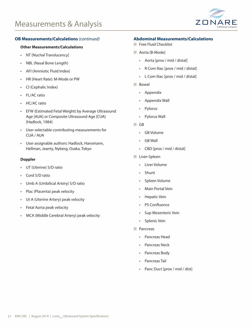
OB Measurements/Calculations (continued)
Other Measurements/Calculations
• NT (Nuchal Translucency]
• NBL (Nasal Bone Length)
• AFI (Amniotic Fluid Index)
• HR (Heart Rate): M-Mode or PW
• CI (Cephalic Index)
• FL/AC ratio
• HC/AC ratio
• EFW (Estimated Fetal Weight) by Average Ultrasound Age [AUA] or Composite Ultrasound Age [CUA] [Hadlock, 1984]
• User selectable contributing measurements for CUA / AUA
• User assignable authors: Hadlock, Hansmann, Hellman, Jeanty, Nyberg, Osaka, Tokyo
Doppler
• UT (Uterine) S/D ratio
• Cord S/D ratio
• Umb A (Umbilical Artery) S/D ratio
• Plac (Placenta) peak velocity
• Ut A (Uterine Artery) peak velocity
• Fetal Aorta peak velocity
• MCA (Middle Cerebral Artery) peak velocity
Abdominal Measurements/Calculationsn Free Fluid Checklist
n Aorta [B-Mode]
• Aorta [prox / mid / distal]
• R Com Iliac [prox / mid / distal]
• L Com Iliac [prox / mid / distal]
n Bowel
• Appendix
• Appendix Wall
• Pylorus
• Pylorus Wall
n GB
• GB Volume
• GB Wall
• CBD [prox / mid / distal]
n Liver-Spleen
• Liver Volume
• Shunt
• Spleen Volume
• Main Portal Vein
• Hepatic Vein
• PS Confluence
• Sup Mesenteric Vein
• Splenic Vein
n Pancreas
• Pancreas Head
• Pancreas Neck
• Pancreas Body
• Pancreas Tail
• Panc Duct [prox / mid / dist]

K90139C | August 2014 | z.onepro Ultrasound System Specifications 22
Measurements & Analysis
Abdominal Measurements/Calculations (continued)n Renal
• Right / Left
• Renal Volume
• Renal Vein
• Adrenal Volume
• Ureter [r plv / prox / mid / dist]
n Bladder Volume
Abdominal Doppler
n Aorta
• Aorta [prox / renl / mid / distal]
• Celiac Artery [1 /2]
• SMA [1 /2]
• CIA (Common Iliac Artery)
• EIA (External Iliac Artery)
• Hepatic Artery
• Splenic Artery
• GDA
• IMA
n Renal
• Right / Left
• Renal RI [orig / prox / mid / distal]
• Renal AT [orig / prox / mid / distal]
• Ren A1 RI [orig / prox / mid / distal]
• Ren A1 AT [orig / prox / mid /distal]
• Ren A2 RI [orig / prox / mid / distal]
• Ren A2 AT [orig / prox / mid /distal]
• Hilum [sup / mid / inf ]
• Interlob RI [sup / mid / inf ]
• Interlob AT [sup / mid / inf ]
• Arcuate RI [sup / mid / inf ]
• Arcuate AT [sup / mid / inf ]
Pediatric Hip Measurementn Right / Left
n Hip Angle [Baseline / Bony / Cart]
• Alpha / Beta angles
B-Mode (Fetal Heart)n Asc Aorta diameter
n MPA (Main Pulmonary Artery) diameter
n Duct Art (Ductus Arteriosis) diameter
n LA (Left Atrium) distance
n RA (Right Atrium) distance
n RV (Right Ventricle) Wall distance (S/D)
n RVID (Right Ventricle Internal Diameter) (S/D)
n IVS (Interventricular Septum) distance (S/D)
n LVID (Left Ventricle Internal Diameter) (S/D)
n LVPW (Left Ventricle Posterior Wall) distance (S/D)
n Heart Circ (circumference)
n Thor Circ (thorax circumference)
PW Doppler (Fetal Heart)n MV (Mitral Valve) peak velocity (E/A)
n TV (Tricuspid Valve) peak velocity (E/A)
n Asc Aorta peak velocity
n Desc Aorta peak velocity
n MR (Mitral Regurgitation) peak velocity
n TR (Tricuspic Regurgitation) peak velocity
n MPA (Main Pulmonary Artery) peak velocity
n Duct Art (Ductus Arteriosis) peak velocity
n IVC (Inferior Vena Cava) peak velocity
n Duct Ven (Ductus venosis) peak velocity

K90139C | August 2014 | z.onepro Ultrasound System Specifications23
Measurements & Analysis
M-Mode (Fetal Heart)n RV (Right Ventricle) Wall (S/D)
n RVID (Right Ventricle Internal Diameter (S/D)
n IVS (Interventricular Septum) distance (S/D)
n LVID (Left Ventricle Internal Diameter) (S/D)
n LVPW (Left Ventricle Posterior Wall) distance (S/D)
GYN Measurements / Calculationsn Uterine Volume
n Endometrial Thickness
n Cervical Length
n Ovary Volume (Right & Left)
n Up to (10) Right + (10) Left Follicles: reports either volumes calculated from 1, 2, or 3 distances or average distance
n Ov RI (Ovarian RI, Right & Left)
n Ov PI (Ovarian PI, Right & Left)
n Ut RI (Uterine RI)
n Ut PI (Uterine PI)
Vascular Measurements / Calculations(R/L = Right and Left assignment)
(P/M/D = Proximal, Mid, and Distal assignment)
n % Stenosis (R/L)
Carotidn CCA (Common Carotid Artery: P/M/D, R/L, PSV/EDV)
n Bulb (R/L, PSV/EDV)
n ICA (Internal Carotid Artery: P/M/D, R/L, PSV/EDV)
n ECA (External Carotid Artery: R/L, PSV/EDV)
n Vertebral (R/L, PSV/EDV)
n ICA/CCA ratio (R/L)
n Subclavian (R/L, PSV/EDV)
n User programmable Carotid Sequence
Upper Extremity Arterial Calc (Right/Left) Stenosis, Diameter, PW Doppler, Report Pagen Subclavian
n Axillary
n Brachial (P/M/D)
n Radial (P/M/D)
n Ulnar (P/M/D)
n Graft Native Inflow
n Graft Prox Anastomosis Pre Velocity
n Graft Prox Anastomosis Max Velocity
n Graft Prox Anastomosis Post Velocity
n Graft (P/M/D)
n Graft Dist Anastomosis Pre Velocity
n Graft Dist Anastomosis Max Velocity
n Graft Dist Anastomosis Post Velocity
n Graft Native Outflow
n Graft Native Inflow Volume Flow
n Graft Prox Anastomosis Volume Flow
n Graft (P/M/D) Volume Flow
n Graft Dist Anastomosis Volume Flow
n Graft Native Outflow Volume Flow

K90139C | August 2014 | z.onepro Ultrasound System Specifications 24
Measurements & Analysis
Lower Extremity Arterial Calc (Right/Left) Stenosis, Diameter, PW Doppler, Report Pagen CIA (Common Iliac Artery)
n EIA (External Iliac Artery)
n CFA (Common Femoral Artery)
n PFA (Profunda Femoris Artery)
n FA (Femoral Artery) (P/M/D)
n Popliteal (P/M/D)
n ATA (Anterior Tibial Artery)
n Peroneal
n PTA (Posterial Tibial Artery)
n Dorsalis Pedis
n Graft Native Inflow
n Graft Prox Anastomosis Pre Velocity
n Graft Prox Anastomosis Max Velocity
n Graft Prox Anastomosis Post Velocity
n Graft (P/M/D)
n Graft Dist Anastomosis Pre Velocity
n Graft Dist Anastomosis Max Velocity
n Graft Dist Anastomosis Post Velocity
n Graft Native Outflow
n Graft Native Inflow Volume Flow
n Graft Prox Anastomosis Volume Flow
n Graft (P/M/D) Volume Flow
n Graft Dist Anastomosis Volume Flow
n Graft Native Outflow Volume Flow
Lower Extremity Venous Measurements Diameter, Checklists, Report Pagen Deep
• IVC (Inferior Vena Cava)
• CIV (Common Iliac Vein)
• EIV (External Iliac Vein)
• CFV (Common Femoral Vein)
• FV (Femoral Vein) (P/M/D)
• DFV (Deep Femoral Vein)
• Popliteal Vein
• PTV (Posterior Tibial Vein)
• Peroneal Vein
• ATV (Anterior Tibial Vein)
n Superficial
• SF Junction (Sapheno-Femoral)
• GSV (Greater Saphenous Vein)Thigh (P/M/D)
• GSV Knee
• GSV Calf (P/M/D)
• SP Junction (Sapheno-Peroneal)
• SSV (Small Saphenous) (P/M/D)
• AASV (Anterior Accessory Saphenous) (P/M/D)
• PASV (Posterior Accessory Saphenous)
n Reflux Time PW Doppler Calc
• Available with all Venous Lower Extremity Presets

K90139C | August 2014 | z.onepro Ultrasound System Specifications25
Measurements & Analysis
Upper Extremity Venous Measurements Diameter, Checklists, Report Pagen Deep
• Internal Jugular Vein
• Innominate Vein
• Subclavian Vein
• Axillary Vein
• Brachial Vein (P/M/D)
• Radial Vein
• Ulnar Vein
• Volar
n Superficial
• CA Junc
• Upper Cephalic (P/M/D)
• Lat Antecubital Vein
• Lower Cephalic Vein (P/M/D)
• BA Junc
• Upper Basilic Vein (P/M/D)
• Medial Antecubital Vein
• Lower Basilic Vein (P/M/D)
• Digital

K90139C | August 2014 | z.onepro Ultrasound System Specifications 26
Specifications
Annotation Package
Text Annotation
n Annotation on live, frozen, & archived images
n Selectable Insert and Overwrite modes
n User-programmable home position
n Selection, modification, & deletion of individual words
n Ability to reposition text groups
n Pre-programmed quick orientation keys:
• Right / Left
• Prox / Mid / Distal
• Long / Trans / Sagittal / Coronal
n (3) user programmable annotation libraries per preset [Factory and User]
Body Pattern Graphics
n 24 body pattern graphics
n Graphics organized by application type
n Default graphic customizable in user imaging presets
n Transducer location graphic with adjustable location and orientation
Arrows
n Available on live, frozen & archived images
n Tint of Arrow is selectable
n Different Arrow Styles are selectable
n Size of Arrow is selectable
n Up to 15 arrow graphics per image
n User controlled location & orientation
Safety and RegulatoryThe z.onepro Ultrasound system has been designed, manufactured, and tested to comply, at a minimum, with the following regulations internationally recognized standards:
n FDA CFR 21 Part 820 QSR
n EU MDD/CE marking (Class IIa)
n ISO 13485
n Health Canada CMDCAS/CSA
n Japan PAL
n IEC 60601-1: Medical Equipment safety
n IEC 60601-1-1: Safety Requirements for Medical Electrical
n IEC 60601-1-2: Electromagnetic compatibility
n IEC 60601-1-4: Programmable medical device
n IEC 60601-2-37: Particular requirements for the safety of ultrasonic medical diagnostic and monitor equipment
n CISPR 11: Industrial, Scientific and Medical (ISM) Radio-Frequency Equipment: Group 1, Class A
n NEMA UD2: Acoustic Output Measurement Standard for Diagnostic Ultrasound Equipment
n NEMA UD3: Standard for Real-Time Display of Thermal and Mechanical Acoustic Output Indices On Diagnostic Ultrasound Equipment
n IEC 61157: Requirement for the declaration of the acoustic output of medical diagnostic ultrasonic equipment
n JIS T 1501: General methods of measuring the performances of ultrasonic Pulse-echo diagnostic equipment
420 N. Bernardo AvenueMountain View, CA 94043-1839
877-966-2731650-230-2800
email: [email protected]
K90139C | z.onepro Ultrasound System Specifications | ©2014 ZONARE Medical Systems, Inc. All rights reserved. Printed in U.S.A.
Caution: Federal law (USA) restricts this device to sale by or on the order of a physician and/or licensed veterinarian. See the appropriate technical manual for detailed information regarding instructions for use, indications, warning and cautions.