Embed Size (px)
Citation preview
ZOONOTIC DISEASES OF LABORATORY, AGRICULTURAL, AND WILDLIFE ANIMALS
July, 2007
Michael S. Rand, DVM, DACLAMUniversity Animal CareUniversity of Arizona
PO Box 245092Tucson, AZ 85724-5092
(520) 626-6705E-mail: [email protected]://www.ahsc.arizona.edu/uac
Table of Contents
Introduction ............................................................................................................................................. 3Amebiasis ............................................................................................................................................... 5B Virus .................................................................................................................................................... 6Balantidiasis ........................................................................................................................................ 6Brucellosis ........................................................................................................................................ 6Campylobacteriosis ................................................................................................................................ 7Capnocytophagosis ............................................................................................................................ 8Cat Scratch Disease ............................................................................................................................... 9Chlamydiosis ...................................................................................................................................... 10Colibacillosis ...................................................................................................................................... 11Cryptosporidiosis .................................................................................................................................. 12Dermatophytosis ................................................................................................................................... 13Echinococcosis ..................................................................................................................................... 14Ectoparasitism ...................................................................................................................................... 15Erysipelas ............................................................................................................................................. 16Giardiasis .............................................................................................................................................. 16Hantaviral Diseases .......................................................................................................................... 17Leptospirosis ...................................................................................................................................... 18Lymphocytic Choriomeningitis .............................................................................................................. 19Orf ......................................................................................................................................................... 20Pasteurellosis ....................................................................................................................................... 21Plague .................................................................................................................................................. 22Q Fever ................................................................................................................................................. 23Rabies .................................................................................................................................................. 24Rat Bite Fever ....................................................................................................................................... 25Salmonellosis ....................................................................................................................................... 26Streptococcosis .................................................................................................................................... 27Toxoplasmosis ...................................................................................................................................... 28Tuberculosis ...................................................................................................................................... 29Tularemia .............................................................................................................................................. 30Viral Hepatitis ....................................................................................................................................... 31Glossary ............................................................................................................................................... 32References ........................................................................................................................................... 41Quiz ...................................................................................................................................................... 42
3
Introduction• The World Health Organization (WHO) defines Zoonoses (Zoonosis, sing.) as "Those
diseases and infections, which are naturally transmitted between vertebrate animals andman.”
In 2000, WHO reported 56 million deaths worldwide: • 25% were due to infectious agents • 61% of the infectious agents were zoonotic • 75% of emerging pathogens were zoonotic
I. Mode of transmission• Feces• Urine• Saliva• Blood• Milk Via aerosol, oral, contact with bedding or animals, etc.
II. The probability of disease transmission from animals to man is influenced by severalfactors:
• Length of time the animal is infective.• Length of the incubation period in animals (this is important in some diseases with long
incubation periods, because the animals may be studied and euthanatized before they becomeinfective for humans).
• The stability of the agent. Most important in direct transmission, where the agent is exposed toenvironmental changes.
• Population density of the animals in the colony.• Husbandry practices.• Maintenance procedures and control of wild rodents and insects.• Virulence of the agent.• Route of transmission.
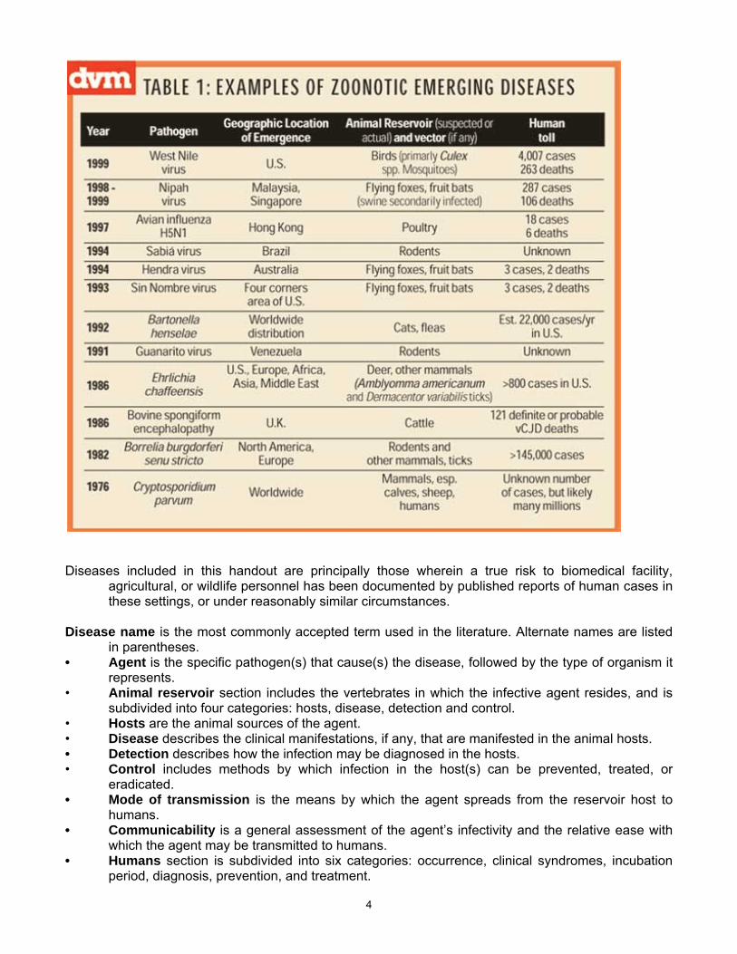
III. "Emerging zoonoses" are defined as zoonotic diseases caused either by apparently newagents, or by previously known microorganisms, appearing in places or in species inwhich the disease was previously unknown. New animal diseases with an unknown hostspectrum are also included in this definition. Natural animal reservoirs represent a morefrequent source of new agents of human disease than the sudden appearance of acompletely new agent. Factors explaining the emergence of a zoonotic or potentially zoonoticdisease are usually complex, involving mechanisms at the molecular level, such as genetic driftand shift, and modification of the immunological status of individuals and populations. Socialand ecological conditions influencing population growth and movement, food habits, theenvironment and many other factors may play a more important role than changes at themolecular level.
4
Diseases included in this handout are principally those wherein a true risk to biomedical facility,agricultural, or wildlife personnel has been documented by published reports of human cases inthese settings, or under reasonably similar circumstances.
Disease name is the most commonly accepted term used in the literature. Alternate names are listedin parentheses.
• Agent is the specific pathogen(s) that cause(s) the disease, followed by the type of organism itrepresents.
• Animal reservoir section includes the vertebrates in which the infective agent resides, and issubdivided into four categories: hosts, disease, detection and control.
• Hosts are the animal sources of the agent. • Disease describes the clinical manifestations, if any, that are manifested in the animal hosts. • Detection describes how the infection may be diagnosed in the hosts. • Control includes methods by which infection in the host(s) can be prevented, treated, or
eradicated. • Mode of transmission is the means by which the agent spreads from the reservoir host to
humans. • Communicability is a general assessment of the agent’s infectivity and the relative ease with
which the agent may be transmitted to humans. • Humans section is subdivided into six categories: occurrence, clinical syndromes, incubation
period, diagnosis, prevention, and treatment.
5
• Occurrence provides brief information on documented cases and includes the source of theinfection and some characteristics of the affected persons.
• Clinical syndromes describe the principal clinical features of the disease, including thosereported in documented cases.
• Incubation period is the time between exposure to the agent and the appearance of clinicalsigns of disease in humans.
• Diagnosis describes the laboratory methods that may be used to detect infection in humans. • Prevention provides information on measures used to avoid or minimize exposure and disease
in humans. • Treatment provides information on the manner in which the disease may be treated. • Reporting requirement under the animal reservoir section are those diseases required to be
reported to the United States Department of Agriculture, if diagnosed in animals; in the humanssection, those required to be reported to the local public health authorities and/or to the Centersfor Disease Control, if diagnosed in humans.
Amebiasis(Humans: Amebic Dysentery, Amebiosis)Agent: Entamoeba histolytica; protozoaAnimal Reservoir:Hosts: Macaques, baboons, squirrel monkeys, and other nonhuman primates; occasionally dogs and
cats.Disease: Asymptomatic to severe diarrhea (hemorrhagic or catarrhal) with weight loss, depending on
strain of organism and invasiveness of the condition; lesions outside of the intestinal tractoccasionally develop.
Detection: Microscopic examination of fresh wet fecal smears for trophozoites or cysts. Repeatedexaminations may be necessary due to periodic fecal shedding. Cysts may be identified by useof zinc sulfate flotation examination of feces. Colonoscopy with scraping or biopsy of ulcers.An ELISA-based test is available and immunostaining is also useful.
Control: Antimicrobial treatment or culling of carriers; strict sanitation practices.
Mode of transmission: Fecal-oral. Flies and cockroaches can spread cyst forms of the agent.Communicability: High, due to potential for asymptomatic carriers (humans) that can act as a source
of infection for nonhuman primates.Reporting Requirement: Not reportable.
Humans:Occurrence: Man is the natural host and is the usual source of infection for animals. Clinical cases in
laboratory animal settings have not been documented.Clinical syndromes: Asymptomatic to acute or fulminating dysentery with fever, chills, and
hemorrhagic or catarrhal diarrhea sometimes alternating with constipation; extraintestinalabscesses may develop especially in the liver.
Incubation period: Ranges from two to four weeks, may be longer.Diagnosis: For intestinal amebiasis: examination of fresh stool smears for trophozoites; colonoscopy;
serologic testing for invasive forms of the disease. For hepatic abscesses: ultrasonography,MRI, or CT with image-guided needle aspiration.
Prevention: Good hygiene; strict sanitation; barrier methods of protection; vermin control programs.Treatment: Tinidazole and metronidazole are unique in that they are effective both in the bowel lumen
and wall and other tissues, both must be used in combination followed by iodoquinol,paromomycin, or diloxanide furoate. For hepatic abscess, must be followed by treatment withchloroquine.
Prognosis: If untreated, mortality rate is high; with treatment, good.Reporting Requirement: Reportable to local health department in Arizona.

6
B VirusAgent: Cercopithecine herpesvirus 1, Herpesvirus simiae, Herpes B; alphaherpesvirus.Animal Reservoir:Host: Macaques.Taught as a separate class for those working with Macaque nonhuman primates.
BalantidiasisAgent: Balantidium coli; ciliated protozoa.Animal Reservoir:Hosts: Pigs, nonhuman primates (macaques, baboons, squirrel monkeys and other nonhuman
primates), guinea pigs.Disease: Asymptomatic to watery diarrhea and ulcerative enterocolitis, weight loss; rectal prolapse.Detection: Examination of fresh wet fecal smears for trophozoites or cysts; histologic examination;
autofluorescence microscopy of feces.Control: Sanitation; treatment of infected animals with antimicrobials.
Mode of transmission: Fecal-oral.Communicability: Humans have a high natural resistance to this parasite, but infection may pose
more substantial health hazards in debilitated individuals.Reporting Requirement: Not reportable.
Humans:Occurrence: In human cases, contact with pigs is the most likely source of infection. Up to 100% of
pigs more than four weeks old on one farm were tested positive for B. coli cysts in their feces.Waterborne epidemics occasionally occur in areas of poor sanitation. Balantidium coli cancomplicate other gastrointestinal tract diseases of humans.
Clinical syndromes: Diarrhea alternating with constipation, gastroenteritis, tenesmus, nausea,vomiting.
Incubation period: Unknown, possibly a few days.Diagnosis: Examination of fresh wet smear of stool for trophozoites or cysts. Colonoscopy with
scraping or biopsy of ulcers.Prevention: Good hygiene and strict sanitation; barrier methods of protection.Treatment: Tetracycline, iodoquinol, paromomycin, metronidazole; supportive care.Prognosis: GoodReporting Requirement: Not reportable.
Brucellosis(Livestock: Contagious Abortion, Bang’s Disease; Humans: Undulant Fever, Malta Fever,
Mediterranean Fever)Agent: Brucella abortus, B. canis, B. melitensis, B. ovis, B. suis; aerobic, gram-negative coccobacilli.Animal Reservoir:Hosts: Brucella melitensis, B. ovis, B. abortus (occasionally)—sheep, B. melitensis—goats, B.
suis—pigs, B. canis—dogs.Disease: Infection of the genital tract causing infertility, abortion with chronic vaginal discharge;
orchitis, prostatitis, epididymitis. Arthritis, lymphadenopathy, uveitis, and diskospondylitis mayoccur.
Detection: Culture, using selective media, of genital discharges, aborted fetuses, udder secretions,and tissues; PCR assay; rose bengal plate agglutination and complement-fixation (CF) tests arerecommended for screening flocks and individuals; enzyme-linked immunosorbent assay(ELISA).
7
Control: Vaccination available (live-agent vaccines superior to inactivated) for cattle, dogs, and goats;animal testing (such as the Brucella milk ring test [BRT] in cattle) and removal programs arefrequently used; long-term antimicrobial therapy has been successful in dogs.
Mode of transmission: Direct contact of broken skin or conjunctiva with genital secretions, abortedfetuses, fetal fluids, and urine from animals; inhalation of aerosols from tissues; ingestion ofunpasteurized milk or cheese.
Communicability: High with exposure to infected livestock or their tissues. The disease is uncommonin countries where control programs have largely eradicated the disease in livestock. Rarely,dogs can be infected with various strains of Brucella spp., and may serve to disseminate theagent to livestock. Brucella abortus and B. melitensis appear to be the agents of most zoonoticimportance, followed by B. suis. It is unclear whether B. ovis has zoonotic importance. Sincebrucellosis in dogs is not uncommon, the risk may be low to moderate with B. canis.
Reporting Requirement: Must be reported to the Veterinary Services National Animal HealthProgram, USDA when diagnosed in cattle, bison, elk, and pigs.
Humans:Occurrence: Rare in the US; almost all cases are imported. A survey of veterinarians in the
Netherlands indicated that four of 89 (5%) had antibodies to B. abortus. Although two of theveterinarians were cattle practitioners; one was a swine practitioner, and the other worked forgovernment, industry, or academia. In a serologic survey of 43 veterinarians in Florida, nonehad antibodies to B. canis, although six had antibodies to other Brucella spp. However, in aseroepidemiologic study in Oklahoma, 53 of 73 (72%) practicing veterinarians had antibodies toB. canis. An animal technician had serologic evidence of B. canis infection. A fatal case ofbrucellosis due to B. suis was reported in a pig farmer that had not been exposed to livestockfor at least 20 years.
Clinical syndromes: Acute or chronic onset with intermittent and persistent fever, anorexia, sweating,headache, myalgia, irritability, arthralgia, weakness; cervical and axillary lymphadenopathy,hepatosplenomegaly, genitourinary infection.
Incubation period: Ranges from five days to five months, average one to two months; may be longer.Diagnosis: Blood culture during the acute phase, using the lysis concentration method; serologic
diagnosis by enzyme immunoassay (EIA). Specialized serologic techniques are needed todetect B. canis antibodies because of cross-reaction with other species within the genus.
Prevention: Personal protective equipment (PPE); strict hygienic measures for disposal of placenta,discharges, fluids, and fetuses after abortions; disinfection of contaminated surfaces; avoidanceof eating unpasteurized dairy products. Satisfactory vaccines are not available for humans.
Treatment: Combination of rifampin or streptomycin, and doxycycline, for at least six weeks.Reporting Requirement: Reportable to local health department in Arizona and the CDC.
Campylobacteriosis(Vibrionic Enteritis)Agent: Campylobacter coli, C. fetus, C. hyointestinalis, C. jejuni; microaerophilic, gram-negative, rod-
shaped bacteria.Animal Reservoir:Hosts: Cattle, pigs, chickens, sheep, dogs, cats, ferrets, hamsters, and nonhuman primates (including
macaques, baboons, and squirrel monkeys).Disease: Asymptomatic to watery or mucohemorrhagic diarrhea; abortion and stillbirths due to C. fetus;
fever, reduced appetite, vomiting. Clinical manifestations may be more severe in young animals.Detection: Culture of feces on selective media; examination of fresh fecal or tissue samples, using
dark-field or phase-contrast microscopy; serial serum samples to document increasing antibodytiter by ELISA.
8
Control: Because C. jejuni and coli are not routinely cited as potential intestinal pathogens in animals(except for diarrhea in young cats and dogs and in several species of primates), efficacy ofantibiotic therapy has been reported infrequently. In certain cases in which animals areseverely affected or are a zoonotic threat, antibiotic treatment may be indicated. Before therapyis instituted, isolation and sensitivity tests should be done to prevent development of a carrierstate.
Mode of transmission: Fecal-oral and food- (via undercooked meat) or waterborne transmission.Unpasteurized milk has been cited as a principal source of infection in several human cases.
Communicability: Low to moderate if proper sanitation measures are followed. Prevalence of thisorganism in laboratory nonhuman primate colonies may be low.
Reporting Requirement: Not reportable.
Humans:Occurrence: Cases of enteritis have been associated with exposure to domestic animals, sheep, and
pigs. Asymptomatic laboratory-housed coyotes transmitted the agent to animal technicians.Clinical syndromes: Often asymptomatic; abdominal pain, malaise, fever, nausea, vomiting, diarrhea;
typhoid-like syndrome or reactive arthritis may develop. C. fetus causes systemic infectionsthat can be fatal, including bacteremia, endocarditis, meningitis, and focal abscesses.
Incubation period: Ranges from one day to 10 days, usually two to five days.Diagnosis: Culture of stool.Prevention: Good hygiene to include washing of hands after handling animals or their feces and
wearing of boots and coveralls when working around farm animals and sanitation; detection andtreatment of infected animals.
Treatment: Supportive care, including rehydration and electrolyte replacement, as needed. Thedisease is self-limited, but its duration can be shortened with antimicrobial therapy. Selectantimicrobial agents (e.g., erythromycin or ciprofloxacin) may be efficacious during initial stagesof disease, but isolation and susceptibility testing should be performed prior to administration.Systemic infections respond to therapy with gentamicin, chloramphenicol, ceftriaxone, orciprofloxacin.
Reporting Requirement: Reportable to local health department in Arizona.
CapnocytophagosisAgent: Capnocytophaga canimorsus, C. cynodegmi (formerly Dysgonic Fermenter-type 2); aerobic
gram-negative rod.Animal Reservoir:Hosts: Dogs, cats, rodents.Disease: Asymptomatic in host species.Detection: Culture, using stringent growth conditions, of saliva and oral mucosa.Control: Impractical, agent considered to be highly associated within the oral cavity of reservoir hosts.Mode of transmission: Animal bites or scratches, or contamination with oral secretions.Communicability: Low if proper animal handling techniques are used. Splenectomy and alcoholism
appear to be strong predisposing factors for disease in human cases.
Humans:Occurrence: No known cases have been reported in animal facilities; however, multiple reports of
transmission to humans from pets exist. Purpura fulminans was noted following a dog bite. Twocases in immunocompetent persons were linked to contact with dogs, involving licks andscratches. In one study, 14 individuals developed fever and septicemia after contact with dogsor dog bites; in another review, 42 of 52 human cases were linked to dog or cat bites, scratchesor contact. Two cases of fever and erythema, one in an asplenic person, were linked to cat bites
and scratches. Septicemia developed in an immunosuppressed asplenic individual after a cat scratch.Clinical syndromes: Cellulitis, fever, septicemia, purulent meningitis, endocarditis and septic arthritis;
can be fatal. Immunosuppressed and splenectomized patients, as well as alcoholics and thosewith various chronic diseases, appear to be at highest risk.
Incubation period: Ranges from one to five days.Diagnosis: Identification of bacteria within neutrophils; isolation by culture.Prevention: PPE, including gloves; proper training in animal handling and restraint; disinfect wounds
following injury or exposure.Treatment: Penicillin G is the antibiotic of choice.
Cat Scratch Disease(Humans: Benign Lymphoreticulosis, Bacillary Angiomatosis, Bacillary Peliosis Hepatitis)Agent: Bartonella henselae; aerobic, gram-negative bacilli.Animal Reservoir:Hosts: Cats, occasionally dogs.Disease: Usually asymptomatic; however, may cause reproductive failure in female cats and peliosis
hepatis in dogs.Detection: Culture; PCR assay of formalin-fixed, paraffin-embedded tissues.Control: Impractical, agent considered to be highly associated within the reservoir host; control of flea
population to reduce transmission between cats.
Mode of transmission: Principally spread by fleas among cats; however, flea-to-human transmissionis unlikely. Infection typically occurs after bites or scratches from healthy young cats andoccasionally dogs.
Communicability: Low with proper animal handling techniques and protective equipment. This is anemerging pathogen among immunosuppressed individuals in the general population, andwarrants appropriate precaution in animal facilities. Veterinary care personnel are generallyconsidered to be at higher risk.
Reporting Requirement: Not reportable.
Humans:Occurrence: Reports of transmission to humans from pet cats; however, no documented cases in
laboratory animal settings. Estimated 22,000-24,000 humans developed cat scratch diseaseduring 1992, of whom 2,000 were hospitalized. A higher rate of suspected cat-scratch diseasewas documented among veterinary surgeons (18.6%) and veterinary technicians (8.9%) than innon-veterinary workers (1.3%). Pet dogs transmitted the pathogen to two persons, in theabsence of any contact with cats, with resulting fever and lymphadenopathy.
Clinical syndromes: Papule at site of lesion within one week after exposure, lymphadenopathy withintwo weeks, fever, malaise, myalgia, bacillary angiomatosis (particularly in immunosuppressedpersons), peliosis hepatis, lymphadenitis, aseptic meningitis with bacteremia; chronicosteomyelitis.
Incubation period: Ranges from three to 14 days for development of primary lesion, up to 50 days fordevelopment of lymphadenopathy.
Diagnosis: History of clinical signs of disease; immunofluorescent antibody (IFA) test; biopsy of lymphnode followed by Warthin-Starry silver stain to demonstrate the organism histologically; PCRassay.
Prevention: Proper training in cat handling and PPE; disinfect wounds following injury or exposurefrom cats.
Treatment: Not generally used or indicated in symptomatic persons since disease is often self-limiting;however, rifampin, erythromycin, or doxycycline can be administered to immunocompromisedindividuals if disease sequelae are more severe.
Reporting Requirement: Not reportable.
10
ChlamydiosisAgent: Chlamydophila abortus (formerly Chlamydophila (Chlamydia) psittaci—non-avian biotypes), C.
pecorum, C. pneumoniae; obligate, intracellular, gram-negative, coccoid, rod-shaped bacteria.Animal Reservoir:Hosts: C. abortus—sheep and goats, dogs, cats and guinea pigs; C. pecorum—sheep and goats; C.
pneumoniae—frogs (Xenopus sp.). Three reports in frogs were published before identification ofC. pneumoniae; the agent was, therefore, only described by genus or as C. psittaci.
Disease: Asymptomatic; fetal loss (abortion, stillbirth), pneumonia, and enteritis due to C. abortus insheep and goats; keratoconjunctivitis and polyarthritis due to C. pecorum in sheep and goats;pneumonia and endocarditis in dogs, keratoconjunctivitis and pneumonitis in cats; conjunctivitis,keratitis, salpingitis in guinea pigs; lethargy, disequilibrium, petechiation, and edema in frogs.
Detection: Histologic examination of tissues; identification of chlamydial inclusions in tissue scrapings;ELISA or PCR or isolation in embryonated chicken eggs or cell culture.
Control: Antimicrobials (e.g., tetracyclines and erythromycin [orally and topically administered]); testand cull infected animals; vaccination in cats and sheep.
Mode of transmission: Contact with animals or their tissues, particularly birth products; inhalation ofdesiccated excretions or secretions.
Communicability: Moderate with exposure to birth fluids and membranes of sheep and goats.Because pregnant women are particularly susceptible, exposure to sheep and goats,particularly parturient animals, should be avoided. Human infection from zoonotic transmissionhas only been reported for C. abortus.
Reporting Requirement: Not reportable.
Humans:Occurrence: Disease occurs sporadically in persons exposed to animals. Numerous case reports of
infections in pregnant women exposed to sheep in an abattoir, and in farm settings, particularlyafter exposure to aborting sheep or goats. In one case, Q fever occurred concurrently.
Clinical syndromes: Flu-like illness, conjunctivitis, pneumonia, encephalitis, myocarditis,thrombophlebitis; febrile illness, and abortion in pregnant women.
Incubation period: Ranges from one week to four weeks.Diagnosis: Detection of increasing IgG antibody titer in paired sera by use of CF or
microimmunofluorescent assay (MIA).Prevention: PPE to prevent inhalation of and direct contact with the agent; disinfection of
contaminated waste and environmental surfaces.Treatment: Antimicrobials, such as tetracycline or doxycycline, erythromycin, levofloxacin, or
trovafloxacin.Reporting Requirement: Not reportable.
Colibacillosis(Livestock: White Scours, Gut Edema of Swine)Agent: Escherichia coli are gram-negative, aerobic, and facultatively anaerobic medium-sized rods.
Strains of Escherichia coli that cause diarrhea are of 6 major categories: (1) enterohemorrhagic(0157:H7); (2) enterotoxigenic; (3) enteroinvasive; (4) enteropathogenic; (5) enteroaggregative;and, (6) diffuse-adherent. However, the only strain where the human is not the reservoir host isenterohemorrhagic. All information following is on this strain.
Animal Reservoir:Hosts: Cattle are the most important reservoir; humans may also serve as a reservoir for person-to-
person transmission. There is increasing evidence that in North America deer may also serveas a reservoir.
Disease: Calf diarrhea (white scours) is an acute disease causing mortality in calves less than 10 daysold. It manifests itself as serious diarrhea, with whitish feces and rapid dehydration. Mastitis

11
caused by E. coli appears especially in older cows with dilated milk ducts. A long-term study ofhorse fetuses and newborn colts found that close to 1% of abortions and 5% of deaths ofnewborns were due to E. coli. Neonatal enteritis caused by E. coli in suckling pigs begins 12hours after birth with a profuse watery diarrhea, and may end with fatal dehydration. Edema insuckling pigs (gut edema) is an acute disease that generally attacks between 6 and 14 weeks ofage. Sudden onset, incoordination, and edema of the eyelids, the cardiac region of thestomach, and sometimes other parts of the body characterize it. During septicemic diseases offowl, such as cases of salpingitis and pericarditis, pathogenic serotypes of E. coli have beenisolated. A colibacillary etiology has also been attributed to Hjärre’s disease (coligranuloma),which is a condition in adult fowl characterized by granulomatous lesions in the liver, cecum,spleen, bone marrow, and lungs.
Detection: Stool culture. Strains are identified by ability or inability to ferment sorbitol in media such asMacConkey-sorbitol, by demonstrating the presence of Shiga-like toxins, PCR, orimmunoassays for enterotoxins.
Control: Vaccines for swine and bovine have been developed.
Mode of transmission: While most people get E. coli O157 from contaminated food (such asundercooked ground beef), it also can be passed in the manure (feces) of young calves andother cattle. Animals do not have to be ill to transmit E. coli O157 to humans.
Communicability: Although the number of organisms required to cause disease is not known, it issuspected to be very small. The organism can be found on a small number of cattle farms andcan live in the intestines of healthy cattle. Meat can become contaminated during slaughter, andorganisms can be thoroughly mixed into beef when it is ground. Bacteria present on the cow'sudders or on equipment may get into raw milk. Children under 5 years old are most frequentlydiagnosed with infection and are at greatest risk of developing hemolytic uremic syndrome. Theelderly also appear to be at increased risk.
Reporting Requirement: Not reportable.
Humans:Occurrence: An estimated 73,000 cases of infection and 61 deaths occur in the United States each
year.Clinical syndromes: E. coli O157:H7 infection often causes severe bloody diarrhea and abdominal
cramps; sometimes the infection causes nonbloody diarrhea or no symptoms. Usually little or nofever is present, and the illness resolves in 5 to 10 days. In some persons, particularly childrenunder 5 years of age and the elderly, the infection can also cause a complication calledhemolytic uremic syndrome, in which the red blood cells are destroyed and the kidneys fail.About 2%-7% of infections lead to this complication. In the United States, hemolytic uremicsyndrome is the principal cause of acute kidney failure in children, and most cases of hemolyticuremic syndrome are caused by E. coli O157:H7.
Incubation period: Can be 2-10 days, usually 3-4.Diagnosis: E. coli 0157:H7 is not identified by routine stool cultures. Isolation requires identification of
sorbitol-negative colonies of E. coli on sorbitol-MacConkey agar followed by serologic testing toconfirm the serotype.
Prevention: With respect to man, control measures include: a) personal cleanliness and hygienicpractices, sanitary waste removal and b) protection of food products.
Treatment: Antimicrobial therapy does not alter the course of the disease, and may increase the risk ofhemolytic-uremic syndrome. Treatment is primarily supportive.
Prognosis: Fair except for the young and elderly. Reporting Requirement: Reportable to local health department in Arizona and the CDC.
12
CryptosporidiosisAgent: Cryptosporidium parvum, C. canis, C. felis; coccidian protozoa.Animal Reservoir:Hosts: Cats, dogs, macaques, baboons, squirrel monkeys, and other nonhuman primates, cattle,
sheep, pigs, ferrets, chickens; frogs; rodents.Disease: Asymptomatic to intractable diarrhea; respiratory tract disease and airsacculitis in chickens;
proliferative gastritis in frogs. Disease can be severe in immunocompromised animals.Detection: Microscopic detection of oocysts in fecal smears that have been stained by use of the acid-
fast procedure; commercially available assay can be used to detect C. parvum-specific antigenin fecal samples; identification of cryptosporidia in intestinal biopsy specimens by use ofhistologic examination; detection of IgG antibodies by use of ELISA.
Control: Environmental sanitation; treatment with paromomycin, although its toxicity has beenreported.
Mode of transmission: Fecal-oral, possibly airborne.Communicability: Low; risk is higher if contact occurs with neonatal animals, which are more
susceptible and can shed high titers of the organism. Infections in dogs and cats are rare.Oocysts are immediately infective on shedding.
Reporting Requirement: Not reportable.
Humans:Occurrence: In general, cases are more frequently associated with exposure to livestock than to pet
animals. A case-control study of human immunodeficiency virus (HIV) infected individuals, withand without cryptosporidiosis, in the United States, found no difference in overall pet ownershipor cat or bird ownership; dog ownership was of borderline significance, indicating that pets werenot a major risk factor. Although C. parvum is the major species involved in human infection, astudy of stool samples from 1,680 patients in England indicated that four were infected with C.felis and one with C. canis (201); in 80 cases from Peru, two were infected with C. canis andone with C. felis, indicating that human infections with these species occur. A person withacquired immune deficiency syndrome (AIDS) developed chronic diarrhea following exposure toa cat.
Clinical syndromes: May be asymptomatic or cause profuse watery diarrhea. The agent can causeprotracted illness in immuno-compromised persons spreading to the respiratory tract, liver,pancreas, spleen, and lymph nodes.
Incubation period: Likely ranges from one day to 12 days, with an average of seven days.Diagnosis: Microscopic detection of oocysts in stool smears, stained by use of the acid-fast procedure;
detection of cryptosporidia in intestinal biopsy specimens by use of histologic examination;direct fluorescent antibody; ELISA.
Prevention: PPE; sanitation; good hygiene.Treatment: Supportive care; nitazoxanide. Treatment of underlying AIDS is necessary.Reporting Requirement: Reportable to local health department in Arizona and the CDC.
Dermatophytosis(Humans: Ringworm, Jock Itch, Athlete’s Foot, Microsporosis)Agent: Trichophyton mentagrophytes, T. verrucosum; Microsporum canis; saprophytic fungi.Animal Reservoir:Hosts: T. mentagrophytes, M. canis—guinea pigs and rabbits; M. canis—cats, dogs, and nonhuman
primates; T. verrucosum—horses, cattle, sheep, and goats.Disease: May be asymptomatic, especially in cats. Lesions usually develop on or about the head,
typically appearing as patchy areas of alopecia and erythema; crusts are present with anunderlying inflammatory reaction. Lesions are pruritic, and may spread to other areas of thebody. Secondary bacterial infection may result in abscessation of hair follicles.
13
Detection: Microscopic examination of hair/skin/fleece scrapings mounted in 10% potassiumhydroxide, culture on suitable dermatophyte test media; despite limited sensitivity, affectedareas can be examined, using a Wood’s UV lamp (for M. canis only).
Control: Infection is probably low in barrier-maintained or specific-pathogen-free (SPF) animals; topicaltreatment with miconazole or clotrimazole; systemic griseofulvin can be used for severe cases,but teratogenic and occasionally hepatotoxic effects are noted; a vaccine for cats is available.Sheep should be shorn prior to their introduction into the animal facility.
Mode of transmission: Direct contact with skin lesions in various animal species; spores can bewidely disseminated and persist in the environment. Chronic asymptomatic carrier animalsrepresent a continuing source for transmission. Contaminated fomites such as furniture,grooming tools, or tack.
Communicability: Unknown, but probably low to moderate.Reporting Requirement: Not reportable.
Humans:Occurrence: A survey of veterinary staff in two governmental departments in the United Kingdom
indicated that ringworm was the most commonly reported zoonosis, with overall prevalence of24% in 1,625 staff interviewed. An animal attendant responsible for handling T.mentagrophytes-infected laboratory animals developed a ringworm lesion on his wrist. Sixpersons in Japan were infected after exposure to laboratory rats and guinea pigs. Three casesin laboratory animal and research technicians that had contact with guinea pigs were reported.
Clinical syndromes: Flat, spreading, ring-shaped lesions in the skin (“ringworm”). The margin isusually reddish, vesicular, or pustular and may be dry and scaly with crusts. As the lesionsexpand peripherally, the central areas often clear, leaving apparently normal skin.
Incubation period: Ranges from 10 to 14 days.Diagnosis: Appearance of characteristic lesions; fluorescence of organisms under UV light (for M.
canis only); culture on appropriate fungal media.Prevention: PPE, including gloves; sanitation agents should be labeled as disinfectant and fungicidal.
Linens and clothing need to be laundered in hot water and bleach.Treatment: Thorough bathing with soap and water; daily topical application of fungicide for at least one
month; Terbinafine and butenafine require shorter courses and lead to the most rapid responseand prolonged remissions. Treatment needs to continue for 2 weeks after clinical clearing; oralfungicides (e.g., griseofulvin, itraconazole, and terbinafine) are effective in treatment ofextensive lesions.
Prognosis: Body ringworm usually responds promptly to conservative topical therapy or to oraltreatment.
Reporting Requirement: Not reportable.
Echinococcosis(Hydatid Disease, Hydatidosis)Agent: Echinococcus granulosus, E. multilocularis; cestodes.Animal Reservoir:Hosts: Echinococcus granulosus -dogs are the definitive host and sheep, cattle and other domestic
livestock are intermediate hosts. A northern, sylvatic strain is maintained in wolves and wildungulates (moose and reindeer). The cycle of E. granulosus involving dogs and sheep isespecially important. E. multilocularis—involves foxes and wolves as definitive host andmicrotine rodents (such as voles) as intermediate host. Domestic dogs and cats becomeinfected when they eat infected wild rodents.
Disease: Infection by strobilar stage typically asymptomatic in final hosts with all species ofEchinococcus. Cysts most commonly develop in liver and lungs of intermediate hosts.
14
Detection: In final hosts, observation of the strobilar stage in feces after anthelminthic treatment.Detection of adult worm products in feces: coproantigen by ELISA or copro-DNA by PCR assay.Fecal flotation is unreliable since echinococcal eggs are indistinguishable from eggs of Taeniaspp. Metacestodes in intermediate hosts usually not discernible before death.
Control: Prevent consumption by dogs of viscera of domestic ungulates and reindeer (E. granulosus),and rodents (E. multilocularis) that harbor metacestodes. Dogs can be treated with praziquantelevery 30 (E. multilocularis) or 45 days (E. granulosus).
Mode of transmission: Ingestion of eggs shed in the feces of the final hosts (dogs and cats).Communicability: Low in biomedical research environments.Reporting Requirement: Not reportable.
Humans:Occurrence: Human cases have not been reported in laboratory animal settings. Cystic
echinococcosis, caused by E. granulosus, is endemic in nearly all livestock-rearing countries.Alveolar echinococcosis, caused by E. multilocularis, occurs widely in North America andEurasia. Dogs originating in rural areas may be infected with E. granulosus or E. multilocularis.
Clinical syndromes: The occurrence of clinical signs of disease depends on the species ofEchinococcus, cyst location, and size. With E. granulosus, cysts can form in the liver, lungs,brain, bones, skeletal muscle, kidneys, spleen, or other tissues. Infection with E. multilocularisresults principally in hepatomegaly, but spread to lungs and brain may occur late in disease.
Incubation period: Ranges from months to 20 or more years, depending on the species ofEchinococcus, parasite-burden, rate of growth of the metacestode, the organ(s) affected, andthe duration of infection.
Diagnosis: The immunoblot test is the test of choice. Prevention: PPE; good hygiene. Incinerate or deeply bury infected organs from intermediate hosts. In
endemic areas, prevention is by prophylactic treatment of dogs with praziquantel at monthlyintervals and preventing feeding of offal to dogs.
Treatment: Surgical resection of cysts; treatment with albendazole.Prognosis: With E. granulosus, about 15% of untreated patients eventually die because of the disease
or its complications. With E. multilocularis, 90% of patients die within 10 years withouttreatment.
Reporting Requirement: Not reportable.
EctoparasitismAgents: Mites: Cheyletiella parasitovorax, Liponyssoides sanguineus, Notoedres cati, Ornithonyssus
bacoti, Sarcoptes scabiei, and others. Fleas: Ctenocephalides canis, C. felis, and others. Ticks:Dermacentor variabilis, Rhipicephalus sanguineus, and others.
Animal Reservoir:Hosts: Cats, dogs, rabbits, rodents, and other common laboratory animal species may occasionally be
infested with these arthropod agents.Disease: Asymptomatic to severe dermatitis with alopecia, skin thickening, and secondary pyoderma;
anemia, debility, decreased reproduction, pruritus. Some of these ectoparasites are importantvectors of various bacterial, rickettsial, and viral diseases. For example, ticks transmitbabesiosis, ehrlichiosis, Rocky Mountain Spotted Fever, and Lyme Disease to humans.
Detection: Microscopic or direct examination of skin scrapings or tufts of hair; manual collection fromthe animals or their bedding.
Control: Prompt elimination of infestation from the animals and their habitats is warranted, usingappropriate insecticides (e.g., pyrethrins and permethrins, avermectins); nontoxic measures(e.g., insect growth regulators and silica gels) should be used wherever possible; isolation orquarantine of random-source animals; prophylactic topical treatment of animals on arrival at the
15
facility; thorough cleaning of the environment and ensuring that an appropriate pest preventionand control program has been established.
Mode of transmission: Direct or indirect contact, including infestation of food, bedding, shippingcontainers, and caging equipment used in conjunction with animal care.
Communicability: Low to moderate; most ectoparasites of laboratory animals are host-specific, andtheir life cycle often cannot be sustained in modern animal care programs.
Reporting Requirement: Not reportable.
Humans:Occurrence: There have been several reports of ectoparasitism among animal husbandry and
research technicians in laboratory animal care settings. Recognition of animal infestations hassometimes initially been prompted by medical complaints from staff members or pet owners.
Clinical syndromes: Moderate to severe, but transient pruritic dermatitis, eczema, pyoderma, orpainful or irritating bites from some arthropods on any area of the skin; more substantialsystemic consequences can result from ectoparasites harboring bacterial, rickettsial, or viralagents of human disease.
Incubation period: Immediately following exposure to arthropods.Diagnosis: Microscopic or direct examination of skin scrapings or tufts of hair.Prevention: PPE, including gloves when handling animals that are suspect for ectoparasites.Treatment: Prescribed antiparasitic medication; cleansing baths; vaccination for ectoparasite-borne
disease agents.Reporting Requirement: Not reportable.
Erysipelas(Livestock: Rose Disease, Diamond Skin Disease in Swine; Nonsuppurative Polyarthritis in
lambs; Post-dipping Lameness in Sheep; Humans: Erysipeloid)Agent: Erysipelothrix rhusiopathiae; gram-positive, rod-shaped bacteria.Animal Reservoir:Hosts: Pigs, chickens, sheep.Disease: Fever, lethargy, septicemia, non-suppurative chronic arthritis in lambs, calves, and kids, post-
dipping lameness in sheep; diskospondylitis, and sudden death; sudden death, fever, arthritis,diamond-shaped skin lesions, necrosis of ear and tail tips in pigs; septicemia in chickens.
Detection: ELISA, culture of blood, tonsils, lymph nodes, or joint fluid; histologic identification oforganism in tissues at necropsy.
Control: Antimicrobials; good sanitation of housing environment; routine vaccination program; testingand elimination of carriers; addition of copper sulfate to sheep dips.
Mode of transmission: Direct contact with animals, tissues, or feces; piglets can be infected throughskin abrasions around the navel (“joint ill”); insect vectors (e.g., Dermanyssus gallinae) havebeen linked to spread of the disease in chicken flocks. Recovered animals may be carriers forlife.
Communicability: Low to moderate in biomedical research environments. Risk of cutaneous infectionincreases if animal handlers have unprotected cuts or abrasions on hands.
Reporting Requirement: Not reportable.
Humans:Occurrence: One case report described concomitant infection with E. rhusiopathiae and orf in a sheep
farmer. Two animal technicians in a chicken-rearing facility were infected after handling sick anddead birds.
Clinical syndromes: Cellulitis, fever, bacteremia, endocarditis, encephalitis, septic arthritis.Incubation period: Ranges from one day to three days.
16
Diagnosis: Culture of blood.Prevention: PPE, including gloves when handling infected animals; good hygiene.Treatment: Penicillins and, less commonly, tetracyclines.Reporting Requirement: Not reportable.
GiardiasisAgent: Giardia duodenalis, G. intestinalis, G. lamblia; flagellate protozoa.Animal Reservoir:Hosts: Most domestic and wild mammals, many birds, and humans. Most commonly found in cats,
dogs, nonhuman primates (macaques, baboons, squirrel monkeys), ruminants, and rare in pigsand horses.
Disease: Usually asymptomatic; may have diarrhea, with weight loss, vomiting, and anorexia.Detection: Isolate cysts by use of zinc sulfate fecal flotation; evaluate using light microscopy. Due to
intermittent cyst shedding, fecal samples from three consecutive days should be examined.Control: Sanitation of environment (cysts are inactivated with quaternary ammonium disinfectants,
household bleach [1:32 or 1:16 dilution], steam, or boiling water). Disinfectants should be lefton surfaces for 5-20 minutes before rinsing. Prompt removal of feces from pens; treatment withantiparasitic agents (fenbendazole for dogs, cats, ruminants, and birds or albendazole forruminants). Dogs and cats should be bathed after treatment to remove cysts from hair.
Mode of transmission: Fecal-oral.Communicability: Moderate to high when working with livestock obtained from infected herds. The
agent may be shed by asymptomatic animals.Reporting Requirement: Not reportable.
Humans:Occurrence: Infection is common, and may be transmitted from animals to humans and vice-versa.
Organisms infecting humans and ruminants are morphologically and antigenically similar. Onecase of transmission from a dog to an animal technician occurred at the University of Arizonaapproximately 10 years ago.
Clinical syndromes: Usually asymptomatic; may have mild to severe diarrhea with bulky, greasy,frothy, malodorous stools, with cramps, distension, gas, tiredness. Weight loss, vomiting, andanorexia.
Incubation period: Ranges from two to more than 25 days, average of 7 to 10 days.Diagnosis: Identification of cysts or trophozoites in stool samples or via direct search for trophozoites
in the duodenum; document three negative test results in series; ELISA or direct fluorescentantibody assay.
Prevention: Sanitation; personal hygiene. Community chlorination of water does not kill the cyst. Forwildlife workers, water should be boiled for one minute; filtration with a pore size of less than 1μm can also be used.
Treatment: Treatment should be administered even when no symptoms occur since the parasite canbe transmitted to other people or animals. Antiparasitic agents (e.g., tinidazole ormetronidazole); supportive care.
Prognosis: With treatment and successful eradication of the infection, there are no sequelae. Withouttreatment, severe malabsorption may rarely contribute to death from other causes.
Reporting Requirement: Reportable to local health department in Arizona and to the CDC.
Hantaviral Diseases(Humans: Hemorrhagic Fever with Renal Syndrome, Hantavirus Pulmonary Syndrome)Agent: Hantaan virus; Seoul virus; bunyavirus.
17
Animal Reservoir:Hosts: Rats and mice, other wild rodents.Disease: Asymptomatic.Detection: Serologic testing for specific antibodies, using ELISA, IFA test.Control: Exclude wild rodents from laboratory animal facilities; screen rodents prior to acceptance; test
rodent-derived cell lines prior to use, particularly those originating from endemic regions.
Mode of transmission: Virus shed in urine, feces, and saliva of persistently infected rodents formonths; inhalation of infective aerosols from rodent excreta; wound contamination, conjunctivalexposure, ingestion. Rat cell lines have been implicated as a source of virus.
Communicability: Probability of transmission increases during winter months due to lower humidityand closure of circulation system to outside air. Brief periods of exposure have been sufficient tocause human infections.
Reporting Requirement: Not reportable.
Humans:Occurrence: Korean hemorrhagic fever was noted among professional staff (veterinarians and
physicians) at a Japanese university that used rats. A nationwide survey of research institutionsin Japan reported that 126 cases of hemorrhagic fever and renal syndrome (HFRS) occurredbetween 1970 and 1986. The HFRS also occurred in several laboratory staff workers exposedto rats at a university in Belgium and at a cancer research institute in the United Kingdom.Screening of laboratory animal personnel for antibodies to Hantaan virus in Japan, France, theUnited Kingdom, and Singapore indicated that several persons had experienced subclinicalinfections. The Belgian university conducted a serologic survey of 60 staff members that hadcontact with laboratory animals, particularly rats, and found that 30 (50%) of them had evidenceof subclinical infection with Hantaan virus. At two other institutions that had no cases of HFRS,only one of 34 (3%) personnel with similar exposures to laboratory animals had antibodiesagainst the virus. Approximately 300 cases of Hantavirus Pulmonary Syndrome have beenreported from 31 states since 1993.
Clinical syndromes: HFRS is of variable severity (mortality <5%), with clinical signs of disease relatedto the strain of virus involved; acute onset of fever, lower back pain, sometimes associated withhemorrhage and nephropathy. Hantavirus pulmonary syndrome (HPS), caused by anotherspecies of Hantavirus, Sin Nombre virus, begins as a nonspecific febrile illness followed by asevere increase in pulmonary vascular permeability and rapid progression to a shock-like state.The HPS has not been associated with rats of the genus Rattus or mice of the genus Mus.
Incubation period: Ranges from a few days to months, average two to four weeks.Diagnosis: Serologic testing for specific antibodies, using ELISA and the IFA test; RT-PCR assay is
the molecular diagnostic test of choice, although the genomic heterogeneity of Hantavirusescan complicate interpretation of results.
Prevention: Respiratory tract protection is necessary to prevent inhalation exposure; good hygiene;disinfection of contaminated waste and work surfaces before cleaning. Avoid exposure torodent excreta in rural and wildlife settings. If rodent trapping, Risk Management and Safetyhas a mandatory t rain ing requirement, which can be accessed at :http://fpnew.ccit.arizona.edu/riskmgmt/whatsnew.htm
Treatment: Intravenous fluid therapy; bed rest; ribavirin given intravenously has been beneficial insome cases.
Reporting Requirement: Reportable to local health department in Arizona and to the CDC.
18
Leptospirosis(Livestock: Redwater of Calves; Humans: Weil’s Disease, Hemorrhagic Jaundice, Canicola
Fever, Dairy Worker Fever)Agent: Leptospira canicola, L. hardjo, L. icterohaemorrhagiae, L. interrogans serovar ballum, L.
pomona, L. sejroe, and other Leptospira spp; spirochete bacteria.Animal Reservoir:Hosts: Leptospira interrogans serovar ballum—mice; L. icterohaemorrhagiae— rats; L. canicola, L.
sejroe—dogs; L. pomona, L. hardjo—horses, cattle, pigs, sheep, goats; cats; gerbils, hamsters;rarely, squirrel monkeys, baboons, and other nonhuman primates.
Disease: Asymptomatic in mice; calves may have fever, anorexia, dyspnea, icterus, hemoglobinuria,and hemolytic anemia. In cows, there is a sudden drop in milk production, abortions andstillbirths; fever, hematuria, and hepatic and renal disease in dogs; reproductive failure in sheep,goats, and pigs; icterohemorrhagic disease with abortion in squirrel monkeys. Most commonlyassociated with uveitis or abortions in horses.
Detection: ELISA and MIA test; increasing IgG antibody titer in paired serum samples; detection ofleptospires in urine (using dark-field microscopy) or tissues; PCR assay of urine.
Control: Confinement rearing with sanitation of facilities and appropriate animal waste control,especially of urine; regular vaccination program (cattle, dogs); treatment of infected animals withantimicrobials (e.g., penicillins and aminoglycosides in small animals, streptomycin in pigs);isolation or quarantine of sick animals; immunization with a custom-prepared inactivatedvaccine was used to control the disease in an endemically infected squirrel monkey colony.Vermin control.
Mode of transmission: Oral ingestion; exposure to contaminated urine, placenta, fetal tissues;inhalation. Organisms can also infect hosts through abrasions in the skin or mucosal surfaces.The leptospires are often transmitted to humans by the ingestion of food and drinkcontaminated by the urine of the reservoir animals. Recreational cases have followedswimming or rafting in contaminated water, and occupational cases occur among sewerworkers, rice planters, abattoir workers, and farmers. Sporadic urban cases have been seen inthe homeless exposed to rat urine.
Communicability: Low to moderate.Reporting Requirement: Not reportable.
Humans:Occurrence: An animal technician developed jaundice after bloodborne exposure to a dog with
jaundice. The dog and the technician had antibodies to L. icterohaemorrhagiae and L. canicola.A seroprevalence study of 35 animal technicians indicated that 32 (91%) had antibodies toseveral leptospiral serovars, compared with four of 20 (20%) laboratory personnel withoutanimal exposure. The animal technicians had Leptospira antibody profiles that were similar tothose of mice, rats, guinea pigs, and rabbits in their facility, with prevalence by species rangingfrom 71 to 90%. In a cross-sectional study of university employees working with an infectedswine herd, nine of 110 (8%) were serologically confirmed as cases.
Clinical syndromes: Clinical illness can vary from asymptomatic to fatal liver and kidney disease. Inthe biphasic form, symptoms begin as mild flu-like illness with fever, headaches, rash, andmyalgia; following 1-3 days of improvement symptoms begin again with the addition ofmeningitis, uveitis, rash, adenopathy, and rarely hemorrhagic pneumonia. In the icteric form(Weil’s syndrome), there is impairment of renal and liver function, abnormal mental status,hemorrhagic pneumonia, and hypotension.
Incubation period: Ranges from 2-20 days, average of 10 days.Diagnosis: Serologic testing for specific antibodies, using ELISA, enzyme immunosorbent assay, or
IFA test; PCR; isolation of leptospires from blood or CSF within seven to 10 days of infection;isolation from urine within 10 days of infection.
19
Prevention: PPE to prevent exposure of uncovered skin or mucous membranes in contaminatedsettings; good hygiene.
Treatment: Penicillin, cephalosporins, tetracyclines, erythromycin; more severe infections may requireintravenously administered antimicrobials.
Prognosis: Without jaundice, the disease is almost never fatal. With jaundice, the mortality rate is 5%for those under age 30 and 30% for those over age 60.
Reporting Requirement: Reportable to local health department in Arizona.
Lymphocytic ChoriomeningitisAgent: Lymphocytic choriomeningitis virus (LCMV); arenavirus.Animal Reservoir:Hosts: Mice, rats, hamsters, guinea pigs, dogs, pigs, and monkeys. Nonhuman primates in zoological
settings have experienced outbreaks after the accidental feeding of infected mice.Disease: The pattern of disease in animals depends on age of animals, strain and dose of virus, and
route of inoculation. Asymptomatic to runting and chronic wasting in mice; asymptomatic inguinea pigs and hamsters; dyspnea, anorexia, lethargy, jaundice, and mortality in marmosetsand tamarins.
Detection: Virus isolation in cell culture.Control: Exclude wild rodents from laboratory animal facilities; screen rodents prior to acceptance; test
rodent-derived cell lines prior to use.Reporting Requirement: Not reportable.
Mode of transmission: Contaminated tumors or cell lines are the usual source of LCMV in laboratoryoutbreaks. Transmission to humans by parenteral inoculation, ingestion, inhalation, and splashcontamination of mucous membranes with infective secretions (urine, feces and saliva); otherroutes include contact with contaminated bedding material and infected ectoparasites. Athymicand severe-combined-immunodeficient mice pose a special risk to humans by harboring silent,chronic infections. The large number of outbreaks attributed to hamsters suggests that they maybe amplifying hosts for the virus. Transmission by aerosolization poses a particular hazard forpregnant women. The virus is not spread person to person, though vertical transmissionoccurs.
Communicability: Low if appropriate protective measures are taken.
Humans:Occurrence: A flu-like illness due to LCMV in animal technicians and research personnel exposed to
nude mice and hamsters that were inoculated with infected tumor cells was reported. A fewcases also had aseptic meningitis. Additionally, serologic screening of personnel indicated that10 to 24% experienced subclinical infection with LCMV. Seroconversion to LCMV occurred intwo zoo veterinarians following bite wounds from and necropsy examinations of infectedmarmosets and tamarins. On May 3, 2005, the CDC received a report of severe illness in 4patients who had received organ transplants. 3 of the 4 patients died. The virus was tracedback to an infected hamster belonging to the donor.
Clinical syndromes: Symptoms are biphasic. The prodromal illness is characterized by fever, chills,headache, myalgia, cough, vomiting, lymphadenopathy, and rash with occasional pneumonia.After 3-5 days, the fever subsides and recurs in 2-4 days with the meningeal phase,characterized by headache, nausea, vomiting, lethargy. Arthralgias and chorioretinitis maydevelop later. The LCMV has been recognized as a neuroteratogen in humans.
Incubation period: Ranges from one week to three weeks.Diagnosis: CF; PCR; virus isolation assays.Prevention: PPE, including gloves for handling rodents or their tissues; good hygiene. Pregnant
women should be advised of the dangers to their unborn children inherent in exposure torodents.
20
Treatment: Supportive care.Prognosis: Fatalities are rare. The illness usually lasts 1-2 weeks, though convalescence may be
prolonged.Reporting Requirement: Reportable to local health department in Arizona.
Orf(Contagious Pustular Dermatitis, Contagious Ecthyma)Agent: Orf virus; parapoxvirus.Animal Reservoir:Hosts: Sheep, goats, reindeer.Disease: Pustular lesions principally around the lips; also on gums, nostrils, and occasionally, teats
and udders; interdigital and coronet lesions can lead to lameness; atypical presentation aswarts on distal aspect of limbs; disease in goats more severe than that in sheep. Sheep can bereinfected.
Detection: Clinical lesions; detection of IgG antibodies by use of ELISA.Control: Usually self-limiting; vaccination with live attenuated virus or scarification with a suspension of
infective scab material.
Mode of transmission: Direct contact with mucous membranes of infected animals, with udders ofnursing dams; virus highly resistant to desiccation and can persist in scabs and crusts for years;communal equipment used between animals in sheep flocks.
Communicability: High when exposed to animals with active lesions. The disease is uncommon inlaboratory animal facilities as sheep are generally required to be free of clinical signs of diseaseprior to acceptance.
Reporting Requirement: Not reportable.
Humans:Occurrence: The disease is common in persons with occupational exposure to sheep. In the United
Kingdom, 15 to 29% of farm workers reported having had orf. A study of English farm workersindicated an annual incidence of 2.8%. Several case reports document human infection due tooccupational exposure in agricultural and research settings. Cases usually involved handlinginfected animals without gloves. In a laboratory setting, two researchers were infected afterbeing bitten by an affected lamb during passage of an orogastric tube. In one report of a sheepfarmer, orf was complicated by erysipelas.
Clinical syndromes: Pustular dermatitis, usually on the hands and face.Incubation period: Ranges from three to six days.Diagnosis: Clinical signs of disease; detection of IgG antibody, using a cell culture
immunofluorescence test; histologic examination of skin biopsy specimen.Prevention: PPE, especially gloves; good hygiene.Treatment: None. Typically, disease is self limiting over three to six weeks; secondary bacterial
infections may occur.Reporting Requirement: Not reportable.
Pasteurellosis(Livestock: Shipping Fever, Transport Fever, Hemorrhagic Septicemia; Rabbits: Snuffles)Agent: Pasteurella multocida; facultative anaerobic gram negative rod-shaped bacteria.Animal Reservoir:Hosts: Cats, rabbits, dogs, pigs.Disease: Asymptomatic in cats and dogs; respiratory signs of variable severity, rhinitis, otitis,
subcutaneous and visceral abscesses and genital infections in rabbits. Up to 30 to 90% of
21
healthy rabbits may be carriers in conventional rabbit colonies; in pigs, atrophic rhinitis candevelop in co-infections with Bordetella bronchiseptica.
Detection: Culture; IFA test on nasal swab specimens.Control: Antimicrobials (e.g., enrofloxacin) may only provide temporary remission and alleviation of
clinical signs of disease. Vaccines for rabbits have been developed.
Mode of transmission: Bite wounds, possibly aerosol.Communicability: Low, but may be greater among debilitated or immunocompromised persons.
Between 30-50% of all cat bites become infected; dog bites only 5% of the time with 75% ofinfected cat bites and 50% of dog bites Pasteurella sp.
Reporting Requirement: Not reportable.
Humans:Occurrence: Infection in a pregnant woman from a cat bite resulted in fatal congenital pneumonia.
Other reports describe the development of meningitis and pericardial tamponade in oneindividual, and pneumonia in an immunosuppressed person following a cat bite. An animaltechnician developed a local abscess after a bite sustained from a rabbit with rhinitis. In pigbreeders, culture revealed the presence of P. multocida in the oropharynges of 19 of 49 (39%)examined.
Clinical syndromes: Cellulitis, erythema and painful swelling at site of bite; septicemia, peritonitis.Incubation period: Up to 24 h.Diagnosis: Culture of bite or scratch wounds.Prevention: PPE, including gloves; appropriate restraint of animals; sanitation.Treatment: Vigorous cleansing and irrigation. X-rays to look for foreign bodies and fractures.
Penicillin, tetracycline, cephalosporins, fluoroquinolones, or azithromycin plus clarithromycin.Immunocompromised patients and especially individuals without functional spleens are at riskfor developing overwhelming bacteremia and sepsis following animal bites and should alsoreceive prophylaxis, even for low-risk bites.
Reporting Requirement: Not reportable.
Plague (Humans: Black Death, Yersiniosis)Agent: Yersinia pestis, a small bipolar-staining gram-negative rod.Animal Reservoir:Hosts: Wild rodents.Disease: Plague epizootics cause nearly 100% mortality in affected wild rodent and rabbit populations.
Cats with bubonic plague typically present with fever, anorexia, lethargy, and an enlarged lymphnode that may be abscessed. Oral and lingual ulcers, skin abscesses, ocular discharge,diarrhea, vomiting, and cellulitis have also been documented. Cats with primary septicemicplague present with fever, lethargy, anorexia, diarrhea, vomiting, tachycardia, weak pulse, andrespiratory distress.
Detection: FA test of a glass slide smear. Serologic antibody tests can be confirmatory but requiresamples be taken 2-3 weeks apart to demonstrate a rising titer.
Control: Pet owners in enzootic areas should keep their pets from roaming and hunting, limit contactwith rodent or rabbit carcasses, and use appropriate flea control.
Mode of transmission: Transmitted among rodents and to humans by the bites of fleas or directcontact. The patient with pneumonia can transmit the infection to others by droplets. Cats anddogs are usually exposed to Y. pestis by mucous membrane contact with secretions or tissuesof an infected rodent or rabbit or by the bite of an infected flea.
Communicability: On average, 10 human plague cases are reported each year in the USA.Reporting Requirement: Not reportable.
Humans:Occurrence: It is endemic in California, Arizona., Nevada, and New Mexico. Of the 23 patients who
developed cat-associated plague in the USA between 1977 and 1998, 6 were veterinary staff;the rest were cat owners or others handling a sick cat. In 2004, 2 cats in New Mexico werediagnosed with bubonic plague. Also in 2004, several wood rats and prairie dogs were founddead from the plague in the panhandle of Texas.
Clinical syndromes: Sudden onset with high fever, malaise, tachycardia, intense headache, delirium,myalgia. If pneumonia develops, tachypnea, productive cough, blood-tinged sputum, andcyanosis also occur. Axillary, inguinal, or cervical lymph nodes become enlarged (bubo) andtender and may suppurate and drain. There may be signs of meningitis. With hematogenousspread, the patient may rapidly become toxic and comatose, with purpuric spots (black plague)on the skin. Primary plague pneumonia is a fulminant pneumonitis with bloody, frothy sputumand sepsis. It is usually fatal unless treatment is started within a few hours.
Incubation period: 2-10 days.Diagnosis: Smears and cultures from aspirates of buboes. Rising titers demonstrated by convalescing
patients by agglutination tests.Prevention: Drug prophylaxis with tetracycline for persons exposed to high risk potential. The use of
gloves, surgical masks, eye protection, and standard hygiene and disinfection procedures isrecommended. Plague vaccines have not proven to be effective.
Treatment: Antimicrobials (e.g., streptomycin, gentamicin, or doxycycline) for 10 days. Patients withplague pneumonia need to be isolated.
Prognosis: When pneumonia or meningitis develops, the outcome is often fatal.Reporting Requirement: Reportable to local health department in Arizona and to the CDC.
Q Fever(Humans: Query Fever, Balkan Influenza, Balkan Grippe, Pneumorickettsiosis, Abattoir Fever)Agent: Coxiella burnetii; obligate intracellular rickettsia.Animal Reservoir:Hosts: Cattle, sheep and goats (main reservoirs).Disease: Usually asymptomatic; in cases of severe infection, abortion and reproductive failure may
occur.Detection: Detection of antibodies against phase-I antigen by use of ELISA (more useful on a flock
than an individual basis); PCR assay; immunohistochemical staining of tissues.Control: Coxiella is resistant to heat and drying. Thus, it survives in dust, on the fleece of infected
animals, or in inadequately pasteurized milk. Serologic testing followed by culling of infectedanimals; strict sanitation practices, especially during lambing since the organism is found inextremely high concentrations in the placenta and amniotic fluid; maintenance of C. burnetii-freeanimal populations; antimicrobials (e.g., tetracycline, chloramphenicol).
Mode of transmission: The agent is unique among rickettsiae in that it is usually transmitted tohumans not by arthropods but by inhalation or ingestion. Transmission by cows and goats isprincipally through the milk and placenta and by sheep through feces, placenta, and milk. Dryfeces and milk, dust contaminated with them, and tissues of these animals contain largenumbers of infectious organisms that are spread by the airborne route. Inhalation ofcontaminated dust and of droplets from infected animal tissues is the main source of humaninfection. Outbreaks have been described in association with parturient cats. Spread fromhuman-to-human does not occur despite the presence of pneumonitis but maternal-fetalinfection can occur.
Communicability: Occupational exposure is highly linked to the risk of acquiring infection fromanimals. There is an occupational risk for animal handlers, slaughterhouse workers,veterinarians, and laboratory workers. In research facilities, if sheep are properly screened forinfection, the risk is substantially less.
Reporting Requirement: Not reportable.
23
Humans:Occurrence: A survey in the United Kingdom indicated that 29 of 87 (33%) abattoir workers, 17 of 61
(28%) veterinarians, and 24 of 193 (13%) farm families had antibodies. In contrast, only 11 of697 (2%) of the general population had antibodies. In a serosurvey of personnel at veterinaryschools in California, and Minas Gerais, Brazil, 14 of 138 (10%) and 48 of 219 (22%),respectively, had antibodies. Sporadic cases and outbreaks have been reported in severalbiomedical research institutions. Serosurveys in laboratory animal settings have indicated thatpersonnel potentially exposed to sheep have a greater prevalence of antibodies (16 to 18%)than do non-exposed groups (0.3 to 0.6%). However, during outbreaks, a considerably highernumber of exposed personnel had antibodies. Serosurveys have also uncovered theretrospective occurrence of cases.
Clinical syndromes: Acute infection results in headaches, fevers, chills, and sweats; myalgias,pneumonia with or without hilar lymphadenopathy, fatigue, chest pain, sore throat, nausea,vomiting and diarrhea. Chronic infection results in granulomatous hepatitis, CNS manifestations,and endocarditis. Spontaneous abortion can occur in pregnant women. The course could beacute or chronic and relapsing.
Incubation period: Ranges from one to three weeks.Diagnosis: Detection of increasing IgG antibody titer against phase-II antigen, in paired sera by use of
ELISA or IFA test. PCR is the most useful test.Prevention: PPE, including respiratory tract protection where airborne hazards exist; good hygiene;
human vaccines have undergone clinical trials.Treatment: Tetracyclines administered orally for 15 to 21 days have been effective. The new
macrolides.Prognosis: Even in untreated patients, the mortality rate is low, except when endocarditis develops.Reporting Requirement: Reportable to local health department in Arizona and to the CDC.
Rabies(Humans: Hydrophobia, Lyssa)Agent: Rabies virus; rhabdovirus.Animal Reservoir:Hosts: Biting species that cause rabies in the US are geographically determined and include raccoons
in the East, including New England; skunks in New England, in the Midwest and Southwest, andin California (skunk risk is high and increasing in many areas); coyotes in Texas; and foxes inthe Southwest, in New England, and in Alaska. Dogs and cats are infected often in developingcountries (including the Mexican border), but are rarely infected in the US. Recent fatal casesin the US were acquired from bats and raccoons. Rodents and lagomorphs (e.g., rabbits) areunlikely to have rabies because they cannot survive the disease long enough to transmit it.
Disease: Initial clinical signs of disease are extremely variable with change of behavior usually the firstnoted symptom; hydrophobia, aerophobia, agitation, confusion, limb pain, paresthesia, ataxia,paralysis. The various forms of disease range from “furious” to “paralytic,” on the basis of areasof the central nervous system affected. Death can occur within two to seven days of illness.
Detection: Detection of viral antigen in brain by use of the direct fluorescent antibody test; confinementand daily observation of healthy dogs, cats, and ferrets for 10 days after bite injury to humans toobserve for behavioral signs suggestive of infection that would warrant necropsy. Brain tissuesfor testing must include the hippocampus, medulla oblongata, and cerebellum, and must berefrigerated.
Control: Vaccination of laboratory dogs, cats, and ferrets; quarantine, euthanasia, and diagnostictesting of animals manifesting signs of disease. Guidelines for control and prevention arepublished annually by the National Association of State Public Health Veterinarians.Requirements for vaccination vary by geographic region, depending on endemic status of virus,as determined by public health authorities. There is no treatment for clinical disease.
24
Mode of transmission: Bite of rabid animal or inoculation of infective saliva into fresh wounds ormucous membranes.
Communicability: Low if facility acquires animals with verified vaccination history or with no possibleexposure to reservoir species.
Reporting Requirement: Reportable to the CDC.
Humans:Occurrence: No reported cases among persons in laboratory animal settings. Globally, an estimated
30,000-70,000 deaths occur annually.Clinical syndromes: The prodromal syndrome consists of pain at the site of the bite in association
with fever, malaise, nausea, and vomiting. The skin is sensitive to changes of temperature,especially air currents. About 10 days later, the CNS stage begins, which may be eitherencephalitic (“furious”) or paralytic (“dumb”). The encephalitic form produces the classic rabiesmanifestations of delirium alternating with classic rabies manifestations of delirium alternatingwith periods of calm, when attempts at drinking cause extremely painful laryngeal spasms(hydrophobia). In the less common paralytic form, an acute ascending paralysis resemblingGuillain-Barré syndrome predominates. Both forms progress relentlessly to coma, autonomicnervous system dysfunction, and death despite intensive support.
Incubation period: May range from 10 days to many years but is usually 3-7 weeks.Diagnosis: Animals involved in biting should be kept under observation for up to 10 days. Specific
immunofluorescent antibody staining of brain tissue, skin, or mucosal scrapings; virus isolation.PCR of the CSF or saliva offer definitive diagnosis.
Prevention: Raccoons, skunks, bats, and foxes should be presumed to be rabid. Pre-exposureimmunization series if increased risk of occupational exposure among persons in endemicregions; immediate and thorough postexposure wound disinfection and prophylaxis byadministration of human rabies immune globulin at the site of bite wound, and concurrentadministration of rabies vaccine. National standards address rabies prevention for persons inthe United States.
Treatment: Medical decisions are based on recommendations of the US Public Health ServiceAdvisory Committee. Supportive care with attention to the airway, maintenance of oxygenation,and control of seizures. Treatment modalities include a combination of rabies vaccine, rabiesimmune globulin, monoclonal antibodies, ribavirin, interferon-α and ketamine. Corticosteroidsshould not be used.
Prognosis: Once the symptoms have appeared, death almost inevitably occurs after 7 days, usuallyfrom respiratory failure. There have been 4 documented surviving cases; all receivedpostexposure prophylaxis.
Reporting Requirement: Reportable to local health department in Arizona and to the CDC.
Rat Bite Fever(Humans: Spirillary Fever, Haverhill Fever, Epidemic Arthritis Erythema, Sodoku)Agent: Streptobacillus moniliformis, gram-negative rod-shaped bacteria. Spirillum minus, gram-
negative spiral-shaped bacteria.Animal Reservoir:Hosts: Wild or laboratory rats, mice; rarely, cats and nonhuman primates.Disease: Asymptomatic in rodents because agents are considered to be highly associated within the
oral cavity; endocarditis and arthritis in nonhuman primates.Detection: Isolation of the agents from the oral cavity, nares, or conjunctival sacs on appropriate
culture medium. Animal inoculation is used for the isolation of S. minus; PCR assay.Control: Cesarean-derived rodents are free of the agent; wild rodent control for indoor and outdoor
animal facilities; separation of nonhuman primates from rodents.
25
Mode of transmission: Most frequently occurs from animal bites. Agent transmitted by urine orsecretions of the mouth, nares or conjunctival sacs. Indirect inoculation by contaminated fomitesthrough trauma to unprotected skin.
Communicability: Unknown but probably low.Reporting Requirement: Not reportable.
Humans:Occurrence: An animal technician developed undulating fever and myalgias following a rat bite.
Inhabitants of rat-infested slum dwellings and laboratory workers are at greatest risk.Clinical syndromes: Streptobacillus moniliformis: chills, fever, headache, weakness; regional
lymphadenopathy. S. minus: additional symptoms are: myalgia, arthritis and that the original ratbite, unless secondarily infected, heals promptly, but 1 to several weeks later the site becomesswollen, indurated, and painful; assumes a dusky purplish hue; and may ulcerate. Therelapsing pattern of fever for 3-4 days alternating with afebrile periods lasting 3-9 days maypersist for weeks.
Incubation period: Streptobacillus moniliformis: fewer than 10 days. S. minus: two weeks to twomonths.
Diagnosis: Darkfield examination of exudates or aspirations; animal inoculation since S. minus cannotbe cultured in media.
Prevention: PPE, including gloves; thoroughly wash bite wounds; proper restraint techniques forhandling animals.
Treatment: Penicillin or tetracyclines for seven to 10 days. Tetanus prophylaxis should be considered.Prognosis: Without treatment, the mortality rate is 10%. Prompt diagnosis and treatment markedly
reduces this rate.Reporting Requirement: Not reportable.
SalmonellosisAgent: Human infections are caused almost exclusively by Salmonella enterica subspecies enterica of
which 3 serotypes-typhi, typhimurium, and choleraesuis-are predominantly isolated; facultativeanaerobic gram-negative rod-shaped bacteria.
Animal Reservoir:Hosts: Reptiles, amphibians, guinea pigs, mice, rats, chickens, pigs, cattle, sheep, horses, cats;
rabbits, nonhuman primates (macaques, baboons, squirrel monkeys).Disease: Asymptomatic to septicemia, diarrhea, gastroenteritis; abortion.Detection: Culture of feces.Control: Good programs of sanitation in animal housing areas; strict attention to proper methods of
animal waste disposal; purchase of Salmonella-free animals; broad-spectrum antimicrobialsused parenterally to treat septicemia, with further treatment based on antimicrobial susceptibilitypattern of agent. For agricultural animals, treatment with antimicrobial agents is controversial.Oral antibiotics may deleteriously alter the intestinal microflora, interfere with competitiveantagonism, and prolong shedding of the organism. There is also concern that antibiotic-resistant strains of salmonellae selected by oral antibiotics may subsequently infect humans.Prevalence of the agent in laboratory nonhuman primate colonies may be low. In 1970, theFDA prohibited the distribution and sale of baby turtles with shells 4 inches in length or less,after a quarter million infants and small children developed turtle-associated salmonellosis.Turtles with shells smaller than 4 inches are dangerous because children can put them in theirmouths.
Mode of transmission: Fecal-oral, usually from contaminated food or drink.Communicability: High when exposed to infected animals or birds. To date, human infections have
not been associated with cats.Reporting Requirement: Not reportable.
26
Humans:Occurrence: The CDC estimates that the fecal carriage rate of Salmonella spp in pet reptiles is >90%
and that there are approximately 74,000 cases of reptile-associated salmonellosis in the USAannually. Carriage rate in wild North American turtles is low (<5%). In recent years, there hasbeen a resurgence in the illegal sale of baby turtles.
Clinical syndromes: Serotype typhi causes Typhoid Fever, which is characterized by malaise,headache, cough, sore throat, abdominal pain, and constipation with relapses for up to 2 weeks.A rash commonly appears after 2 weeks. Serotype typhimurium causes fever and chills,nausea and vomiting, cramping, and diarrhea. Serotype choleraesuis causes feversaccompanied by bacteremia and local infection in joints, bone, pleura, pericardium, lungs, orother sites.
Incubation period: For Enteric Fever, 5-14 days; for gastroenteritis, 8-48 hours.Diagnosis: For Typhoid Fever, blood culture; for gastroenteritis, fecal culture.Prevention: Good hygiene; appropriate PPE, including gloves. Adequate waste disposal and
protection of food and water supplies from contamination. Human carriers should not beallowed to handle food supplies or work with animals. Vaccine is available for travelers.
Treatment: For Typhoid Fever, several antibiotics, including ampicillin, azithromycin, chloramphenicol,third-generation cephalosporins, and trimethoprim-sulfamethoxazole are effective. For resistantstrains, fluoroquinolones or cephalosporins are used. For gastroenteritis, treatment is usuallysymptomatic only. Severely ill or those with suspected bacteremia are treated withtrimethoprim-sulfamethoxazole, ampicillin, or ciprofloxacin. In addition, abscesses should bedrained.
Prognosis: The mortality rate of typhoid fever is about 2% in treated cases. Elderly or debilitatedpersons are likely to do poorly. With complications, the prognosis is poor. Relapses occur in upto 15% of cases. A residual carrier state frequently persists. For gastroenteritis, prognosis isgood since it is usually self-limited.
Reporting Requirement: Reportable to local health department in Arizona and to the CDC.
StreptococcosisAgent: Streptococcus suis type 2; facultative anaerobic gram-positive coccoid-shaped bacteria.Animal Reservoir:Hosts: Pigs.Disease: Asymptomatic to severe disease; septicemia; arthritis, polyserositis, meningitis, endocarditis;
pneumonia; abortions; abscessation.Detection: Culture of tonsil swab specimens; detection of IgG antibodies by use of ELISA.Control: Good programs of sanitation.
Mode of transmission: Direct contact with pigs mainly through skin abrasions or cuts. At highest riskare farm workers, butchers, abattoir workers, veterinarians, and those handling pork products.
Communicability: Low.Reporting Requirement: Not reportable.
Humans:Occurrence: In The Netherlands, the prevalence of antibodies to S. suis among pig farmers was two of
190 (1%) whereas veterinarians had a prevalence of six of 100 (6%). The estimated annual riskof developing S. suis meningitis in Dutch abattoir workers and pig breeders was three of100,000. In New Zealand, 0 of 16 (0%) veterinary students, 11 of 107 (10%) meat inspectors,and 15 of 70 (21%) pig farmers had antibodies. The annual incidence of seroconversion in pigfarmers was estimated at 28%. A comparison of 30 cases in The Netherlands and 30 casesreported in literature indicated that 50 (83%) were employed in the pig industry.
Clinical syndromes: Septicemia, meningitis, arthritis, endocarditis, endophthalmitis; deafness andataxia are frequent sequelae of meningitis.
27
Incubation period: Unknown, probably one day to three days.Diagnosis: Culture; isolation of viridans streptococci or Group-D Streptococcus should arouse
suspicion of S. suis infection in cases with pig exposure.Prevention: PPE, including gloves; sanitation.Treatment: Antimicrobials (e.g., penicillin); supportive care.Reporting Requirement: Not reportable.
ToxoplasmosisAgent: Toxoplasma gondii; intracellular coccidian protozoa.Animal Reservoir:Hosts: Cats (definitive host); mice, rats, dogs, cattle, sheep, goats, pigs, chickens (intermediate hosts).Disease: Asymptomatic in adult cats, but occasionally vomiting, diarrhea, dyspnea, coughing,
anorexia, uveitis, pancreatitis, especially in young or immunocompromised cats; hepaticnecrosis in dogs; abortions and stillbirths in sheep, goats, and pigs.
Detection: In cats, fecal examination for oocysts; isolation of tachyzoites (rapidly multiplying form oforganism) from CSF or aqueous humor. There are no practical methods to detect the presenceof bradyzoites (slowly multiplying form of organism) encysted in tissues (e.g., brain, liver, andskeletal and cardiac muscle) of intermediate hosts. Serologic testing: ELISA, IFA test.
Control: Prevent exposure of cats to potentially contaminated meat by feeding only commerciallyprocessed cat food; dispose of cat feces daily before oocysts sporulate and become infective;house cats indoors and apart from other species; antimicrobials (e.g., clindamycin) for dogs andcats; acquisition of cats from T. gondii-free sources.
Mode of transmission: A 2005 survey of physicians conducted by the Humane Society of the USrevealed that physicians in the USA continue to promote pet cats as the major source oftoxoplasmosis infection in pregnant women despite evidence that they are more likely tocontract the disease from eating raw or undercooked meat and from gardening without wearinggloves. Human infection results (1) from ingestion of cysts in raw or undercooked meat; (2)from ingestion of oocysts in contaminated food or water, by careless handling of contaminatedcat litter, or from soil by soil-eating children; (3) from transplacental transmission; or (4) fromdirect inoculation via blood transfusions or organ transplants. Intermediate hosts can becomelifelong carriers of infection due to encysted bradyzoites.
Communicability: Low; oocysts shed from cats become infective within 12 hours to several days andmay remain viable in water or soil for more than 12 months.
Reporting Requirement: Not reportable.
Humans:Occurrence: The prevalence of Toxoplasma antibodies in veterinarians was similar to that of other
occupational groups. High prevalence of Toxoplasma antibodies among animal technicians hasbeen reported, but was attributed to their exposure outside of occupational settings.Comparison of the prevalence of Toxoplasma antibodies in employees with exposure to cats ata research institution revealed no increase in risk of infection.
Clinical syndromes: 80% are asymptomatic. Mild flu-like illness; can develop more severe clinicalsigns of disease, including fever, lymphadenopathy, pneumonia, chorioretinitis, and rashes.Immunosuppressed individuals can develop focal lesions of encephalitis following reactivationof bradyzoites. Infections in pregnant women can lead to birth defects, chorioretinitis, blindness,and severe neurologic sequelae, with mental retardation in infants.
Incubation period: 1-2 weeks.Diagnosis: Detection of increase in titer between acute and convalescent serum samples via Sabin-
Feldman dye test, indirect hemagglutination, IFA, ELISA, Western blot, PCR, or IgG avidity;isolation of T. gondii tachyzoites from blood, tissue, or body fluids.
28
Prevention: Irradiated meat or meat cooked to 66°C (151°F) kills cysts in tissues. Hands, kitchensurfaces, and cooking utensils must be thoroughly cleaned with soap and water after contactwith raw meat. Children’s play areas, including sandboxes, should be protected from cat (anddog) feces; hand washing is indicated after contact with soil potentially contaminated by animalfeces. Indoor cats should be fed only dry, canned, or cooked meat. Litter boxes should bechanged daily as freshly deposited oocysts are not infective for 48 hours. PPE, includinggloves, good hygiene when handling potentially infective material. Pregnant women can havetheir serum examined for antibody. If negative, preventive measures such as having no furthercontact with cats and cat litter, thorough cooking of meat, handwashing after handling raw meatand before eating and wearing gloves when gardening, and thorough washing of vegetablesshould be taken.
Treatment: Not routinely indicated for immunocompetent individuals; antimicrobial therapy (e.g.,sulfadiazine and pyrimethamine) for immunosuppressed persons manifesting specificsymptoms.
Prognosis: Excellent for immunocompetent and fatal for immunosuppressed people not receivingtreatment.
Reporting Requirement: Not reportable.
Tuberculosis(Humans: Consumption)Agent: Mycobacterium avium complex, M. bovis, M. tuberculosis; acid-fast rod-shaped bacteria.Animal Reservoir:Hosts: Mycobacterium avium, M. bovis, M. tuberculosis—macaques, baboons, squirrel monkeys, and
other nonhuman primates. These agents do not occur naturally in monkeys, but are acquiredfrom humans or other species (M. bovis), or directly from environmental sources, such as soiland water (M. avium). Transmission occurs between monkeys, with secondary spread back tohumans. Mycobacterium paratuberculosis, the causative agent of Johne’s disease, has beenrecognized in one species of macaque, and is a suspected zoonotic agent. M. bovis, M.tuberculosis—dogs, cats, pigs; M. bovis—cattle, sheep, goats; M. avium—pigs, chickens,pigeons.
Disease: Asymptomatic to sudden death; pulmonary disease, anorexia, chronic weight loss; peripherallymphadenopathy, with or without chronic draining tracts to the skin; cutaneous abscesses;nodular lesions may form in multiple organs, including gastrointestinal tract, vertebrae, brainand spinal cord.
Detection: Intradermal tuberculin skin test (Mantoux test); thoracic radiography; gross and histologicexamination of tissues; ELISA; PCR assay. False-negative skin test results may occur due toconcurrent disease, early stage disease, or immunosuppression, requiring repeated testingduring quarantine and thereafter. Culture usually takes four to eight weeks for confirmation.
Control: Testing and elimination of infected animals; rigorous quarantine programs for arriving animals;continuous tuberculosis surveillance; good sanitation programs.
Mode of transmission: For M. tuberculosis, exposure is via airborne spread from people sneezing orcoughing. For M. bovis, via ingestion of unpasteurized dairy products or airborne spread frominfected cattle to farmers and animal handlers. For M. avium, via fecal-oral via watercontamination.
Communicability: Moderate, depending on the host species.Reporting Requirement: Reportable to the USDA’s Veterinary Services National Animal Health
Program.
Humans:Occurrence: Cases have followed occupational exposure to infected nonhuman primates in research
settings. New diagnostic technologies such as DNA fingerprinting suggest that as many as 1/3
of new cases of tuberculosis in urban populations are primary infections resulting from person-to-person transmission. Prior to this finding, it was thought that most cases of active TB wereas a result of activation of latent disease.
Clinical syndromes: The patient with pulmonary TB typically presents with slowly progressivesymptoms of malaise, anorexia, weight loss, fever, and night sweats. Chronic coughing is themost common symptom. Pulmonary, meningeal, visceral organs, and other body systems canbe involved; chronic cough, fatigue, fever, weight loss, and hemoptysis during advanced stagesof pulmonary disease. Lifelong latent (non-progressive) infections within calcified pulmonarynodules also occur. Crohn’s disease is linked to M. paratuberculosis infection.
Incubation period: Ranges from two to 10 weeks for development of primary lesions or skin test-positivity.
Diagnosis: May perform dual Mantoux tests (using avian and human precipitin) due to false-negativereactions for non-tuberculous mycobacteria with the use of only the human Mantoux; PCRassay; RT-PCR assay; pulmonary radiography; acid-fast stain of sputum samples.
Prevention: PPE, including respiratory tract protection; good hygiene; appropriate animal restraint;separate animal facilities from human work areas; semiannual tuberculin testing for facilitypersonnel. Millions of individuals worldwide have been vaccinated with BCG but it is notgenerally given in the US because of the low prevalence of TB, the vaccine’s interference withthe ability to determine latent TB using TST, and its variable effectiveness against pulmonaryTB.
Treatment: Nonadherence to antituberculous treatment is a major cause of treatment failure, continuedtransmission of tuberculosis, and the development of drug resistance. Most patients withpreviously untreated pulmonary TB can be effectively treated with either a 6-month or a 9-monthregimen. The initial phase of a 6-month program consists of daily isoniazid, rifampin,pyrazinamide, and ethambutol. Treatment is adjusted based on culture results.
Prognosis: Almost all properly treated patients with TB can be cured. Relapse rates are less than 5%with current regiments. The main cause of treatment failure is nonadherence to therapy.
Reporting Requirement: Reportable to local health department in Arizona and to the CDC.
Tularemia(Francis' disease, deer-fly fever, rabbit fever, O’Hara disease)Agent: Francisella tularensis, a highly pleomorphic, small, aerobic, gram-negative, nonmotile rod or
coccobacillus less than 1 micron that can survive several weeks in the external environment.Animal Reservoir:Hosts: Wild rodents and rabbits. Hamster and prairie dogs may carry the organism.Disease: Clinical signs usually occur alongside heavy infestation with ticks, and include sudden high
fever, anorexia, and stiffness, eventually leading to prostration and death. In sheep, pregnantewes may abort. Affected dogs have soft nodular swellings under the skin. Miliary foci ofnecrosis occur in the liver, spleen, and lymph nodes. Severe lesions in the lung involvewidespread consolidation with edema and pleurisy.
Detection: Culture and serologic testing: IFA or agglutination test. Control: Control is difficult and is limited to tick control and to rapid diagnosis and treatment.
Mode of transmission: (1) handling tissue of infected animals (direct contact with UNBROKEN skin issufficient); (2) human infections due to a cat bite and scratch and a NHP bite also reported; (3)transmitted by biting insects; (4) inhalation, ingestion.
Communicability: All ages are susceptible, and long-term immunity follows recovery; reinfection israre and has been reported in only laboratory staff.
Reporting Requirement: Not reportable.
Humans:Occurrence: Approximately 200 cases of tularemia in humans are reported annually in the
United States, mostly in persons living in the south-central and western states. Tularemiais an occupational risk for farmers, foresters, and veterinarians, and is listed by the US Centers
30
for Disease Control and Prevention (CDC) as one of the six category A, or high-priority,biological warfare agents.
Clinical syndromes: Fever, headache, nausea, and prostration. Papule progressing to ulcer at site ofinoculation.
Incubation period: 2-10 days.Diagnosis: Serologic testing: Agglutination test. Culturing requires special media and for this reason
and because culturing may be hazardous to laboratory personnel, the diagnosis is usually madeserologically.
Prevention: Avoid tick, fly, and mosquito bites; avoid contact with untreated water; use imperviousgloves when skinning or handling animals; cook meat thoroughly; wear face masks, imperviousgloves, and use biosafety cabinets when performing laboratory cultures.
Treatment: Streptomycin is drug of choice.Prognosis: The mortality rate for untreated cases is 5-7%.Reporting Requirement: Reportable to local health department in Arizona and to the CDC.
Viral HepatitisAgent: Hepatitis A virus, a RNA hepatovirus in the picornavirus family.Animal Reservoir:Hosts: Humans, rarely nonhuman primates.Disease: Asymptomatic.Detection: Serologic testing for specific antibodies, using ELISA.Control: Vaccinate monkeys with Havrix® (GlaxoSmithKline) 360 units IM, repeat in 6 months, check
for positive titer one month later.
Mode of transmission: Fecal-oral. Spread is favored by overcrowding and poor sanitation. Commonsource is from contaminated food or water.
Communicability: Low in biomedical settings due to required quarantining and testing of nonhumanprimates.
Reporting Requirement: Not reportable.
Humans:Occurrence: In the US, about 30% of the population has serologic evidence of previous infection. A
large outbreak among patrons of a restaurant in Monaca, Pennsylvania in 2003 was traced tocontaminated green onions from Mexico. One case of transmission from pigtailed macaques toa cage washer occurred at the University of Arizona approximately 10 years ago. Clinicalsyndromes: Prodrome of anorexia, nausea, vomiting, malaise. Fever, enlarged and tenderliver, jaundice may develop 5-10 days later.
Incubation period: 30 days.Diagnosis: Detection of IgM anti-HAV is an excellent test for diagnosing HAV since antibody occurs
early in the course of the disease.Prevention: Handwashing; thorough cleaning of utensils and bedding. Two effective inactivated
hepatitis A vaccines are available. Treatment: Bed rest is recommended if symptoms are marked; 10% glucose given IV if there is
pronounced nausea and vomiting. Dietary management consists of palatable meals astolerated. Avoid strenuous physical exertion, alcohol, and hepatoxic agents. Corticosteroidsshould be avoided.
Prognosis: In most patients, clinical recovery is complete in 3-6 weeks. The mortality rate is less than0.2%.
Reporting Requirement: Reportable to local health department in Arizona and to the CDC.
31
Glossary
Abattoir - A slaughterhouse
Aerophobia - An unusual fear of drafts of air
Agglutination - The process by which suspended bacteria, cells, or other particles are caused toadhere and form into clumps; similar to precipitation, but the particles are larger and are insuspension rather than being in solution
Airsacculitis - Inflammation of the mucous membrane of the air sacs of birds
Alopecia - Loss or the absence of hair, especially from the human head
Amniotic - Of or pertaining to the amnion; characterized by an amnion; as, the amniotic fluid; theamniotic sac
Antigen - A substance, usually a protein, on the surface of a cell or bacterium that stimulates theproduction of an antibody
Antimicrobial - Tending to destroy microbes, to prevent their multiplication or growth, or to preventtheir pathogenic action
Aqueous Humor - The transparent fluid that circulates in the eye chamber between the back of thecornea and the front of the iris and pupil and permeates the vitreous humor behind the lens
Arthralgia - Pain in a joint
Arthropod - An invertebrate animal that has jointed limbs, a segmented body, and an exoskeletonmade of chitin, e.g. an insect, arachnid, centipede, or crustacean
Asplenic - Having no spleen
Asymptomatic - Not showing or producing indications of a disease or other medical condition
Ataxia - The inability to coordinate the movements of muscles
Athymic - Lacking a thymus gland (A lymphoid organ situated in the center of the upper chest justbehind the sternum (breastbone). It is in the thymus that lymphocytes mature, multiply, andbecome T cells)
Atrophic - A wasting away, a diminution in the size of a cell, tissue, organ or part
Axillary - Relating to or near the armpit
Bacillary Angiomatosis - A skin disease characterized by raised, red lesions, caused by bacterialinfection in individuals with weakened immune systems, and treatable with antibiotics, althoughpotentially fatal if untreated.
Bacteremia - The presence of bacteria in the blood
Bradyzoite - A sessile (slow-growing), form of a zoonotic microorganism, typically, Toxoplasma gondii,including others responsible for parasitic infections. In chronic (latent) toxoplasmosis, bradyzoite
32
microscopically presents as clusters enclosed by an irregular crescent-shaped wall (apseudocyst), in infected muscle and brain tissues. Bradyzoite is the antonym of tachyzoite,which is a stage of rapid growth.
Bubo - A tender, enlarged, and inflamed lymph node, particularly in the armpit or groin.
Catarrhal - Inflammation of a mucous membrane; especially: one chronically affecting the human noseand air passages
Cellulitis - Infection and inflammation of the tissues beneath the skin
Cervical - Relating to a neck, or cervix, in any sense
Choanal - Pertaining to the choana, the passageway from the back of one side of the nose to thethroat. The choana is shaped like an inverted funnel which opens into the upper throat. Thechoanae must be open to permit breathing through the nose
Chorioretinitis - An inflammation of the choroid and retina of the eye. The symptoms are the presenceof floating black spots and blurry vision
Ciliated - Provided with cilia or with a fringe of hairs
Cloacal - Pertaining to the cloaca (A common passage for faecal, urinary and reproductive discharge inmost lower vertebrates)
Colostrum - A yellowish fluid rich in antibodies and minerals that a mother's breasts produce aftergiving birth and before the production of true milk. It provides newborns with immunity toinfections
Complement-Fixation (CF) - A diagnostic test in which serum drawn from the animal or human ismixed with antiserum containing antigen in the presence of complement, which causes the lysisof antibody coated sheep red cells. Lack of hemolysis indicates a positive test result
Congenital - Existing at birth, referring to certain mental or physical traits, anomalies, malformations,diseases, etc. which may be either hereditary or due to an influence occurring during gestationup to the moment of birth
Conjunctiva - The mucous membrane investing the anterior surface of the eyeball and the posteriorsurface of the lids
Coronet - The line of junction between the skin and the hoof or claw
Cull - When people cull animals, they kill them, especially the weaker members of a particular group ofthem, in order to reduce or limit their number
Cutaneous - Relating to the skin
Cyanosis - A condition in which the skin and mucous membranes take on a bluish color because thereis not enough oxygen in the blood
Cysts - A round growth, just under the skin or deeper in the body, which contains liquid
Dams - A female parent; used of beasts, especially of quadrupeds
33
Definitive Host - The plant or animal in or on which a parasitic organism reaches sexual maturity
Dermatitis - Inflammation of the skin from any cause, resulting in a range of symptoms such asredness, swelling, itching, or blistering
Discospondylitis - Inflammation of intervertebral disks in animals, often with osteomyelitis of adjacentvertebrae, caused by a bacterial infection or occasionally trauma. Resultant compression of thespinal cord can cause paralysis. It is most commonly seen in adult dogs but also affects pigs,horses, and cattle
Disequilibrium - A state of instability or imbalance
Duodenum - The first short section of the small intestine immediately beyond the stomach
Dysentery - A disease marked by frequent watery stools, often with blood and mucus, andcharacterized clinically by pain, tenesmus, fever, and dehydration
Dyspnea - Difficult or labored breathing; shortness of breath
Ectoparasitism - A parasite, such as a flea, that lives on the exterior of another organism
Eczema - An inflammation of the skin characterized by reddening and itching and the formation ofscaly or crusty patches that may leak fluid
Edema - An accumulation of an excessive amount of watery fluid in cells or intercellular tissues
ELISA (Abbreviation for enzyme-linked immunosorbent assay) - A widely used technique fordetermining the presence or amount of protein in a biological sample, using an enzyme thatbonds to an antibody or antigen and causes a color change
Endemic - Denoting a temporal pattern of disease occurrence in a population in which the diseaseoccurs with predictable regularity with only relatively minor fluctuations in its frequency over time
Endocarditis - Inflammation of the thin membranous lining endocardium of the heart's cavities
Endophthalmitis - Inflammation of the ocular cavities, caused by infection, trauma, or allergic reaction
Enterocolitis - Inflammation of the small and large intestine as a result of infection
Epididymitis - Inflammation of the epididymis (Convoluted tubule connecting the vas efferens, thatcomes from the seminiferous tubules of the mammalian testis, to the vas deferens. Maturationand storage of sperm occur in the epididymis)
Erythema - Redness of the skin as a result of a widening of the small blood vessels near its surface. Ithas various causes, including fever and inflammation
Exanthema - A skin eruption occurring as a symptom of an acute viral or coccal disease, as in scarletfever or measles
Facultative - Able to live under more than one specific set of environmental conditions; possessing analternative pathway
Flagellate - A microorganism with long thin cellular appendages flagella. Some flagellates arepathogenic parasites that cause diseases such as giardiasis in humans
34
Flatulence - Presence of an excessive amount of gas in the stomach and intestines
Fomites - Inanimate objects capable of carrying germs from an infected person to another person
Fulminating - Running a rapid course, worsening quickly
Gastroenteritis - Inflammation of the mucous membrane of both stomach and intestine
Genital - Relating to the external sexual organs or to reproduction
Genitourinary - Relating to or affecting the genital and urinary organs
Genomic - The total genetic content contained in a haploid set of chromosomes in eukaryotes, in asingle chromosome in bacteria, or in the DNA or RNA of viruses.
Glottis - The long opening between the vocal cords at the upper part of a vertebrate's windpipe larynx Granulomatous - A mass or nodule of chronically inflamed tissue with granulations that is usually
associated with an infective process
Hemoglobinuria - The presence of free hemoglobin in the urine, an abnormal finding, that may makethe urine look dark. Hemoglobin is the protein in the red blood cells which carries oxygen fromthe lungs to the tissues of the body and returns carbon dioxide from the tissues to the lungs.The iron contained in hemoglobin gives red blood cells their characteristic color
Hematuria - The presence of blood in the urine
Hemolytic - Destructive to blood cells, resulting in liberation of hemoglobin
Hemoptysis - The coughing up of blood or mucus containing blood
Hepatic - Relating to or affecting the liver
Hepatosplenomegaly - Enlargement of the liver and the spleen
Hepatoxic - Any substance which is toxic to the liver
Heterogeneity - The condition or state of being different in kind or nature
Hilar - A depression or pit at the part of an organ where structures such as blood vessels and nervesenter
Histologic - A branch of anatomy that deals with the minute structure of animal and plant tissues asdiscernible with the microscope
Histiocytic - A Histiocyte is a cell that is part of the human immune system. All categories ofHistiocytes are derived from the bone marrow by multiplication from a stem cell. The derivedcells migrate from the bone marrow to the blood as monocytes. They circulate through the bodyand stop in various organs where they undergo differentiation into histiocytes which are part ofthe mononuclear phagocyte system
Hydrophobia - An extremely intense aversion to water, especially the fear of drinking water or otherliquids
35
Iatrogenic - Denoting response to medical or surgical treatment, induced by the treatment itself;usually used for unfavorable responses
Icterohemorrhagic - A mixture of icterus and hemolysis
Icterus - Yellowish discoloration of the whites of the eyes, skin, and mucous membranes caused bydeposition of bile salts in these tissues
Incubation Period - The period between the time somebody is infected with a disease and theappearance of its first symptoms
Indurated - Hardened, usually used with reference to soft tissues becoming extremely firm but not ashard as bone
Jejunum - The section of the small intestine situated between the duodenum and the ileum, whosemain function is the absorption of nutrients from digested food
Keratitis - Inflammation of the corneaKeratoconjunctivitis - Combined inflammation of the cornea and conjunctiva
Laryngeal - Belonging to, relating to, situated in, or affecting the larynx
Lingual - Relating to the tongue or any tongue like part; Next to or toward the tongue
Lumen - The space in the interior of a tubular structure, such as an artery or the intestine
Lymphadenopathy - Any disease process affecting a lymph node or lymph nodes
Malaise - A general feeling of illness or sickness of no diagnostic significance
Metacestodes - The larval stages of a tapeworm, including the metamorphosis of the oncosphere tothe first evidence of sexuality in the adult worm, differentiation of the scolex, and beginning ofproglottid formation; it includes the procercoid and plerocercoid stages of pseudophyllidcestodes, and the cysticercus, cysticercoid, coenurus, and hydatid stages of cyclophyllideancestodes
Microscopy - Investigation of minute objects by means of a microscope
Miliary - Resembling a millet seed in size (about 2 mm)Marked by the presence of nodules of millet seed size on any surface
Morphologically - The branch of biology that deals with the form and structure of organisms withoutconsideration of function
Mortality - Death or the number of deaths
Mucosal - Of or relating to mucous membranes
Mucosa - A mucous tissue lining various tubular structures, consisting of epithelium, lamina, propria,and, in the digestive tract, a layer of smooth muscle
Myalgia - Pain or tenderness in a muscle or group of muscles
Myelitis - Inflammation of the spinal cord or bone marrow
36
Myocarditis - Acute or chronic inflammation of the heart muscle
Nares - The nostrils or nasal openings, the anterior nares being the external or proper nostrils, and theposterior nares, the openings of the nasal cavities into the mouth or pharynx
Necrosis - The death of cells in a tissue or organ caused by disease or injury
Nematodes - Any of several worms of the phylum Nematoda, having unsegmented, cylindrical bodies,often narrowing at each end, and including parasitic forms such as the hookworm and pinworm.Also called roundworm
Neonatal - Relating to the period immediately succeeding birth and continuing through the first 28 daysof extrauterine life
Neonates - A newborn infant, especially one less than four weeks oldNephropathy - A disease or medical disorder of the kidney
Neuroteratogen - Any agent or factor that induces or increases the incidence of defects in the nervoussystem or brain of the developing embryo.
Ocular - Relating to, perceived by, or performed by the eye
Offal - The edible, mainly internal organs of an animal
Oocyst - A fertilized gamete of sporozoan: a fertilized gamete of parasitic organisms’ sporozoans thatis enclosed in a thick wall
Orchitis - Inflammation of the testis
Oropharynges - The part of the pharynx between the soft palate and the epiglottis
Osteomyelitis - Inflammation of the bone marrow and adjacent bone
Palatable - Food or drink that has a pleasant taste
Papule - A small hard round protuberance on the skin
Parenteral - Describes drug administration other than by the mouth or the rectum, e.g. by injection,infusion, or implantation
Parthenogenetic - Reproduction by development of an unfertilized usually female gamete that occursespecially among lower plants and invertebrate animals
Parturient - Relating to the process or time of childbirth
Pathogens - Any small organism, such as a virus or a bacterium which can cause disease
PCR - Abbreviation for polymerase chain reaction
Peliosis Hepatis - The presence throughout the liver of blood-filled cavities that may become lined byendothelium or become organized
Peripherally - Of, relating to, involving, or forming a periphery or surface part
37
Peritonitis - Inflammation of the membrane that lines the abdomen peritoneum. Symptoms can includeswelling of the abdomen, severe pain, and weight loss
Petechiation - The state of being covered with petechiae <spotty petechiation of the intestinal mucosa
Photophobia - very low tolerance of the eye for light, sometimes a symptom of disease or migraine; Anirrational fear and avoidance of light or lighted spaces
Pleocytosis - Presence of a greater than normal number of cells in the cerebrospinal fluid
Pleomorphic - This term refers to cells that vary in their size, shape, and inside structure
Pleurisy - Inflammation of the membrane pleura surrounding the lungs, usually involving painfulbreathing, coughing, and the buildup of fluid in the pleural cavity
Polyserositis - Chronic inflammation of several serous membranes with effusions in serous cavitiesresulting in fibrous thickening of the serosa and constrictive pericarditis
Pock - A pustule
Prodromal - An early symptom indicating the onset of an attack or a disease Prolapse - A sinking of an organ or other part, especially its appearance at a natural or artificial orifice
Prophylactic - Protecting against infection or disease
Prostatitis - Inflammation of the prostate gland
Prostration - A marked loss of strength, as in exhaustion
Protozoa - Any of various types of very small, usually single-celled animal which do not have a spine
Pruritus - An intense feeling of itchiness
Purpura Fulminans - A severe and fatal form of idiopathic thrombocytopenic purpura that occursespecially in children, usually following an infectious illness, and that is characterized by lowblood pressure, fever, and disseminated intravascular coagulation.
Purulent - Relating to, containing, or consisting of pus
Pyoderma - A skin infection causing the development of pus or pustules
Reservoir - An alternate or passive host or carrier that harbors pathogenic organisms, without injury toitself, and serves as a source from which other individuals can be infected
Rhinitis - Inflammation of the nasal mucous membrane
Ruminants - Any of various hoofed, even-toed, usually horned mammals of the suborder Ruminantia,such as cattle, sheep, goats, deer, and giraffes, characteristically having a stomach divided intofour compartments and chewing a cud consisting of regurgitated, partially digested food.
Salpingitis - Inflammation of the uterine tube or inflammation of the auditory tube
Saprophytic - Obtaining nourishment from the products of organic breakdown and decay
38
Scarification - The making of a number of superficial incisions in the skin
Sepsis - The condition or syndrome caused by the presence of microorganisms or their toxins in thetissue or the bloodstream
Septicemia - Body-wide disease caused by microorganisms or their toxins in the blood. Also calledblood poisoning
Sequelae - A pathological condition resulting from a disease, injury, or other trauma
Serotype (serovar) - A taxonomic subdivision of bacteria based on the kinds and combinations ofconstituent antigens present in the cell
Serpiginous - Creeping; denoting an ulcer or other cutaneous lesion that extends with an arciformborder; the margin has a wavy or serpent-like border.
Shorn - With hair cut short
Spirochete - A microscopic bacterial organism in the Spirochaeta family. Spirochetes have a worm-like, spiral-shaped form, and wiggle vigorously when viewed under a microscope
Splenectomy - Surgical removal of the spleen
Sporulate - To produce or release spores
Sputum - A substance coughed up from the respiratory tract and usually ejected by mouth, e.g. saliva,phlegm, or mucus
Stellate - Shaped like a star and arranged as a rosette.
Strobilar - A part or structure that buds to form a series of segments, as the main body part of atapeworm or the polyp stage in certain jellyfish
Sylvatic - Occurring in or affecting wild animals
Tachycardia - An excessively rapid heartbeat, typically regarded as a heart rate exceeding 100 beatsper minute in a resting adult
Tachypnea - Unusually fast breathing, generally considered to be over 20 breaths per minute in aresting adult
Tamponade - The insertion of a tampon during surgery to check bleeding
Teats - The raised part of the female breast of a mammal through which an animal takes its mother'smilk; a nipple
Tenesmus - A painful spasm of the urogenital diaphragm with an urgent desire to evacuate the bowelor bladder, involuntary straining, and the passage of little fecal matter or urine
Teratogenic - Of, relating to, or causing malformations of an embryo or fetus
Thrombophlebitis - Inflammation of a vein with the formation of a blood clot
Trophozoites - A protozoan, especially of the class Sporozoa, in the active stage of its life cycle
Ungulates - A mammal with hooves
Urticarial - A transient condition of the skin, usually caused by an allergic reaction, characterized bypale or reddened irregular, elevated patches and severe itching, hives
Uveitis - Inflammation of the uvea of the eye
Vascular - Composed of, containing, or having to do with tubes or ducts that carry a fluid, such asblood or lymph in animals or sap in plants, or with a system of such tubes or ducts
Vesicle - A small circumscribed elevation in the skin containing clear fluid. Virulence - The ability of any agent of infection to produce disease. The virulence of a microorganism
(such as a bacterium or virus) is a measure of the severity of the disease it is capable ofcausing
Viscera - The internal organs of the body, especially those of the abdomen such as the intestines
References
Hankenson FC, Johnston NA, Weigler BJ, Di Giacomo RF. 2003. Zoonoses of occupational healthimportance in contemporary laboratory animal research. Comparative Medicine. 53:579-601.
Heymann DL. 2004. Control of communicable diseases manual, 18th ed. Baltimore: United Book Press.
Kahn CM, Line S. 2005. The Merck veterinary manual, 9th ed. Whitehouse Station: Merck & Co.
Tierney LM, McPhee SJ, Papadakis MA. 2006. Current medical diagnosis & treatment, 45th ed. NewYork: McGraw-Hill.
40
Zoonotic Diseases of Research Animals Quiz (Version 2007A)
Your Name(print): Department: Employee ID# Investigator: Lab phone: E-mail: Confirm I passed (Must provide email address)
CERTIFICATION NOT GIVEN IF INFORMATION IS NOT READABLE OR INCOMPLETE
In order to become certified, you may miss FIVE or less questions. The notes may be consultedwhen answering this quiz. Please circle the "T" if "true", or the "F" if "false".
1. T F The World Health Organization defines zoonoses as diseases and infections, which arenaturally transmitted between vertebrate animals and man.
2. T F Mode of transmission include exposure to: feces, urine, saliva, blood, and milk.3. T F Probability of disease transmission from animals to man is influenced by several factors
including population density of the animals in the colony.4. T F Emerging zoonoses does not include new animal diseases with an unknown host
spectrum.5. T F Natural animal reservoirs represent a less frequent source of new agents of human
disease than the sudden appearance of a completely new agent.6. T F Agent is the specific pathogen(s) that cause the disease.7. T F Animal reservoir section includes the invertebrates in which the infective agent resides,
and is subdivided into four categories: hosts, disease, detection and control. 8. T F Hosts are the animal sources of the specific pathogen(s) that cause the disease.9. T F Disease describes the clinical manifestations, if any, that are manifested in the human
hosts.10. T F Detection describes how the infection may be diagnosed in the hosts.11. T F Control includes methods by which infection in the host(s) can be prevented, treated, or
eradicated. 12. T F Mode of transmission is the means by which the agent spreads from the reservoir host
to humans. 13. T F Communicability is a general assessment of the agent’s infectivity and the relative ease
with which the agent may be transmitted to humans. 14. T F Occurrence provides brief information on documented cases and includes the source of
the infection and some characteristics of the affected persons. 15. T F Clinical syndromes describe the principal clinical features of the disease, including those
reported in documented cases. 16. T F Incubation period is the time between exposure to the agent and the appearance of
clinical signs of disease in animals. 17. T F Prevention provides information on measures used to maximize exposure and disease
in humans. 18. T F Reporting requirement under the animal reservoir section are those diseases required to
be reported to the United States Department of Agriculture, if diagnosed in animals.19. T F Amebic Dysentery, Amebiosis, found in macaques, baboons, squirrel monkeys, and
other nonhuman primates; occasionally dogs and cats, is reportable to local healthdepartment in Arizona.
20. T F Cats are the natural host for Amebiosis and is the usual source of infection for humans.21. T F B Virus is taught as a separate class for those working with Macaque nonhuman
primates.22. T F Humans have a natural resistance to balantidiasis which is found in pigs, but can be
prevented with good hygiene and strict sanitation.
41
23. T F Brucellosis or contagious abortion is carried in sheep, goats, pigs and dogs and is verycommon in the US.
24. T F Camplyobacteriosis, enteritis, is carried mainly in horses and is rampant even withproper sanitation.
25. T F Capnocytophagosis is carried by dogs, cats and rodents. No known cases have beenreported in animal facilities; however, multiple reports of transmission to humans frompets exist.
26. T F Cat Scratch Disease is principally spread by fleas among cats; however, flea-to-humantransmission is unlikely. Infection typically occurs after bites or scratches from healthyyoung pet cats and occasionally dogs.
27. T F Chlamydiosis is found in sheep, goats, dogs, cats, guinea pigs and frogs. Only the kindcommunicated by sheep and goats is dangerous to human women.
28. T F Colibacillosis, white scours, is most often found in rodents.29. T F Cryptosporidiosis is carried by cats, dogs, macaques, baboons, squirrel monkeys, and
other nonhuman primates, cattle, sheep, pigs, ferrets, chickens; frogs; rodents. Agentcauses no clinical syndrome even in immunocompromised individuals and is notreportable in AZ or to the CDC.
30. T F Dermatophytosis, ringworm or athlete’s foot, is carried by guinea pigs, rabbits, cats,dogs, non-human primates and some livestock. Disease can cause flat, spreading, ring-shaped lesions in the skin.
31. T F Dogs are the definitive host and sheep, cattle and other domestic livestock areintermediate hosts of Echinococcosis. Domestic dogs and cats become infected whenthey eat infected wild rodents.
32. T F Ectoparasitism is found in cats, dogs, rabbits, rodents, and other common laboratoryanimal species may occasionally be infested with these arthropod agents. There havebeen several reports of ectoparasitism among animal husbandry and researchtechnicians in laboratory animal care settings. Recognition of animal infestations hassometimes initially been prompted by medical complaints from staff members or petowners.
33. T F Erysipelas is found in pigs, chickens and sheep. Risk of cutaneous infection increases ifanimal handlers have unprotected cuts or abrasions on hands and can cause cellulitis,fever and septic arthritis.
34. T F Giardiasis is found in most domestic and wild mammals, birds and humans. Infection isuncommon.
35. T F Hantaviral Diseases is carried in rats, mice and other wild rodents. Virus shed in urine,feces, and saliva of persistently infected rodents for months; inhalation of infectiveaerosols from rodent excreta; wound contamination, conjunctival exposure, ingestion. Ifrodent trapping, Risk Management and Safety has a mandatory training requirement.
36. T F Leptospirosis is carried in many species. Without jaundice, the disease is almostalways fatal.
37. T F Lymphocytic Choriomeningitis is found in rodents, dogs, pigs and monkeys.Contaminated tumors or cell lines are the usual source of LCMV in laboratory outbreaks.Transmission to humans by parenteral inoculation, ingestion, inhalation, and splashcontamination of mucous membranes with infective secretions.
38. T F Orf is found mainly in rodents and is tramitted to humans through direct contact withmucous membranes of infected animals, and with with udders of nursing dams.
39. T F Pasteurellosis is found in cats, rabbits, dogs and pigs. Infection causes almost nosymptoms in humans so there is no need for special procedures.
40. T F Plague is carried in wild rodents. It is transmitted among rodents and to humans by thebites of fleas or direct contact. The patient with pneumonia can transmit the infection toothers by droplets. When pneumonia or meningitis develops, the outcome is often fatal.
41. T F Q Fever is carried by cattle, sheep and goats. Occupational exposure is highly linked tothe risk of acquiring infection from animals. In research facilities, if sheep are properlyscreened for infection, the risk is substantially higher.
42
42. T F Rabies is possible in most mammals. Rodents and lagomorphs are unlikely to haverabies because they cannot survive the disease long enough to transmit it. Once thesymptoms have appeared, death almost inevitably occurs after 7 days, usually fromrespiratory failure.
43. T F Rat Bite Fever is found in rodents, and sometimes in cats and primates. Withouttreatment, the mortality rate is extremely high.
44. T F Salmonella typhi causes Typhoid Fever. The mortality rate of typhoid fever is about 2%in treated cases. Elderly or debilitated persons are likely to do poorly. Withcomplications, the prognosis is poor. Relapses occur in up to 15% of cases. A residualcarrier state frequently persists.
45. T F Streptococcosis is transmitted to humans through direct contact with pigs mainly throughskin abrasions or cuts. At highest risk are farm workers, veterinarians, and thosehandling pork products.
46. T F Cats are the definitive hosts for Toxoplasmosis. Litter boxes should be changed daily asfreshly deposited oocysts are not infective for 48 hours. Infections in pregnant womencan lead to birth defects, chorioretinitis, blindness, and severe neurologic sequelae, withmental retardation in infants.
47. T F Tuberuculosis is carried by humans and the environment and can be transmitted toprimates. Reportable to the USDA’s Veterinary Services National Animal HealthProgram.
48. T F Tularemia is carried by wild rodents and rabbits. Tularemia is an occupational risk for labanimal workers.
49. T F Viral Hepatitis is found in humans and rarely in nonhuman primates. Transmission ishigh in biomedical settings due to required quarantining and testing of nonhumanprimates.
50. T F Viral Hepatitis can be prevented through handwashing; thorough cleaning of utensilsand bedding. Two effective inactivated hepatitis A vaccines are available.
DON'T LEAVE ANY QUESTIONS UNANSWERED, THEY WILL BE MARKED INCORRECT!
2007 Evaluation of "Take Home" Zoonotic Disease of Research Animals Module - V. A
Thank you for completing this "self-instruction" module. Please take time to complete the shortevaluation form. Your comments are valuable in designing the style and substance of futurecertification courses to make this process effective for research staff. (circle one)
Was this packet useful? YES NO
Did you learn anything new? YES NO
Was the training adequate? YES NO
Was the packet too long? YES NO If so, suggested items to cut:
Were the questions too difficult or was it hard to find answers to the questions? YES NO
Did you like the self-instruction packet? YES NO, I would you prefer to attend a lecture.
Do you have any other comments or suggestions to improve this module? Topics not covered?