Journal of Theoretical and Applied Information Technology 20
th August 2014. Vol. 66 No.2
© 2005 - 2014 JATIT & LLS. All rights reserved.
ISSN: 1992-8645 www.jatit.org E-ISSN: 1817-3195
638
GRAY CO-EFFICIENT MASS ESTIMATION BASED IMAGE
SEGMENTATION TECHNIQUE FOR LUNG CANCER
DETECTION USING GABOR FILTERS
1K.SANKAR,
2DR.M.PRABAKARAN
1Research Scholar, Department of Computer Science, Karpagam University, Coimbatore, Tamil Nadu,
India 2Assistant Professor, Department of Computer Science, Government Arts College, Ariyalur, Tamil Nadu,
India
Email: [email protected]
ABSTRACT
Lung cancer detection techniques have been discussed widely in the medical domain; where the location
and presence of cancer has to be identified from low level X-Rays or medium level Scans. Still the
accuracy of detection depends on the medical practitioner or the automatic detection system. Whatever it is,
the process has defects in identifying LCD and the accuracy of identification is highly questionable due to
the false positive results provided. There are many features and techniques have been proposed earlier for
detection of LCD, we propose a new mass estimation technique with gray co-efficient values based on
which segmentation is performed. The proposed method removes the noise present in the input image using
Gabor filter. The efficiency of Gabor filter helps to improve the image quality, and then we compute the
gray co-efficient mass estimation for each of the pixel from image. Based on computed mass value of each
pixel, the segmentation is performed. The segmentation process uses mass threshold, using which the pixel
is selected for LCD process. The selected pixels are used to form the region for LCD and to produce results
to the user. The proposed approach has produced efficient results with less false results, also reduces the
time complexity.
Key Terms: Gabor Filter, Image segmentation, Mass Estimation, LCD Detection.
1. INTRODUCTION
The lung cancer, which is a dangerous
disease happens to the human beings where there
is no medical support exist, unless it is identified
in the early stage. The presence of lung cancer is
diagnosed using either X-ray of human chest, CT
or MRI scans. These are preliminary diagnostic
procedures which cannot be identified exactly
but an important procedure which uses the
biopsy of lung portion which is taken using a
needle on the portion of lung. The lung biopsies
are more difficult to analyze and needs an
experienced pathologist which is difficult to get
one.
The segmentation is the process of
separating a set of similar pixels to form a group
and represent them in different color values than
other pixels. The segmentation techniques uses
various metrics to separate the pixels from
others, for example color values, region
properties and etc.. The segmentation techniques
have been used for variety of medical problems
and produces good results for most cases.
Whatever the scans used to diagnose, the
medical practitioner could not identify or locate a
region exactly where the cancer present or he
may ignore the presence of cancer. This is where
segmentation plays and performs grouping of
pixels and deviate them from other normal pixels
to identify lung cancer.
The input image contains noise, blur so
that the image has to be noise removed. The
Gabor filter is the most efficient linear filter
which could be used to remove noise from the
Journal of Theoretical and Applied Information Technology 20
th August 2014. Vol. 66 No.2
© 2005 - 2014 JATIT & LLS. All rights reserved.
ISSN: 1992-8645 www.jatit.org E-ISSN: 1817-3195
639
image. The noise removed image can be further
segmented to achieve the task of segmentation.
We use the Gabor filter at different levels to get
the quality image from the input image.
The input X-ray image contains noise
and blurs, so that it has to be removed to achieve
the required results. We compute mass
estimation here, where the mass estimation is the
process of computing the gray level density
value or mass. The region of cancer affected
pixels must have more gray levels and the mass
of gray co-efficient will be higher in that region.
We use this property of pixels to identify the
location of lung cancer.
The chapter 2 discusses the related
works, and Chapter 3 discusses the proposed
approach, chapter 4 discusses the results
achieved and so on.
2. RELATED WORKS
A novel approach for detection of cancerous
cells from Lungs CT scan images is proposed in
[1], where Locating lung cancer at an early stage
is a challenging task since there are few or no
symptoms in this stage of the disease and
majority of the cases are diagnosed in the later
stages of the disease. Treating cancer in the early
stages can provide more treatment options, less
invasive surgery, and increases the survival rate.
The majority of lung cancers originate as a small
growth or nodule in the lung. Screening CT
scans are extremely sensitive in detecting
nodules as small as 2 or 3mm within the lungs.
CT screening is efficient in locating majority of
lung cancers. Lung CT Scan helps in detecting
lung cancers at an early stage. This present work
proposes a method to detect the cancerous cells
effectively from the CT scan images by reducing
the detection error made by the physicians’
naked eye for medical study based on Sobel edge
detection and label matrix.
Lung Nodule Detection in CT Images
Using Thresholding and Morphological
Operations [2], the lung CT image is subjected
to various processing steps and features are
extracted for a set of images. The processing
steps include thresholding, morphological
operations and feature extraction. By using these
steps the nodules are detected and segmented and
some features are extracted. The extracted
features are tabulated for future classification.
Cell extraction from sputum images for
early lung cancer detection [3], address this
problem using two different methods, namely, a
Rule-based method, and Bayesian classification.
We describe the two methods and we compare
their performances in terms of their behaviors
with respect to color representation and color
quantization.
A noval assignment of various bioimaging
methods for lung tumor detection and treatment
using 4-D and 2-D images [6], describes fully
Automatic Decision Support system for Lung
Cancer diagnostic from CT Lung images. Most
traditional medical diagnosis systems are
founded on huge quantity of training data and
takes long processing time. However, on the
occasion that very little volume of data is
available, the traditional diagnosis systems
derive defects such as larger error, Time
complexity. Focused on the solution to this
problem, a Medical Diagnosis System based on
Hidden Markov Model (HMM) is presented. In
this paper we describe a pre-processing stage
involving some Noise removal techniques help
to solve this problem, we preprocess an images
(by Mean Error Square Filtering and Histogram
analysis)obtained after scanning the Lung CT
images. Secondly separate the lung areas from an
image by a segmentation process (by
Thresholding and region growing techniques).
Finally we developed HMM for the classification
of Cancer Nodule. Results are checked for 2D
and 4D CT images. This automation process
reduces the time complexity and increases the
diagnosis confidence.
Detection of lung tumor in CE CT images by using weighted Support Vector Machines [7], ropose a novel method for assigning optimal weights for the calculated features. This proposed technique is tested on CE CT Lung images. Simulation results and analysis showed
Journal of Theoretical and Applied Information Technology 20
th August 2014. Vol. 66 No.2
© 2005 - 2014 JATIT & LLS. All rights reserved.
ISSN: 1992-8645 www.jatit.org E-ISSN: 1817-3195
640
that our proposed system has shown better classification accuracy than the conventional SVM.
Extraction and Segmentation of Sputum Cells for Lung Cancer Early Diagnosis [8], present here a framework for the extraction and segmentation of sputum cells in sputum images using, respectively, a threshold classifier, a Bayesian classification and mean shift segmentation. Our methods are validated and compared with other competitive techniques via a series of experimentation conducted with a data set of 100 images. The extraction and segmentation results will be used as a base for a CAD system for early detection of lung cancer which will improve the chances of survival for the patient.
Lung cancer detection by using artificial neural network and fuzzy clustering methods [9], presents two segmentation methods, Hopfield Neural Network (HNN) and a Fuzzy C-Mean (FCM) clustering algorithm, for segmenting sputum color images to detect the lung cancer in its early stages. The manual analysis of the sputum samples is time consuming, inaccurate and requires intensive trained person to avoid diagnostic errors. The segmentation results will be used as a base for a Computer Aided Diagnosis (CAD) system for early detection of lung cancer which will improves the chances of survival for the patient. The two methods are designed to classify the image of N pixels among M classes. In this study, we used 1000 sputum color images to test both methods, and HNN has shown a better classification result than FCM, the HNN succeeded in extracting the nuclei and cytoplasm regions.
Automatic detection of abnormalities in lung radiographs caused by planocellular lung cancer
[10], propose an automatic algorithm for early
planocellular lung cancer detection in lung X ray
images. Considering the fact that lung cancer is
one of the most lethal cancers and that it is
usually diagnosed too late, the solution is to
attempt early diagnosis at general practitioners
level, using cheapest diagnostic tools, chest
radiography. The proposed algorithm determines
and segments the suspected area in lung X ray
images. It consists of two steps: comparison
between extracted planocellular lung cancer
structure and the analyzed lung X ray image by
calculating similarity coefficients and finding the
maximum similarity coefficient which will
indicate the suspected cancer affected area in the
lung image.
Lung Cancer Detection using Curvelet Transform and Neural Network [12], propose a new technique for LCD identification where curvelet transform can extract the features of lung cancer CT scan images proficiently. All extracted feature by curvelet transform are applied to the neural network for training and testing. The performance of proposed work show efficient results.
Computer Aided Detection of Large Lung Nodules using Chest Computer Tomography Images [13], present an automatic Computer Aided Detection (CAD) system to detect a large lung nodule from lateral Chest Radiographs of computed tomography (CT) images to reduce false positive rates. Basic image processing techniques such as Bit-lane Slicing, Erosion, Median Filter, Dilation, Outlining, radon transform and edge detection are applied to the CT scan images in order to detect the lung region. A total of 22 image features were extracted from the enhanced image based on statistical features such as standard deviation, average and mean. A fisher score ranking method is used as a feature selection method to select best ten features (standard deviation, variance, range, maximum grey level, seven invariant moments except the second, sixth and seventh invariant moments and 5th percentile, 9th percentile).
3. PROPOSED METHOD
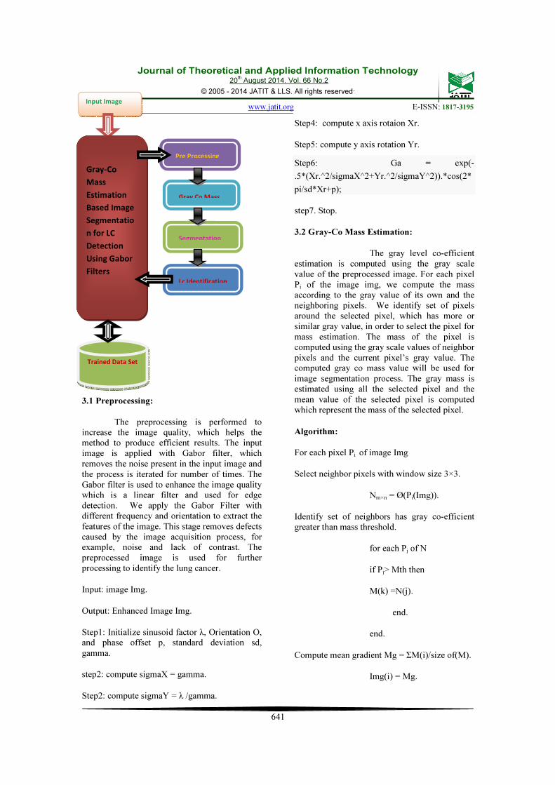
The proposed method has three steps namely; Preprocessing, Gray-Co Mass Estimation, Segmentation, LC Detection. We discuss each of the stages deeply in the next chapters.
Journal of Theoretical and Applied Information Technology 20
th August 2014. Vol. 66 No.2
© 2005 - 2014 JATIT & LLS. All rights reserved.
ISSN: 1992-8645 www.jatit.org E-ISSN: 1817-3195
641
3.1 Preprocessing:
The preprocessing is performed to increase the image quality, which helps the method to produce efficient results. The input image is applied with Gabor filter, which removes the noise present in the input image and the process is iterated for number of times. The Gabor filter is used to enhance the image quality which is a linear filter and used for edge detection. We apply the Gabor Filter with different frequency and orientation to extract the features of the image. This stage removes defects caused by the image acquisition process, for example, noise and lack of contrast. The preprocessed image is used for further processing to identify the lung cancer.
Input: image Img.
Output: Enhanced Image Img.
Step1: Initialize sinusoid factor λ, Orientation O, and phase offset p, standard deviation sd, gamma.
step2: compute sigmaX = gamma.
Step2: compute sigmaY = λ /gamma.
Step4: compute x axis rotaion Xr.
Step5: compute y axis rotation Yr.
Step6: Ga = exp(-
.5*(Xr.^2/sigmaX^2+Yr.^2/sigmaY^2)).*cos(2*
pi/sd*Xr+p);
step7. Stop.
3.2 Gray-Co Mass Estimation:
The gray level co-efficient estimation is computed using the gray scale value of the preprocessed image. For each pixel Pi of the image img, we compute the mass according to the gray value of its own and the neighboring pixels. We identify set of pixels around the selected pixel, which has more or similar gray value, in order to select the pixel for mass estimation. The mass of the pixel is computed using the gray scale values of neighbor pixels and the current pixel’s gray value. The computed gray co mass value will be used for image segmentation process. The gray mass is estimated using all the selected pixel and the mean value of the selected pixel is computed which represent the mass of the selected pixel.
Algorithm:
For each pixel Pi of image Img
Select neighbor pixels with window size 3×3.
Nm×n = Ø(Pi(Img)).
Identify set of neighbors has gray co-efficient greater than mass threshold.
for each Pj of N
if Pj> Mth then
M(k) =N(j).
end.
end.
Compute mean gradient Mg = ΣM(i)/size of(M).
Img(i) = Mg.
Input Image
Gray-Co
Mass
Estimation
Based Image
Segmentatio
n for LC
Detection
Using Gabor
Filters
Pre Processing
Gray Co Mass
Segmentation
Lc Identification
Trained Data Set
Journal of Theoretical and Applied Information Technology 20
th August 2014. Vol. 66 No.2
© 2005 - 2014 JATIT & LLS. All rights reserved.
ISSN: 1992-8645 www.jatit.org E-ISSN: 1817-3195
642
End.
3.3 Segmentation:
The segmentation is the process of grouping similar pixels of image according to some criteria. We segment the image based on computed mass value of each pixel. The segmentation process groups the pixels with more mass value to form a cluster and represent them with different color. The segmentation process generates number of regions and groups the similar pixels with more mass ratio to form a group.
Procedure:
Step1: Intialize Cluster set Cs.
For each pixel Pi of image Img
if Pi > Mth then
for each neighbor of Pj
if Pj > Mth then
Add to cluster cl.
end.
end.
end.
Add cl to Cs = ΣCl+cl.
End.
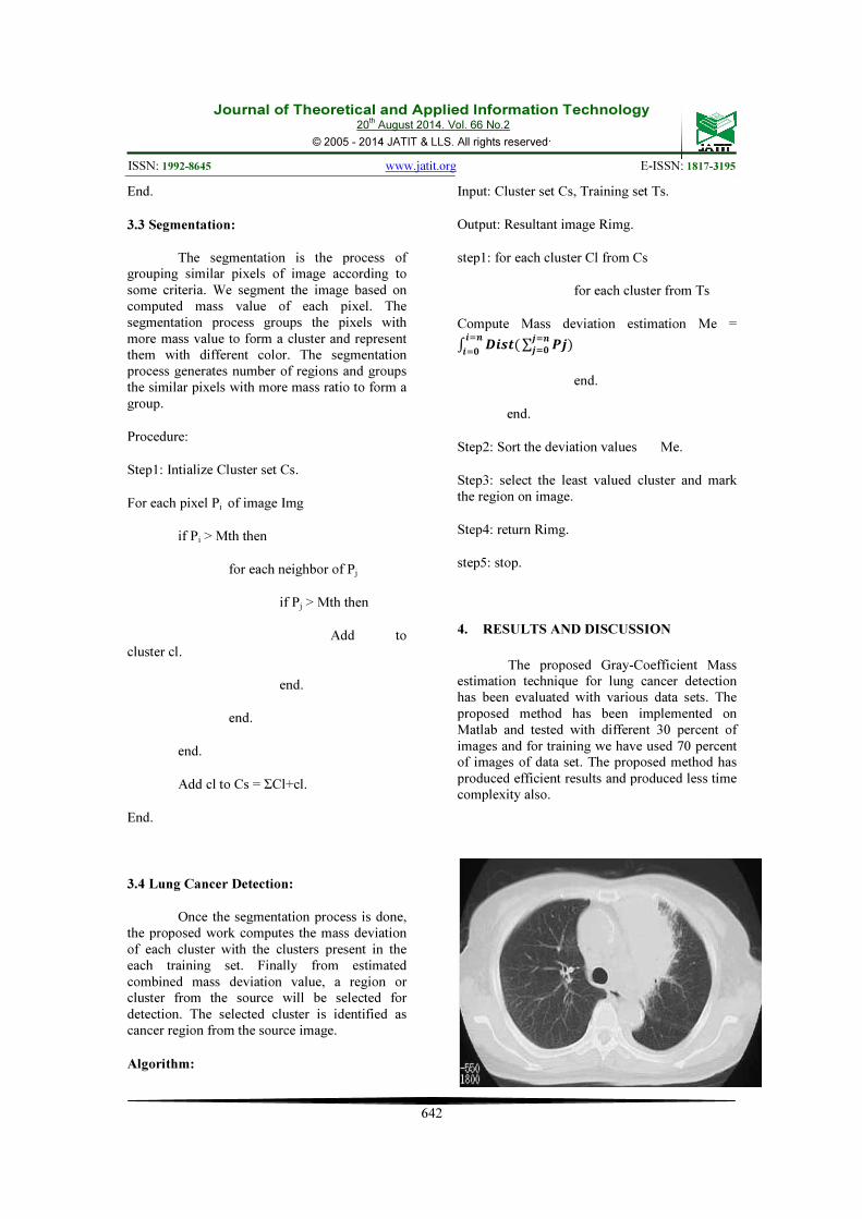
3.4 Lung Cancer Detection:
Once the segmentation process is done, the proposed work computes the mass deviation of each cluster with the clusters present in the each training set. Finally from estimated combined mass deviation value, a region or cluster from the source will be selected for detection. The selected cluster is identified as cancer region from the source image.
Algorithm:
Input: Cluster set Cs, Training set Ts.
Output: Resultant image Rimg.
step1: for each cluster Cl from Cs
for each cluster from Ts
Compute Mass deviation estimation Me =
� ��������
���∑ ����
���
end.
end.
Step2: Sort the deviation values Me.
Step3: select the least valued cluster and mark the region on image.
Step4: return Rimg.
step5: stop.
4. RESULTS AND DISCUSSION
The proposed Gray-Coefficient Mass estimation technique for lung cancer detection has been evaluated with various data sets. The proposed method has been implemented on Matlab and tested with different 30 percent of images and for training we have used 70 percent of images of data set. The proposed method has produced efficient results and produced less time complexity also.
Journal of Theoretical and Applied Information Technology 20
th August 2014. Vol. 66 No.2
© 2005 - 2014 JATIT & LLS. All rights reserved.
ISSN: 1992-8645 www.jatit.org E-ISSN: 1817-3195
643
Figure 2: shows the gray scaled input image
Figure 3: shows the edge detected image.
Figure 4: shows the cancer detected image.
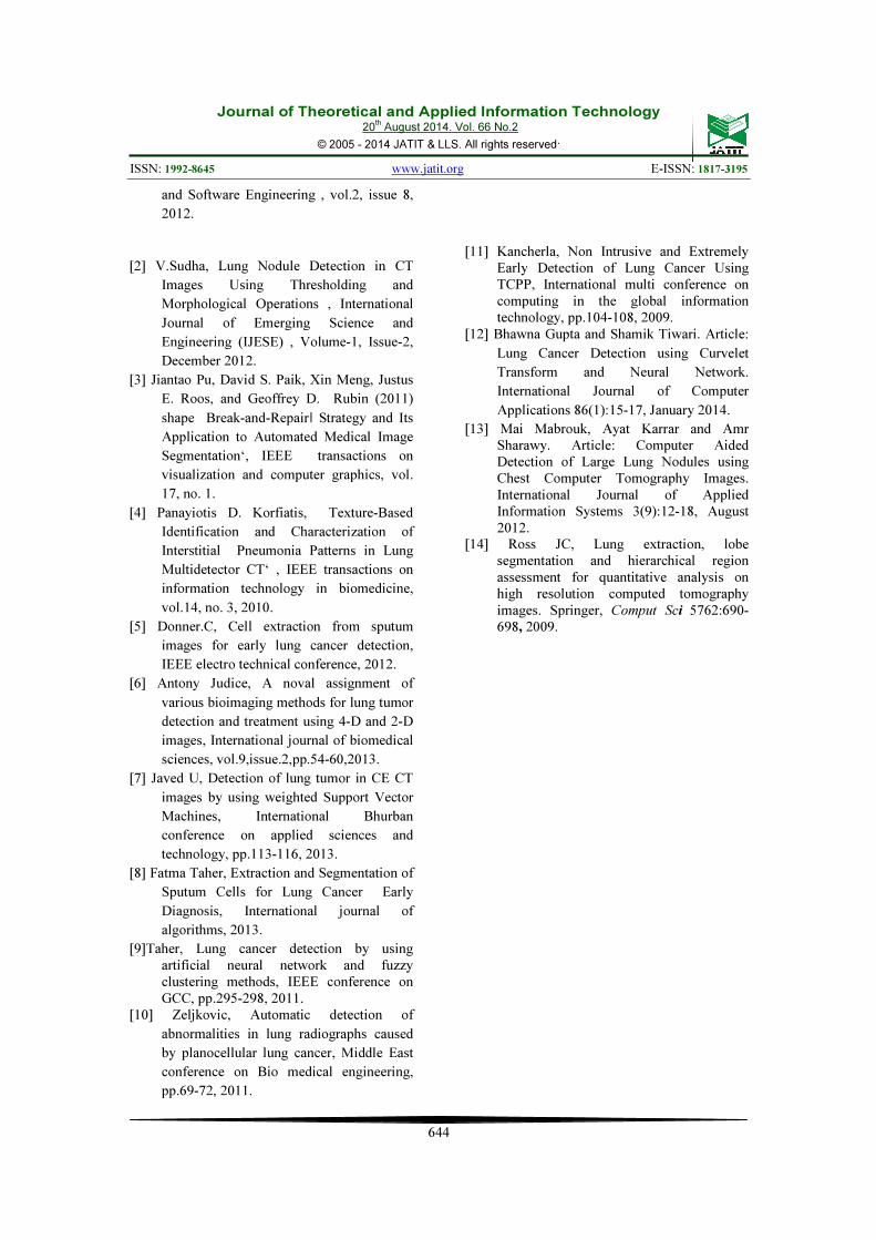
Table1: shows the LCD accuracy
The table 1 shows the lung cancer detection accuracy with number of samples and
it shows that the proposed method has good impact on LCD with growing size.
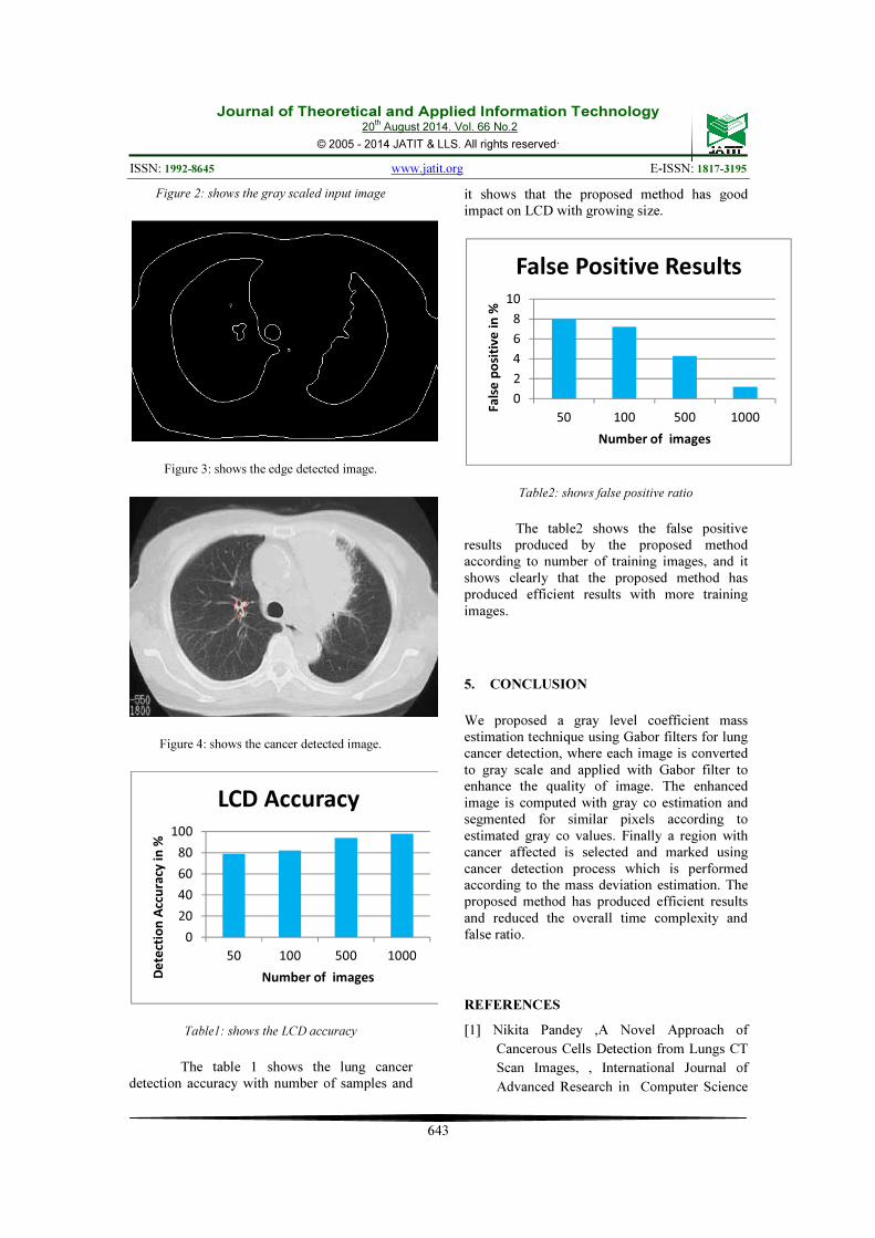
Table2: shows false positive ratio
The table2 shows the false positive results produced by the proposed method according to number of training images, and it shows clearly that the proposed method has produced efficient results with more training images.
5. CONCLUSION
We proposed a gray level coefficient mass estimation technique using Gabor filters for lung cancer detection, where each image is converted to gray scale and applied with Gabor filter to enhance the quality of image. The enhanced image is computed with gray co estimation and segmented for similar pixels according to estimated gray co values. Finally a region with cancer affected is selected and marked using cancer detection process which is performed according to the mass deviation estimation. The proposed method has produced efficient results and reduced the overall time complexity and false ratio.
REFERENCES
[1] Nikita Pandey ,A Novel Approach of
Cancerous Cells Detection from Lungs CT
Scan Images, , International Journal of
Advanced Research in Computer Science
0
20
40
60
80
100
50 100 500 1000
De
tect
ion
Acc
ura
cy i
n %
Number of images
LCD Accuracy
0
2
4
6
8
10
50 100 500 1000
Fa
lse
po
siti
ve
in
%
Number of images
False Positive Results
Journal of Theoretical and Applied Information Technology 20
th August 2014. Vol. 66 No.2
© 2005 - 2014 JATIT & LLS. All rights reserved.
ISSN: 1992-8645 www.jatit.org E-ISSN: 1817-3195
644
and Software Engineering , vol.2, issue 8,
2012.
[2] V.Sudha, Lung Nodule Detection in CT
Images Using Thresholding and
Morphological Operations , International
Journal of Emerging Science and
Engineering (IJESE) , Volume-1, Issue-2,
December 2012.
[3] Jiantao Pu, David S. Paik, Xin Meng, Justus
E. Roos, and Geoffrey D. Rubin (2011)
shape Break-and-Repair‖ Strategy and Its
Application to Automated Medical Image
Segmentation‘, IEEE transactions on
visualization and computer graphics, vol.
17, no. 1.
[4] Panayiotis D. Korfiatis, Texture-Based
Identification and Characterization of
Interstitial Pneumonia Patterns in Lung
Multidetector CT‘ , IEEE transactions on
information technology in biomedicine,
vol.14, no. 3, 2010.
[5] Donner.C, Cell extraction from sputum
images for early lung cancer detection,
IEEE electro technical conference, 2012.
[6] Antony Judice, A noval assignment of
various bioimaging methods for lung tumor
detection and treatment using 4-D and 2-D
images, International journal of biomedical
sciences, vol.9,issue.2,pp.54-60,2013.
[7] Javed U, Detection of lung tumor in CE CT
images by using weighted Support Vector
Machines, International Bhurban
conference on applied sciences and
technology, pp.113-116, 2013.
[8] Fatma Taher, Extraction and Segmentation of
Sputum Cells for Lung Cancer Early
Diagnosis, International journal of
algorithms, 2013.
[9]Taher, Lung cancer detection by using artificial neural network and fuzzy clustering methods, IEEE conference on GCC, pp.295-298, 2011.
[10] Zeljkovic, Automatic detection of
abnormalities in lung radiographs caused
by planocellular lung cancer, Middle East
conference on Bio medical engineering,
pp.69-72, 2011.
[11] Kancherla, Non Intrusive and Extremely Early Detection of Lung Cancer Using TCPP, International multi conference on computing in the global information technology, pp.104-108, 2009.
[12] Bhawna Gupta and Shamik Tiwari. Article:
Lung Cancer Detection using Curvelet
Transform and Neural Network.
International Journal of Computer
Applications 86(1):15-17, January 2014.
[13] Mai Mabrouk, Ayat Karrar and Amr Sharawy. Article: Computer Aided Detection of Large Lung Nodules using Chest Computer Tomography Images. International Journal of Applied Information Systems 3(9):12-18, August 2012.
[14] Ross JC, Lung extraction, lobe segmentation and hierarchical region assessment for quantitative analysis on high resolution computed tomography images. Springer, Comput Sci 5762:690-698, 2009.
Recommended