Overview of fMRI Analysis Software
McConnell BICOpen Methods Meetup
January 13th 2013
FSLMichael Ferreira
Outline
IntroductionMRI scanners and fMRI equipment
FSLImage formats and conversionFSLView and FEATFSL at the BIC
Support Links
Introduction
● Research Assistant● maintain fMRI stimulation and subject monitoring
equipment for the MRI scanners● assist fMRI researchers with the development and
implementation of new protocols● introduce researchers to fMRI statistical analysis
software packages and provide ongoing support● maintain laboratory facilities for, and assist in the
preparation of, MRI phantoms
Hardware
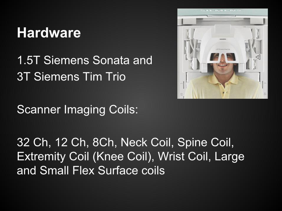
1.5T Siemens Sonata and3T Siemens Tim Trio
Scanner Imaging Coils:
32 Ch, 12 Ch, 8Ch, Neck Coil, Spine Coil, Extremity Coil (Knee Coil), Wrist Coil, Large and Small Flex Surface coils
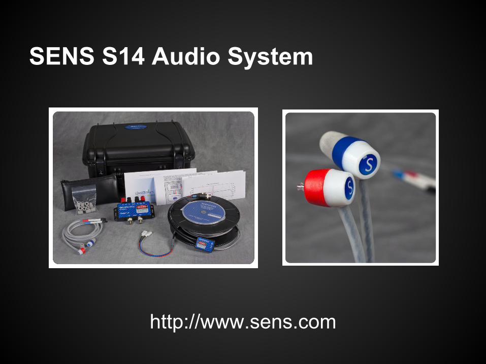
SENS S14 Audio System
http://www.sens.com
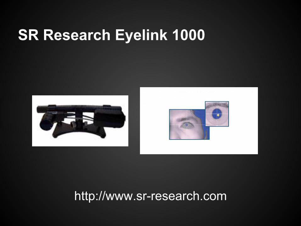
SR Research Eyelink 1000
http://www.sr-research.com
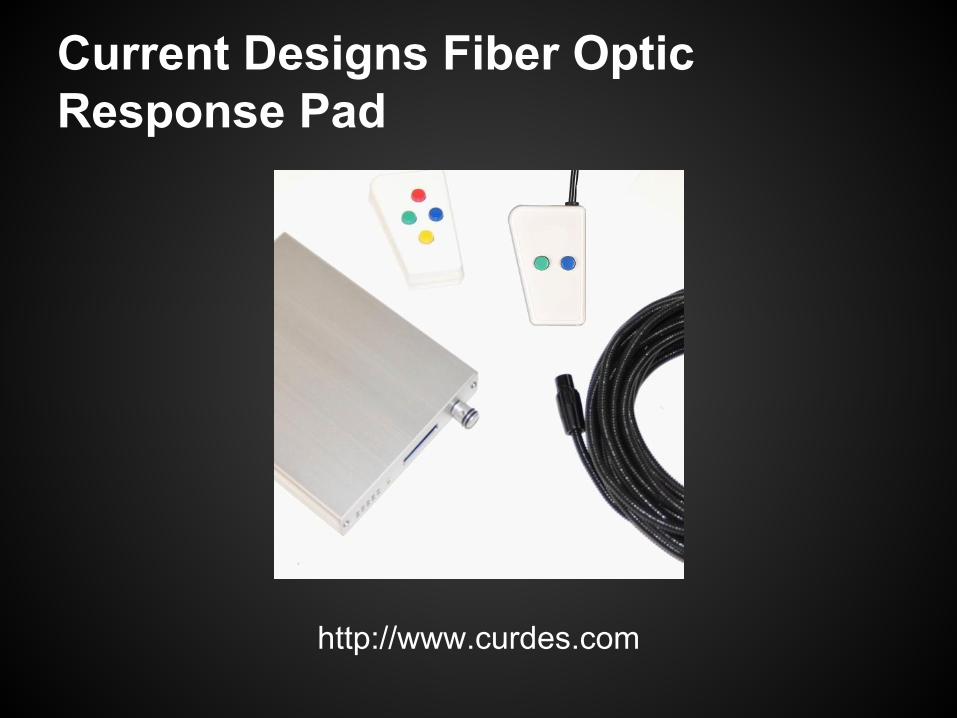
Current Designs Fiber Optic Response Pad
http://www.curdes.com
Hardware ...
● SENS SR14 ear insert audio system● SR research Eyelink 1000 Eye tracker● Current Designs Response Pads● 2 XGA (1024x768) LCD projectors● Siemens Physiological Monitoring Unit
○ ECG○ Pulse○ Respiration○ log files with scan markers
FSL
● Imaging data provided in DICOM and MINC formats
● FSL uses the NIFTI (.nii) format● conversion to NIFTI from DICOM
○ MRIcro http://www.mccauslandcenter.sc.edu/mricro● conversion to NIFTI from MINC
○ mnc2nii
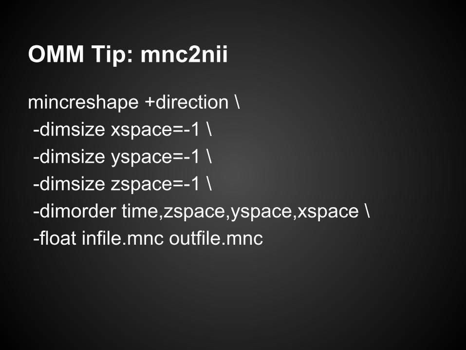
OMM Tip: mnc2nii
mincreshape +direction \ -dimsize xspace=-1 \ -dimsize yspace=-1 \ -dimsize zspace=-1 \ -dimorder time,zspace,yspace,xspace \ -float infile.mnc outfile.mnc
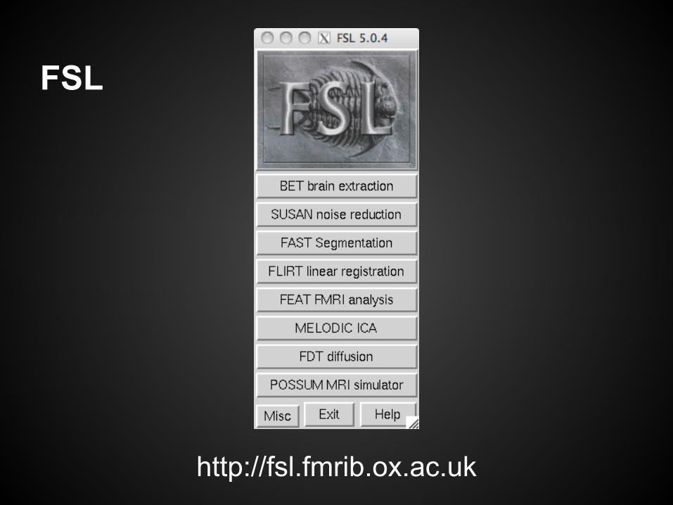
http://fsl.fmrib.ox.ac.uk
FSL
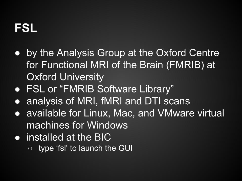
FSL
● by the Analysis Group at the Oxford Centre for Functional MRI of the Brain (FMRIB) at Oxford University
● FSL or “FMRIB Software Library”● analysis of MRI, fMRI and DTI scans● available for Linux, Mac, and VMware virtual
machines for Windows● installed at the BIC
○ type ‘fsl’ to launch the GUI
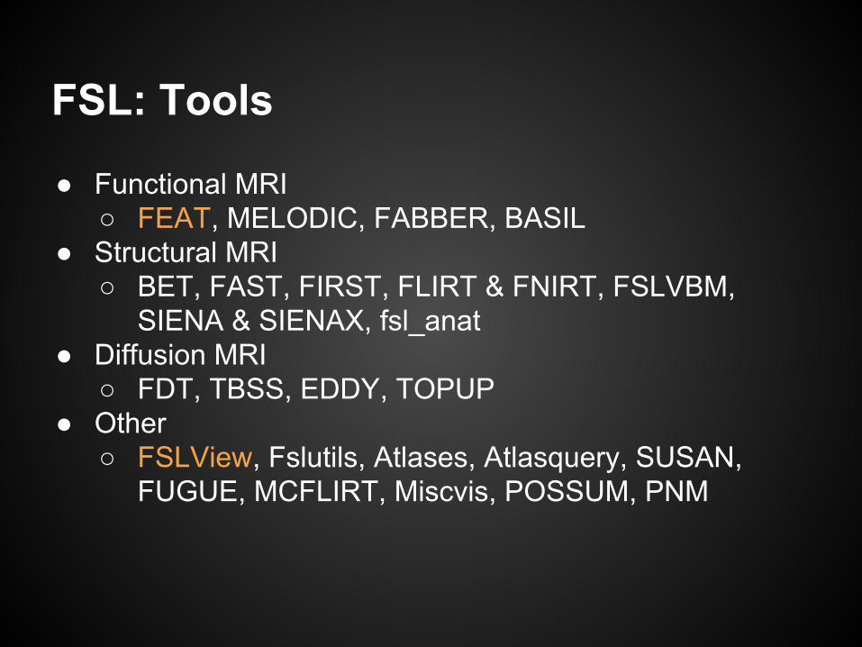
FSL: Tools
● Functional MRI○ FEAT, MELODIC, FABBER, BASIL
● Structural MRI○ BET, FAST, FIRST, FLIRT & FNIRT, FSLVBM,
SIENA & SIENAX, fsl_anat● Diffusion MRI
○ FDT, TBSS, EDDY, TOPUP● Other
○ FSLView, Fslutils, Atlases, Atlasquery, SUSAN, FUGUE, MCFLIRT, Miscvis, POSSUM, PNM
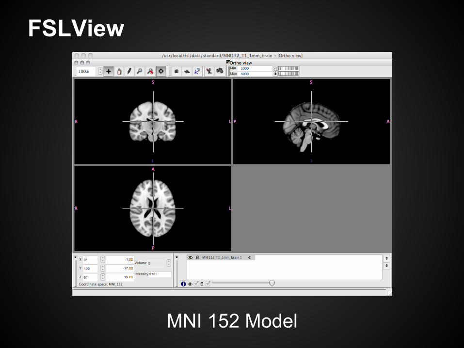
FSLView
MNI 152 Model
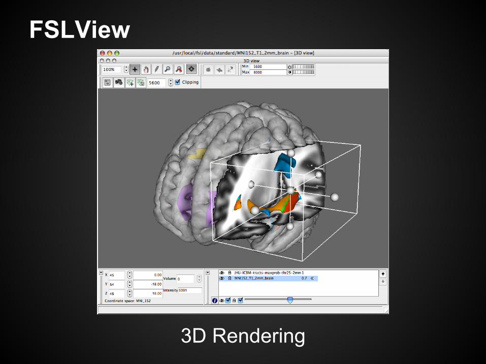
FSLView
3D Rendering
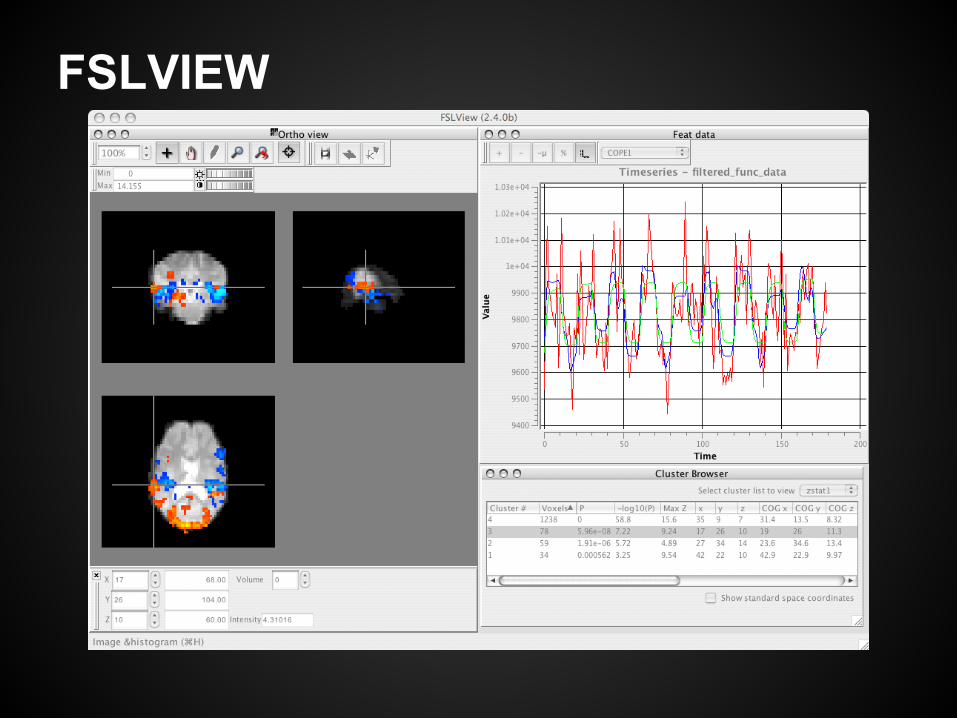
FSLVIEW
FSLView
● view 3D/4D Nifti images○ fmri, dti, anatomical, atlases, 3D rendering
● check your raw data● “movie” mode to cycle through a timeseries
○ motion, ghosting, artefacts● overlays (anatomicals, stats, atlases)
○ multiple overlays, used like layers in GIMP/PS○ must have the same dimensions as the main image
● variety of colour maps available○ lacks spectral mode like Display/register
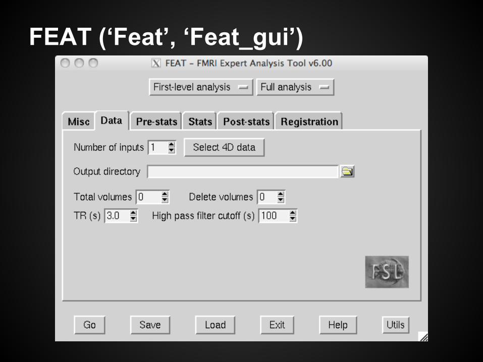
FEAT (‘Feat’, ‘Feat_gui’)
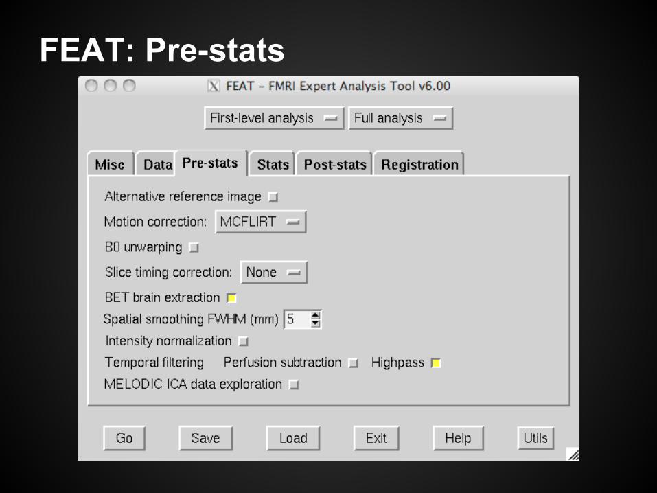
FEAT: Pre-stats
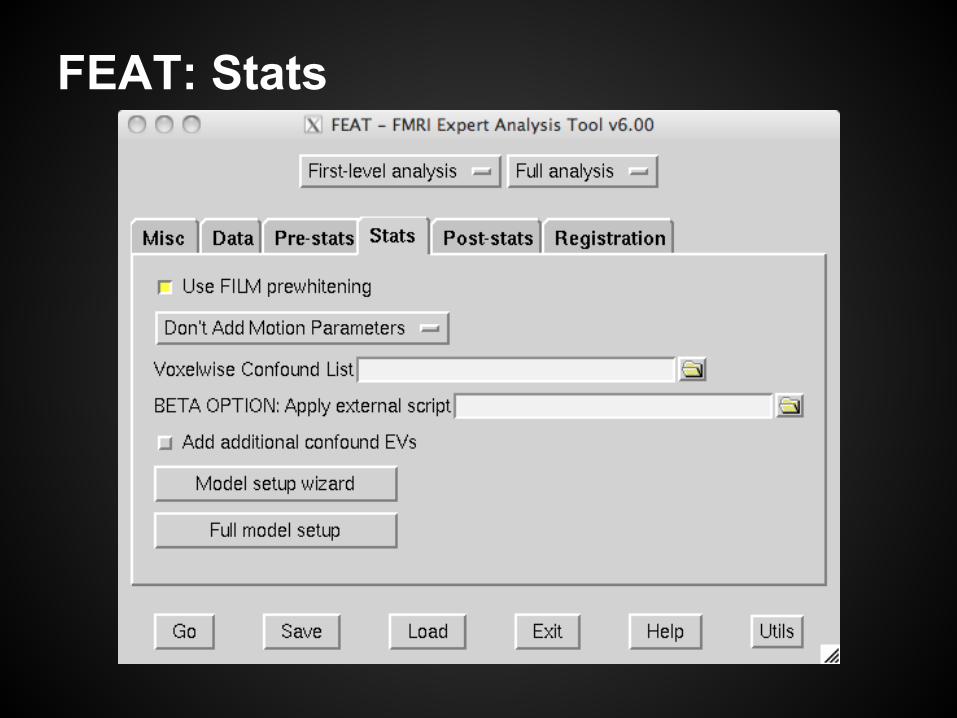
FEAT: Stats
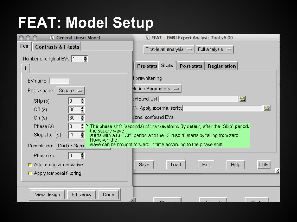
FEAT: Model Setup
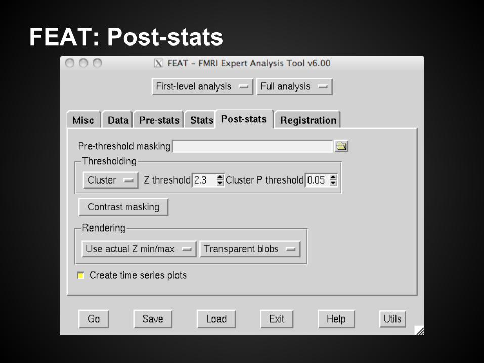
FEAT: Post-stats
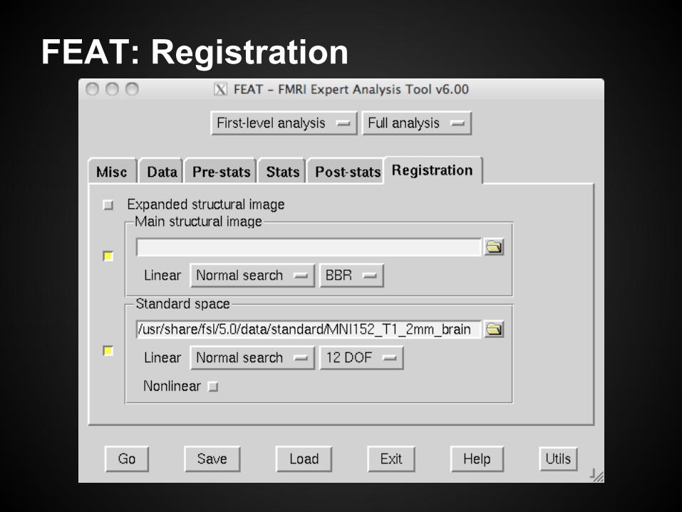
FEAT: Registration
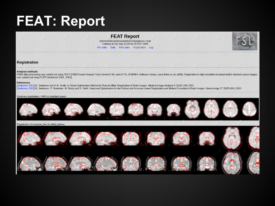
FEAT: Report
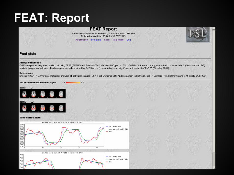
FEAT: Report
FEAT at the BIC: SGE batch
● execution is scheduled on a system of networked computers○ faster than running only on your local machine
● for local execution, disable batch:○ tcsh: unsetenv SGE_ROOT○ bash: unset SGE_ROOT
● try using bash instead of a tcsh shell○ tcsh users may need a custom fsl.csh settings file
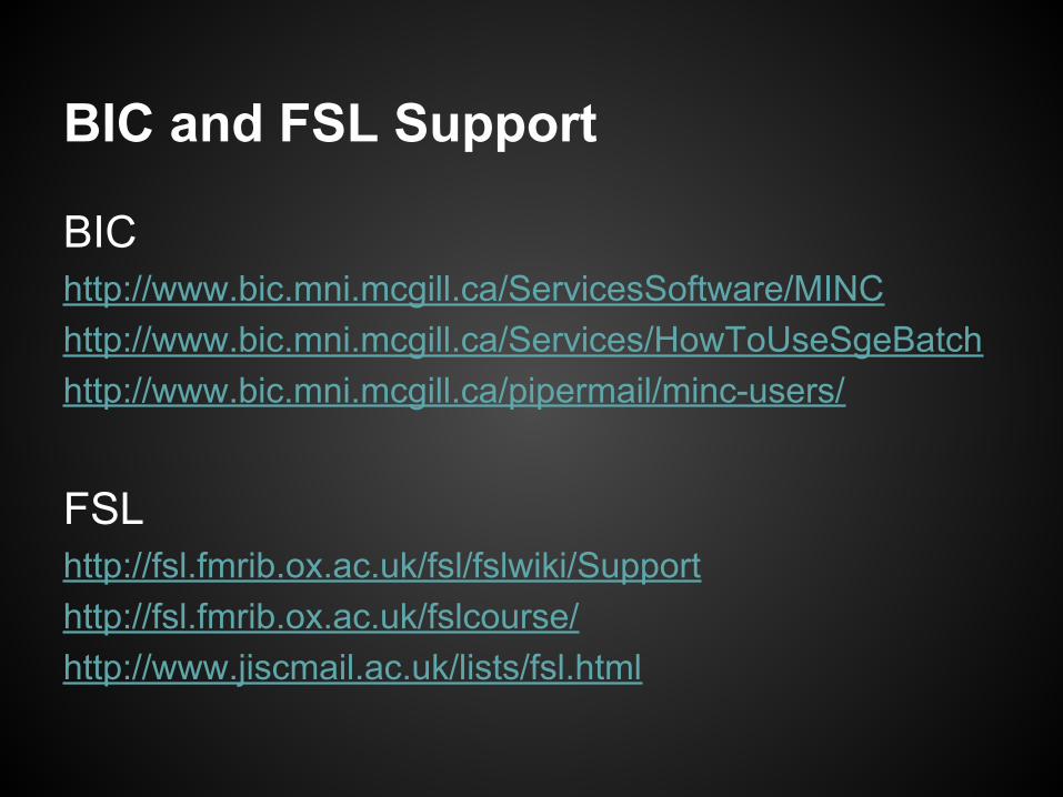
BIC and FSL Support
BIChttp://www.bic.mni.mcgill.ca/ServicesSoftware/MINChttp://www.bic.mni.mcgill.ca/Services/HowToUseSgeBatchhttp://www.bic.mni.mcgill.ca/pipermail/minc-users/
FSL http://fsl.fmrib.ox.ac.uk/fsl/fslwiki/Supporthttp://fsl.fmrib.ox.ac.uk/fslcourse/http://www.jiscmail.ac.uk/lists/fsl.html
AFNIBenjamin Elgie
AFNI Intro
AFNI is an fMRI software suite developed by a group at the NIH
http://afni.nimh.nih.gov/
It runs on Linux systems using libXP, tcsh, PyQt4 and RIt runs on Mac OSX using Xcode, fink, tcsh, PyQt, and R
AFNI components
AFNI consists of 3 principal components:
A GUI used primarily for viewing data
A collection of command-line scripts
A parallel surface analysis package (SUMA)
AFNI GUI
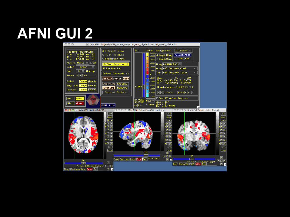
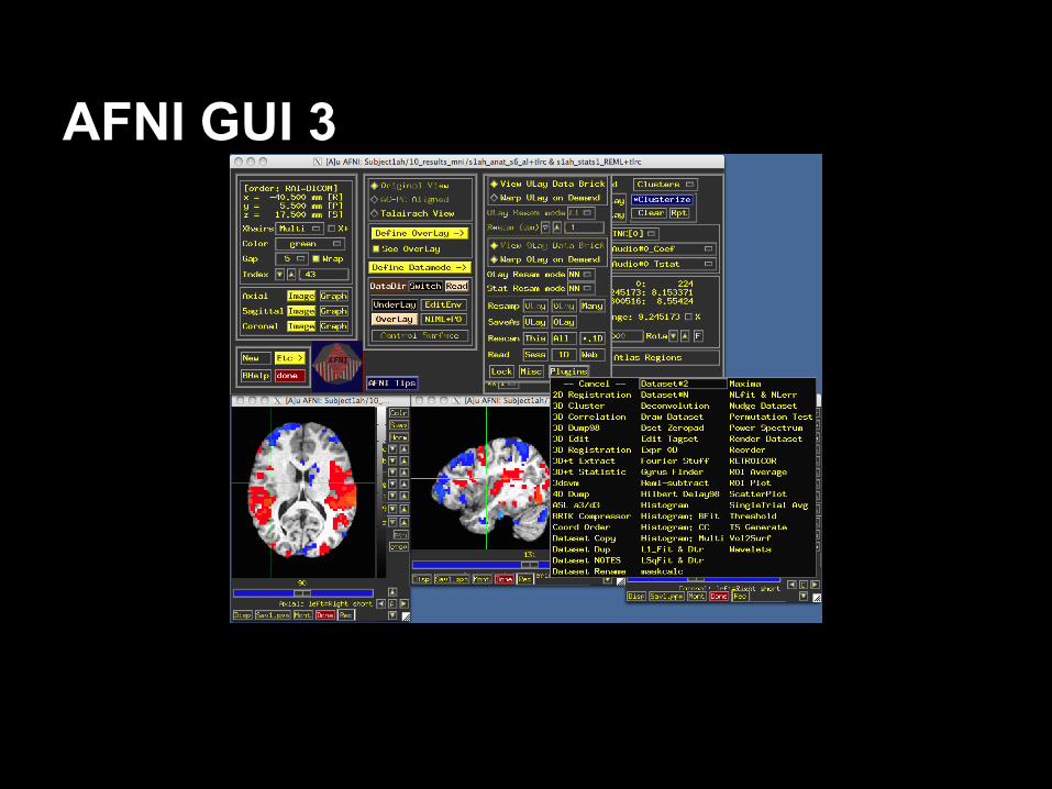
The AFNI GUI is a major part of the software
Can be used for viewing data
Can also conduct certain “on the fly” analyses
AFNI GUI 2
AFNI GUI 3
AFNI Scripts
This is a set of scripts which can either be run from the command line or from a script using tcsh
They typically take as input .1D files, or .head & .brik pairs.
AFNI is increasingly able to process NIFTI format files as well.
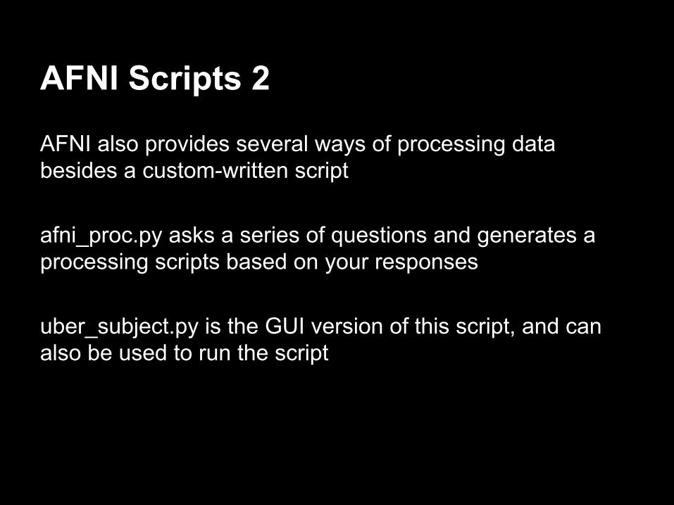
AFNI Scripts 2
AFNI also provides several ways of processing data besides a custom-written script
afni_proc.py asks a series of questions and generates a processing scripts based on your responses
uber_subject.py is the GUI version of this script, and can also be used to run the script
AFNI processing
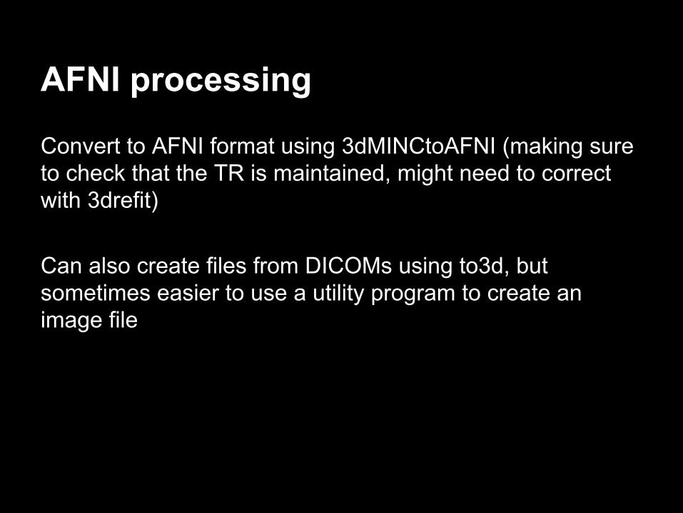
Convert to AFNI format using 3dMINCtoAFNI (making sure to check that the TR is maintained, might need to correct with 3drefit)
Can also create files from DICOMs using to3d, but sometimes easier to use a utility program to create an image file
AFNI processing 2
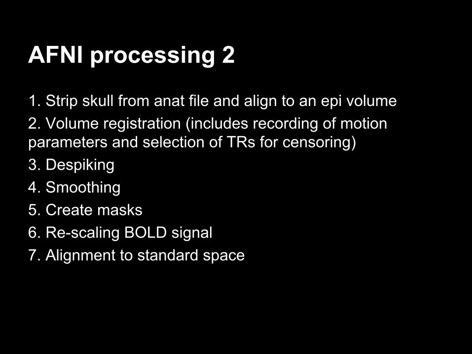
1. Strip skull from anat file and align to an epi volume2. Volume registration (includes recording of motion parameters and selection of TRs for censoring)3. Despiking4. Smoothing5. Create masks6. Re-scaling BOLD signal7. Alignment to standard space
AFNI Ordinary/Generalized LS
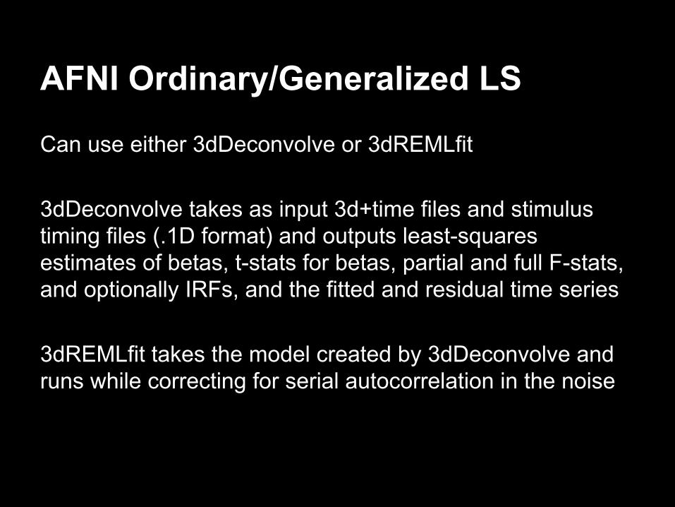
Can use either 3dDeconvolve or 3dREMLfit
3dDeconvolve takes as input 3d+time files and stimulus timing files (.1D format) and outputs least-squares estimates of betas, t-stats for betas, partial and full F-stats, and optionally IRFs, and the fitted and residual time series
3dREMLfit takes the model created by 3dDeconvolve and runs while correcting for serial autocorrelation in the noise
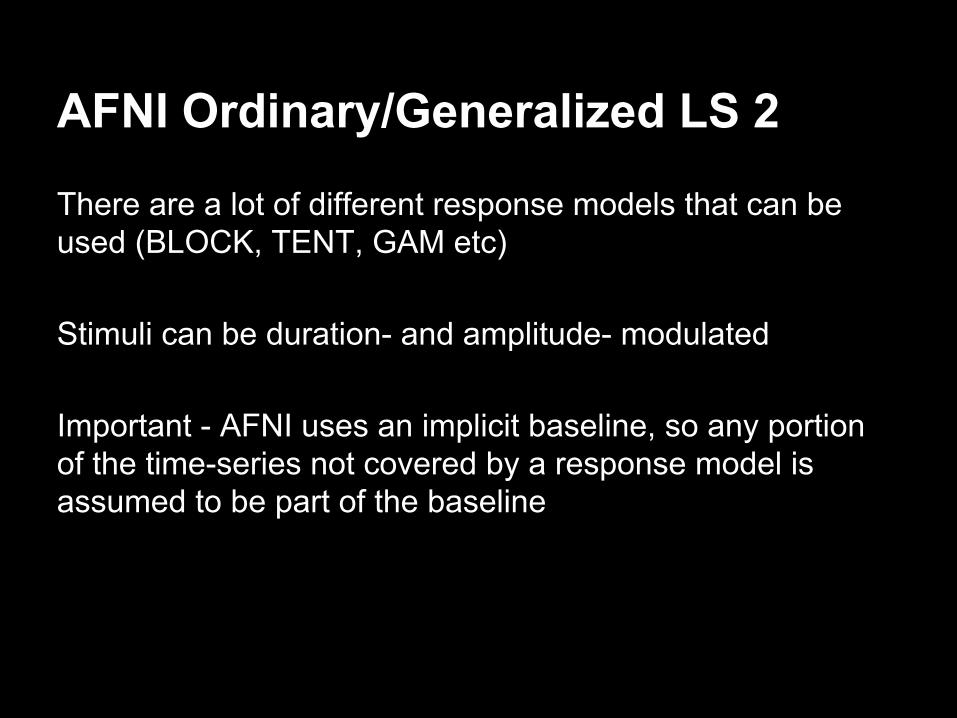
AFNI Ordinary/Generalized LS 2
There are a lot of different response models that can be used (BLOCK, TENT, GAM etc)
Stimuli can be duration- and amplitude- modulated
Important - AFNI uses an implicit baseline, so any portion of the time-series not covered by a response model is assumed to be part of the baseline
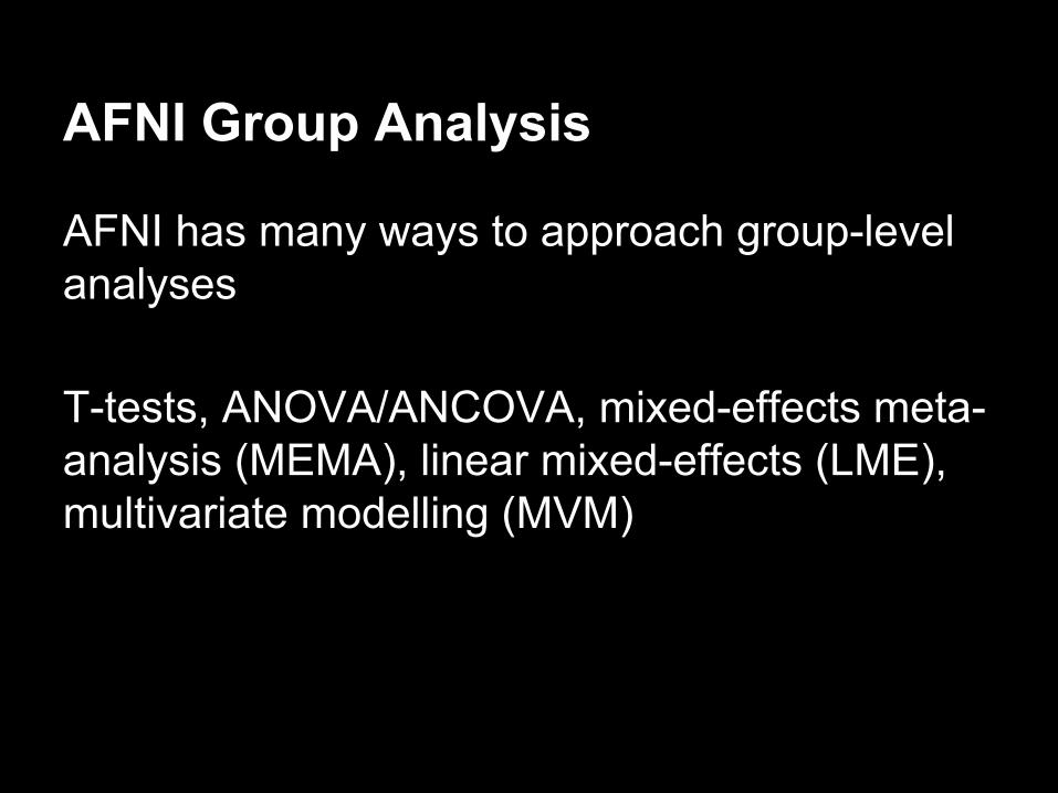
AFNI Group Analysis
AFNI has many ways to approach group-level analyses
T-tests, ANOVA/ANCOVA, mixed-effects meta-analysis (MEMA), linear mixed-effects (LME), multivariate modelling (MVM)
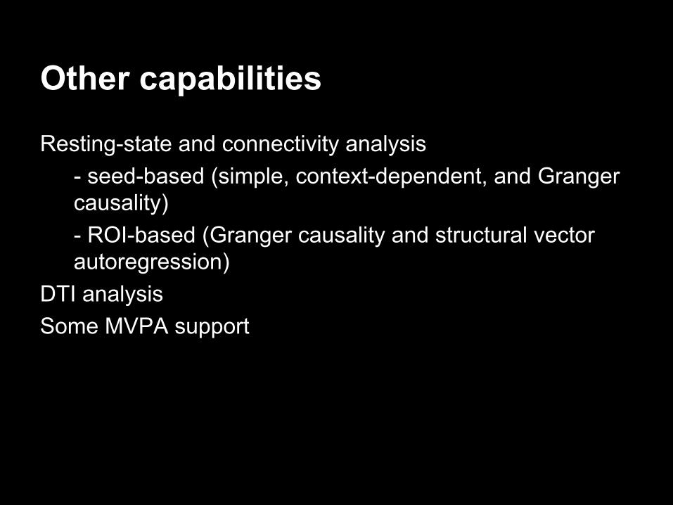
Other capabilities
Resting-state and connectivity analysis- seed-based (simple, context-dependent, and Granger causality)- ROI-based (Granger causality and structural vector autoregression)
DTI analysisSome MVPA support
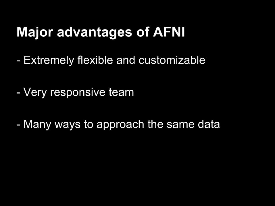
Major advantages of AFNI
- Extremely flexible and customizable
- Very responsive team
- Many ways to approach the same data
Disadvantages of AFNI
- Steep learning curve
- Easy to use the wrong function or approach
- Requires a good understanding of each step in a processing pathway
SPMLaureline Arnaud
fMRI analysis with SPM
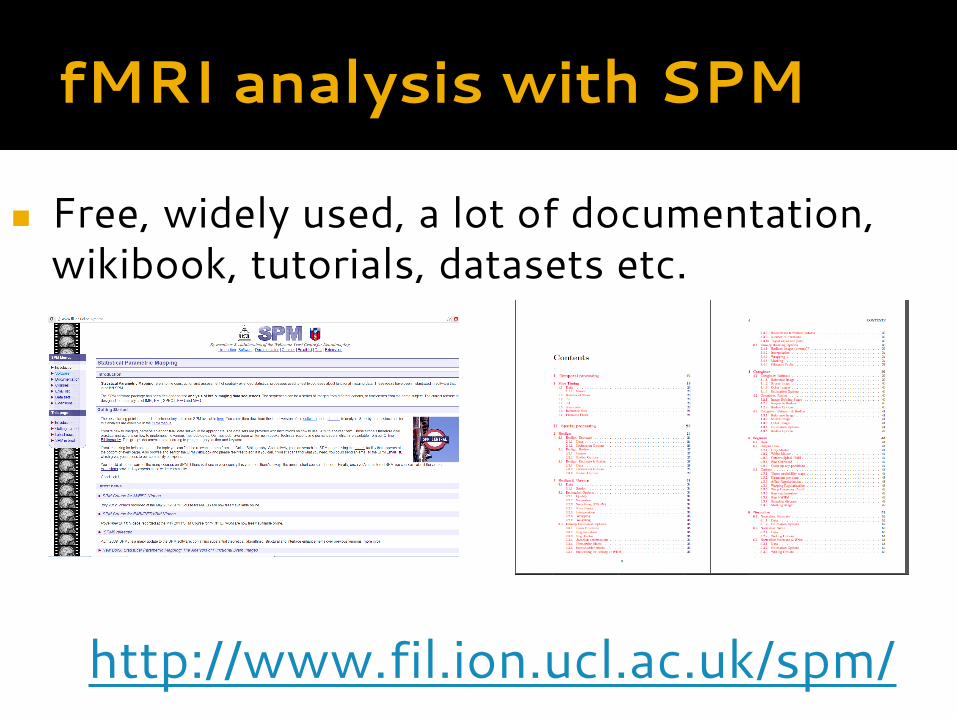
◼ Free, widely used, a lot of documentation, wikibook, tutorials, datasets etc.
http://www.fil.ion.ucl.ac.uk/spm/
fMRI analysis with SPM
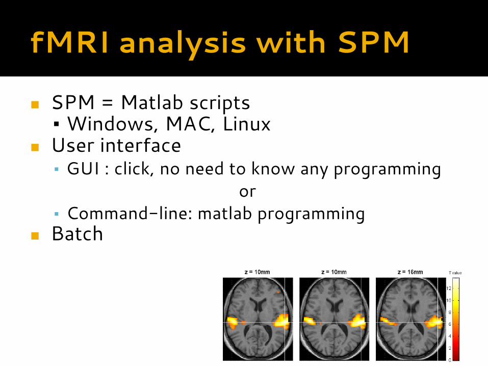
◼ SPM = Matlab scripts ▪ Windows, MAC, Linux
◼ User interface▪ GUI : click, no need to know any programming
or▪ Command-line: matlab programming
◼ Batch
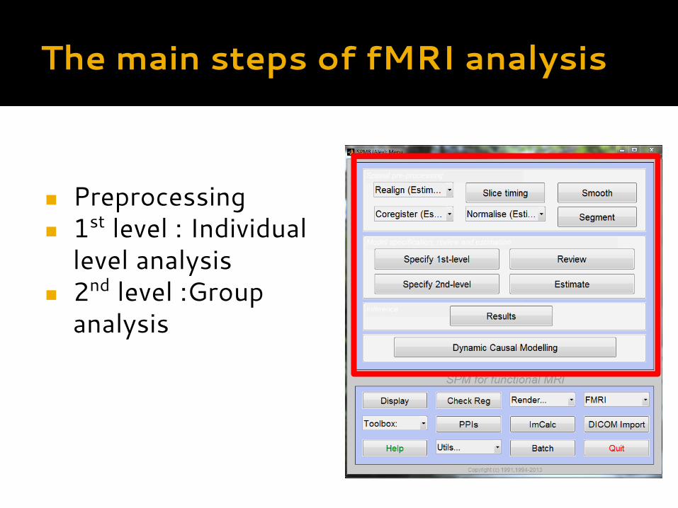
The main steps of fMRI analysis
◼ Preprocessing◼ 1st level : Individual
level analysis◼ 2nd level :Group
analysis
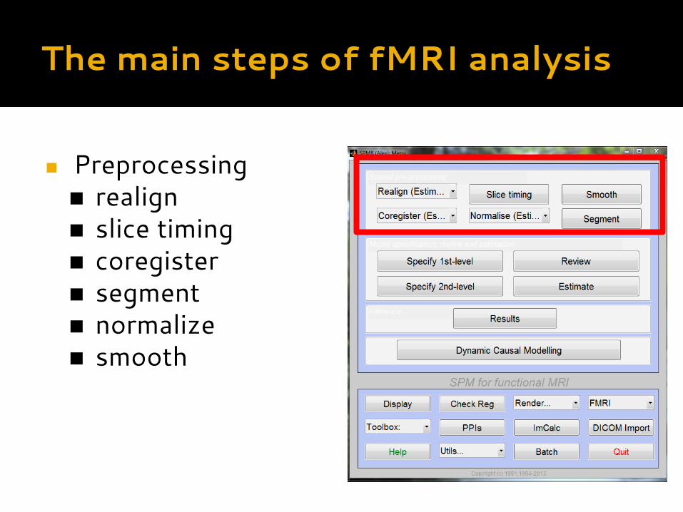
The main steps of fMRI analysis
◼ Preprocessing◼ realign◼ slice timing◼ coregister◼ segment◼ normalize◼ smooth
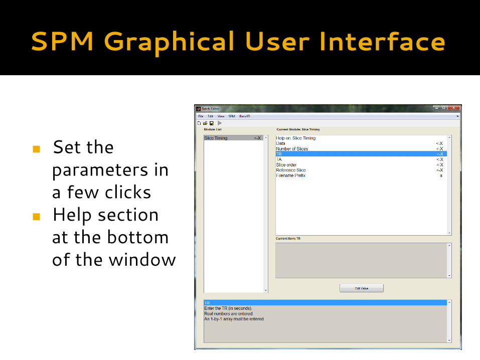
SPM Graphical User Interface
◼ Set the parameters in a few clicks
◼ Help section at the bottom of the window
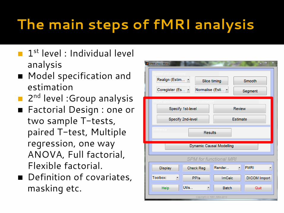
The main steps of fMRI analysis
◼ 1st level : Individual level analysis
◼ Model specification and estimation
◼ 2nd level :Group analysis◼ Factorial Design : one or
two sample T-tests, paired T-test, Multiple regression, one way ANOVA, Full factorial, Flexible factorial.
◼ Definition of covariates, masking etc.
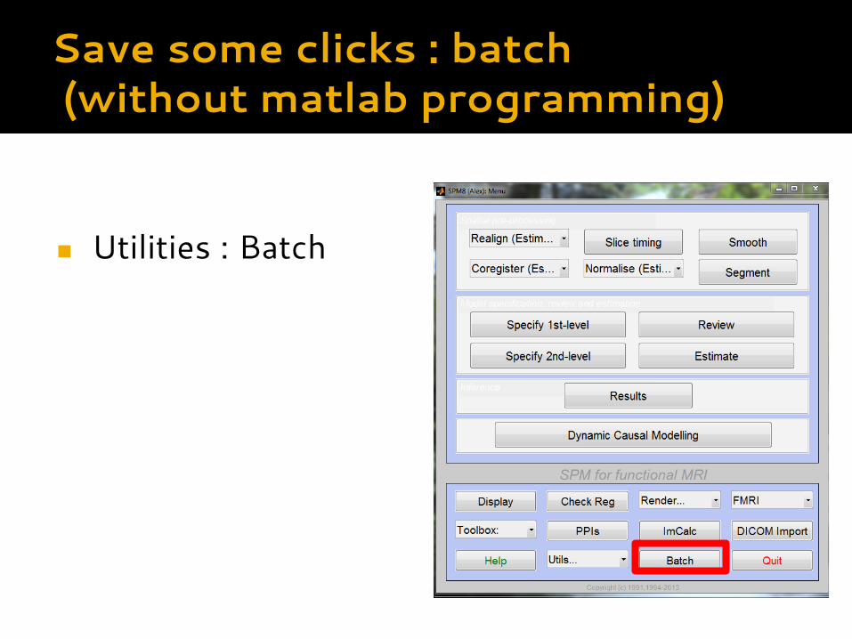
Save some clicks : batch (without matlab programming)
◼ Utilities : Batch
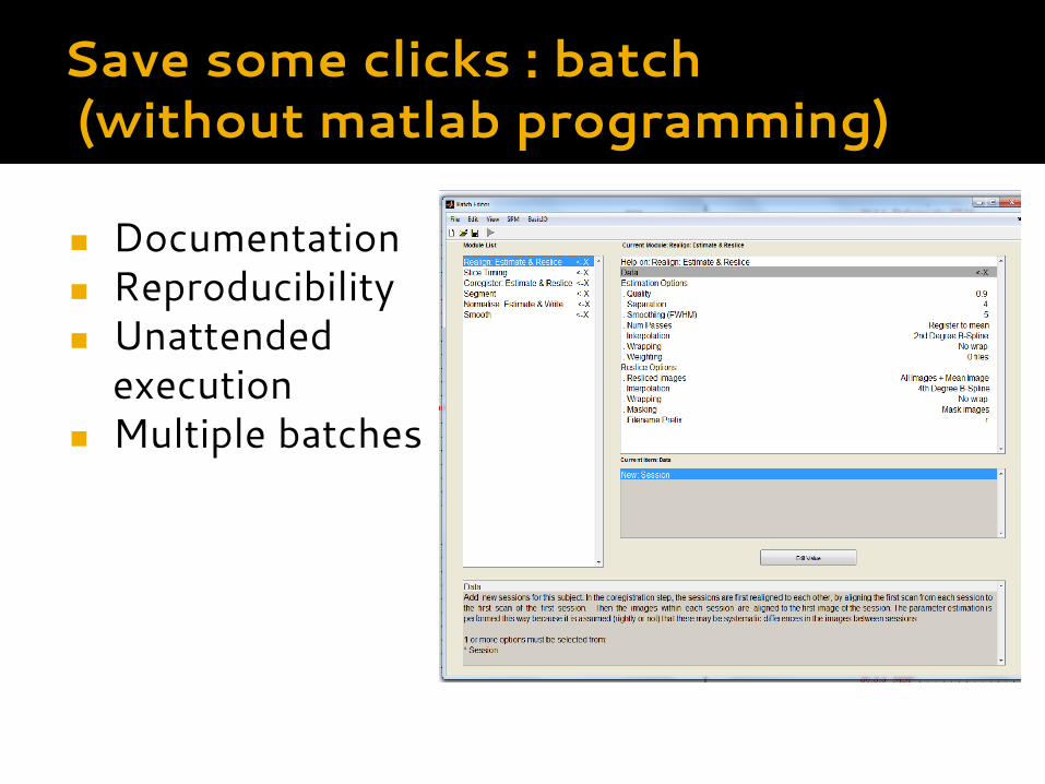
Save some clicks : batch (without matlab programming)
◼ Documentation◼ Reproducibility◼ Unattended
execution◼ Multiple batches
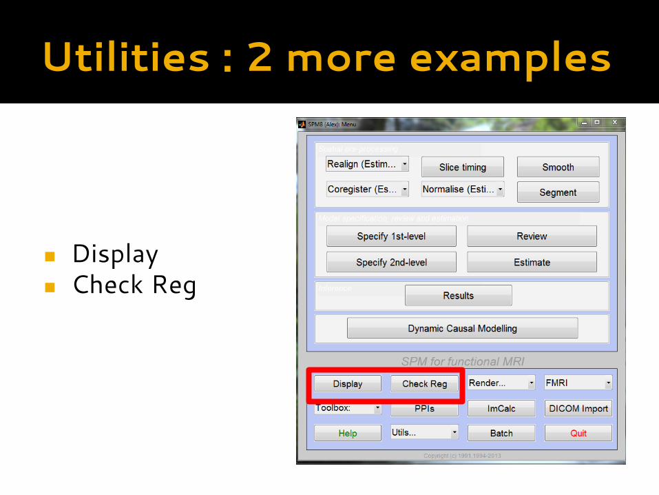
Utilities : 2 more examples
◼ Display◼ Check Reg
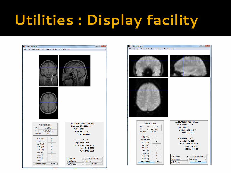
Utilities : Display facility
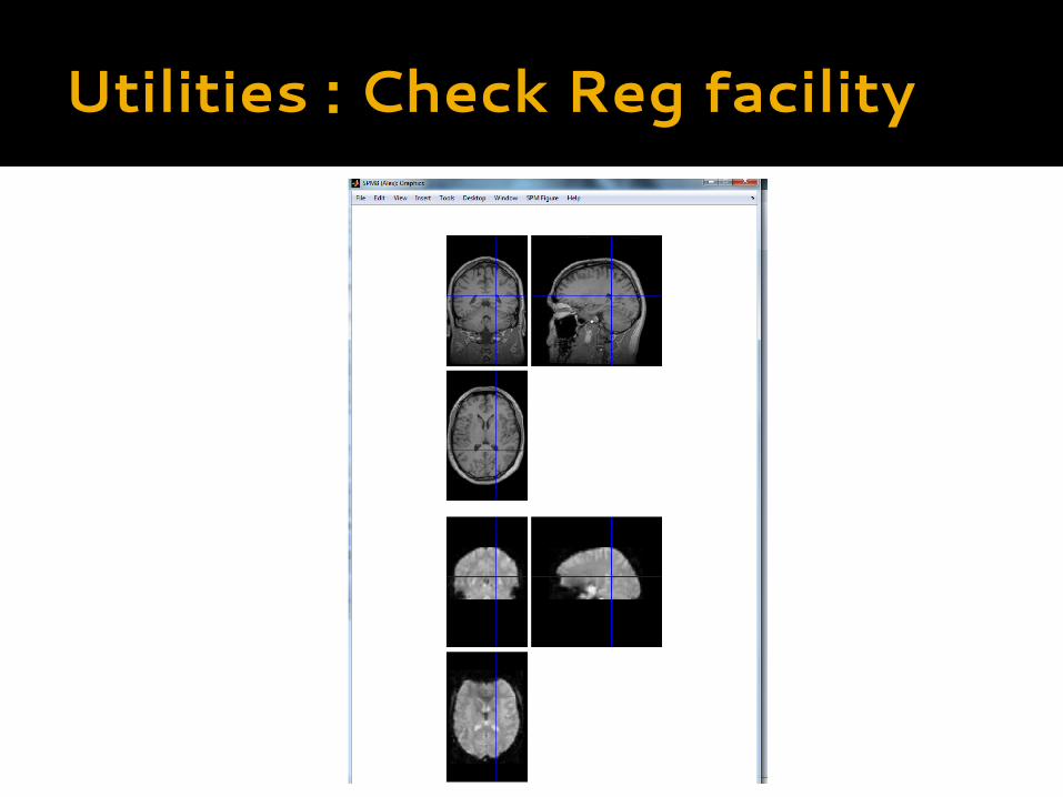
Utilities : Check Reg facility
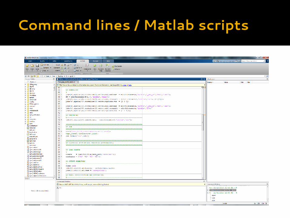
Command lines / Matlab scripts
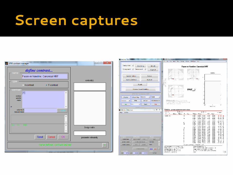
Screen captures
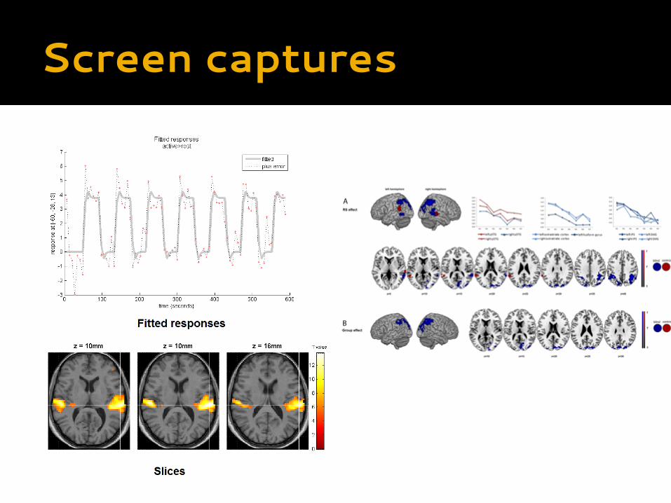
Screen captures
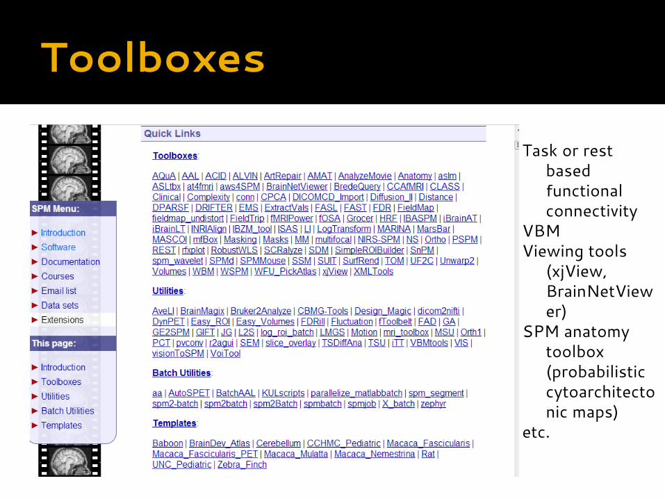
Task or rest based functional connectivity
VBMViewing tools
(xjView, BrainNetViewer)
SPM anatomy toolbox (probabilistic cytoarchitectonic maps)
etc.
Toolboxes
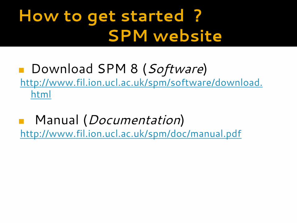
How to get started ?SPM website
◼ Download SPM 8 (Software)http://www.fil.ion.ucl.ac.uk/spm/software/download.
html
◼ Manual (Documentation)http://www.fil.ion.ucl.ac.uk/spm/doc/manual.pdf
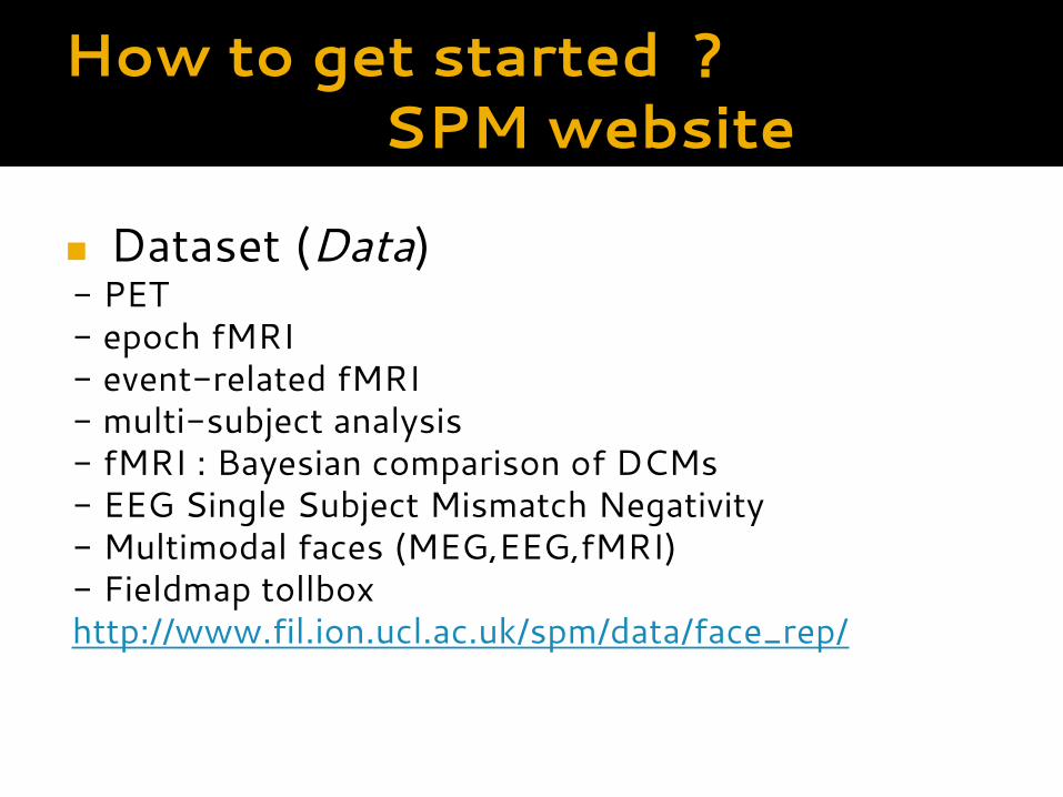
How to get started ?SPM website
◼ Dataset (Data)- PET- epoch fMRI- event-related fMRI - multi-subject analysis- fMRI : Bayesian comparison of DCMs- EEG Single Subject Mismatch Negativity- Multimodal faces (MEG,EEG,fMRI)- Fieldmap tollbox http://www.fil.ion.ucl.ac.uk/spm/data/face_rep/
Thanks !
◼ Interested in creating a SPM user group ?
Recommended