Bioenergetic cost of making an adenosinetriphosphate molecule in animal mitochondriaIan N. Watta, Martin G. Montgomerya, Michael J. Runswicka, Andrew G. W. Leslieb,1, and John E. Walkera,1
aThe Medical Research Council Mitochondrial Biology Unit, Hills Road, Cambridge, CB2 0XY, United Kingdom; and bThe Medical Research CouncilLaboratory of Molecular Biology, Hills Road, Cambridge, CB2 0QH, United Kingdom
Contributed by John E. Walker, August 3, 2010 (sent for review July 9, 2010)
The catalytic domain of the F-ATPase in mitochondria protrudesinto the matrix of the organelle, and is attached to the membranedomain by central and peripheral stalks. Energy for the synthesis ofATP from ADP and phosphate is provided by the transmembraneproton-motive-force across the inner membrane, generated byrespiration. The proton-motive force is coupled mechanically toATP synthesis by the rotation at about 100 times per second ofthe central stalk and an attached ring of c-subunits in the mem-brane domain. Each c-subunit carries a glutamate exposed aroundthe midpoint of the membrane on the external surface of the ring.The rotation is generated by protonation and deprotonation suc-cessively of each glutamate. Each 360° rotation produces three ATPmolecules, and requires the translocation of one proton per gluta-mate by each c-subunit in the ring. In fungi, eubacteria, and plantchloroplasts, ring sizes of c10–c15 subunits have been observed,implying that these enzymes need 3.3–5 protons to make eachATP, but until now no higher eukaryote has been examined. Asshown here in the structure of the bovine F1-c-ring complex, thec-ring has eight c-subunits. As the sequences of c-subunits are iden-tical throughout almost all vertebrates and are highly conserved ininvertebrates, their F-ATPases probably contain c8-rings also.Therefore, in about 50,000 vertebrate species, and probably inmany or all of the two million invertebrate species, 2.7 protonsare required by the F-ATPase to make each ATP molecule.
ATP synthase ∣ rotational catalysis ∣ c-ring structure ∣ protons per ATP ∣vertebrates
Almost all ATP in respiring cells is made by the membranebound enzyme F-ATPase (F-ATP synthase). In the F-ATPase
in the inner membranes of mitochondria, the energy of thetransmembrane proton-motive-force, generated by respiration,is coupled mechanically to the synthesis of ATP from ADPand phosphate in its membrane extrinsic catalytic domain by ro-tating the asymmetrical central stalk in a clockwise direction (asviewed from the membrane) at about 100 times per second (1–4).The spherical catalytic domain, which protrudes into the matrixof the organelle, has three catalytic sites in β-subunits at inter-faces with α-subunits (5). The rotational torque is resisted bythe peripheral stalk which links the surface of the catalytic do-main to subunit a (ATPase-6) in the membrane domain; togetherthey constitute the stator (6). The asymmetry of the central stalkimposes different conformations on the three catalytic sites. In aground state structure of the catalytic domain, two of them, theβDP and the βTP sites, have similar but significantly differentclosed conformations. Both bind nucleotides, but catalysis occursat the βDP site. The third, or βE site, has a different open confor-mation with low nucleotide affinity (5). These three catalyticconformations correspond to “tight,” “loose,” and “open” statesin a binding change mechanism of ATP hydrolysis and synthesis(7). Each 360° rotation of the central stalk takes each catalytic sitethrough these conformations in which substrates bind, and threeATP molecules are made and released. The turning of the rotor isimpelled by protons, driven across the inner membrane into themitochondrial matrix by the transmembrane proton-motive force.The transmembrane pathway for protons in the a-subunit has not
been defined structurally. This pathway allows protons in the in-termembrane space to access an essential ionized carboxylate of aglutamate residue, midmembrane on the C-terminal α-helix ofsubunit c. Once protonated, this carboxylate moves to a morehydrophobic environment by Brownian motion generating a ro-tation of the ring. As succeeding c-subunits become protonated,each neutralized carboxylate reaches an environment in subunit awhere it reionises, releasing the proton into the mitochondrialmatrix (8). According to current models based on structures,the number of translocated protons for generation of each 360°rotation is the same as the number of c-subunits in the ring, aseach c-subunit carries a carboxylate involved in protonation anddeprotonation events. In the yeast F-ATPase, the ring has tenc-subunits, and so ten protons are translocated per three ATPmolecules made during a 360° cycle; therefore, the bioenergeticcost to the enzyme is 3.3 protons per ATP (9). However, the c-ringsizes differ between species; c10–c15 rings have been found inyeast, eubacterial, and plant chloroplast F-ATPases (10–13).Therefore, the bioenergetic cost of these F-ATPases making anATP molecule ranges from 3.3–5 protons per ATP.
Until now, the c-ring symmetry and the bioenergetic cost ofmaking an ATP in a mammalian F-ATPase has been unknown.As described here, we have determined the ring size in the struc-ture of the bovine F1-c-ring complex at 3.5 Å resolution.
Results and DiscussionIsolation of the Bovine F1-c-ring Complex. The complex was pre-pared from the purified bovine ATP synthase by dissociationof the peripheral stalk, subunit a, and other minor membranesubunits (subunits A6L, e, f, and g) with detergents. The purifiedcomplex consisted of the α-, β-, γ-, δ- and ϵ-subunits that consti-tute the catalytic F1-domain plus the membrane protein, sub-unit c. All of these subunits were present in crystals of thecomplex (Fig. S1).
Structure Determination. The structure of the bovine F1-c-ringcomplex (Fig. 1) was solved by molecular replacement with datato 3.5 Å resolution. Data processing and statistics are summa-rized in Table 1. The final model contains the following residues:19-510 of the three α-subunits, 9-475 of the three β-subunits, re-sidues 1-61, 67-96, and 101-272 of the γ-subunit, residues 15-145of the δ-subunit, residues 1-47 of the ϵ-subunit, and residues 2-73of c-subunits. The c-ring contains eight c-subunits. During refine-
Author contributions: J.E.W. designed research; I.N.W., M.G.M., M.J.R., and J.E.W.performed research; I.N.W., A.G.W.L., and J.E.W. analyzed data; and J.E.W. wrotethe paper.
The authors declare no conflict of interest.
Freely available online through the PNAS open access option.
Data deposition: The atomic coordinates have been deposited in the Protein Data Bank,www.pdb.org (PDB ID code 2xnd).
See Commentary on page 16755.1To whom correspondence may be addressed. E-mail: [email protected] [email protected].
This article contains supporting information online at www.pnas.org/lookup/suppl/doi:10.1073/pnas.1011099107/-/DCSupplemental.
www.pnas.org/cgi/doi/10.1073/pnas.1011099107 PNAS ∣ September 28, 2010 ∣ vol. 107 ∣ no. 39 ∣ 16823–16827
BIOCH
EMISTR
YSE
ECO
MMEN
TARY
ment, an unsuccessful attempt was made to model a c9-ring intothe density (see Fig. S2).
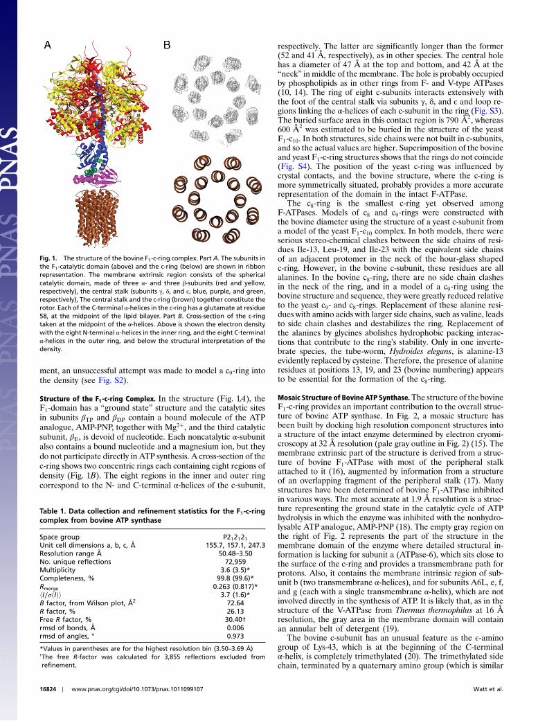
Structure of the F1-c-ring Complex. In the structure (Fig. 1A), theF1-domain has a “ground state” structure and the catalytic sitesin subunits βTP and βDP contain a bound molecule of the ATPanalogue, AMP-PNP, together with Mg2þ, and the third catalyticsubunit, βE, is devoid of nucleotide. Each noncatalytic α-subunitalso contains a bound nucleotide and a magnesium ion, but theydo not participate directly in ATP synthesis. A cross-section of thec-ring shows two concentric rings each containing eight regions ofdensity (Fig. 1B). The eight regions in the inner and outer ringcorrespond to the N- and C-terminal α-helices of the c-subunit,
respectively. The latter are significantly longer than the former(52 and 41 Å, respectively), as in other species. The central holehas a diameter of 47 Å at the top and bottom, and 42 Å at the“neck” in middle of the membrane. The hole is probably occupiedby phospholipids as in other rings from F- and V-type ATPases(10, 14). The ring of eight c-subunits interacts extensively withthe foot of the central stalk via subunits γ, δ, and ϵ and loop re-gions linking the α-helices of each c-subunit in the ring (Fig. S3).The buried surface area in this contact region is 790 Å2, whereas600 Å2 was estimated to be buried in the structure of the yeastF1-c10. In both structures, side chains were not built in c-subunits,and so the actual values are higher. Superimposition of the bovineand yeast F1-c-ring structures shows that the rings do not coincide(Fig. S4). The position of the yeast c-ring was influenced bycrystal contacts, and the bovine structure, where the c-ring ismore symmetrically situated, probably provides a more accuraterepresentation of the domain in the intact F-ATPase.
The c8-ring is the smallest c-ring yet observed amongF-ATPases. Models of c8 and c9-rings were constructed withthe bovine diameter using the structure of a yeast c-subunit froma model of the yeast F1-c10 complex. In both models, there wereserious stereo-chemical clashes between the side chains of resi-dues Ile-13, Leu-19, and Ile-23 with the equivalent side chainsof an adjacent protomer in the neck of the hour-glass shapedc-ring. However, in the bovine c-subunit, these residues are allalanines. In the bovine c8-ring, there are no side chain clashesin the neck of the ring, and in a model of a c9-ring using thebovine structure and sequence, they were greatly reduced relativeto the yeast c9- and c8-rings. Replacement of these alanine resi-dues with amino acids with larger side chains, such as valine, leadsto side chain clashes and destabilizes the ring. Replacement ofthe alanines by glycines abolishes hydrophobic packing interac-tions that contribute to the ring’s stability. Only in one inverte-brate species, the tube-worm, Hydroides elegans, is alanine-13evidently replaced by cysteine. Therefore, the presence of alanineresidues at positions 13, 19, and 23 (bovine numbering) appearsto be essential for the formation of the c8-ring.
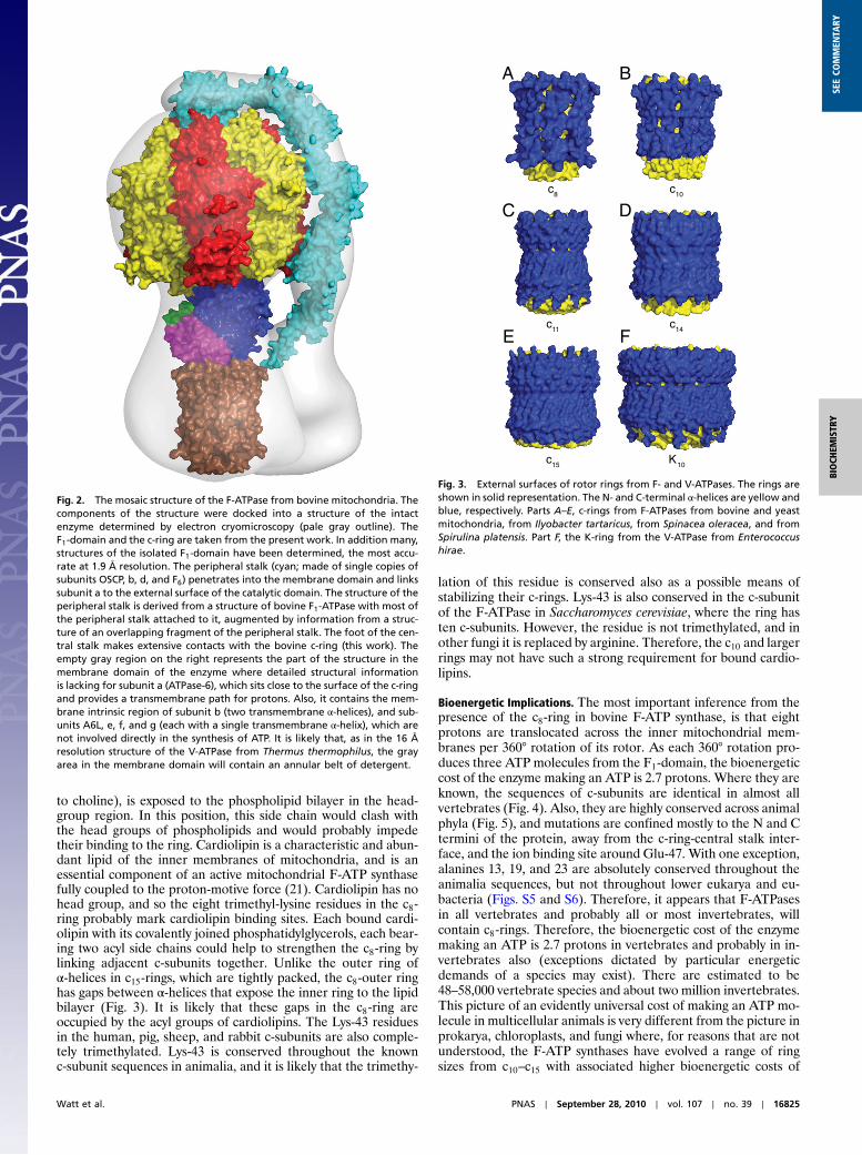
Mosaic Structure of Bovine ATP Synthase.The structure of the bovineF1-c-ring provides an important contribution to the overall struc-ture of bovine ATP synthase. In Fig. 2, a mosaic structure hasbeen built by docking high resolution component structures intoa structure of the intact enzyme determined by electron cryomi-croscopy at 32 Å resolution (pale gray outline in Fig. 2) (15). Themembrane extrinsic part of the structure is derived from a struc-ture of bovine F1-ATPase with most of the peripheral stalkattached to it (16), augmented by information from a structureof an overlapping fragment of the peripheral stalk (17). Manystructures have been determined of bovine F1-ATPase inhibitedin various ways. The most accurate at 1.9 Å resolution is a struc-ture representing the ground state in the catalytic cycle of ATPhydrolysis in which the enzyme was inhibited with the nonhydro-lysable ATP analogue, AMP-PNP (18). The empty gray region onthe right of Fig. 2 represents the part of the structure in themembrane domain of the enzyme where detailed structural in-formation is lacking for subunit a (ATPase-6), which sits close tothe surface of the c-ring and provides a transmembrane path forprotons. Also, it contains the membrane intrinsic region of sub-unit b (two transmembrane α-helices), and for subunits A6L, e, f,and g (each with a single transmembrane α-helix), which are notinvolved directly in the synthesis of ATP. It is likely that, as in thestructure of the V-ATPase from Thermus thermophilus at 16 Åresolution, the gray area in the membrane domain will containan annular belt of detergent (19).
The bovine c-subunit has an unusual feature as the ϵ-aminogroup of Lys-43, which is at the beginning of the C-terminalα-helix, is completely trimethylated (20). The trimethylated sidechain, terminated by a quaternary amino group (which is similar
Fig. 1. The structure of the bovine F1-c-ring complex. Part A. The subunits inthe F1-catalytic domain (above) and the c-ring (below) are shown in ribbonrepresentation. The membrane extrinsic region consists of the sphericalcatalytic domain, made of three α- and three β-subunits (red and yellow,respectively), the central stalk (subunits γ, δ, and ϵ, blue, purple, and green,respectively), The central stalk and the c-ring (brown) together constitute therotor. Each of the C-terminal α-helices in the c-ring has a glutamate at residue58, at the midpoint of the lipid bilayer. Part B. Cross-section of the c-ringtaken at the midpoint of the α-helices. Above is shown the electron densitywith the eight N-terminal α-helices in the inner ring, and the eight C-terminalα-helices in the outer ring, and below the structural interpretation of thedensity.
Table 1. Data collection and refinement statistics for the F1-c-ringcomplex from bovine ATP synthase
Space group P212121Unit cell dimensions a, b, c, Å 155.7, 157.1, 247.3Resolution range Å 50.48–3.50No. unique reflections 72,959Multiplicity 3.6 (3.5)*Completeness, % 99.8 (99.6)*Rmerge 0.263 (0.817)*hI∕σðIÞi 3.7 (1.6)*B factor, from Wilson plot, Å2 72.64R factor, % 26.13Free R factor, % 30.40†rmsd of bonds, Å 0.006rmsd of angles, ° 0.973
*Values in parentheses are for the highest resolution bin (3.50–3.69 Å)†The free R-factor was calculated for 3,855 reflections excluded fromrefinement.
16824 ∣ www.pnas.org/cgi/doi/10.1073/pnas.1011099107 Watt et al.
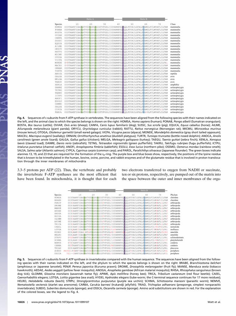
to choline), is exposed to the phospholipid bilayer in the head-group region. In this position, this side chain would clash withthe head groups of phospholipids and would probably impedetheir binding to the ring. Cardiolipin is a characteristic and abun-dant lipid of the inner membranes of mitochondria, and is anessential component of an active mitochondrial F-ATP synthasefully coupled to the proton-motive force (21). Cardiolipin has nohead group, and so the eight trimethyl-lysine residues in the c8-ring probably mark cardiolipin binding sites. Each bound cardi-olipin with its covalently joined phosphatidylglycerols, each bear-ing two acyl side chains could help to strengthen the c8-ring bylinking adjacent c-subunits together. Unlike the outer ring ofα-helices in c15-rings, which are tightly packed, the c8-outer ringhas gaps between α-helices that expose the inner ring to the lipidbilayer (Fig. 3). It is likely that these gaps in the c8-ring areoccupied by the acyl groups of cardiolipins. The Lys-43 residuesin the human, pig, sheep, and rabbit c-subunits are also comple-tely trimethylated. Lys-43 is conserved throughout the knownc-subunit sequences in animalia, and it is likely that the trimethy-
lation of this residue is conserved also as a possible means ofstabilizing their c-rings. Lys-43 is also conserved in the c-subunitof the F-ATPase in Saccharomyces cerevisiae, where the ring hasten c-subunits. However, the residue is not trimethylated, and inother fungi it is replaced by arginine. Therefore, the c10 and largerrings may not have such a strong requirement for bound cardio-lipins.
Bioenergetic Implications. The most important inference from thepresence of the c8-ring in bovine F-ATP synthase, is that eightprotons are translocated across the inner mitochondrial mem-branes per 360° rotation of its rotor. As each 360° rotation pro-duces three ATP molecules from the F1-domain, the bioenergeticcost of the enzyme making an ATP is 2.7 protons. Where they areknown, the sequences of c-subunits are identical in almost allvertebrates (Fig. 4). Also, they are highly conserved across animalphyla (Fig. 5), and mutations are confined mostly to the N and Ctermini of the protein, away from the c-ring-central stalk inter-face, and the ion binding site around Glu-47. With one exception,alanines 13, 19, and 23 are absolutely conserved throughout theanimalia sequences, but not throughout lower eukarya and eu-bacteria (Figs. S5 and S6). Therefore, it appears that F-ATPasesin all vertebrates and probably all or most invertebrates, willcontain c8-rings. Therefore, the bioenergetic cost of the enzymemaking an ATP is 2.7 protons in vertebrates and probably in in-vertebrates also (exceptions dictated by particular energeticdemands of a species may exist). There are estimated to be48–58,000 vertebrate species and about two million invertebrates.This picture of an evidently universal cost of making an ATP mo-lecule in multicellular animals is very different from the picture inprokarya, chloroplasts, and fungi where, for reasons that are notunderstood, the F-ATP synthases have evolved a range of ringsizes from c10–c15 with associated higher bioenergetic costs of
c8 c10
c11
c15 K10
A B
C D
E Fc14
Fig. 3. External surfaces of rotor rings from F- and V-ATPases. The rings areshown in solid representation. The N- and C-terminal α-helices are yellow andblue, respectively. Parts A–E, c-rings from F-ATPases from bovine and yeastmitochondria, from Ilyobacter tartaricus, from Spinacea oleracea, and fromSpirulina platensis. Part F, the K-ring from the V-ATPase from Enterococcushirae.
Fig. 2. The mosaic structure of the F-ATPase from bovine mitochondria. Thecomponents of the structure were docked into a structure of the intactenzyme determined by electron cryomicroscopy (pale gray outline). TheF1-domain and the c-ring are taken from the present work. In addition many,structures of the isolated F1-domain have been determined, the most accu-rate at 1.9 Å resolution. The peripheral stalk (cyan; made of single copies ofsubunits OSCP, b, d, and F6) penetrates into the membrane domain and linkssubunit a to the external surface of the catalytic domain. The structure of theperipheral stalk is derived from a structure of bovine F1-ATPase with most ofthe peripheral stalk attached to it, augmented by information from a struc-ture of an overlapping fragment of the peripheral stalk. The foot of the cen-tral stalk makes extensive contacts with the bovine c-ring (this work). Theempty gray region on the right represents the part of the structure in themembrane domain of the enzyme where detailed structural informationis lacking for subunit a (ATPase-6), which sits close to the surface of the c-ringand provides a transmembrane path for protons. Also, it contains the mem-brane intrinsic region of subunit b (two transmembrane α-helices), and sub-units A6L, e, f, and g (each with a single transmembrane α-helix), which arenot involved directly in the synthesis of ATP. It is likely that, as in the 16 Åresolution structure of the V-ATPase from Thermus thermophilus, the grayarea in the membrane domain will contain an annular belt of detergent.
Watt et al. PNAS ∣ September 28, 2010 ∣ vol. 107 ∣ no. 39 ∣ 16825
BIOCH
EMISTR
YSE
ECO
MMEN
TARY
3.3–5 protons per ATP (22). Thus, the vertebrate and probablythe invertebrate F-ATP synthases are the most efficient thathave been found. In mitochondria, it is thought that for each
two electrons transferred to oxygen from NADH or succinate,ten or six protons, respectively, are pumped out of the matrix intothe space between the outer and inner membranes of the orga-
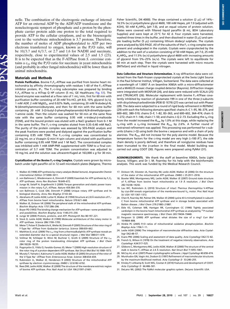
Fig. 4. Sequences of c-subunits from F-ATP synthase in vertebrates. The sequences have been aligned from the following species with their names indicated onthe left, and the animal class to which the species belongs is shown on the right: HOMSA, Homo sapiens (human); PONAB, Pongo albelii (Sumatran orangutan);BOSTA, Bos taurus (cattle); OVIAR; Ovis aries (sheep); CANFA, Canis lupus familiaris (dog); SUSSC, Sus scrofa (pig); EQUCA, Equus caballus (horse); AILME,Ailuropoda melanoleuca (giant panda); ORYCU, Oryctolagus cuniculus (rabbit); RATTU, Rattus norvegicus (Norwegian rat); MICMU, Microcebus murinus(mouse lemur); OTOGA, Otolemur garnettii (small eared galago); VICPA, Vicugna pacos (alpaca); MONDE, Mondelphis domestica (gray short tailed opposum);MACEU,Macropus eugenii (wallaby); ORNAN; Ornithorhynchus anatinus (duckbill platypus); TURTR, Tursiops truncates (bottle nosed dolphin); ANOCA, Anoliscarolinesis (green anole lizard); GALGA, Gallus gallus (chicken); MELGA, Meleagris gallopavo (turkey); TAEGU, Taenio guttat (zebra finch); XENLA, Xenopuslaevis (clawed toad); DANRE, Danio rerio (zebrafish); TETNG, Tetraodon nigroviridis (green pufferfish); TAKRU, Takifugu rubripes (fugu pufferfish); ICTPU,Ictalurus punctatus (channel catfish); ANOFI, Anoplopoma fimbria (sablefish); ESOLU, Esox lucius (northern pike); OSSMO, Osmerus mordax (rainbow smelt);SALSA, Salmo salar (Atlantic salmon); CYPCA, Cyprinus carpio (common carp); and PAROL, Paralichthys olivaceus (Japanese flounder). The green boxes indicatealanines 13, 19, and 23 that are required for the formation of the c8-ring. The purple box and blue boxes show, respectively, the positions of the lysine residuethat is known to be trimethylated in the human, bovine, ovine, porcine, and rabbit enzymes and of the glutamate residue that is involved in proton transloca-tion through the inner membranes of mitochondria
Fig. 5. Sequences of c-subunits from F-ATP synthase in invertebrates compared with the human sequence. The sequences have been aligned from the follow-ing species with their names indicated on the left, and the phylum to which the species belongs is shown on the right: BRABE, Branchiostoma belcheri(amphioxus or Japanese lancelet); PENJP, Peneus japonica (Kuruma prawn); DROME, Drosophila melanogaster (fruit fly); MANSE, Manduca sexta (tobaccohawkmoth); AEDAE, Aedes aegypti (yellow fever mosquito); ANOGA, Anopheles gambiae (African malarial mosquito); RHISA, Rhicephalus sanguineus (browndog tick); GLOMM, Glossina morsitans (savannah tsetse fly); APIME, Apis mellifera (honey bee); TRICA, Tribolium castaneum (red flour beetle); CAEEL,Caenorhabditis elegans; LOTGA, Lottia gigantea (sea snail); HYDEI, Hydroides elegans (tube-worm; the C-terminal sequence continues for 17 more residues);HELRO, Helobdella robusta (leech); STRPU, Stronglycentrotus purpuratus (purple sea urchin); SCHMA, Schistosoma mansoni (parasitic worm); NEMVE,Nematostella vectensis (starlet sea anenome); CARBA, Carukia barnesi (Irukandji jellyfish); TRIAD, Trichoplax adhaerans (presponge, simplest nonparasiticinvertebrate); SUBDO, Suberites domuncula (sponge); and OSSCA, Oscarella carmela (sponge). Amino acid substitutions are shown in red. For the explanationof the colored boxes, see the legend to Fig. 4.
16826 ∣ www.pnas.org/cgi/doi/10.1073/pnas.1011099107 Watt et al.
nelle. The combination of the electrogenic exchange of internalATP for an external ADP by the ADP/ATP translocase and thenonelectrogenic symport of phosphate and a proton by the phos-phate carrier protein adds one proton to the total required toprovide ATP to the cellular cytoplasm, and so the bioenergeticcost to the vertebrate mitochondrion is 3.7 protons. Therefore,the number of moles of ADP phosphorylated to ATP per twoelectrons transferred to oxygen, known as the P∕O ratio, willbe 10∕3.7 and 6∕3.7, or 2.7 and 1.6 for NADH and succinate,respectively, close to experimental values of 2.5 and 1.5 (23).It is to be expected that as the F-ATPase from S. cerevisiae con-tains a c10 ring the P∕O ratio for succinate in yeast mitochondria(and probably in other fungal mitochondria) will be lower than inanimal mitochondria.
Materials and MethodsProtein Purification. Bovine F1Fo-ATPase was purified from bovine heart mi-tochondria by affinity chromatography with residues 1–60 of the F1-ATPaseinhibitor protein, IF1. The F1-c-ring subcomplex was prepared by bindingF1Fo-ATPase to a HiTrap Q HP column (5 mL; GE Healthcare; Fig. S1). Thebound enzyme was washed at a flow rate of 1 mL∕min, first for 60 min withpurification buffer consisting of 20 mM Tris-HCl, pH 8.0, 10% glycerol (v∕v),1 mM ADP, 2 mM MgSO4, and 0.02% NaN3 containing 20 mM N-dodecyl-N,N-(dimethylammonio)butyrate, and then for 60 min with the same buffercontaining 20 mM 3-(3-butyl-3-phenylheptanamido)-N,N-dimethylpropan-1-amine oxide. Then the column was washed for 50 min at the same flowrate with the same buffer containing 0.95 mM n-tridecyl-β-maltoside(TDM), and the bound protein was eluted with a NaCl gradient from 0–1 Min the same buffer. The F1-c-ring complex eluted from 0.35–0.40 mM NaCl.The purity of the protein complex was assessed by SDS-PAGE analysis andthe peak fractions were pooled and dialyzed against the purification buffercontaining 0.95 mM TDM. The F1-c-ring complex was concentrated to22 mg∕mL on a Vivaspin Q mini H spin column and eluted with purificationbuffer containing 0.95 mM TDM and 1 M NaCl. The concentrated complexwas inhibited with 1 mM AMP-PNP, supplemented with TDM to a final con-centration of 5.7 mM TDM. The protein concentration was adjusted to10 mg∕mL and the solution was ultracentrifuged at 163,000 x g for 30 min.
Crystallization of the Bovine F1-c-ring Complex. Crystals were grown by micro-batch under light paraffin oil in 72-well microbatch plates (Nalgene, Thermo
Fisher Scientific, DK-4000). The drops contained a solution (2 μL) of 14%–16.5% (w∕v) polyethylene glycol 4600, 100 mM Hepes, pH 7.0 (adjusted withKOH), 50 mM K2HPO4 (pH 7.0), and an equal volume of protein solution.Plates were covered with filtered liquid paraffin (6 mL; BDH LaboratorySupplies) and were kept at 23 °C for 42 d. Four crystals were harvested,washed three times in the buffer, and then dissolved in water (5 μL) and sam-ple loading buffer (5 μL) containing lithium dodecyl sulphate. The crystalswere analyzed by SDS-PAGE. All of the subunits of the F1-c-ring complex werepresent and undegraded in the crystals. Crystals were cryoprotected by theaddition to the well of a solution consisting of 50 mM Hepes, pH 7.0, 25 mMK2HPO4, and 11.5% (w∕v) polyethylene glycol 4600 with increasing amountsof glycerol from 5%–25% (w∕v). The crystals were left to equilibrate for60 min at each step. Then the crystals were harvested with micro mounts(MiTeGen) and vitrified in liquid nitrogen.
Data Collection and Structure Determination. X-ray diffraction data were col-lected from the flash-frozen cryoprotected crystals at the Swiss Light Sourcein the Paul Scherrer Institut, Villigen, Switzerland. The data were collected ata wavelength of 1.0007 Å on beamline X06SA with a micro-diffractometerand a MAR225 mosaic charge coupled detector (Rayonix). Diffraction imageswere integrated with MOSFLM (24), and data were reduced with SCALA (25)and CTRUNCATE (26). Molecular replacement with bovine F1-ATPase cova-lently inhibited by reaction of glutamate-199 in one of the three β-subunitswith dicyclohexylcarbodiimide (PDB ID 1E79) (27) was carried out with Phaser(28). The data were subjected to a round of rigid body refinement in REFMAC(29) with each the following domains specified: chains A–C 1–95, 96–379, and600–602, 380–510; chains D–F, 1–82, 83–363, and 600–602, 364–474; chain G1–272; chain H 1–146; chain I 1–50; and chains J–Q 2–73. Excluding the c8-ringfrom the model increased the Rfree by 1.0% at this stage, while replacing thec8-ring with a c9-ring increased the Rfree by 2.6%. A further round ofrestrained refinement was applied. This processing was performed for c-sub-units (chains J–Q) using both the bovine c sequence and with a chain of polyalanines. The Rfree did not increase for the poly alanine model. Because thetemperature factors for the c-ring model are very high (>100 Å2), the sidechain density is poorly defined, and therefore all nonglycine residues havebeen truncated to the β-carbon in the final model. Model building wascarried out using COOT (30). Figures were prepared using PyMol (31).
ACKNOWLEDGMENTS. We thank the staff at beamline X06SA, Swiss LightSource, Villigen; and Dr I. M. Fearnley for his help with the bioinformaticanalyses. This work was funded by the Medical Research Council.
1. Walker JE (1998) ATP synthesis by rotary catalysis (Nobel lecture).Angewandte ChemieInternational Edition 37:5000–5011.
2. von Ballmoos C, Wiedenmann A, Dimroth P (2009) Essentials for ATP synthesis by F1F0ATP synthases. Annu Rev Biochem 78:649–672.
3. Junge W, Sielaff H, Engelbrecht S (2009) Torque generation and elastic power trans-mission in the rotary FOF1-ATPase. Nature 459:364–370.
4. von Ballmoos C, Cook GM, Dimroth P (2008) Unique rotary ATP synthase and itsbiological diversity. Annu Rev Biophys 37:43–64.
5. Abrahams JP, Leslie AGW, Lutter R, Walker JE (1994) Structure at 2.8 Å resolution of F1-ATPase from bovine heart mitochondria. Nature 370:621–668.
6. Walker JE, Dickson VK (2006) The peripheral stalk of the mitochondrial ATP synthase.Biochim Biophys Acta 1757:286–296.
7. Boyer PD (1993) The binding change mechanism for ATP synthase—some probabilitiesand possibilities. Biochim Biophys Acta 1140:215–250.
8. Junge W (2004) Protons, proteins, and ATP. Photosynth Res 80:197–221.9. Stock D, Leslie AGW, Walker JE (1999) Molecular architecture of the rotary motor in
ATP synthase. Science 286:1700–1705.10. Meier T, Polzer P, Diederichs K,WelteW, Dimroth P (2005) Structure of the rotor ring of
F-Type Naþ-ATPase from Ilyobacter tartaricus. Science 308:659–662.11. Matthies D, et al. (2009) The c13 ring from a thermoalkaliphilic ATP synthase reveals an
extended diameter due to a special structural region. J Mol Biol 388:611–618.12. Vollmar M, Schlieper D, Winn M, Buchner C, Groth G (2009) Structure of the c14
rotor ring of the proton translocating chloroplast ATP synthase. J Biol Chem284:18228–18235.
13. Pogoryelov D, Yildiz O, Faraldo-Gomez JD, Meier T (2009) High-resolution structure ofthe rotor ring of a proton-dependent ATP synthase. Nat Struct Mol Biol 16:1068–1073.
14. Murata T, Yamato I, Kakinuma Y, Leslie AGW,Walker JE (2005) Structure of the rotor ofthe V-Type Naþ-ATPase from Enterococcus hirae. Science 308:654–659.
15. Rubinstein JL, Walker JE, Henderson R (2003) Structure of the mitochondrial ATPsynthase by electron cryomicroscopy. EMBO J 22:6182–6192.
16. Rees DM, Leslie AGW,Walker JE (2009) The structure of themembrane extrinsic regionof bovine ATP synthase. Proc Natl Acad Sci USA 106:21597–21601.
17. Dickson VK, Silvester JA, Fearnley IM, Leslie AGW, Walker JE (2006) On the structureof the stator of the mitochondrial ATP synthase. EMBO J 25:2911–2918.
18. Bowler MW, Montgomery MG, Leslie AGW, Walker JE (2007) Ground state structureof F1-ATPase from bovine heart mitochondria at 1.9 Å resolution. J Biol Chem282:14238–14242.
19. Lau WC, Rubinstein JL (2010) Structure of intact Thermus thermophilus V-ATPaseby cryo-EM reveals organization of the membrane-bound VO motor. Proc Natl AcadSci USA 107:1367–1372.
20. Chen R, Fearnley IM, Palmer DN, Walker JE (2004) Lysine 43 is trimethylated in subunitC from bovine mitochondrial ATP synthase and in storage bodies associated withBatten disease. J Biol Chem 279:21883–21887.
21. Eble KS, Coleman WB, Hantgan RR, Cunningham CC (1990) Tightly associatedcardiolipin in the bovine heart mitochondrial ATP synthase as analyzed by 31P nuclearmagnetic resonance spectroscopy. J Biol Chem 265:19434–19440.
22. Ferguson SJ (2000) ATP synthase: what dictates the size of a ring? Curr Biol10:R804–808.
23. Hinkle PC (2005) P∕O ratios of mitochondrial oxidative phosphorylation. BiochimBiophys Acta 1706:1–11.
24. Leslie AGW (2006) The integration of macromolecular diffraction data. Acta Crystal-logr D62:48–57.
25. Evans PRE (2006) Scaling and assessment of data quality. Acta Crystallogr D62:72–82.26. French G, Wilson K (1978) On the treatment of negative intensity observations. Acta
Crystallogr A34:517–525.27. Gibbons C, MontgomeryMG, Leslie AGW,Walker JE (2000) The structure of the central
stalk in bovine F1-ATPase at 2.4 Å resolution. Nat Struct Biol 7:1055–1061.28. McCoy AJ, et al. (2007) Phaser crystallographic software. J Appl Crystallogr 40:658–674.29. Murshudov GN, Vagin AA, Dodson EJ (1997) Refinement of macromolecular structures
by the maximum-likelihood method. Acta Crystallogr D 53:240–255.30. Emsley P, Lohkamp B, Scott WG, Cowtan K (2010) Features and development of COOT.
Acta Crystallogr D 66:486–501.31. DeLano WL (2002) The PyMol molecular graphics system. DeLano Scientific USA.
Watt et al. PNAS ∣ September 28, 2010 ∣ vol. 107 ∣ no. 39 ∣ 16827
BIOCH
EMISTR
YSE
ECO
MMEN
TARY
Recommended


![Increased Rate of Adenosine Triphosphate …...(CANCER RESEARCH 55, 4352-4360, October 1, 1995] Increased Rate of Adenosine Triphosphate-dependent Etoposide (VP-16) Efflux in a Murine](https://img.pdfslide.net/doc/110x75/5e7e8d68c5d0407f2447f2a9/increased-rate-of-adenosine-triphosphate-cancer-research-55-4352-4360-october.jpg)