CDK2 and PKA Mediated-Sequential Phosphorylation IsCritical for p19INK4d Function in the DNA DamageResponseMariela C. Marazita1, M. Florencia Ogara1, Silvina V. Sonzogni1, Marcelo Martı1, Nelson J. Dusetti2,
Omar P. Pignataro3, Eduardo T. Canepa1*
1 Laboratorio de Biologıa Molecular, Departamento de Quımica Biologica, Facultad de Ciencias Exactas y Naturales, Universidad de Buenos Aires, Ciudad de Buenos Aires,
Argentina, 2 INSERM, U624 Stress Cellulaire, Marseille, France, 3 Laboratorio de Endocrinologıa Molecular y Transduccion de senales, Instituto de Biologıa y Medicina
Experimental-CONICET, Ciudad de Buenos Aires, Argentina
Abstract
DNA damage triggers a phosphorylation-based signaling cascade known as the DNA damage response. p19INK4d, amember of the INK4 family of CDK4/6 inhibitors, has been reported to participate in the DNA damage response promotingDNA repair and cell survival. Here, we provide mechanistic insight into the activation mechanism of p19INK4d linked to theresponse to DNA damage. Results showed that p19INK4d becomes phosphorylated following UV radiation, b-amyloidpeptide and cisplatin treatments. ATM-Chk2/ATR-Chk1 signaling pathways were found to be differentially involved inp19INK4d phosphorylation depending on the type of DNA damage. Two sequential phosphorylation events at serine 76and threonine 141 were identified using p19INK4d single-point mutants in metabolic labeling assays with 32P-orthophosphate. CDK2 and PKA were found to participate in p19INK4d phosphorylation process and that they wouldmediate serine 76 and threonine 141 modifications respectively. Nuclear translocation of p19INK4d induced by DNAdamage was shown to be dependent on serine 76 phosphorylation. Most importantly, both phosphorylation sites werefound to be crucial for p19INK4d function in DNA repair and cell survival. In contrast, serine 76 and threonine 141 weredispensable for CDK4/6 inhibition highlighting the independence of p19INK4d functions, in agreement with our previousfindings. These results constitute the first description of the activation mechanism of p19INK4d in response to genotoxicstress and demonstrate the functional relevance of this activation following DNA damage.
Citation: Marazita MC, Ogara MF, Sonzogni SV, Martı M, Dusetti NJ, et al. (2012) CDK2 and PKA Mediated-Sequential Phosphorylation Is Critical for p19INK4dFunction in the DNA Damage Response. PLoS ONE 7(4): e35638. doi:10.1371/journal.pone.0035638
Editor: Ferenc Gallyas, University of Pecs Medical School, Hungary
Received January 9, 2012; Accepted March 19, 2012; Published April 25, 2012
Copyright: � 2012 Marazita et al. This is an open-access article distributed under the terms of the Creative Commons Attribution License, which permitsunrestricted use, distribution, and reproduction in any medium, provided the original author and source are credited.
Funding: This work was supported by research grants from Consejo Nacional de Investigaciones Cientıficas y Tecnicas (CONICET), Agencia Nacional dePromocion Cientıfica y Tecnologica (ANCYPT) and Universidad de Buenos Aires. The funders had no role in study design, data collection and analysis, decision topublish, or preparation of the manuscript.
Competing Interests: The authors have declared that no competing interests exist.
* E-mail: [email protected]
Introduction
DNA damage response (DDR) mechanisms are essential for
maintaining genomic integrity and an accurate transmission of
genetic information. DDR consists of an intricate signaling
network in which complex DNA surveillance programs play a
key role [1–3]. These control programs or checkpoints respond to
a variety of lesions including stalled replication forks and DNA
damage induced by both internal and external sources like reactive
cellular metabolites, ionizing or UV radiation and chemothera-
peutic agents [2,4,5]. After sensing the damage, the activation of
the checkpoints modulate cell cycle arrest, DNA repair systems
and cell death mechanisms to repair or to eliminate hazardous,
genetically unstable cells [6,7]. Although DDR components have
not yet been completely described the canonical checkpoint
signaling is composed by two major transduction pathways
initiated by the upstream PI3K-like kinases Ataxia-telangiectasia
Mutated (ATM) and ATM and Rad3-related (ATR). ATM is
predominantly activated by double strand break lesions (DSBs)
while ATR responds fundamentally to single strand breaks or
bulky lesions. ATM and ATR activate their downstream kinases
Chk1 and Chk2 amplifying the initial signal and modulating the
G1/S, intra-S and G2/M checkpoints [4,8]. While ATM and ATR
were initially reported to activate Chk2 and Chk1 respectively, this
concept was challenged by studies that show crosstalks between
these kinases [9]. Chk1 activation by ATM was reported in cells
exposed to ionizing radiation treatment [10,11] and ATM and
ATR were required for Chk2 activation in response to replication
stress [12]. Moreover, it was shown that both ATR and ATM
were able to target the SQ-rich C terminus of Chk1 on serine 317
and 345 leading to its activation [10,13–15]. Following Chk1 and
Chk2 activation, these kinases phosphorylate a wide range of
downstream effectors which prevent further progression through
the cell cycle and initiate DNA repair mechanisms but also
modulate the trigger of cell death pathways if the insult exceeds the
repair capacity [2,16]. Among these effector proteins, Chk1
phosphorylates TLK12 and RAD51, while BRCA, PIK3, PML
and E2F1 are Chk2 substrates. They also share target proteins like
Mdm2, p53, cdc25A and cdc25C [5,17–20]
The cell cycle progression is driven by the activity of cyclin-
dependent kinases (CDKs) and is negatively regulated by INK4
PLoS ONE | www.plosone.org 1 April 2012 | Volume 7 | Issue 4 | e35638

and Cip/Kip inhibitory proteins [21–24]. INK4 family consists of
four members, p16INK4a, p15INK4b, p18INK4c and p19INK4d
which play a redundant role as CDK4/6 inhibitors. However,
novel cell cycle independent functions were recently described for
some of them [25]. Interestingly, p16INK4a and p19INK4d (p19)
were linked to the cellular response to genotoxic agents [26–28]. In
particular, extensive data points out that p19 is a critical factor in
the maintenance of genomic integrity and cell survival. It was
reported that UV light, cisplatin and b-amyloid peptide promoted
p19INK4d transcriptional induction and nuclear translocation
[27]. Adding to this, p19 overexpression significantly enhanced
DNA repair and diminished apoptosis in different cell lines. More
important, physiological p19 levels are necessary for an appropri-
ate response to the damage. In this way, p19 deficient cells display
an impaired DNA repair activity and enhanced apoptosis [27–29].
Consistent with these findings, other studies described enhanced
sensitivity of cells to apoptosis and autophagic cell death in p19
null mice [30]. p19 expression status directly correlates with cell
resistance and survival to DNA damage. Finally, p19 activity
protects from UV-induced chromosomal aberrations and sponta-
neous chromosome abnormalities as well [27,29]. These facts
uncovered a novel p19 function in regulating genomic stability and
overall cell viability under conditions of genotoxic stress.
Despite these findings, the regulation of p19 activity in the DDR
remains unknown. We hypothesized that post-translational
modifications on p19 could be taking part in regulating this
specific function. Here, we report the activation mechanism of p19
in response to DNA damage. It is shown that this event is
dependent on the ATM/ATR signaling pathways and occurs in a
sequential manner at serine 76 (S76) and threonine 141 (T141).
These two identified phosphorylation sites would be direct
substrates for CDK2 and PKA kinases respectively. Moreover,
DNA damage induced p19 nuclear translocation requires S76
phosphorylation. And finally, both phosphorylation sites are
shown to be crucial for p19 function in DNA repair and cell
survival but dispensable for CDK4/6 inhibition. These results
position p19 in a novel context as a downstream target of the
DDR signaling pathways, provide mechanistic insight into the
activation mechanism of p19INK4d in response to DNA damage
and demonstrate the functional relevance of this activation
following genotoxic stress.
Materials and Methods
Cell culture, transfections and antibodiesWI-38 cell line was grown in minimum essential medium
supplemented with 10% fetal bovine serum (FBS) and 1%
penicillin-streptomycin, 100 mM non-essential aminoacids, and
2 mM glutamine at 37uC in a humidified 5% CO2 atmosphere.
HEK-293 cells were grown in Dulbecco’s modified Eagle medium
supplemented as described above. Transfections were performed
using Lipofectamine 2000 Reagent (Invitrogen). Approximately,
26106 cells were seeded in 60-mm plates and transfected with
4 mg of wild type or mutant p19 expression plasmids. 24 h after
transfection, cells were treated as indicated in each experiment.
For UDS, [3H]thymidine incorporation and caspase-3 activity
assays, WI-38 cells were plated in 35-mm dishes at a density of
16106 cells/well in 2.5 ml of medium. After a 24 h attachment
period, each well was transfected with a mixture containing 2 mg
of wild type or mutant p19 expression plasmids and 0.5 mg of
pBabePuro per plate. Twenty-four hours after transfection,
2.5 mg/ml puromycin were added for 48 h to select for transfected
cells.
Antibodies: p19 (P1000-38, USBiological), p19 (sc-1063, Santa
Cruz Biotechnology, INC), V5 (R960-25, Invitrogen), V5 (sc-
83849-R, Santa Cruz Biotechnology, INC). CDK2 (sc-163, Santa
Cruz Biotechnology, INC), PKAc(C-20, Santa Cruz Biotechnol-
ogy), histone H3 (sc-8654–R, Santa Cruz Biotechnology). GAPDH
(AB8245, ABCAM). Anti-mouse and anti-rabbit secondary
antibodies conjugated with horseradish peroxidase were purchased
from SIGMA (Saint Louis, USA).
UV irradiationCells were irradiated in open dishes with UV (4 mJ/cm2),
254 nm (range 240–280 nm) at room temperature using a Philips
ultraviolet lamp (TUV15WG15T8) calibrated to deliver 0.25 mJ/
cm2 s. Following UV irradiation, medium was replaced and cells
were further incubated for the indicated times. For each
experiment, control cells were treated identically except for UV
light exposure.
Metabolic labeling of p19 in WI-38 cells with sodiumorthophosphate - 32P
WI-38 cells were grown in 60-mm dishes and treated as
indicated. Before treatment, cells were incubated with 0,5 mCi
sodium orthophosphate-32P for 3 h. At indicated time points, cells
were washed, collected in cold PBS and lysed in RIPA buffer. The
lysates (100 mg) were incubated with the appropiate antibody for
2 h, followed by O.N. incubation with protein AG agarose beads
at 4uC. After washing three times with RIPA buffer, samples were
analyzed by immunoblotting or SDS-PAGE. Dried gels were
exposed to a radiographic intensifying screen by Fujifilm and
scanned directly using a Bio-Imaging Analyzer Fujifilm BAS-
1800II.
PlasmidsConstruction of p19 mutants is described in Supporting
Information (Materials and Methods S1).
Downregulation of CDK1 and CDK2Antisense oligonucleotides (AS) complementary to either human
CDK1 or CDK2 (corresponding to bases +129 to +155 and +46 to
+73 respectively) were transfected with Lipofectamine 2000. At a
final concentration of the AS was 1 mM. After 12 h, the medium
was replaced by fresh medium containing 1 mM of the
corresponding AS. Cells were treated for in vivo phosphorylation
as described previously. ASCDK1: 59 tattttggtattatcttccatagttag 39;
ASCDK2: 59ccaacttgaaacaatcttgccgcctccc 39.
p19 structure and Molecular Dynamics SimulationsAnalysis of p19 structure was performed using the VMD
programmed (Visual Molecular Dynamics, http://www.ks.uiuc.
edu/Research/vmd/). Simulations of p19 phosphorylations were
obtained using AMBER software. XY graphs were done using
XMGRACE utility. (CA positions, alpha carbon positions).
Analysis of potential phosphorylation sites and kinase-specific prediction of phosphorylation sites
Netphos 2.0 server [31], a neural network-based method for
predicting potential phosphorylation sites at serine, threonine or
tyrosine residues, was used to analyze p19 protein sequence
(http://www.cbs.dtu.dk/services/NetPhos/). NetphosK 1.0 server
was used for kinase-specific prediction of phosphorylation sites
[32] (http://www.cbs.dtu.dk/services/NetPhosK/).
Activation Mechanism of p19 following DNA Damage
PLoS ONE | www.plosone.org 2 April 2012 | Volume 7 | Issue 4 | e35638

Alignment of protein sequencesProtein sequences were aligned using T-Coffee multiple
sequence alignment tool [33].
RNA extraction and Northern blot analysisTotal cellular RNA was isolated from cultures as described
previously [34]. Ten micrograms of total RNA were denatured,
electrophoresed in 1% glyoxal/agarose gels, and transferred to
nylon membranes (GeneScreen Plus, PerkinElmer). The mem-
branes were sequentially hybridized with 32P-labeled probes to
CDK1, CDK2 and b-ubulin. To detect CDK1 mRNA, a 28-mer
ODN was synthesized complementary to bases +124 to +151 of
human p19 mRNA. To detect CDK2 mRNA, a 29-mer ODN
was synthesized complementary to bases +45 to +73 of human p19
mRNA. To detect b-tubulin a 22-mer ODN was synthesized
complementary to bases +174 to +195 of human tubulin beta 3
mRNA. ODN were 5-end-labeled using [c-32P] ATP and T4
polynucleotide kinase. Hybridization was carried as previously
described [27]. Membranes were exposed to a radiographic
intensifying screen by Fujifilm and scanned directly using a Bio-
Imaging Analyzer Fujifilm BAS-1800II.
CDK2 kinase assayCDK2 kinase assay was performed as described by Giono et. al.
[35]. Briefly, HEK 293 cells were washed with PBS and lysed in
HB buffer Proteins (1 mg) were immunoprecipitated with 10 ml of
anti-CDK2 antibody and 50 ml of a 50% slurry of protein A/G-
agarose beads (SIGMA) and rocked at 4uC for 2 h. For control
sample containing only the substrate (GST-p19) but no enzyme,
proteins were incubated with the beads without anti-CDK2
antibody. The beads were washed and resuspended in 15 ml of HB
buffer. To each sample was added 15 ml of 26 kinase reaction
buffer (HB buffer containing 200 mM ATP, 1 mg/ml GST-p19,
and 1 ml of [c-32P]ATP (6,000 Ci/mmol [10 mCi/ml]) per 10 ml of
26 reaction buffer). Samples were incubated for 20 min at 37uC.
The reaction was stopped by addition of 7.5 ml of 56 sample
buffer and 5 min incubation at 95uC. Histone H1 from calf
thymus (Calbiochem) was used for kinase activity control. Samples
were electrophoresed on 12% denaturing gels. Gels were dried on
Whatman paper, exposed to a radiographic intensifying screen by
Fujifilm and scanned directly using a Bio-Imaging Analyzer
Fujifilm BAS-1800II. In a similar assay, phosphorylation of a p19
derived peptide containing the surrounding sequence of S76 (p-
S76; RGTSPVHDAART; 200 mM) was tested. As control for
CDK2 activity, an histone H1 derived peptide (p-H1;
PKTPKKAKKL). Following the reaction, samples were processed
according to the phosphocellulose paper method. As a negative
control, the reaction was conducted without substrate.
PKA kinase assayThe in vitro phosphorylation assays were performed in a final
volume of 40 ml of reaction buffer (50 mM potassium phosphate,
pH 7.5, 0.1 mM EGTA, 0.1 mM EDTA, 15 mM MgCl2, 10 mM
2-mercaptoethanol, 0.1 mM [c-32P]ATP (700 dpm/pmol)
0,1 mM ATP, 100 mg/ml PMSF, 60 mg/ml aprotinin, 1 mM
sodium orthovanadate) plus 5 mg of GST-p19 with or without H-
89 (100 mM). After 15 min at 30uC, samples were electrophoresed
on 12% denaturing gels. Gels were dried on Whatman paper,
exposed to a radiographic intensifying screen by Fujifilm and
scanned directly using a Bio-Imaging Analyzer Fujifilm BAS-
1800II. In a similar assay, phosphorylation of a p19 derived
peptide containing the surrounding sequence of threonine 141 (p-
T141; RDARGLTPLELA; 200 mM) was tested. As control for
PKA activity, Kemptide was used as substrate (kemp; LRRASLG).
Following the reaction, samples were processed according to the
phosphocellulose paper method. As a negative control, the
reaction was conducted without substrate.
PKA-p19 co-immunoprecipitationCo-immunoprecipitation assays were performed by transfection
of pcDNA4cp19wt in WI-38 cells. A total of 500 mg of proteins
were immunoprecipitated with 3 ml of anti-V5 antibody and 30 ml
of 50% slurry of protein A/G agarose beads (SIGMA). The beads
were washed three times with RIPA, resuspended in 30 ml of 26sample buffer, and heated to 95uC for 5 min. Proteins were
resolved in 15% polyacrylamide gels and analyzed by immuno-
blotting.
Caspase-3 activityCells were treated with UV (4 mJ/cm2) or 10 mM b-amyloid
peptide for 12 h. Cells were then harvested with lysis buffer
(50 mM Tris–HCl, pH 7.4, 1 mM EDTA, 10 mM EGTA,
10 mM digitonine, 0.5 mM PMSF, 10 mg/ml pepstatin, and
10 mg/ml aprotinin) incubated for 30 min at 37uC and centrifuged
at 12,0006g for 20 min. The activity of caspase-3 in 150 ml of cell
lysate was determined using 100 mM of the synthetic caspase-3
substrate Ac-DEVD-pNA (Sigma) in reaction buffer (100 mM
HEPES pH 7.5, 0.5 mM EDTA, 5 9 mM dithiothreitol and
glicerol 20% v/v) in a final volume of 300 ml and incubated at
37uC during 4 h. Color development was measured at 405 nm.
Caspase activity was estimated as A405/mg protein.
Subcellular fractionationCells were seeded on 100 mm-plates and transfected with the
indicated p19 expression plasmids. After 24 h, 1 mM H-89 was
added for 1 hour treatment. Cells were treated with 4 mJ/cm2
UV and harvested at the indicated times. Subcellular fractionation
was performed as described by Schreiber et al [36] with minor
modifications. Briefly, cells were washed with 10 ml PBS, collected
in 1 ml PBS and collected by centrifugation at 15006g for 5 min.
Cell pellets were resuspended in 400 ml of cold buffer A (10 mM
HEPES pH 7.9; 10 mM KCl; 0.1 mM EDTA; 0.1 mM EGTA;
1 mM DTT; 0.5 mM PMSF) and cells were allowed to swell on
ice for 15 min. Twenty five ml of a 10% solution of Nonidet NP-40
were added and the tube was vigorously vortexed for 10 sec.
Homogenates were centrifuged for 30 sec in a microcentrifuge.
Supernatants (cytoplasmic fraction) were transferred to a fresh
tube and pellets (nuclear fraction) were resuspended in 90 m1 ice-
cold buffer C (20 mM HEPES pH 7.9; 0.4 M NaCl; 1 mM
EDTA; 1 mM EGTA; 1 mM DTT; 1 mM PMSF) and the tube
was vigorously rocked at 4uC for 15 min on a shaking platform.
Nuclear extracts were centrifuged for 5 min and supernatants
transferred to a clean tube. Cytoplasmic and nuclear extracts were
analyzed for in vivo p19 or p19-V5 phosphorylation as mentioned
before.
[3H]thymidine incorporationTwenty four hours after transfection, cells were incubated with
1 mCi/ml [3H]-thymidine (81 Ci/mmol) (Amersham Biosciences)
for 6 h. Cells were washed three times with cold PBS, harvested,
and centrifuged at 30006g for 5 min. The cellular pellet was lysed
with 5% trichloroacetic acid (TCA) for 30 min, centrifuged and
washed twice with cold H2O2. The pellet was resuspended in
150 ml 1 M NaOH for 1 h at room temperature. Incorporated
radioactivity was quantified by scintillation counting and DNA
synthesis expressed as dpm/mg protein.
Activation Mechanism of p19 following DNA Damage
PLoS ONE | www.plosone.org 3 April 2012 | Volume 7 | Issue 4 | e35638

Unscheduled DNA synthesisTwenty four hours after transfection, cells were washed with
PBS and growth medium was replaced by arginine-free medium
containing 1% FBS which was renewed after 24 h. Inhibition of
DNA semiconservative synthesis was confirmed under these
conditions. Cells were treated with UV (4 mJ/cm2), or 10 mM
b-amyloid peptide and further cultured in UDS medium and
10 mCi/ml [3H]thymidine. At the indicated times, cells were
washed three times with cold PBS, harvested and collected at
30006g for 5 min. Cells were lysed with 5% TCA for 30 min. and
centrifuged at 10,0006g for 10 min. Pellet was washed twice with
cold PBS and resuspended in 1 M NaOH. The incorporated
radioactivity was quantified by scintillation counting. Unscheduled
DNA synthesis was expressed as dpm/mg protein.
Results
p19INK4d phosphorylation in response to DNA damageWe have previously reported that p19 is involved in DNA
repair, genome maintenance and cell survival [27–29]. Then, we
aimed to study the mechanism by which p19 is activated in
response to DNA insults. It was hypothesized that p19 could be
target of the phosphorylation pathways activated by DNA
damage. To test this, p19 phosphorylation status was examined
after treatment with three different genotoxic agents: UV light,
cisplatin and b-amyloid peptide. In vivo phosphorylation analyses
were performed by metabolic labeling of WI-38 fibroblasts. Under
basal conditions, no phosphorylation of endogenous p19 was
observed. In contrast, p19 rapidly became phosphorylated
20 minutes after b-amyloid treatment or UV exposure
(Figure 1A). The phosphorylation signal remained elevated for at
least 8 hours after treatment with all three damaging agents
(Figure 1B, Figure S1). These results show that p19 becomes
phosphorylated following DNA damage.
p19INK4d is sequentially phosphorylated in serine 76 andthreonine 141 upon DNA damage
To further study p19 phosphorylation, the protein sequence was
analyzed for the presence of potential phosphorylation residues
(Figure S2). Five p19 mutants were constructed replacing serine or
threonine by alanine at the predicted phosphorylation sites and the
phosphorylation capacity of these mutants was assessed in vivo by
metabolic labeling.
Phosphorylation of overexpressed p19 was absent in untreated
cells and was induced after UV radiation (Figure 2A). p19S13A,
p19S66A and p19T89A mutants showed phosphorylation levels
comparable to p19wt. In contrast, p19T141A and p19S76A
displayed relevant differences. While p19T141A phosphorylation
was significantly reduced, phosphorylation of p19S76A was
completely abolished (Figure 2B). These results strongly suggested
that S76 and T141 were actual target sites for phosphorylation in
vivo. In addition, the lack of phosphorylation on p19S76A raised
the hypothesis of a two-step process in which the modification of
T141 would be dependent on the phosphorylation of S76. To
study this possibility, two glutamic acid mutants were generated
mimicking the phosphorylation effect at S76 (p19S76E) or at both
sites, S76 and T141 (p19S76E/T141E). In accordance with the
hypothesis of a sequential phosphorylation, the phosphomimetic
mutation at S76 enabled the phosphorylation of p19S76E mutant
at T141 (Figure 2C). Interestingly, no p19S76E phosphorylation
was observed in the absence of UV irradiation. Then, an active
DNA damage response pathway is required to undergo a second
modification at a site different from S76. Moreover, no
phosphorylation was detected in p19S76E/T141E after genotoxic
treatment. These results are in agreement with those showing
decreased and lack of signal in p19T141A and p19S76A
respectively and hence support S76 and T141 as the only
phosphorylation residues.
The potential effects of the phosphorylation on p19 structure
were analyzed by Molecular Dynamics Simulation. p19 is
composed of five ankyrin repeats of about 30–35 residues long.
Each repeat consists of a b-hairpin followed by two anti parallel a-
helices. S76 and T141 are located in the third and fifth ankyrin
domain respectively, at the end of the b-hairpin. When
phosphorylation at S76 was simulated (p19p) direct comparison
between p19 and p19p average structures showed significant
differences (Figure 2D). Up to 8 A between the CA positions were
observed for key structural regions. The main structural changes
were found in the b-hairpins of the third ankyrin repeat, where the
phosphoserine is positioned, and also in the fourth repeat. In p19
Figure 1. p19 phosphorylation is induced in response to DNA damage. (A, B) WI-38 fibroblasts were labeled with [32P]-orthophosphate andtreated with b-amyloid peptide (20 mM), cisplatin (10 mM) or UV light (4 mJ/cm2) for the indicated times. Equal amounts of whole cell extracts weresubjected to immunoprecipitation with anti-p19 antibody and the immune complexes were analyzed by SDS-PAGE and autoradiography (upperpanels; P-p19, phosphorylated p19) or immunoblotting (lower panels; p19). (C; Control, untreated cells).doi:10.1371/journal.pone.0035638.g001
Activation Mechanism of p19 following DNA Damage
PLoS ONE | www.plosone.org 4 April 2012 | Volume 7 | Issue 4 | e35638

structure both loops are close together but the presence of the
phosphate pushed them away. An increase in mobility was also
found in the loop between helices I and II of the fifth repeat in
which T141 is situated. Supporting p19 sequential phosphoryla-
tion, the structural changes induced by S76 modification could be
necessary for the interaction between p19 and the second kinase
responsible for T141 phosphorylation. When both sites were
phosphorylated (p19pp) the main differences compared to p19p
were located in the fifth repeat (Figure 2E). The change in the b-
hairpin loop of the repeat was smaller than the change observed
for the loop between both helices. The presence of the phosphate
broke the R135-E144 interaction. E144 was pushed away and a
strong interaction between R135 and the phosphate group was
established. These additional changes resulted particularly inter-
esting since they might promote the interaction between p19 and
proteins related to its function in the DDR.
Overall, we propose a sequential phosphorylation model for p19
in which modification at S76 would enable a second phosphor-
ylation event at T141. The phosphorylation-induced structural
changes could have functional implicancies for p19 in the DNA
damage response.
ATM-Chk2 and ATR-Chk1 checkpoint pathways aredifferentially involved in p19 phosphorylation
ATM and ATR kinases are members of the PIKKs family that
play a major role in the DDR. It was previously reported that UV
radiation and cisplatin cause DNA damage which promotes
predominantly ATR activation, while b-amyloid peptide mainly
stimulates ATM activity [37,38]. Since we demonstrated that p19
phosphorylation is induced by all three DNA damaging agents, we
hypothesized that the ATM/ATR pathway could be involved in
this process. To examine this possibility, caffeine, a specific PIKKs
inhibitor, was used in the analysis of p19 phosphorylation in vivo.
Caffeine treatment effectively prevented p19 phosphorylation
(Figure 3A). Additionally, wortmannin, a PIKKs and PI3K
inhibitor, was used to conduct a similar analysis. It was reported
that different wortmannin concentrations are needed to inhibit the
diverse kinases (PI3K and DNA-PKc IC50: 0.016 mM; ATM
IC50: 0.15 mM; ATR IC50: 1.8 mM) [39]. A dose-response curve
of wortmannin was performed to assess its inhibitory effect on p19
phosphorylation. As expected, the concentration of wortmannin
required to block p19 phosphorylation depended on the genotoxic
agent tested. The phosphorylation promoted by b-amyloid peptide
was diminished by 0.15 mM wortmannin and totally suppressed at
the concentration of 1 mM, doses reported to inhibit ATM but not
Figure 2. Sequential phosphorylation of p19 at S76 and T141 following DNA damage. (A, B) Phosphorylation ability of p19 mutants. WI-38 fibroblasts were transfected with expression vectors encoding the V5 epitope tag in frame with wild type p19 (p19wt) or p19 mutants, in whichthe potential phosphorylation sites were replaced by alanine (p19S13A, p19S66A, p19S76A, p19T89A, p19T141A). Transfected cells were labeled with[32P]-orthophosphate, treated with UV light (4 mJ/cm2) and collected 3 hours after treatment. Extracts were subjected to immunoprecipitation withanti-V5 antibody and analyzed by autoradiography (upper panels, P-p19) or immunoblotting (lower panels, V5). Unstransfected cells were used as acontrol to monitor immunoprecipitation specificity. (C) In a similar experiment, in vivo phosphorylation of two p19 phosphomimetic mutants(p19S76E and p19S76E/T141E) was tested. (D, E) Structural changes promoted by the sequential phosphorylation were analyzed by moleculardynamics simulation. The images show the comparison between the structures of p19 (cyan) and p19 phosphorylated on S76 (p19p, red) (D), orbetween p19p and p19 phosphorylated on sites S76 and T141 (p19pp, yellow) (E). Graphs show CA-distance between p19 and p19p (D), or p19p andp19pp (E) average structures. Arrows indicate domains with predicted structural changes. (ANK 1–5, ankyrin domains 1 to 5).doi:10.1371/journal.pone.0035638.g002
Activation Mechanism of p19 following DNA Damage
PLoS ONE | www.plosone.org 5 April 2012 | Volume 7 | Issue 4 | e35638

ATR (Figure 3B, right panel). In contrast, cisplatin-induced p19
phosphorylation was completely abolished only at high wortman-
nin concentrations (2 mM) necessary to inhibit ATR (Figure 3B,
left panel). Therefore, these results suggested that both ATM and
ATR signaling pathways promote p19 phosphorylation and that
they act in response to different types of DNA damage.
Chk1 and Chk2 kinases amplify the signals initiated by ATM/
ATR. Then, in vivo p19 phosphorylation was examined after
treatment with Chk1 or Chk2 inhibitors. Results showed that p19
phosphorylation promoted by UV light or cisplatin was impaired
by Chk1 inhibition (Figure 3C, left panel). In contrast, Chk2
inhibitor suppressed p19 phosphorylation only when the damage
was induced by b-amyloid peptide. These results are consistent
with the fact that Chk1 and Chk2 are predominantly activated by
ATR and ATM respectively and further support the data
presented in figure 3B.
We conclude that there is a differential involvement of ATM-
Chk2 and ATR-Chk1 pathways in p19 phosphorylation which
depends on the type of lesion in the DNA.
p19 phosphorylation requires CDK and PKA activitiesATM-Chk2 and ATR-Chk1 activates numerous phosphoryla-
tion pathways in response to DNA insults leading to the repair of
the damage or ultimately to cell death. We aimed to investigate
which pathways and particularly which kinases were directly
involved in p19 phosphorylation. As an initial approach, a search
for potential kinases predicted CDK5 and PKA acting at S76 and
T141 respectively (Figure S3). CDK5 is a serine/threonine kinase
with high sequence homology to CDK1 and CDK2 [40–42]. The
brain is the only tissue that shows CDK5 histone H1 kinase
activity and no equivalent kinase activity has been found in other
tissue culture cell lines [43]. The substrate specificity of CDK1 and
CDK2 is similar to that of CDK5 phosphorylating the (S/
T)PX(K/H/R) consensus sequence motif [44,45]. In p19, S76
corresponds to a perfect consensus site constituted by the sequence
SPVH. To evaluate the involvement of these enzymes, specific
kinase inhibitors were used in phosphorylation assays in vivo. H-89
treatment, a specific inhibitor of PKA, partially decreased
endogenous p19 phosphorylation induced by UV radiation, b-
amyloid peptide and cisplatin treatment (Figure 4A). A concen-
tration of H-89 20 times higher than the one used in figure 4A and
reported to abolish PKA activity in various cell types was unable to
further diminish the phosphorylation (Figure S4). Interestingly, the
decrease in p19 phosphorylation after PKA inhibition was similar
to that observed for p19T141A (Figure 2B). This fact is consistent
with the in silico analysis which predicted PKA as the kinase acting
on T141. Adding to this, roscovitine, a potent inhibitor of CDK1,
CDK2 and CDK5 kinases, completely blocked p19 phosphory-
lation induced by the three DNA damaging treatments tested,
supporting the prediction of the CDK activity on S76 (Figure 4A).
Figure 3. ATM/ATR signaling pathways are differentially involved in p19 phosphorylation. (A) Inhibition of p19 phosphorylation bycaffeine treatment. WI-38 fibroblasts were incubated with caffeine (5 mM) for 1 hour, then treated with cisplatin (10 mM) or b-amyloid peptide(20 mM) for the indicated times and endogenous p19 phosphorylation analyzed by autoradiography. (B) Evaluation of ATM/ATR involvement in p19phosphorylation by wortmannin treatment. WI-38 fibroblasts were incubated with the indicated doses of wortmannin for 1 hour, followed bytreatment with cisplatin (10 mM) or b-amyloid peptide (20 mM) for 2 hours. (C) Effect of Chk1 and Chk2 inhibitors on p19 phosphorylation. WI-38fibroblasts were incubated with SB-218078 (SB, 15 nM) or dopamine ß-hidroxylase inhibitor (DBH, 3 mM), both Chk1 inhibitors, or with Chk2 InhibitorCalbiochem (ICHK2, 20 nM) for 1 hour before treatment with UV light (4 mJ/cm2), cisplatin (10 mM) or b-amyloid peptide (20 mM). After 2 hours, cellextracts were analyzed as in A.doi:10.1371/journal.pone.0035638.g003
Activation Mechanism of p19 following DNA Damage
PLoS ONE | www.plosone.org 6 April 2012 | Volume 7 | Issue 4 | e35638

Moreover, PKA inhibition did not affect neither p19T141A nor
p19ANKless (a p19 mutant lacking the last ankyrin repeat where
T141 is positioned) phosphorylation with the genotoxic drugs
tested (Figure 4B–C). These results suggest that there is no other
site different from threonine 141 where PKA activity might be
involved. Furthermore, roscovitine treatment completely blocked
the phosphorylation of both mutants, p19T141A and p19ANKless
(Figure 4B–C). Since only two residues become phosphorylated
after DNA injury, these observations indicate that S76 should be
the specific target site for the CDK activity. These results also
support the hypothesis of a sequential phosphorylation which
would be dependent on CDK and PKA activities.
We next aimed to identify the CDK family member necessary
for p19 phosphorylation acting in this process. Since CDK5
activity was only reported in neural cells, the involvement of
CDK1 and CDK2 kinases was examined in the cell lines tested.
Specific antisense oligonucleotides were used to down-regulate
either CDK1 (ASCDK1) or CDK2 (ASCDK2). The efficiency
and specificity of the antisense oligonucleotides was first tested by
Northern blot (Figure 4D, lower panels). In vivo p19 phosphor-
ylation was not affected by ASCDK1 treatment. In contrast, a
decrease in the phosphorylation was found following ASCDK2
treatment suggesting a role for CDK2 in this process.
In summary, these observations are consistent with the
sequential phosphorylation of p19 involving CDK2 function on
S76 that would enable the activity of PKA on T141.
CDK2 and PKA phosphorylates p19 in vitroWe have demonstrated that CDK2 and PKA activities are
required for p19 phosphorylation. To investigate whether p19 is a
direct target of CDK2 and PKA we carried out in vitro kinase
assays using specific p19 derived peptides or recombinant GST-
p19 protein as substrates. In the first approach, two synthetic
peptides containing either the sequence of S76 (p-S76:
RGTSPVHDAART) or T141 (p-T141: RDARGLTPLELA)
phosphorylation sites were employed to test the direct activity of
CDK2 or PKA correspondingly. Results showed that CDK2 was
able to efficiently phosphorylate the p-S76 peptide (Figure 5A).
PKA catalytic subunit (cPKA) was capable of phosphorylating the
p-T141 peptide as observed by 32P incorporation (Figure 5B).
Taken together, the data indicates that S76 and T141 are
contained within a suitable consensus phosphorylation site for
CDK2 and PKA respectively and support the direct action on
p19.
It was examined whether CDK2 and PKA could phosphorylate
p19 in vitro using bacterially expressed and purified GST-p19. In
vitro phosphorylation assays were performed incubating GST-p19
either with bovine heart-purified cPKA or CDK2 immunopre-
Figure 4. CDK2 and PKA participate in p19 sequential phosphorylation. (A) CDK and PKA involvement in endogenous p19 phosphorylation.WI-38 fibroblasts were incubated with roscovitine (RSC, 10 mM), or with H-89 (1 mM) for 1 hour before the damaging treatments (4 mJ/cm2 UV light,10 mM cisplatin or 20 mM ß-amyloid peptide). p19 phosphorylation was analyzed by autoradiography. (B, C) Effect of CDK and PKA inhibition on thephosphorylation of T141 mutants. WI-38 cells were transfected with the indicated p19 constructs expression plasmids, incubated with roscovitine orH-89 for 1 hour and then treated with UV light (4 mJ/cm2) or b-amyloid peptide (20 mM) for 2 hours. p19wt or the mutants were immunoprecipitatedwith anti-V5 antibody and the immunocomplexes were analyzed by autorradiography and immunoblotting. (D) Measurement of CDK1 and CDK2activities in the phosphorylation process of endogenous p19. WI-38 fibroblasts were incubated for 24 hours with specific CDK1 or CDK2 antisenseoligonucleotides before treatment with UV radiation (4 mJ/cm2). After 2 hours, p19 was immunoprecipitated and phosphorylation observed byautoradiography as mentioned before (upper panel). Northern blot results show the efficiency of the antisense oligonucleotides (lower panel).doi:10.1371/journal.pone.0035638.g004
Activation Mechanism of p19 following DNA Damage
PLoS ONE | www.plosone.org 7 April 2012 | Volume 7 | Issue 4 | e35638
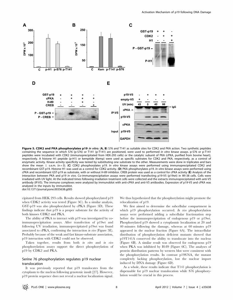
cipitated from HEK 293 cells. Results showed phosphorylated p19
when CDK2 activity was tested (Figure 5C). In a similar analysis,
GST-p19 was also phosphorylated by cPKA (Figure 5D). These
findings indicate that p19 is a proper substrate for the activity of
both kinases CDK2 and PKA.
The ability of PKA to interact with p19 was investigated by co-
immunoprecipitation assays. After transfection of p19wt and
following UV irradiation, immunoprecipitated p19wt was found
associated to cPKA, confirming the interaction in vivo (Figure 5E).
Probably because of the weak and fast kinase-substrate association,
p19 interaction with CDK2 could not be observed.
Taken together, results from both in vitro and in vivo
phosphorylation assays support the direct phosphorylation of
p19 by CDK2 and PKA.
Serine 76 phosphorylation regulates p19 nucleartranslocation
It was previously reported that p19 translocates from the
cytoplasm to the nucleus following genotoxic insult [27]. However,
p19 protein sequence does not reveal a nuclear localization signal.
We thus hypothesized that the phosphorylation might promote the
relocalization of p19.
We first aimed to determine the subcellular compartment in
which p19 phosphorylation occurred. In vivo phosphorylation
assays were performed adding a subcellular fractionation step
before the immunoprecipitation of endogenous p19 or p19wt.
Phosphorylated p19 showed a cytoplasmic localization at 20 and
40 minutes following the damage, whereas at 60 minutes p19
appeared in the nuclear fraction (Figure 6A). The intracellular
distribution of phosphorylation deficient mutants showed that
p19T141A conserved the ability to translocate into the nucleus
(Figure 6B). A similar result was observed for endogenous p19
when PKA was inhibited by H-89 (Figure 6C). The analyses of
protein distribution patterns by western blot were consistent with
the phosphorylation results. In contrast p19S76A, the mutant
completely lacking phosphorylation, lost the nuclear import
induced by DNA damage (Figure 6D).
As a whole, these results indicate that T141 phosphorylation is
dispensable for p19 nuclear translocation while S76 phosphory-
lation would be crucial in this process.
Figure 5. CDK2 and PKA phosphorylates p19 in vitro. (A, B) S76 and T141 as suitable sites for CDK2 and PKA action. Two synthetic peptidescontaining the sequence in which S76 (p-S76) or T141 (p-T141) are positioned, were used to performed in vitro kinase assays. p-S76 or p-T141peptides were incubated with CDK2 (immunoprecipitated from HEK-293 cells) or the catalytic subunit of PKA (cPKA, purified from bovine heart),respectively. A histone H1 peptide (p-H1) or kemptide (Kemp) were used as specific subtrates for CDK2 and PKA, respectively, as a control ofenzymatic activity. Kinase activity specificity was tested by substituting one substrate to the other. Measurements were done in triplicates and barsshow the mean 6 s.e.m. (n = 3). (C) CDK2 phosphorylates p19. In vitro kinase assays were performed using immunoprecipitated CDK2 andrecombinant GST-p19. Histone H1 was used as a control for CDK2 activity. (D) PKA phosphorylates p19. In vitro kinase assays were performed usingcPKA and recombinant GST-p19 as substrate, with or without H-89 inhibitor. CREB protein was used as a control for cPKA activity (E) Analysis of theinteraction between PKA and p19 in vivo. Co-immunoprecipitation assays were performed transfecting p19-V5 (p19wt) in WI-38 cells. Cells wereirradiated with UV light. At the indicated times following irradiation treatment cells were collected and the extracts immunoprecipitated with anti-V5antibody (IP:V5). The immune complexes were analyzed by immunoblot with anti-cPKA and anti-V5 antibodies. Expression of p19-V5 and cPKA wasanalyzed in the inputs by immunoblot.doi:10.1371/journal.pone.0035638.g005
Activation Mechanism of p19 following DNA Damage
PLoS ONE | www.plosone.org 8 April 2012 | Volume 7 | Issue 4 | e35638

Serine 76 and threonine 141 phosphorylation is criticalfor p19 function linked to the response to DNA damage
We next examined the functional relevance of p19 phosphor-
ylation. As previously mentioned, p19 is a cell cycle inhibitor
which has also a role in the DDR. Then, the ability to inhibit cell
cycle progression was first assessed for p19 mutants. The results
showed that all of the mutants (p19S13A, p19S66A, p19S76A,
p19T89A, p19T141A, p19S76A/T141A) displayed similar abili-
ties to block cell proliferation compared to p19wt (Figure S5).
Therefore, neither S76 nor T141 are necessary for inhibiting
CDK4/6.
The DDR involves complex signal transduction pathways that
regulate DNA repair and cell death mechanisms to restore DNA
integrity or to eliminate the damaged cell. We have previously
reported that p19 participates in the DDR being necessary for an
efficient repair of the DNA damage [25,27–29]. Particularly, down
regulation of endogenous p19 resulted in decreased DNA repair
after treatment with different genotoxic drugs. In contrast, p19
overexpression showed enhanced DNA repair activity compared
to non transfected cells. To study the functional role of p19
phosphorylation, the DNA repair ability of the cells overexpressing
p19 mutants was analyzed by Unscheduled DNA Synthesis (UDS).
The overexpression of those which maintain a complete
phosphorylation capability (p19S13A, p19S66A or p19T89A)
achieved similar levels of DNA repair compared to those observed
for p19wt (Figure 7A and Figure S6A). However, when any of the
phosphorylation deficient-mutants were tested (p19S76A,
p19T141A, p19S76A/T141A), DNA repair levels were signifi-
cantly diminished after UV light or b-amyloid treatment, reaching
those values obtained for control cells (Figure 7A and Figure S6A).
In contrast, the glutamic acid mutants mimicking the phosphor-
ylation at S76 and T141 (p19S76E, p19S76E/T141E) recovered
the DNA repair function displaying levels comparable to p19wt
(Figure 7B and Figure S6B). These results show that the
phosphorylation on both sites, S76 and T141, are strictly necessary
for p19 role in DNA repair. Since S76 and T141 are dispensable
for the inhibition of the cell cycle, these findings also support that
the role of p19 as a cell cycle regulator is dissociated from its DNA
repair function (27).
Two mechanisms are essential in response to genotoxic stress to
maintain genome integrity: DNA repair and apoptosis. As part of
the DDR, when the DNA damage is too severe to be repaired, cell
death programs are activated to eliminate the cell irreversibly
affected. It was previously reported that p19 overexpression
significantly decreases the apoptosis induced by UV light, cisplatin
and b-amyloid peptide [27,28]. Then, we tested whether p19
phosphorylation mutants, lacking the DNA repair activity, also lost
Figure 6. DNA damage induced p19 nuclear translocation is dependent on S76 phosphorylation. (A) Distribution of phosphorylated p19in the cytoplasmic and nuclear fractions after DNA damage. In vivo phosphorylation assays were performed in WI-38 fibroblasts. Cells were treatedwith UV (4 mJ/cm2), collected at the indicated times, and the extracts subjected to a subcellular fractionation protocol. Either the cytoplasmic (C) ornuclear fractions (N) were immunoprecipitated with anti-p19 antibody, and the immunocomplexes analyzed by SDS-PAGE and autoradiography(upper panel). (B) Subcellular distribution of the phosphorylation deficient mutant p19T141A. For in vivo phosphorylation assays, WI-38 cells weretransfected with p19wt or p19T141A, treated with UV radiation and collected at the indicated times. After subcellular fractionation, extracts wereimmunoprecipitated with an anti-V5 antibody and analyzed as in (A). p19wt or p19T141A subcellular distributions were also studied by immunoblot(C) Subcellular localization of endogenous deficiently phosphorylated-p19 after PKA inhibition. For in vivo phosphorylation assays, cells wereprocessed as in (A) but, before UV irradiation, they were incubated with H-89 for 1 hour. Endogenous distribution of p19 was also studied byimmunoblot. (D) Subcellular localization of p19S76A mutant following DNA damage. WI-38 cells were transfected with p19S76A and treated with UVradiation. At the indicated times, extracts were prepared by subcellular fractionation and analized by immunoblot with anti V5-antibody.doi:10.1371/journal.pone.0035638.g006
Activation Mechanism of p19 following DNA Damage
PLoS ONE | www.plosone.org 9 April 2012 | Volume 7 | Issue 4 | e35638

the ability to protect cells against apoptosis. All the mutants with
full phosphorylation capability reduced caspase-3 activity to a level
similar to that observed for p19wt. Conversely, cells overexpress-
ing the phosphorylation-deficient mutants displayed higher levels
of caspase-3 activity that were comparable to those measured in
control cells (Figure 7C and Figure S7). In contrast, the mutants
mimicking the phosphorylation on S76 and T141 (p19S76E,
p19S76E/T141E) showed values similar to those found for p19wt
(Figure 7D). We then concluded that those mutations which
disrupt p19 phosphorylation also affect the protection conferred by
p19 from apoptosis.
Figure 7. Phosphorylation of serine 76 and threonine 141 is required for p19 function linked to the response to DNA damage (A)DNA repair ability of cells overexpressing p19wt or p19 phosphorylation deficient mutants. WI-38 fibroblasts were transfected with p19wt or theindicated p19 mutants. Cells were maintained in an arginine-free medium containing 1% fetal bovine serum during 48 h, damage with 4 mJ/cm2 UVand incubated with [3H]-thymidine. Following 10 h, cell lysates were tested for Unscheduled DNA Synthesis assay (UDS). Bars represent the mean 6s.e.m of three independent experiments performed in triplicate. Student’s t-test was used to compare UV-treated control sample (none) with UV-treated p19wt or p19 mutant samples. (*p,0,005). Protein expression was analyzed by immunoblot. (B) Similarly as in (A) but overexpressing thephosphomimetic p19 mutants. (C) UV-dependent apoptotic response of cells overexpressing p19wt or phosphorylation deficient mutants of p19. WI-38 fibroblasts were transfected with p19wt or the indicated p19 mutants. Twelve hours following UV irradiation, cell lysates were tested for caspase-3activity. Results are expressed as percentage of caspase-3 activity with respect to basal activity of cell lysates nontransfected and without UV-treatment, which was set to 100. Bars represent the mean 6 s.e.m. of three independent experiments performed in triplicate. Student t-test was usedto compare, UV-treated control sample (none) with UV-treated p19wt or p19 mutant samples (*p,0.005). (D) Similarly as in (C) but overexpressingthe phosphomimetic p19 mutants.doi:10.1371/journal.pone.0035638.g007
Activation Mechanism of p19 following DNA Damage
PLoS ONE | www.plosone.org 10 April 2012 | Volume 7 | Issue 4 | e35638

Altogether, these findings are consistent with a regulation
pathway in which S76 and T141 phosphorylation is critical for the
reported function of p19 linked to the response to DNA damage.
Discussion
In response to DNA damage, conserved checkpoint mecha-
nisms trigger multiple events that coordinate cell cycle progression,
DNA repair and cell death in order to restore DNA integrity or to
eliminate the irreversibly damaged cell. The present study
uncovers the activation mechanism of p19 in response to DNA
damage. The results show that p19 is a downstream target of the
main DDR signaling pathways, ATM/Chk2 and ATR/Chk1. It
was demonstrated that p19 function involved in DNA repair and
cell survival is modulated by a sequential phosphorylation at serine
76 and threonine 141 dependent on CDK2 and PKA. A previous
work reported p19 phosphorylation under basal conditions in
U2OS cell line and suggested serines 66 and 76 as phosphorylation
sites [46]. Although in our study p19 phosphorylation was not
observed without genotoxic treatment in any of the cell lines
tested, the previous findings are consistent with S76 as a potential
phosphorylation aminoacid. Remarkably, both identified residues,
S76 and T141, are conserved in p19 protein sequences of different
mammal species but not throughout the other INK4 family
members (Figure S8, S9). Then, the phosphorylation and
activation mechanism presented herein might explain the singular
function in the DDR of this INK4 protein.
The structural and dynamic consequences caused by the
phosphorylations on p19 were analyzed by Molecular Dynamics
(MD). Simulation of S76 phosphorylation (p19p) showed a main
increase in the mobility of the third ankyrin motif, where the
negative charge was located, but also mobility differences in the
fourth and fifth ankyrin repeat were observed. The experimental
data indicated a sequential phosphorylation at S76 and T141
aminoacids. Supporting the sequential phosphorylation results, the
structural changes in the fifth ankyrin repeat induced in p19p
might be necessary to enable T141 phosphorylation. The main
differences found when both, S76 and T141, phosphorylations
were simulated are located in the fifth repeat. We propose that the
additional changes in the structure promoted by the second
phosphorylation could be involved in the interaction of p19 with
proteins related to DDR mechanisms and then might be critical
for p19 function. Further experimental studies are being
conducted to identify p19 interactors and verify this possibility.
It is well established that inhibition of CDK activity, one of the
main actions promoted by checkpoint responses, leads to cell cycle
arrest and provides time for DNA repair. However, increasing
evidence supports an active role for CDKs in the DDR. Overall
CDK activity was reported to be necessary for an efficient DDR
activation after c-irradiation induced DNA damage [47]. In yeasts
and mammalian cells, CDK activity is essential for DNA resection
and progression of homologous recombination repair during S and
G2 phases [19,48–50]. For instance, CDK2 targets several
substrates in the DDR pathway such us BRCA1 and BRCA2,
Ku70 and ATRIP [51,52]. Cyclin A1 which promotes CDK2
activation is transcriptionally induced by c and UV-irradiation via
a p53-mediated mechanism [51]. Interestingly, this fact contrasts
with the inhibition and/or repression of the other CDK2-
associated cyclins. Consistent with these reports, the sequence
analysis of S76 matched an exact CDK2 phosphorylation motif,
suggesting that p19 might be a putative substrate for this kinase. In
vitro assays confirmed direct CDK2-mediated phosphorylation of
p19. Adding to this, CDK inhibition prevented DNA-damage-
induced phosphorylation of endogenous p19. Specific down-
regulation of CDK2 impaired p19 phosphorylation. Together,
these results show the dependence of p19 phosphorylation on
CDK2 function and strongly suggest the direct action of this kinase
on this protein. In vivo CDK inhibition also blocked the
phosphorylation of p19T141A and p19ANKless mutants. Since
only two residues, S76 and T141, become phosphorylated after
DNA injury these observations also indicate that S76 might be the
specific target site for CDK2. T141 in p19 was shown to be
embedded within a PKA consensus motif. PKA has been shown to
exert an antiapoptotic effect in different cell lines. In addition,
PKA activity was implicated in the activation of the processivity
factor PCNA and in the nuclear translocation of DNA-PK, two
critical proteins in DNA repair [52–54]. Herein, phosphorylation
and interaction assays performed in vitro and in vivo supported the
direct action of PKA on p19. Moreover, the decreased
phosphorylation observed for endogenous p19 after H-89
treatment was consistent with a reduced phosphorylation of
p19T141A and p19ANKless mutants. Even more, no further
reduction in p19T141A or p19ANKless phosphorylation was
found by PKA inhibition, suggesting that the action site of this
kinase had already been eliminated in these mutants. Taken
together, these findings support p19 phosphorylation by PKA in
response to DNA damage and point out to T141 as the target site
for this kinase.
The regulation of protein localization provides cells with a
convenient way to modulate their functions. p19 does not contain
the standard basic monopartite or bipartite nuclear localization
signal usually found in nuclear proteins [55]. However, protein
phosphorylation also serves as an essential mechanism for
modulating subcellular localization. To analyze this possibility,
the cellular compartment in which p19 phosphorylation takes
place was explored. Phosphorylation assays and immunoblot
analysis showed phosphorylated p19 in the cytoplasm followed by
a translocation into the nucleus. Moreover, p19T141A was also
able to translocate into the nucleus in spite of its phosphorylation
deficiency. In contrast, p19S76A lost the nuclear import induced
by DNA damage. Consequently, these results suggest that the first
phosphorylation event on serine 76 would allow p19 nuclear
translocation while modification of T141 would be dispensable in
this matter. In view of the results discussed before, these findings
imply the presence of active CDK2-cyclin A complexes in the
cytoplasm. During cell cycle progression, the activity of CDKs is
located in the nucleus. However, consistent with our findings
recent works showed cytoplasmic translocation of active CDK2 in
response to UV irradiation and chemotherapeutic agents [56]. In
addition, cytoplasmic CDK2 activity was related to apoptotic cell
death [57]. There is accumulating evidence supporting the fact
that some proteins involved in DNA repair may also be taking part
in apoptosis. [25,58]. Thus, CDK2 might also be among these
proteins playing a dual role in the DDR, modulating the activity of
both anti apoptotic and pro-apototic proteins. Since p19 nuclear
translocation was only dependent on S76, it is tempting to
speculate that the phosphorylation on T141 might occur in the
nucleus. In addition to the structural changes promoted by S76
phosphorylation, the nuclear import preceding T141 phosphory-
lation further supports the sequential phosphorylation of p19.
Protein phosphorylation is a widely used mechanism to
selectively modulate protein activity. We then investigated if
phosphorylation had a functional relevance on p19. The
expression of p19 mutants lacking S76 and/or T141 promoted
cell cycle arrest at similar levels to those observed for wild type
p19. These results indicate that neither S76 nor T141 are
necessary for p19 inhibition of CDK4/6 kinases. Previous works
based on crystal structure analysis showed that binding to CDK6
Activation Mechanism of p19 following DNA Damage
PLoS ONE | www.plosone.org 11 April 2012 | Volume 7 | Issue 4 | e35638

involves mainly ankyrin domains I–III of p19. In accordance with
our findings, threonine 141 is positioned within the fifth ankyrin
repeat and then would not participate in the interaction with
CDK. Moreover, S76, located in the third ankyrin repeat, was not
described to be implicated in CDK binding by NMR studies. [59–
61]. In contrast, both S76 and T141 phosphorylation were found
to be crucial for p19 function related to the response to DNA
damage. Since the phosphorylation-deficient mutants keep the
ability to block cell cycle progression, the results suggest that p19
activity linked to the DDR is not associated with inhibiting cell
proliferation. In fact, these findings denote the independence
between the functions of p19 in the cell cycle and in the DDR, in
agreement with our previous works [27,29].
In summary, our results uncover the activation mechanism of
p19 implicated in the response to DNA damage. We propose that
the phosphorylation of specific sites might induce conformational
changes in p19 necessary for the correct subcellular localization
and for the interaction with DDR proteins. Mutations in DDR
critical genes that lead to impaired genome stability, increased
cancer susceptibility or enhanced cell death reflect the importance
of a proper DDR. Consequently, a comprehensive knowledge of
the DDR pathways becomes essential to understand disease
development and might contribute to establish more efficient
therapeutic approaches.
Supporting Information
Materials and Methods S1 Description of the mutagen-esis strategy used to construct p19 mutants.(DOC)
Figure S1 p19 immunoprecipitation specificity. WI-38
fibroblasts were labeled with [32P]-orthophosphate and treated
with b-amyloid peptide (20 mM), cisplatin (10 mM) or UV light
(4 mJ/cm2) for 3 hours. Equal amounts of whole cell extracts were
subjected to immunoprecipitation with anti-p19 antibody (+,
rabbit IgG, Santa Cruz Biotechnology) or anti-V5 antibody as a
control antibody (2, rabbit IgG, Santa Cruz Biotechnology). The
immune complexes were analyzed by SDS-PAGE and autoradi-
ography (upper panels; P-p19, phosphorylated p19) or immuno-
blotting (lower panels; p19).
(TIF)
Figure S2 Prediction of p19 phosphorylation sites. p19
protein sequence was analyzed for the presence of potential
phosphorylation sites using the bioinformatic tool Netphos 2.0
server. Tables show serine predictions (A) or threonine predictions
(B), no putative tyrosine phoshorylation sites were found. (C)
Graph shows the score of the predicted phosphorylation sites. Pos,
position of the potential phosphorylation site.
(TIF)
Figure S3 Prediction of kinase specific phosphorylationsites in p19. p19 protein sequence was analyzed for the presence
of kinase specific phosphorylation sites using the bioinformatic tool
NetphosK 1.0 server with evolutionary stable sites filter (ESS
filter). Table shows the position of the putative phosphorylation
sites for the indicated kinases. (Pos, position in p19 protein
sequence).
(TIF)
Figure S4 p19 phosphorylation is not abolished by highconcentrations of PKA inhibitor. WI-38 fibroblasts were
incubated with the indicated concentrations of H-89 for 1 hour,
and then treated with cisplatin (10 mM) for 2 hours and
endogenous p19 phosphorylation analyzed by autoradiography.
(TIF)
Figure S5 S76 and T141 are not involved in the cell cyclefunction of p19. Proliferation status of cells overexpressing
p19wt or p19 phosphorylation deficient mutants. WI-38 fibroblasts
were transfected with p19wt or the indicated p19 mutants. Cells
were incubated with [3H]-thymidine for 5 hours and the lysates
were tested for tritium incorporation. Bars represent the mean 6
s.e.m of three independent experiments performed in triplicate.
Student’s t-test was used to compare control sample (none) with
p19wt or p19 mutant samples. (*p,0,005).
(TIF)
Figure S6 Phosphorylation of S76 and T141 is requiredfor p19 function in DNA repair. (A) DNA repair ability of
cells overexpressing p19wt or p19 phosphorylation deficient
mutants. WI-38 fibroblasts were transfected with p19wt or the
indicated p19 mutants. Cells were maintained in an arginine-free
medium containing 1% fetal bovine serum during 48 h. b-amyloid
peptide (20 mM) was added to the medium and cells were
incubated with [3H]-thymidine for 10 hours. Cell lysates were
tested for Unscheduled DNA Synthesis assay (UDS). Bars
represent the mean 6 s.e.m of three independent experiments
performed in triplicate. Student’s t-test was used to compare b-
amyloid peptide-treated control sample (none) with b-amyloid
peptide-treated p19wt or p19 mutant samples. (*p,0,005). Protein
expression was analyzed by immunoblot. (B) Similarly as in (A) but
overexpressing the phosphomimetic p19 mutants.
(TIF)
Figure S7 Phosphorylation of S76 and T141 is necessaryfor p19 function in apoptosis. b-amyloid peptide-dependent
apoptotic response of cells overexpressing p19wt or the phosphor-
ylation deficient mutants, p19S76A and p19T141A. WI-38
fibroblasts were transfected with p19wt or the indicated p19
mutants. b-amyloid peptide (20 mM) was added to the medium
and following 12 hours cell lysates were tested for caspase-3
activity. Results are expressed as percentage of caspase-3 activity
with respect to basal activity of cell lysates nontransfected and
without b-amyloid peptide-treatment, which was set to 100. Bars
represent the mean 6 s.e.m of three independent experiments
performed in triplicate. Students t-test was used to compare, b-
amyloid peptide-treated control sample (none) with b-amyloid
peptide-treated p19wt or p19 mutant samples (*p,0.005).
(TIF)
Figure S8 Conservation of p19 phosphorylation sites indifferent mammalian species. p19 protein sequences from
the indicated mammals were align using T-Coffee multiple
sequence alignment tool. Arrows indicate the position of S76
and T141 from p19 human sequence.
(TIF)
Figure S9 Alignment of protein sequences of the INK4family members. Protein sequences were align using T-Coffee
multiple sequence alignment tool. Arrows indicate the position of
S76 and T141 from p19 protein sequence. (p15, p15INK4b; p16,
p16INK4a; p18, p18INK4c; p19, p19INK4d).
(TIF)
Acknowledgments
We thank Dr. Luciana E. Giono for help with language editing.
Author Contributions
Conceived and designed the experiments: MCM ETC. Performed the
experiments: MCM MFO SVS. Analyzed the data: MCM ETC MM NJD
OPP. Contributed reagents/materials/analysis tools: NJD OPP. Wrote the
paper: MCM ETC.
Activation Mechanism of p19 following DNA Damage
PLoS ONE | www.plosone.org 12 April 2012 | Volume 7 | Issue 4 | e35638

References
1. Lobrich M, Jeggo PA (2007) The impact of a negligent G2/M checkpoint on
genomic instability and cancer induction. Nat Rev Cancer 7: 861–869.
2. Kastan MB, Bartek J (2004) Cell-cycle checkpoints and cancer. Nature 432:
316–323.
3. Jackson SP, Bartek J (2009) The DNA-damage response in human biology and
disease. Nature 461: 1071–1078.
4. Abraham RT (2001) Cell cycle checkpoint signaling through the ATM and
ATR kinases. Genes Dev 15: 2177–2196.
5. Shiloh Y (2003) ATM and related protein kinases: safeguarding genome
integrity. Nat Rev Cancer 3: 155–168.
6. Lukas J, Lukas C, Bartek J (2004) Mammalian cell cycle checkpoints: signalling
pathways and their organization in space and time. DNA Repair (Amst) 3:
997–1007.
7. Bartek J, Lukas J (2007) DNA damage checkpoints: from initiation to recovery or
adaptation. Curr Opin Cell Biol 19: 238–245.
8. Harper JW, Elledge SJ (2007) The DNA damage response: ten years after. Mol
Cell 28: 739–745.
9. Shiotani B, Zou L (2009) Single-stranded DNA orchestrates an ATM-to-ATR
switch at DNA breaks. Mol Cell 33: 547–558.
10. Gatei M, Sloper K, Sorensen C, Syljuasen R, Falck J, et al. (2003) Ataxia-
telangiectasia-mutated (ATM) and NBS1-dependent phosphorylation of Chk1
on Ser-317 in response to ionizing radiation. J Biol Chem 278: 14806–14811.
11. Sorensen CS, Syljuasen RG, Falck J, Schroeder T, Ronnstrand L, et al. (2003)
Chk1 regulates the S phase checkpoint by coupling the physiological turnover
and ionizing radiation-induced accelerated proteolysis of Cdc25A. Cancer Cell
3: 247–258.
12. Stiff T, Walker SA, Cerosaletti K, Goodarzi AA, Petermann E, et al. (2006)
ATR-dependent phosphorylation and activation of ATM in response to UV
treatment or replication fork stalling. Embo J 25: 5775–5782.
13. Liu Q, Guntuku S, Cui XS, Matsuoka S, Cortez D, et al. (2000) Chk1 is an
essential kinase that is regulated by Atr and required for the G(2)/M DNA
damage checkpoint. Genes Dev 14: 1448–1459.
14. Jiang K, Pereira E, Maxfield M, Russell B, Goudelock DM, et al. (2003)
Regulation of Chk1 includes chromatin association and 14-3-3 binding following
phosphorylation on Ser-345. J Biol Chem 278: 25207–25217.
15. Zhao H, Piwnica-Worms H (2001) ATR-mediated checkpoint pathways
regulate phosphorylation and activation of human Chk1. Mol Cell Biol 21:
4129–4139.
16. Bensimon A, Schmidt A, Ziv Y, Elkon R, Wang SY, et al. ATM-dependent and
-independent dynamics of the nuclear phosphoproteome after DNA damage. Sci
Signal 3: rs3.
17. Bartek J, Lukas J (2003) Chk1 and Chk2 kinases in checkpoint control and
cancer. Cancer Cell 3: 421–429.
18. Donzelli M, Draetta GF (2003) Regulating mammalian checkpoints through
Cdc25 inactivation. EMBO Rep 4: 671–677.
19. Jazayeri A, Falck J, Lukas C, Bartek J, Smith GC, et al. (2006) ATM- and cell
cycle-dependent regulation of ATR in response to DNA double-strand breaks.
Nat Cell Biol 8: 37–45.
20. Stucki M, Jackson SP (2006) gammaH2AX and MDC1: anchoring the DNA-
damage-response machinery to broken chromosomes. DNA Repair (Amst) 5:
534–543.
21. Massague J (2004) G1 cell-cycle control and cancer. Nature 432: 298–306.
22. Sherr CJ, Roberts JM (1999) CDK inhibitors: positive and negative regulators of
G1-phase progression. Genes Dev 13: 1501–1512.
23. Pei XH, Xiong Y (2005) Biochemical and cellular mechanisms of mammalian
CDK inhibitors: a few unresolved issues. Oncogene 24: 2787–2795.
24. Ortega S, Malumbres M, Barbacid M (2002) Cyclin D-dependent kinases, INK4
inhibitors and cancer. Biochim Biophys Acta 1602: 73–87.
25. Canepa ET, Scassa ME, Ceruti JM, Marazita MC, Carcagno AL, et al. (2007)
INK4 proteins, a family of mammalian CDK inhibitors with novel biological
functions. IUBMB Life 59: 419–426.
26. Al-Mohanna MA, Al-Khalaf HH, Al-Yousef N, Aboussekhra A (2007) The
p16INK4a tumor suppressor controls p21WAF1 induction in response to
ultraviolet light. Nucleic Acids Res 35: 223–233.
27. Ceruti JM, Scassa ME, Flo JM, Varone CL, Canepa ET (2005) Induction of
p19INK4d in response to ultraviolet light improves DNA repair and confers
resistance to apoptosis in neuroblastoma cells. Oncogene 24: 4065–4080.
28. Ceruti JM, Scassa ME, Marazita MC, Carcagno AC, Sirkin PF, et al. (2009)
Transcriptional upregulation of p19INK4d upon diverse genotoxic stress is
critical for optimal DNA damage response. Int J Biochem Cell Biol 41:
1344–1353.
29. Scassa ME, Marazita MC, Ceruti JM, Carcagno AL, Sirkin PF, et al. (2007) Cell
cycle inhibitor, p19INK4d, promotes cell survival and decreases chromosomal
aberrations after genotoxic insult due to enhanced DNA repair. DNA Repair
(Amst) 6: 626–638.
30. Tavera-Mendoza LE, Wang TT, White JH (2006) p19INK4D and cell death.
Cell Cycle 5: 596–598.
31. Blom N, Gammeltoft S, Brunak S (1999) Sequence and structure-based
prediction of eukaryotic protein phosphorylation sites. J Mol Biol 294:
1351–1362.
32. Blom N, Sicheritz-Ponten T, Gupta R, Gammeltoft S, Brunak S (2004)
Prediction of post-translational glycosylation and phosphorylation of proteins
from the amino acid sequence. Proteomics 4: 1633–1649.
33. Notredame C, Higgins DG, Heringa J (2000) T-Coffee: A novel method for fast
and accurate multiple sequence alignment. J Mol Biol 302: 205–217.
34. Chomczynski P, Sacchi N (1987) Single-step method of RNA isolation by acid
guanidinium thiocyanate-phenol-chloroform extraction. Anal Biochem 162:
156–159.
35. Giono LE, Manfredi JJ (2007) Mdm2 is required for inhibition of Cdk2 activity
by p21, thereby contributing to p53-dependent cell cycle arrest. Mol Cell Biol
27: 4166–4178.
36. Schreiber E, Matthias P, Muller MM, Schaffner W (1989) Rapid detection of
octamer binding proteins with ‘mini-extracts’, prepared from a small number of
cells. Nucleic Acids Res 17: 6419.
37. Shell SM, Li Z, Shkriabai N, Kvaratskhelia M, Brosey C, et al. (2009)
Checkpoint kinase ATR promotes nucleotide excision repair of UV-induced
DNA damage via physical interaction with xeroderma pigmentosum group A.
J Biol Chem 284: 24213–24222.
38. Jung CG, Uhm KO, Miura Y, Hosono T, Horike H, et al. (2011) Beta-amyloid
increases the expression level of ATBF1 responsible for death in cultured cortical
neurons. Mol Neurodegener 6: 47.
39. Sarkaria JN, Tibbetts RS, Busby EC, Kennedy AP, Hill DE, et al. (1998)
Inhibition of phosphoinositide 3-kinase related kinases by the radiosensitizing
agent wortmannin. Cancer Res 58: 4375–4382.
40. Hellmich MR, Pant HC, Wada E, Battey JF (1992) Neuronal cdc2-like kinase: a
cdc2-related protein kinase with predominantly neuronal expression. Proc Natl
Acad Sci U S A 89: 10867–10871.
41. Lew DJ, Kornbluth S (1996) Regulatory roles of cyclin dependent kinase
phosphorylation in cell cycle control. Curr Opin Cell Biol 8: 795–804.
42. Meyerson M, Enders GH, Wu CL, Su LK, Gorka C, et al. (1992) A family of
human cdc2-related protein kinases. Embo J 11: 2909–2917.
43. Tsai LH, Delalle I, Caviness VS, Jr., Chae T, Harlow E (1994) p35 is a neural-
specific regulatory subunit of cyclin-dependent kinase 5. Nature 371: 419–
423.
44. Songyang Z, Blechner S, Hoagland N, Hoekstra MF, Piwnica-Worms H, et al.
(1994) Use of an oriented peptide library to determine the optimal substrates of
protein kinases. Curr Biol 4: 973–982.
45. Songyang Z, Lu KP, Kwon YT, Tsai LH, Filhol O, et al. (1996) A structural
basis for substrate specificities of protein Ser/Thr kinases: primary sequence
preference of casein kinases I and II, NIMA, phosphorylase kinase, calmodulin-
dependent kinase II, CDK5, and Erk1. Mol Cell Biol 16: 6486–6493.
46. Thullberg M, Bartkova J, Khan S, Hansen K, Ronnstrand L, et al. (2000)
Distinct versus redundant properties among members of the INK4 family of
cyclin-dependent kinase inhibitors. FEBS Lett 470: 161–166.
47. Cerqueira A, Santamaria D, Martinez-Pastor B, Cuadrado M, Fernandez-
Capetillo O, et al. (2009) Overall Cdk activity modulates the DNA damage
response in mammalian cells. J Cell Biol 187: 773–780.
48. Deans AJ, Khanna KK, McNees CJ, Mercurio C, Heierhorst J, et al. (2006)
Cyclin-dependent kinase 2 functions in normal DNA repair and is a therapeutic
target in BRCA1-deficient cancers. Cancer Res 66: 8219–8226.
49. Ira G, Pellicioli A, Balijja A, Wang X, Fiorani S, et al. (2004) DNA end
resection, homologous recombination and DNA damage checkpoint activation
require CDK1. Nature 431: 1011–1017.
50. Sonoda E, Hochegger H, Saberi A, Taniguchi Y, Takeda S (2006) Differential
usage of non-homologous end-joining and homologous recombination in double
strand break repair. DNA Repair (Amst) 5: 1021–1029.
51. Muller-Tidow C, Ji P, Diederichs S, Potratz J, Baumer N, et al. (2004) The
cyclin A1-CDK2 complex regulates DNA double-strand break repair. Mol Cell
Biol 24: 8917–8928.
52. Akbar S, Minor T (2001) Significance and molecular targets of protein kinase A
during cAMP-mediated protection of cold stored liver grafts. Cell Mol Life Sci
58: 1708–1714.
53. Tortora G, Ciardiello F (2002) Protein kinase A as target for novel integrated
strategies of cancer therapy. Ann N Y Acad Sci 968: 139–147.
54. Schnoke M, Midura SB, Midura RJ (2009) Parathyroid hormone suppresses
osteoblast apoptosis by augmenting DNA repair. Bone 45: 590–602.
55. Dingwall C, Laskey RA (1991) Nuclear targeting sequences–a consensus?
Trends Biochem Sci 16: 478–481.
56. Hiromura K, Pippin JW, Blonski MJ, Roberts JM, Shankland SJ (2002) The
subcellular localization of cyclin dependent kinase 2 determines the fate of
mesangial cells: role in apoptosis and proliferation. Oncogene 21: 1750–
1758.
57. Maddika S, Ande SR, Wiechec E, Hansen LL, Wesselborg S, et al. (2008) Akt-
mediated phosphorylation of CDK2 regulates its dual role in cell cycle
progression and apoptosis. J Cell Sci 121: 979–988.
58. Hickman MJ, Samson LD (2004) Apoptotic signaling in response to a single type
of DNA lesion, O(6)-methylguanine. Mol Cell 14: 105–116.
59. Renner C, Baumgartner R, Noegel AA, Holak TA (1998) Backbone dynamics of
the CDK inhibitor p19(INK4d) studied by 15N NMR relaxation experiments at
two field strengths. J Mol Biol 283: 221–229.
Activation Mechanism of p19 following DNA Damage
PLoS ONE | www.plosone.org 13 April 2012 | Volume 7 | Issue 4 | e35638

60. Brotherton DH, Dhanaraj V, Wick S, Brizuela L, Domaille PJ, et al. (1998)
Crystal structure of the complex of the cyclin D-dependent kinase Cdk6 boundto the cell-cycle inhibitor p19INK4d. Nature 395: 244–250.
61. Russo AA, Tong L, Lee JO, Jeffrey PD, Pavletich NP (1998) Structural basis for
inhibition of the cyclin-dependent kinase Cdk6 by the tumour suppressorp16INK4a. Nature 395: 237–243.
Activation Mechanism of p19 following DNA Damage
PLoS ONE | www.plosone.org 14 April 2012 | Volume 7 | Issue 4 | e35638
Recommended