Chapter 22— The Respiratory System
22-1
Ch. 22 (Respiratory Sys.) Study Guide
1. Critically read Chapter 22 pp. 864-886 right before 22.3 “Gas Exchange and Transport” section
2. Comprehend Terminology (those in bold)3. Study-- Figure questions, Think About It
questions, and Before You Go On (section-ending) questions
4. Do end-of-the-chapter questions:– Testing Your Recall— 1-5, 7, 10, 11-18– True or False– 1, 2, 4-6, 8– Testing Your Comprehension– 1, 4, 5
2
Breathe/Breath (1 or 2)
Fear less, hope more;Whine less, breathe more;Talk less, say more; Hate less, love more;And all good things are yours.
--Swedish proverb
22-3
Breathe/Breath (2 of 2)Every day brings a chance for you to draw in a breath,
kick off your shoes, and dance.
--Oprah Winfrey
22-4
§ I. Anatomical Consideration
Self-Check Question: As we breathe in, what respiratory organs, in order, does air pass through?
Answer: Nose (mouth) . . .
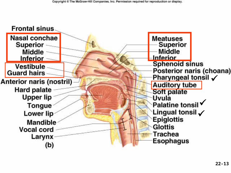
Fig. 22.1
22-5
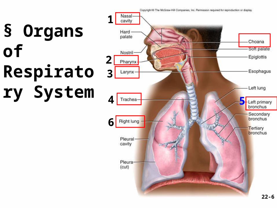
§ Organs of Respiratory System
22-6
1
4
32
5
6
General Aspects1. Airflow in lungs
– bronchi bronchioles alveoli
2. Conducting & Rspiratory (C/R) divisions--– (C) passages ONLY for airflow, nostrils to
bronchioles– (R) distal gas-exchange regions and ________
3. Upper/lower (U/L) respiratory tracts– (U) organs in head and neck, nose through
larynx– (L) organs of trachea through lungs
22-7
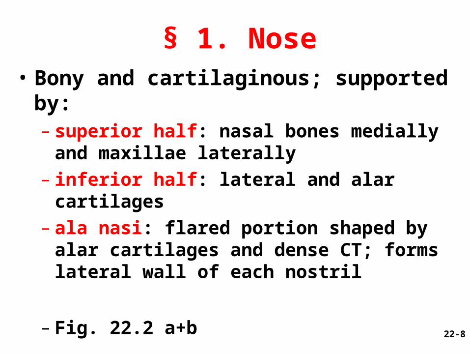
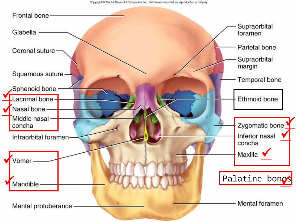
• Bony and cartilaginous; supported by:– superior half: nasal bones medially and
maxillae laterally– inferior half: lateral and alar cartilages– ala nasi: flared portion shaped by alar
cartilages and dense CT; forms lateral wall of each nostril
– Fig. 22.2 a+b
22-8
§ 1. Nose
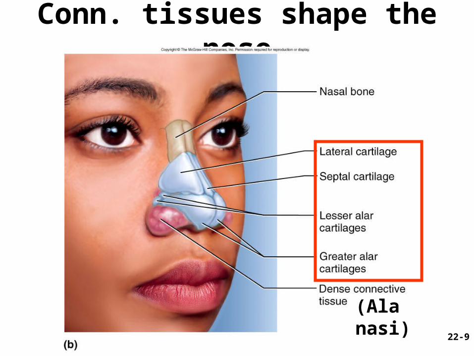
Conn. tissues shape the nose
22-9
(Ala nasi)
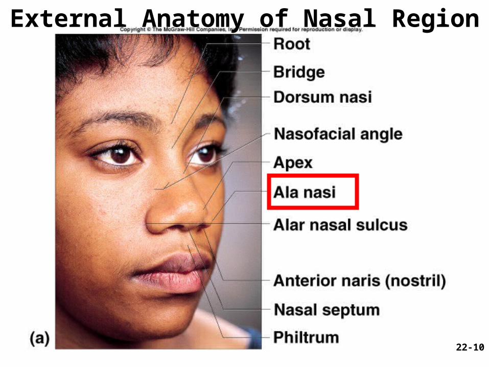
External Anatomy of Nasal Region
22-10
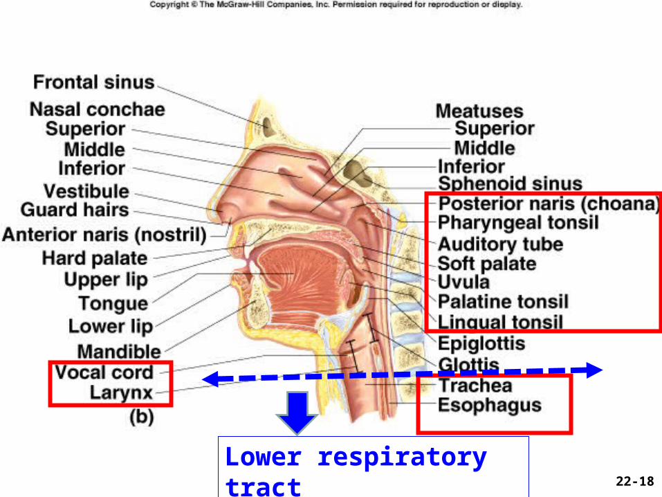
Nasal Cavity (1)
1. Extends from nostrils to posterior nares
2. Vestibule: dilated chamber inside ala nasi (just inside the nostril)– stratified squamous epithelium and vibrissae
(guard hairs)
3. Nasal septum divides cavity into right and left chambers called nasal fossae– Makes up of = Perpendicular plate of ethmoid
bone + . . .
22-11
Nasal Cavity (2) - Conchae and Meatuses
1. Superior, middle and inferior nasal conchae– 3 folds of tissue on lateral wall of nasal fossa– mucous membranes lines the cavity
2. Meatuses:– narrow air passages beneath each conchae– narrowness and turbulence ensures most air
contact the mucous membrane.
Fig. 22.3
22-12
22-13
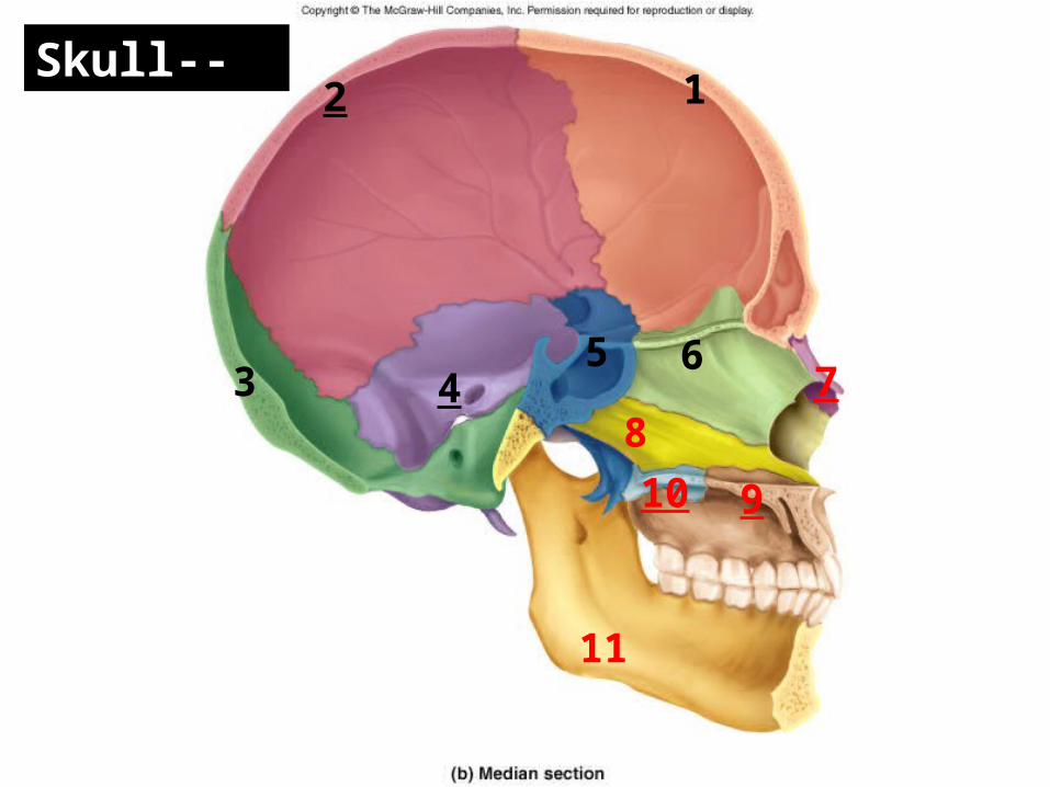
Figure 8.4b 12
3 45 6
Skull--
8
910
7
11
Palatine bones
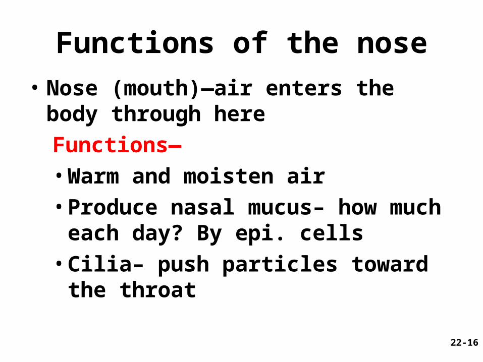
Functions of the nose
• Nose (mouth)—air enters the body through here
Functions—
• Warm and moisten air
• Produce nasal mucus– how much each day? By epi. cells
• Cilia– push particles toward the throat
22-16
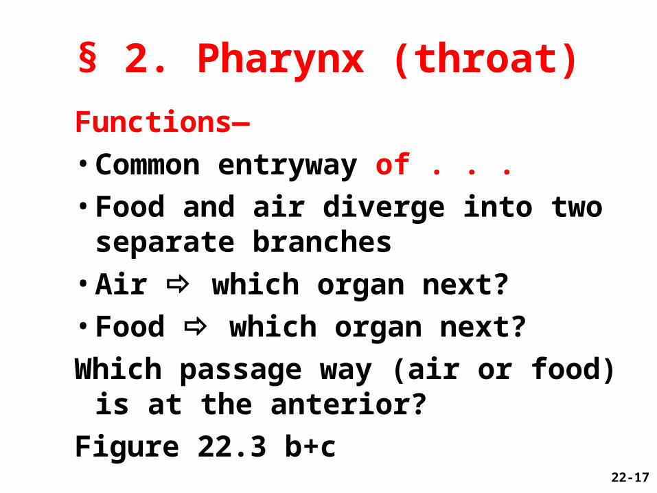
Functions—
• Common entryway of . . .
• Food and air diverge into two separate branches
• Air which organ next?
• Food which organ next?
Which passage way (air or food) is at the anterior?
Figure 22.3 b+c22-17
§ 2. Pharynx (throat)
22-17
22-18
Lower respiratory tract
Hyoid bone
Cricoid cartilage
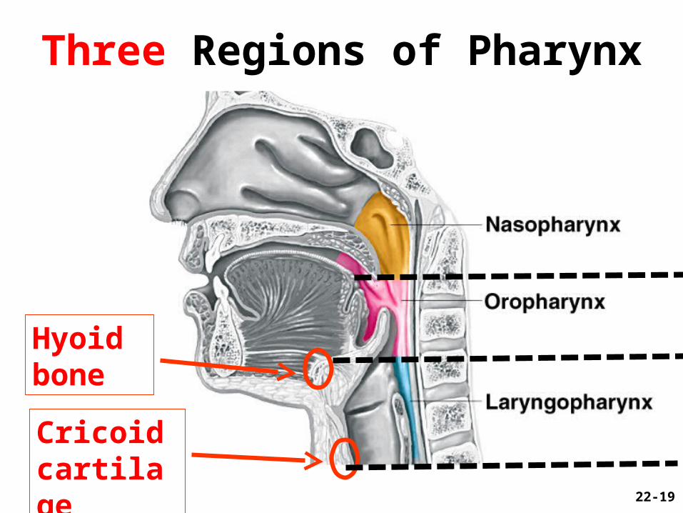
Three Regions of Pharynx
22-19
§ 2. Pharynx (continued)• Nasopharynx (pseudostratified epithelium)
– posterior to choanae, dorsal to soft palate– receives auditory tubes; houses _____ tonsil– 90 downward turn; traps large particles (>10m)
• Oropharynx (stratified squamous epithelium)
– space between soft palate and root of tongue, inferiorly as far as hyoid bone, contains palatine and lingual tonsils
• Laryngopharynx (stratified squamous epi.)
– hyoid bone to level of cricoid cartilage22-20
– Anatomy—anterior protrusion called ?– Functions—• Air passageway with cilia• Epiglottis– superior opening of larynx• Voice production by ____________
• Laryngitis—Inflammation of the vocal cords; symptoms? Three major causes?
22-21
§ 3. Larynx (Voice box)
Larynx• Glottis – vocal cords and opening between
them
• Epiglottis – flap of tissue that guards glottis, directs food
and drink to esophagus
• Infant larynx; epiglottis touches soft palate– higher in throat, forms a continuous airway
from nasal cavity to the larynx that allows breathing while swallowing
– by age 2, more muscular tongue, forces larynx down to lower position
22-22
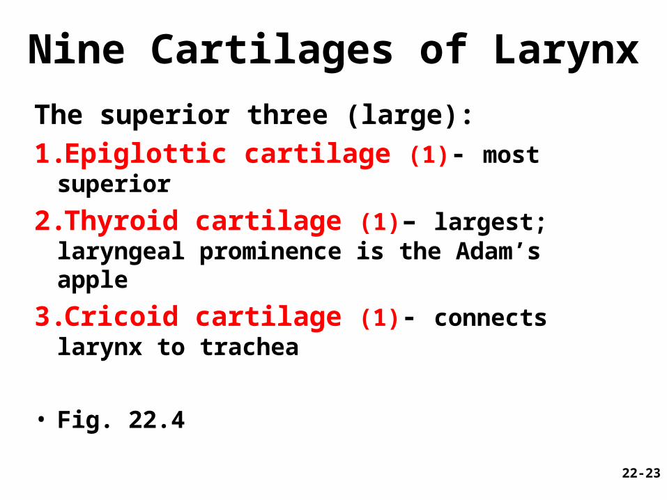
Nine Cartilages of Larynx
The superior three (large):
1.Epiglottic cartilage (1)- most superior
2.Thyroid cartilage (1)– largest; laryngeal prominence is the Adam’s apple
3.Cricoid cartilage (1)- connects larynx to trachea
• Fig. 22.4
22-23
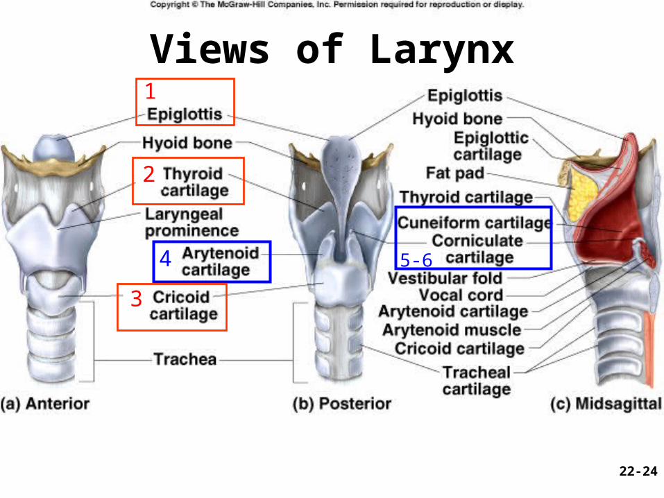
Views of Larynx1
2
3
4 5-6
22-24
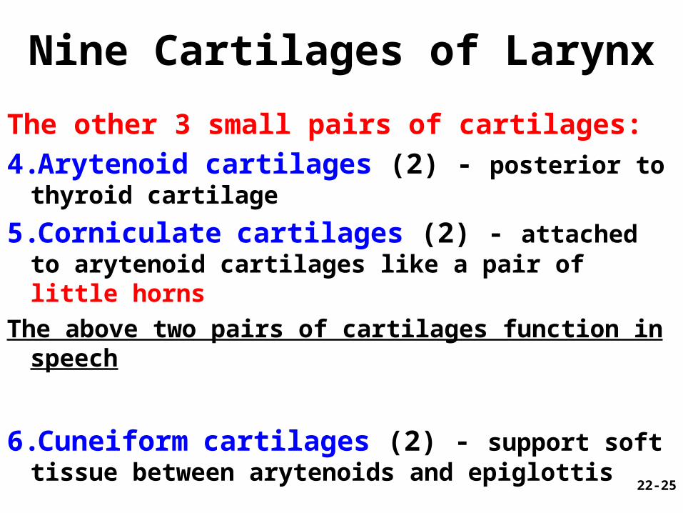
Nine Cartilages of Larynx
The other 3 small pairs of cartilages:
4.Arytenoid cartilages (2) - posterior to thyroid cartilage
5.Corniculate cartilages (2) - attached to arytenoid cartilages like a pair of little horns
The above two pairs of cartilages function in speech
6.Cuneiform cartilages (2) - support soft tissue between arytenoids and epiglottis
22-25
Walls of Larynx• Interior wall has 2 muscular folds on each
side, from thyroid to arytenoid cartilages– Vestibular folds (superior pair) and vocal
cords/folds (inferior) (produce sound)
• Intrinsic muscles (deep)- rotate corniculate and arytenoid cartilages (Fig. 22.6)– adducts (tightens: high pitch sound) or abducts (loosens: low
pitch sound) vocal cords
• Extrinsic muscles (superficial)- connect larynx to hyoid bone, elevate larynx during swallowing
22-26
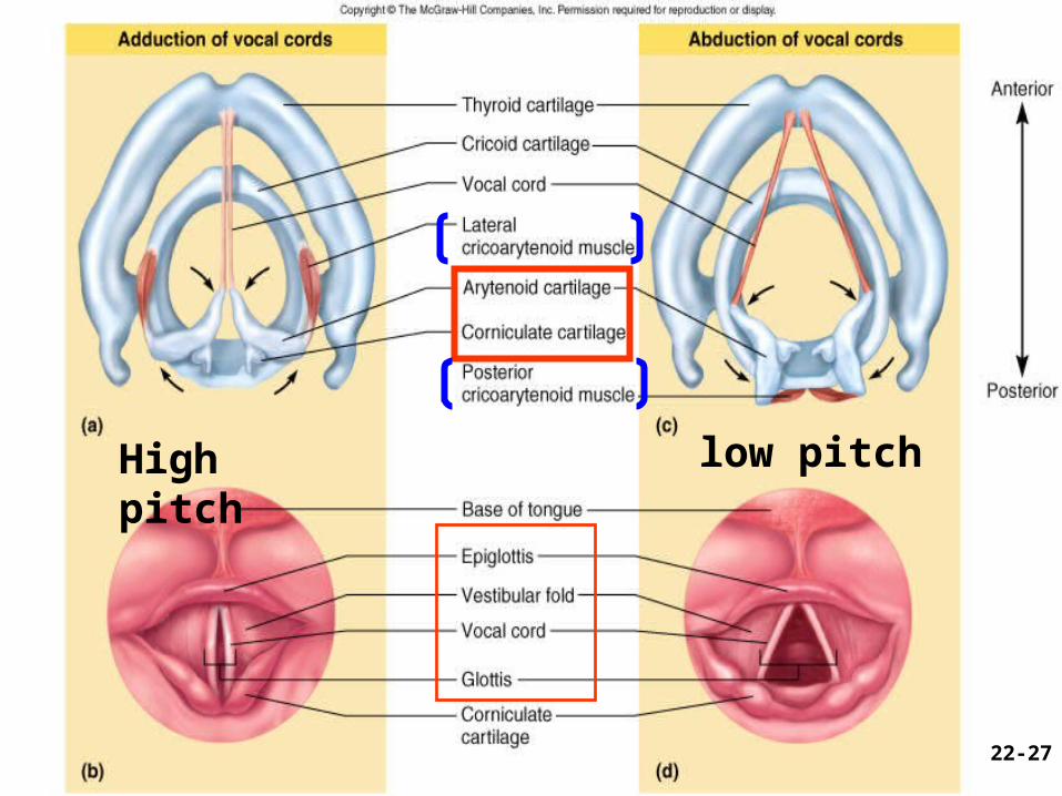
22-27
High pitch low pitch
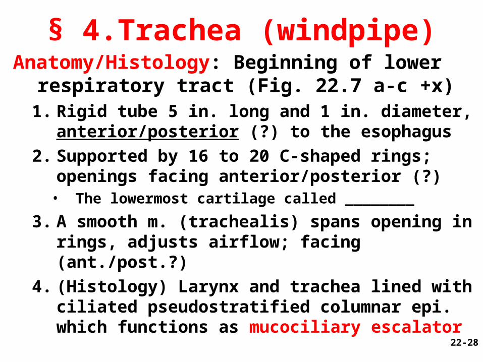
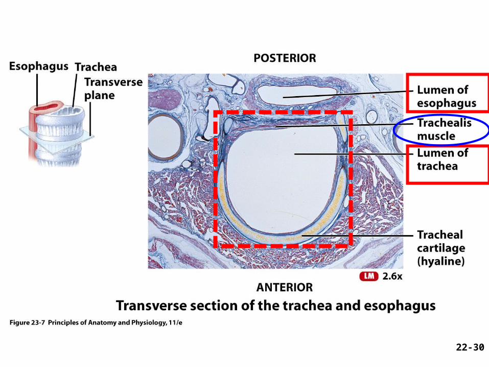
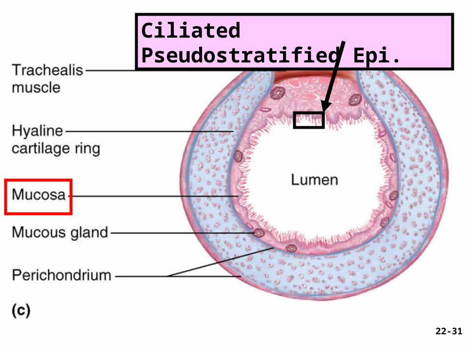
§ 4.Trachea (windpipe)Anatomy/Histology: Beginning of lower
respiratory tract (Fig. 22.7 a-c +x)1. Rigid tube 5 in. long and 1 in. diameter,
anterior/posterior (?) to the esophagus
2. Supported by 16 to 20 C-shaped rings; openings facing anterior/posterior (?)
• The lowermost cartilage called ________
3. A smooth m. (trachealis) spans opening in rings, adjusts airflow; facing (ant./post.?)
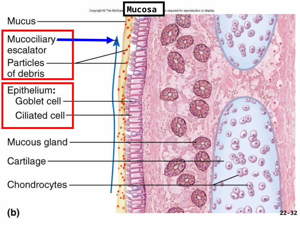
4. (Histology) Larynx and trachea lined with ciliated pseudostratified columnar epi. which functions as mucociliary escalator 22-28
See next three slides
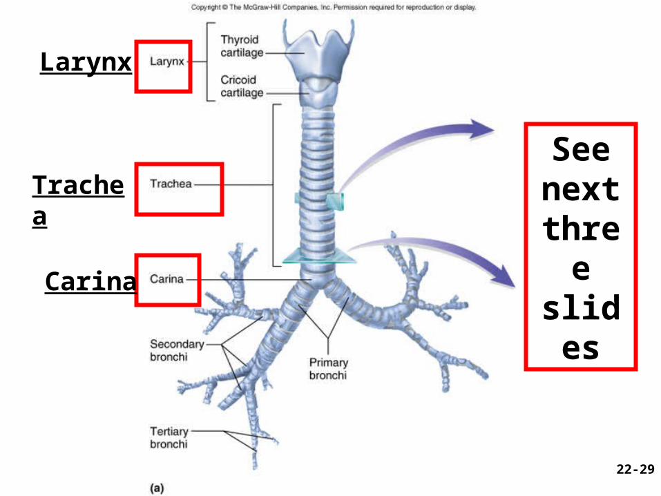
22-29
Larynx
Trachea
Carina
22-30
22-31
Ciliated Pseudostratified Epi.
22-32
Mucosa
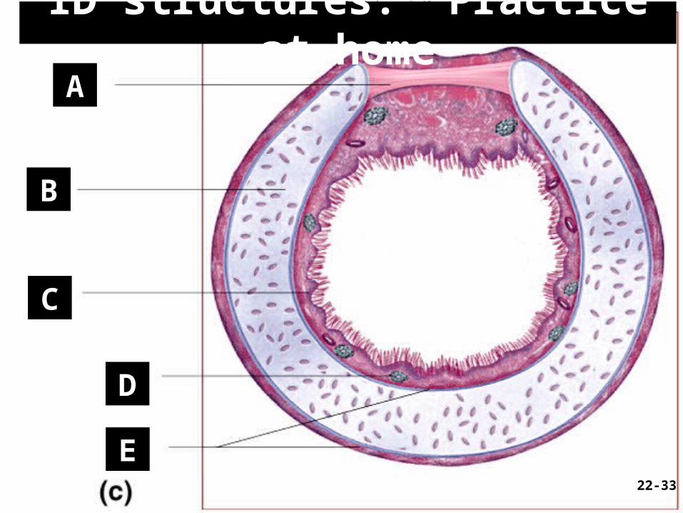
ID structures: Practice at homeA
B
C
D
E22-33
Functions:– Air passageway– Warm and moisten air– Remove particles & debris Clinical applications:– Trachea obstruction and Heimlich
Maneuver– Tracheostomy (Insight 22.1) when the
obstruction is superior to the level of the larynx; pitfall?
§ 4. Trachea (continued)
22-34
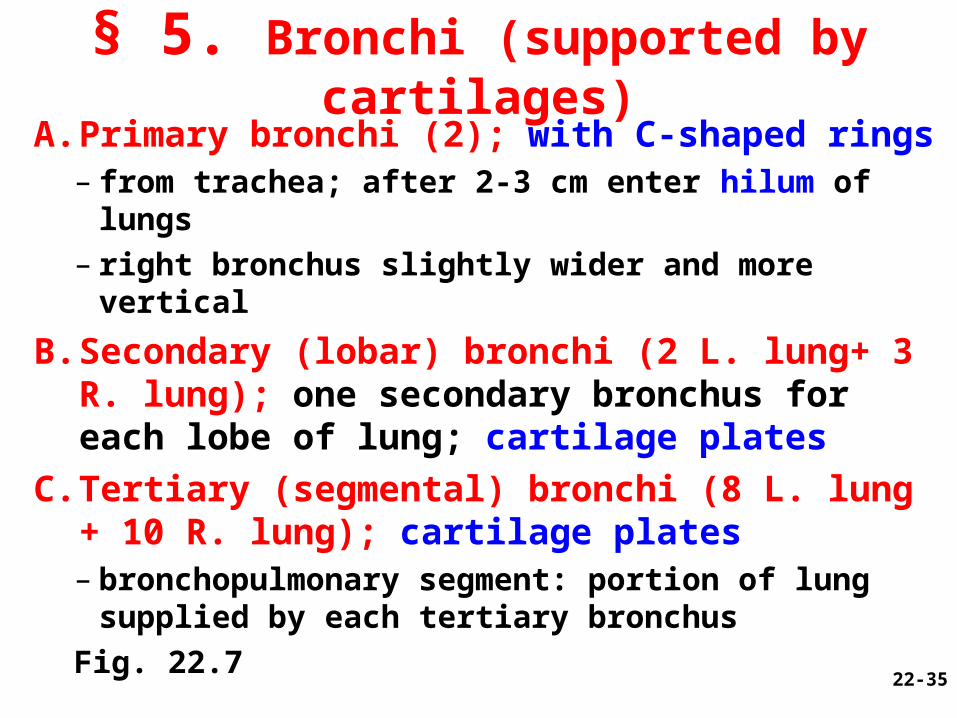
§ 5. Bronchi (supported by cartilages)
A. Primary bronchi (2); with C-shaped rings– from trachea; after 2-3 cm enter hilum of lungs– right bronchus slightly wider and more vertical
B. Secondary (lobar) bronchi (2 L. lung+ 3 R. lung); one secondary bronchus for each lobe of lung; cartilage plates
C. Tertiary (segmental) bronchi (8 L. lung + 10 R. lung); cartilage plates– bronchopulmonary segment: portion of lung
supplied by each tertiary bronchus
Fig. 22.7 22-35
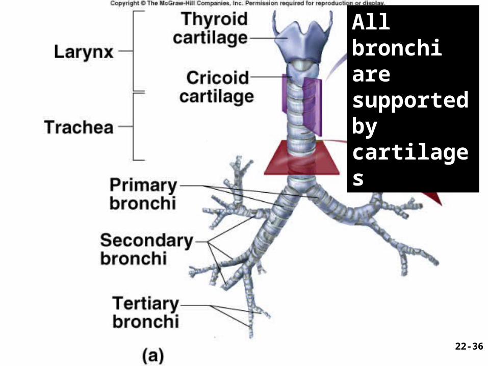
All bronchi are supported by cartilages
22-36
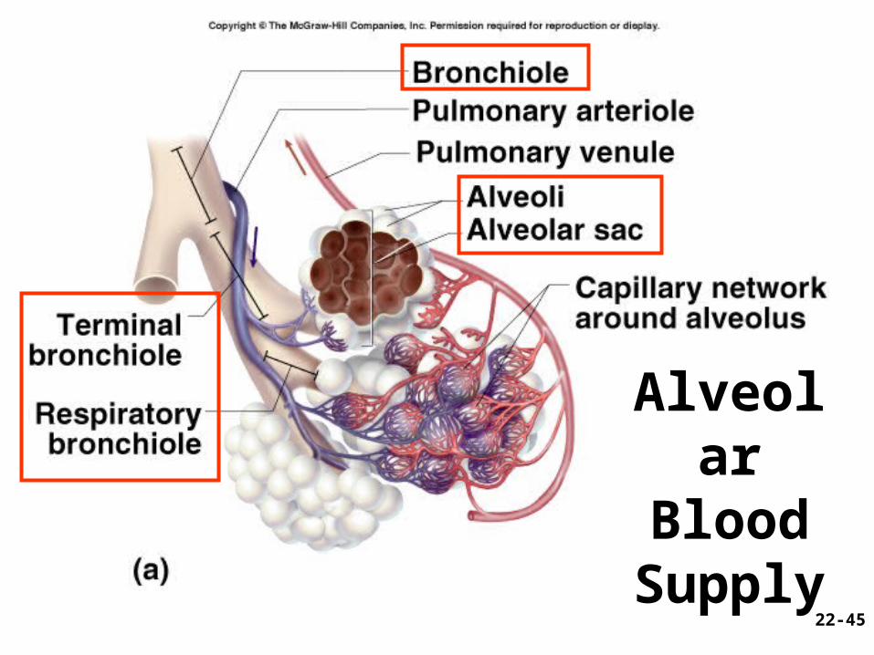
Bronchioles (lack cartilage; 1 mm or less in diameter; ciliated simple columnar to ciliated simple cuboidal epi.)
A. Each divides into 50 - 80 terminal bronchioles• Mostly nonciliated simple cuboidal; end of
conducting division
B. Each terminal bronchiole branches into respiratory bronchioles (respiratory div. now); smallest ones are nonciliated epi.
C. Each divides into 2-10 alveolar ducts (nonciliated simple squamous epi.); end in alveolar sacs• Fig. 22.11 22-37
§ 6. Bronchioles
Def. --Highly branched system of air tubes from the primary bronchi to about 65,000 terminal bronchioles
Resemble inverted trees
Fig. 22.0 + X
22-38
§ Bronchial tree
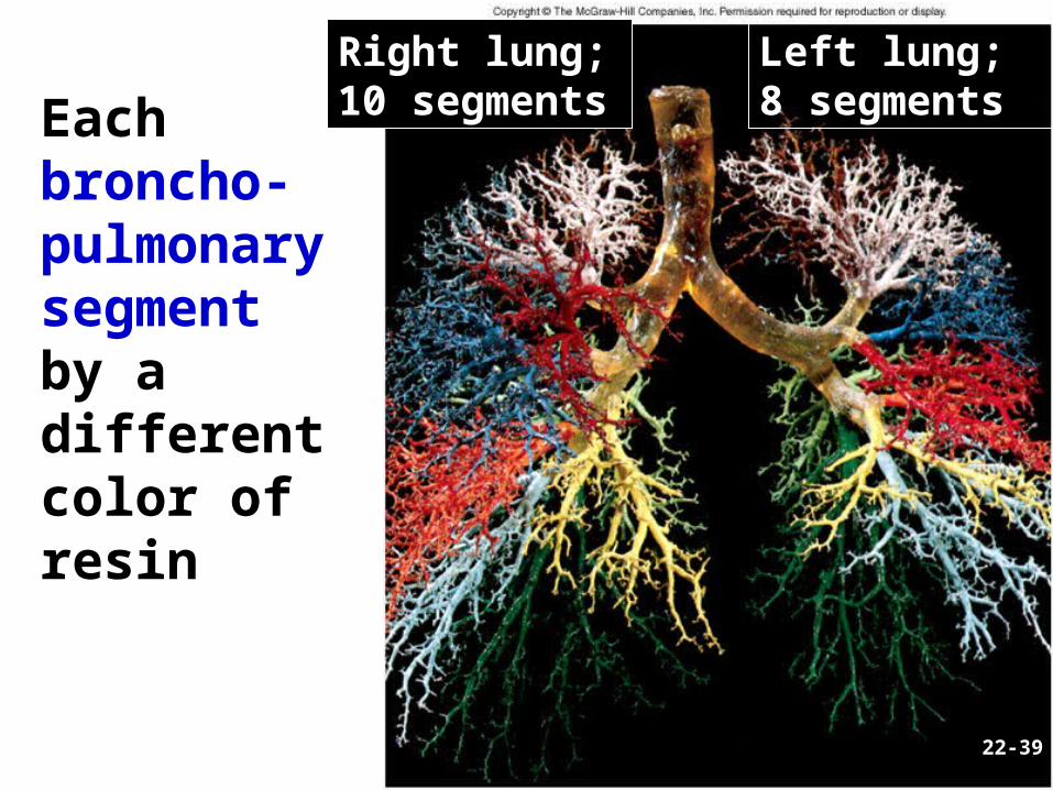
CO 22
Each broncho-pulmonary segment by a different color of resin
22-39
Left lung; 8 segments
Right lung; 10 segments
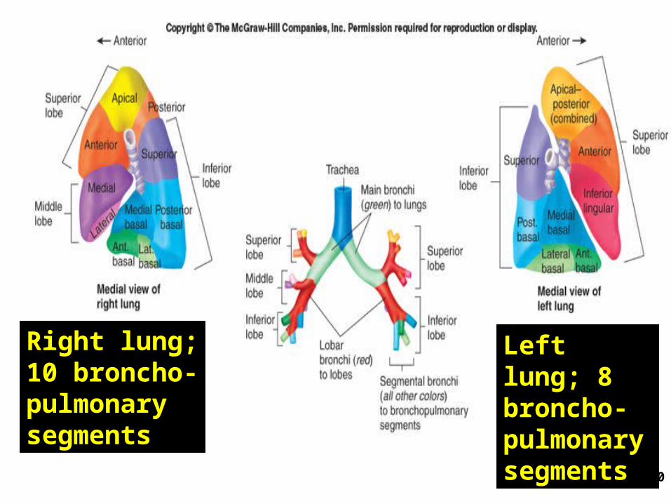
22-40
Left lung; 8 broncho-pulmonary segments
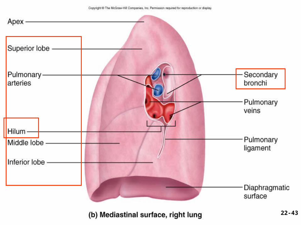
Right lung; 10 broncho-pulmonary segments
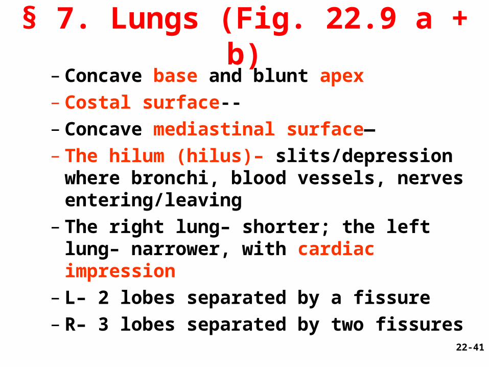
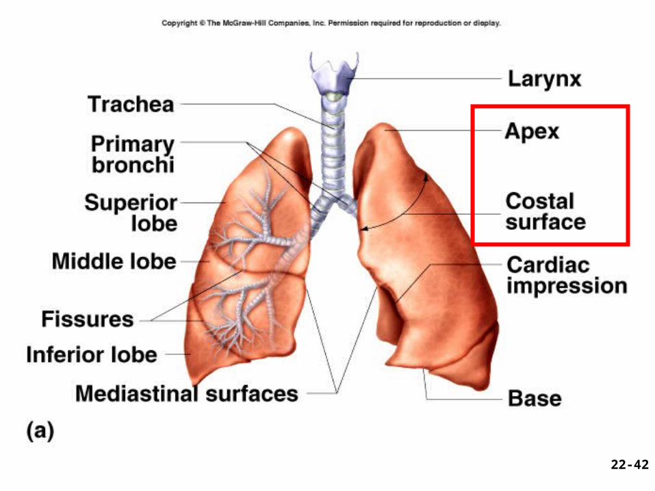
– Concave base and blunt apex– Costal surface--– Concave mediastinal surface—– The hilum (hilus)– slits/depression where
bronchi, blood vessels, nerves entering/leaving
– The right lung– shorter; the left lung– narrower, with cardiac impression
– L– 2 lobes separated by a fissure– R– 3 lobes separated by two fissures
22-41
§ 7. Lungs (Fig. 22.9 a + b)
22-42
22-43
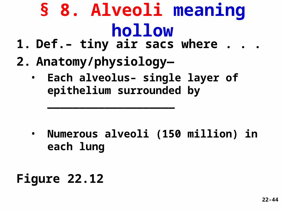
§ 8. Alveoli meaning hollow1. Def.– tiny air sacs where . . .
2. Anatomy/physiology—• Each alveolus– single layer of epithelium
surrounded by ____________________
• Numerous alveoli (150 million) in each lung
Figure 22.12
22-44
Alveolar Blood Supply
22-45
§ Alveoli—Pore of Kohn
Pore of Kohn• Location?
• Function?
• Analogy—
Fig. x22-46
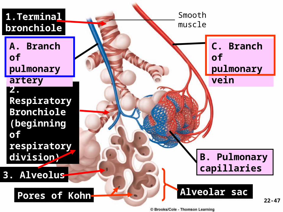
1.Terminalbronchiole
2. RespiratoryBronchiole(beginning of respiratory division)
A. Branch ofpulmonaryartery
3. Alveolus
Pores of Kohn
Smoothmuscle
C. Branch ofpulmonaryvein
B. Pulmonarycapillaries
Alveolar sac22-47
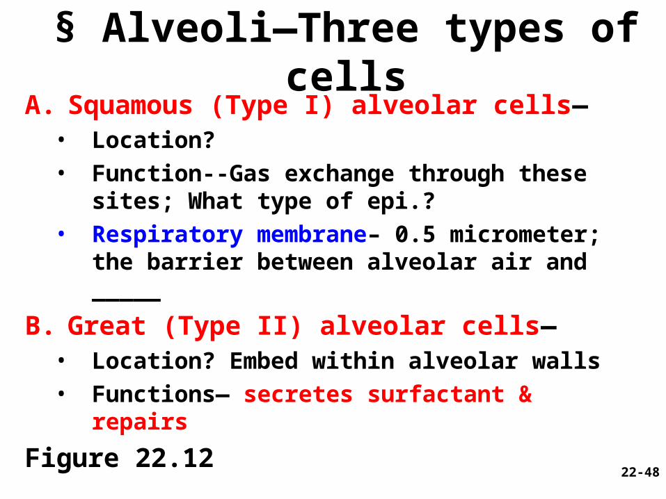
§ Alveoli—Three types of cellsA. Squamous (Type I) alveolar cells—
• Location? • Function--Gas exchange through these
sites; What type of epi.?• Respiratory membrane– 0.5 micrometer;
the barrier between alveolar air and _____
B. Great (Type II) alveolar cells—• Location? Embed within alveolar walls• Functions— secretes surfactant & repairs
Figure 22.1222-48
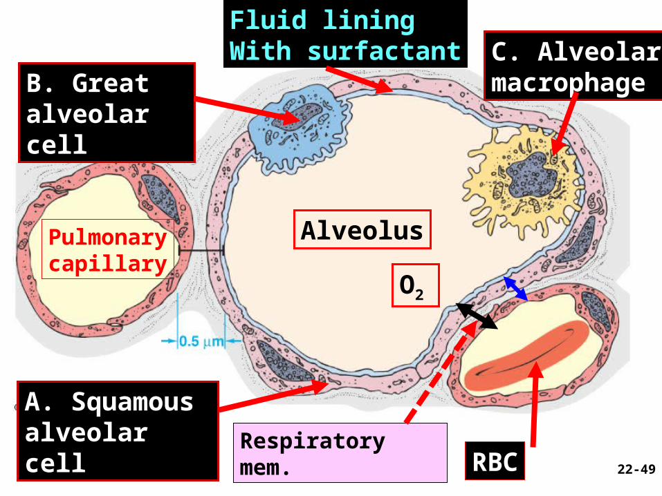
Fluid liningWith surfactant
B. Great alveolar cell
A. Squamous alveolar cell
Alveolus
C. Alveolarmacrophage
RBC
Pulmonarycapillary
O2
22-49
Respiratory mem.
§ Alveoli—Three types of cells
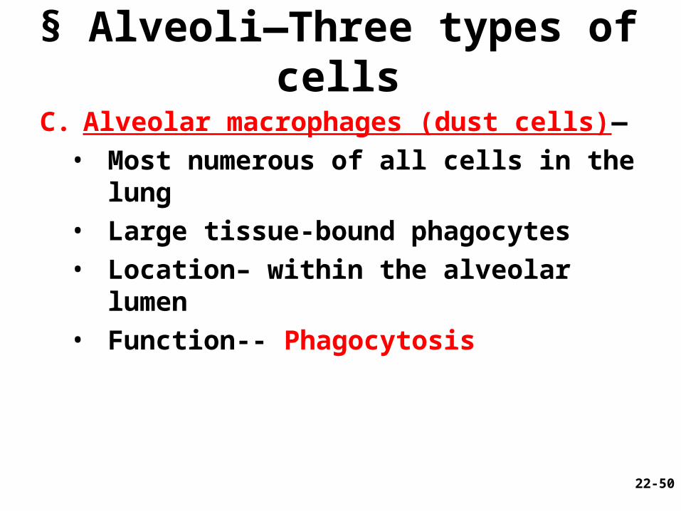
C. Alveolar macrophages (dust cells)—
• Most numerous of all cells in the lung
• Large tissue-bound phagocytes
• Location– within the alveolar lumen
• Function-- Phagocytosis
22-50
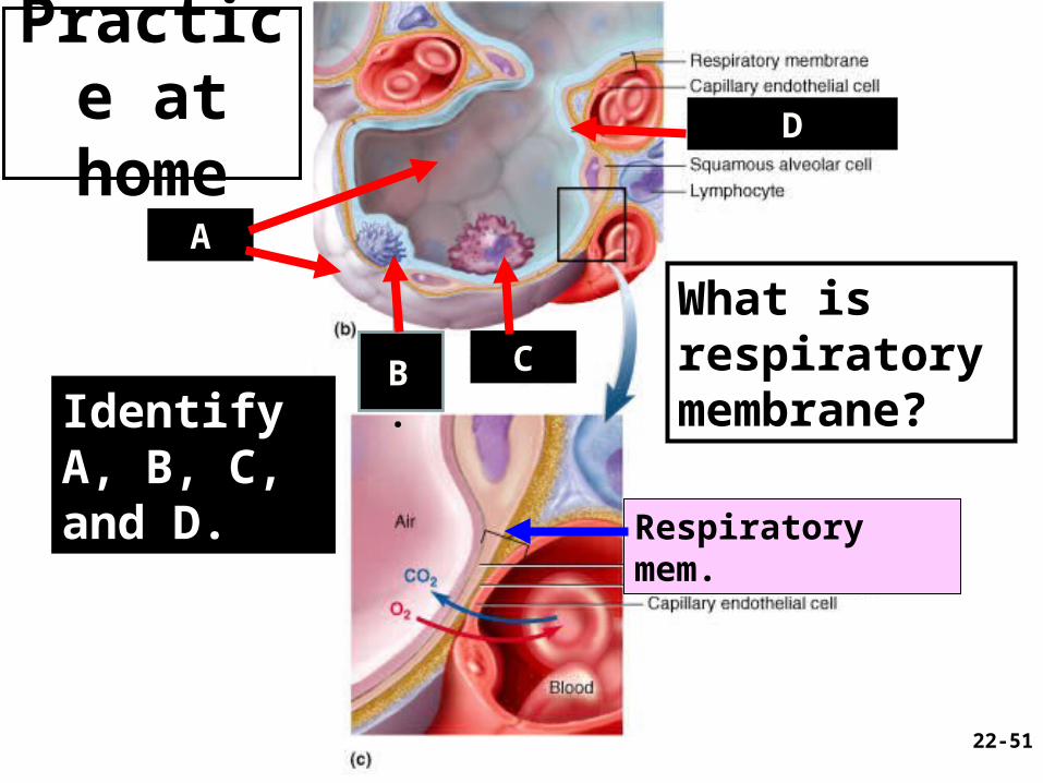
Practice at home
Fig. 22.11
b and c
22-51
B. C
A
D
Identify A, B, C, and D.
What is respiratory membrane?
Respiratory mem.

Questions (muddiest points)?
22-52
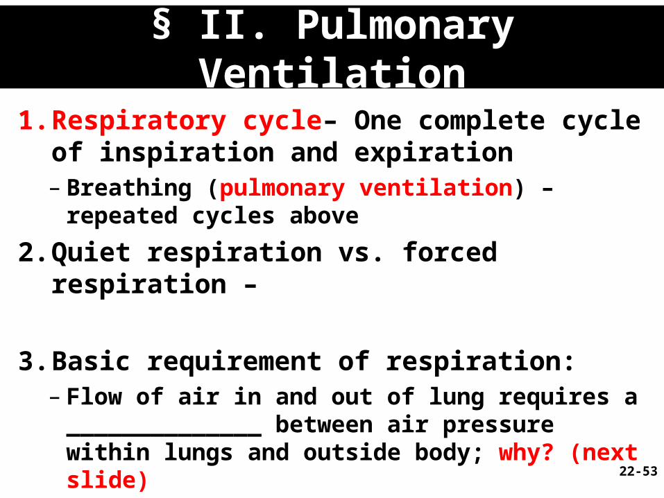
§ II. Pulmonary Ventilation
1. Respiratory cycle– One complete cycle of inspiration and expiration– Breathing (pulmonary ventilation) – repeated
cycles above
2. Quiet respiration vs. forced respiration –
3. Basic requirement of respiration: – Flow of air in and out of lung requires a
______________ between air pressure within lungs and outside body; why? (next slide)
22-53
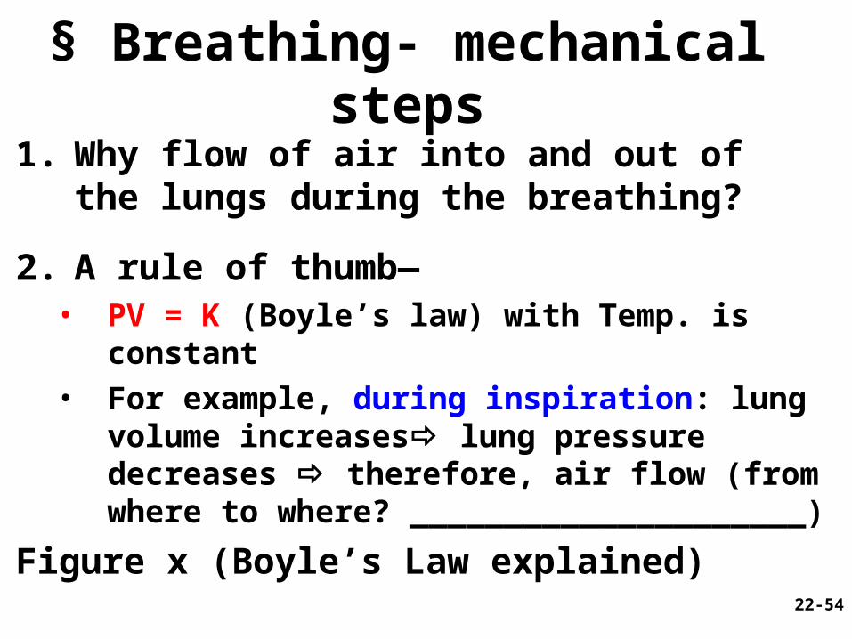
§ Breathing- mechanical steps
1. Why flow of air into and out of the lungs during the breathing?
2. A rule of thumb—• PV = K (Boyle’s law) with Temp. is constant• For example, during inspiration: lung volume
increases lung pressure decreases therefore, air flow (from where to where? _____________________)
Figure x (Boyle’s Law explained)
22-54
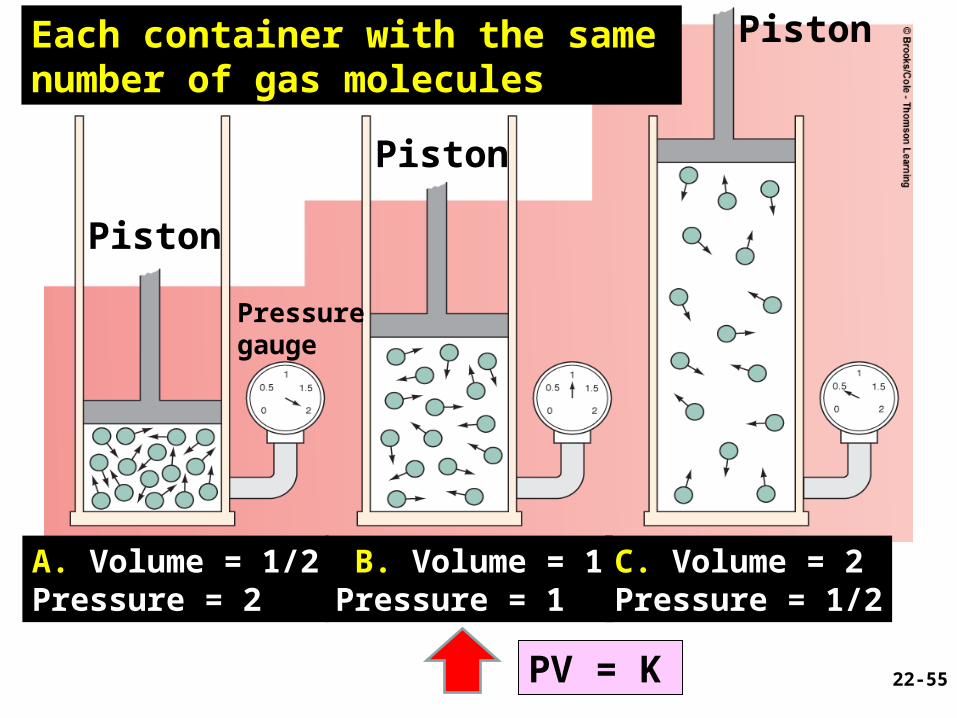
Figure 13.10Page 467
A. Volume = 1/2Pressure = 2
B. Volume = 1Pressure = 1
C. Volume = 2Pressure = 1/2
Piston
Pressuregauge
Each container with the same number of gas molecules
22-55PV = K
Piston
Piston
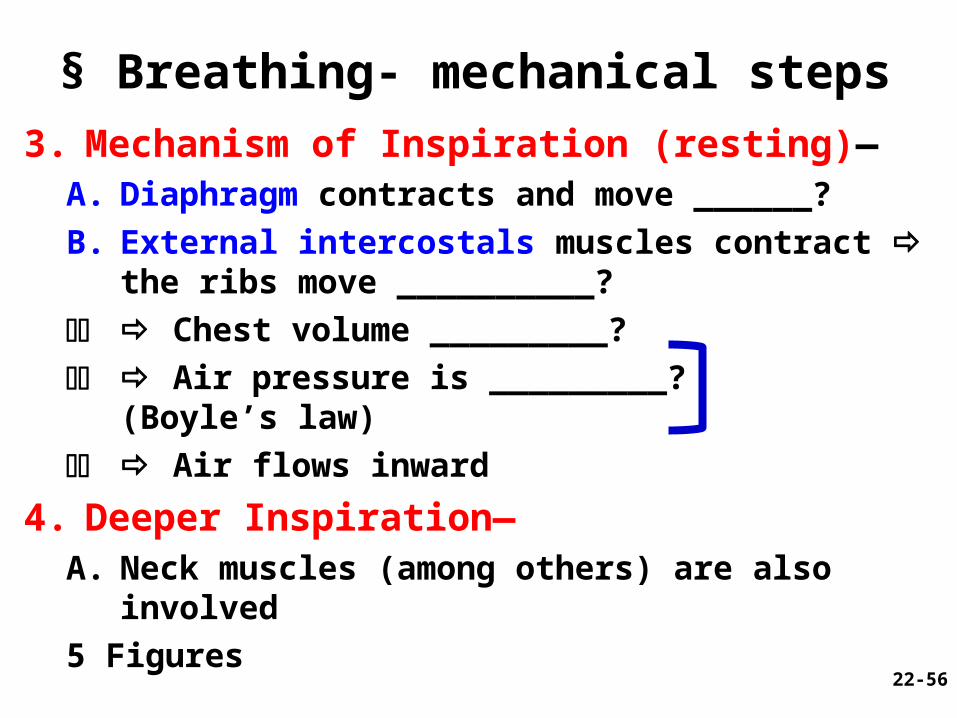
§ Breathing- mechanical steps
3. Mechanism of Inspiration (resting)—A. Diaphragm contracts and move ______?
B. External intercostals muscles contract the ribs move __________?
CC Chest volume _________?
CD Air pressure is _________? (Boyle’s law)
CC Air flows inward
4. Deeper Inspiration—A. Neck muscles (among others) are also involved
5 Figures
22-56
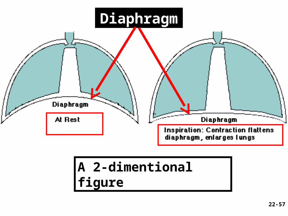
22-57
Diaphragm
A 2-dimentional figure
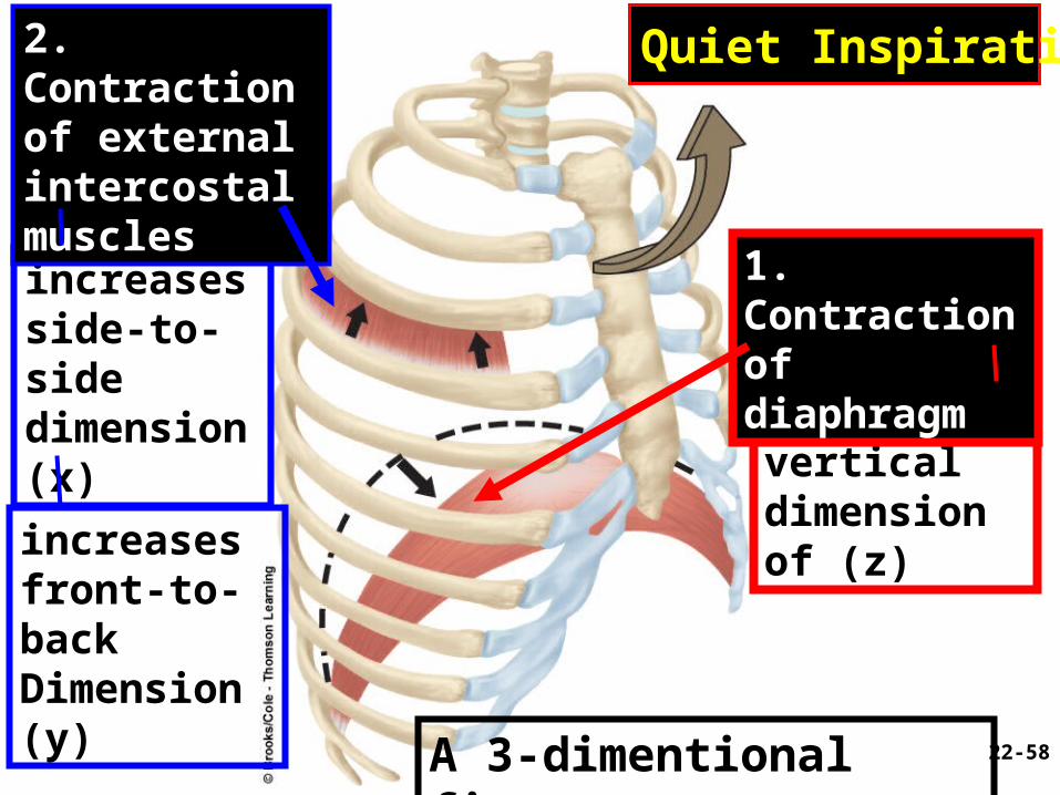
increases side-to-sidedimension (x) increases
verticaldimension of (z)increases
front-to-back Dimension (y)
Quiet Inspiration2. Contractionof externalintercostalmuscles
1. Contractionof diaphragm
22-58A 3-dimentional figure
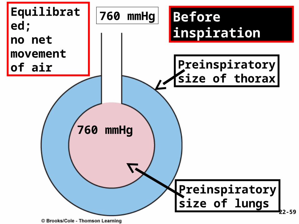
Equilibrated;no net movement of air
760 mmHg
Preinspiratorysize of thorax
Preinspiratorysize of lungs
Before inspiration
22-59
760 mmHg
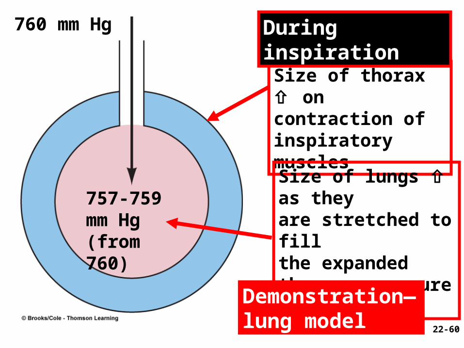
757-759 mm Hg (from 760)
Size of thorax oncontraction ofinspiratory muscles
Size of lungs as theyare stretched to fillthe expanded thorax; pressure
During inspiration760 mm Hg
22-60
Demonstration—lung model
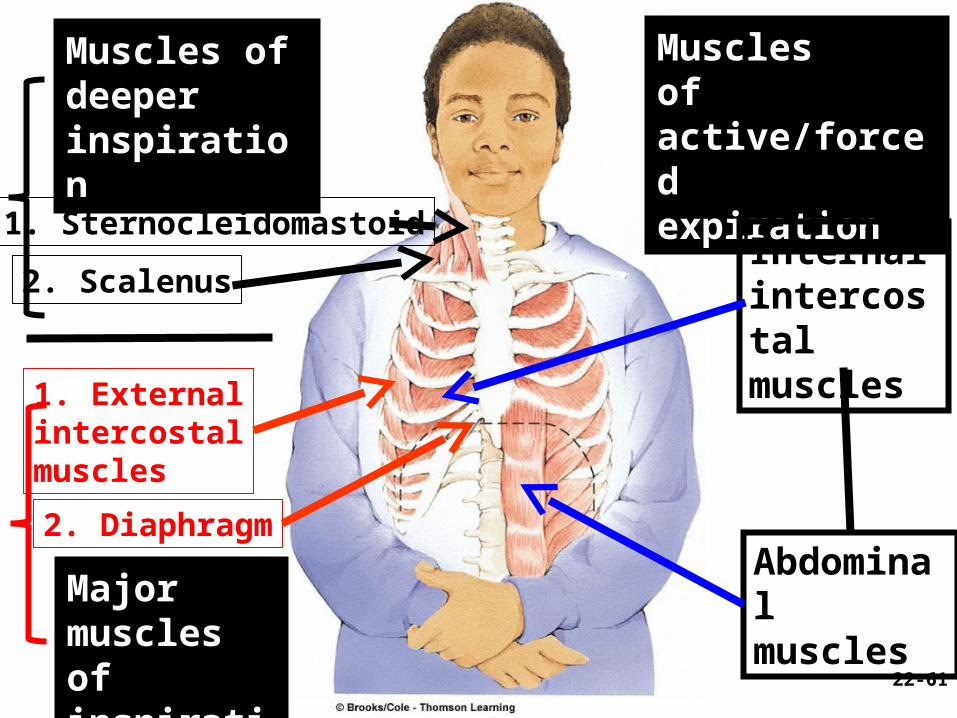
Muscles of deeperinspiration
Musclesof active/forcedexpiration
Majormuscles ofinspiration
1. Sternocleidomastoid
2. Scalenus
1. Externalintercostalmuscles
2. Diaphragm
Internalintercostalmuscles
Abdominalmuscles
22-61
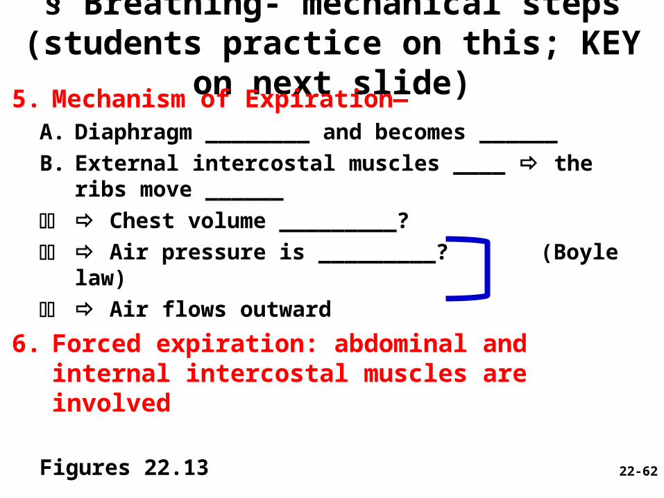
§ Breathing- mechanical steps (students practice on this; KEY on next slide)
5. Mechanism of Expiration—A. Diaphragm ________ and becomes ______
B. External intercostal muscles ____ the ribs move ______
CC Chest volume _________?
CD Air pressure is _________? (Boyle law)
CC Air flows outward
6. Forced expiration: abdominal and internal intercostal muscles are involved
Figures 22.13 22-62
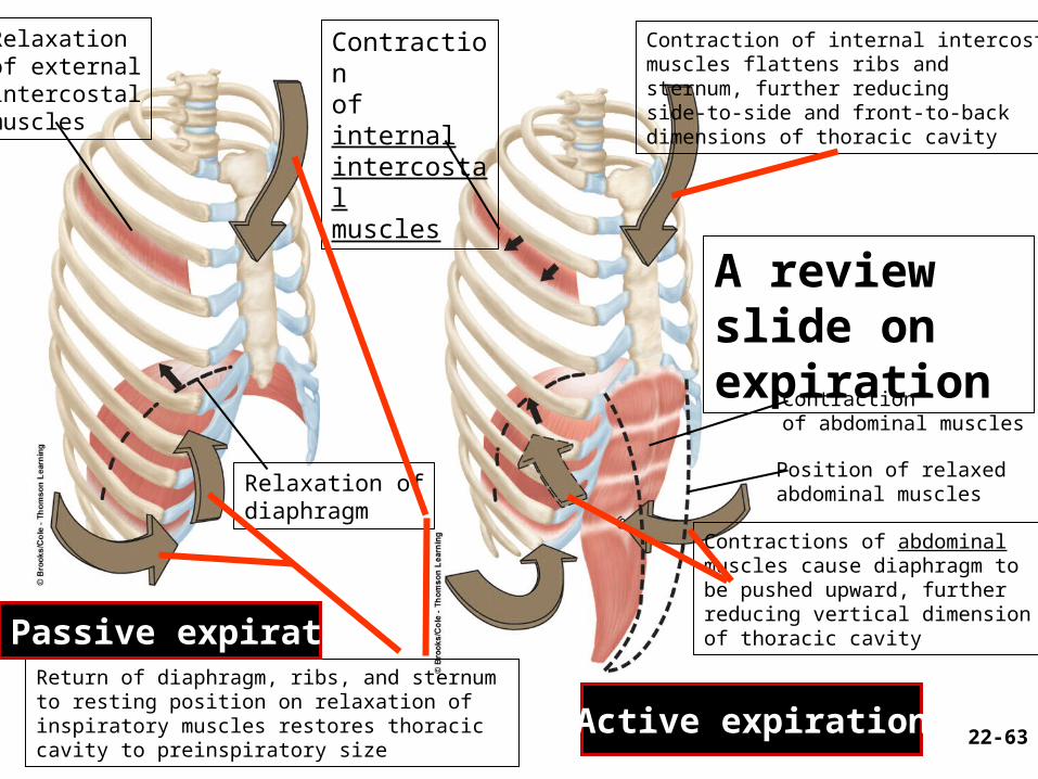
Relaxationof externalintercostalmuscles
Return of diaphragm, ribs, and sternum to resting position on relaxation of inspiratory muscles restores thoracic cavity to preinspiratory size
Contractions of abdominalmuscles cause diaphragm tobe pushed upward, furtherreducing vertical dimension of thoracic cavity
Contraction of internal intercostal muscles flattens ribs and sternum, further reducingside-to-side and front-to-back dimensions of thoracic cavity
Passive expiration
Active expiration
Contractionof internalintercostalmuscles
Relaxation ofdiaphragm
Contractionof abdominal muscles
Position of relaxedabdominal muscles
22-63
A review slide on expiration
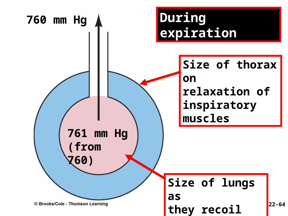
761 mm Hg (from 760)
Size of thorax onrelaxation ofinspiratory muscles
Size of lungs asthey recoil
During expiration760 mm Hg
22-64

§ Summary of respiratory muscles (This slide for review with Fig. x next)
1. Diaphragm (dome shaped) – contraction flattens diaphragm
2. External intercostals– increases X&Y diameter; stiffen thoracic cage
3. Scalenes - hold first 2 pair of ribs stationary
4. Pectoralis minor, sternocleidomastoid and erector spinae muscles– used in forced inspiration
5. Abdominals, internal intercostals, and latissimus dorsi– forced expiration (to sing, cough, sneeze)
– Valsalva maneuver– raise abdominal pressure . . . 22-65
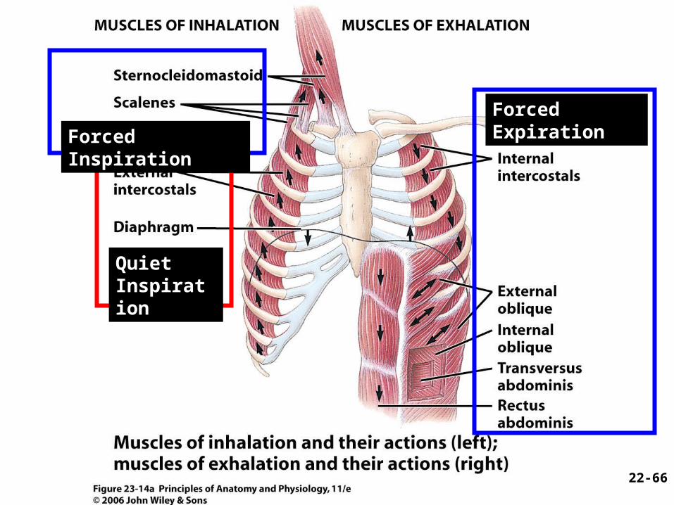
Quiet Inspiration
Forced Inspiration
Forced Expiration
22-66
§ III. Neural Control of Breathing
22-67
§ Neural Control of Breathing (1)1. Breathing depends on repetitive stimuli from the
brain—controlled at two levels (A & B below):
A. Neurons in medulla oblongata and pons control unconscious breathing
• Ondine’s curse – brainstem damage• Causes– Poliomyelitis etc.• Symptoms– disabled automatic respiratory
functions• Cure--
B. Voluntary control provided by motor cortex is cerebral and consciously controlled
22-68
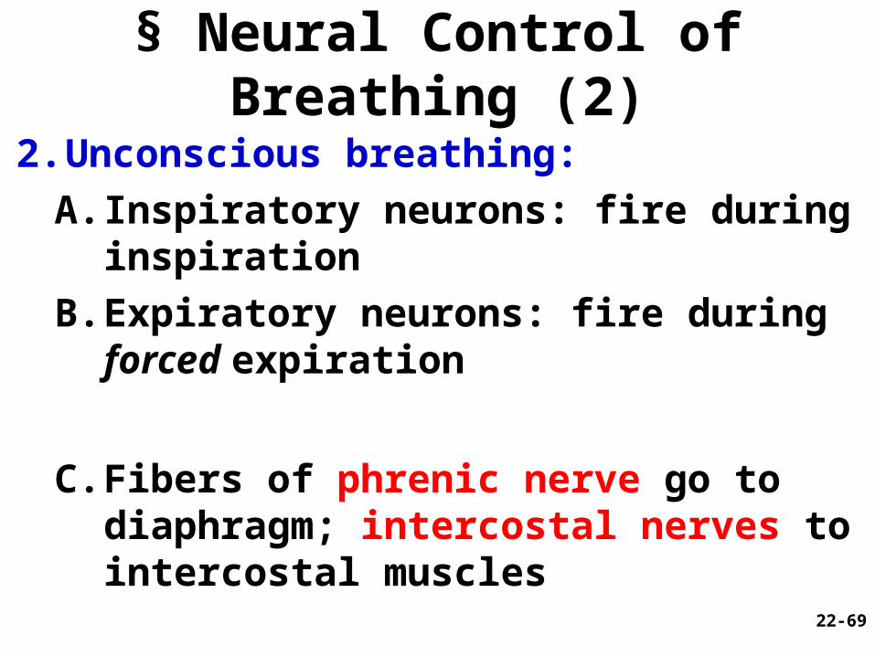
§ Neural Control of Breathing (2)
2. Unconscious breathing:A. Inspiratory neurons: fire during
inspiration
B. Expiratory neurons: fire during forced expiration
C. Fibers of phrenic nerve go to diaphragm; intercostal nerves to intercostal muscles
22-69
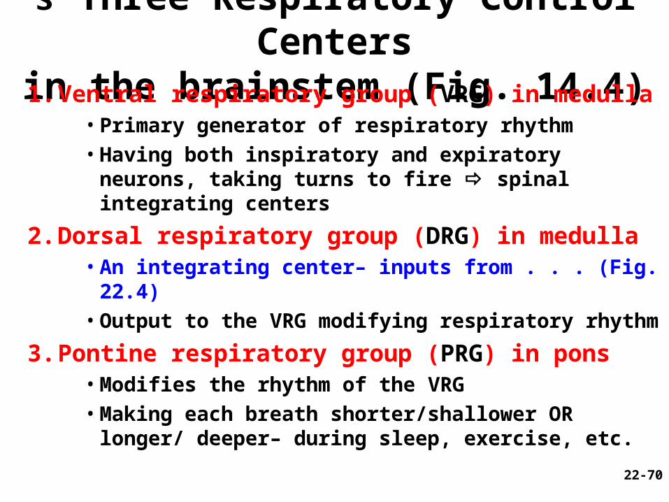
§ Three Respiratory Control Centersin the brainstem (Fig. 14.4)
1. Ventral respiratory group (VRG) in medulla•Primary generator of respiratory rhythm• Having both inspiratory and expiratory neurons,
taking turns to fire spinal integrating centers
2. Dorsal respiratory group (DRG) in medulla• An integrating center– inputs from . . . (Fig. 22.4) • Output to the VRG modifying respiratory rhythm
3. Pontine respiratory group (PRG) in pons• Modifies the rhythm of the VRG• Making each breath shorter/shallower OR longer/
deeper– during sleep, exercise, etc.
22-70
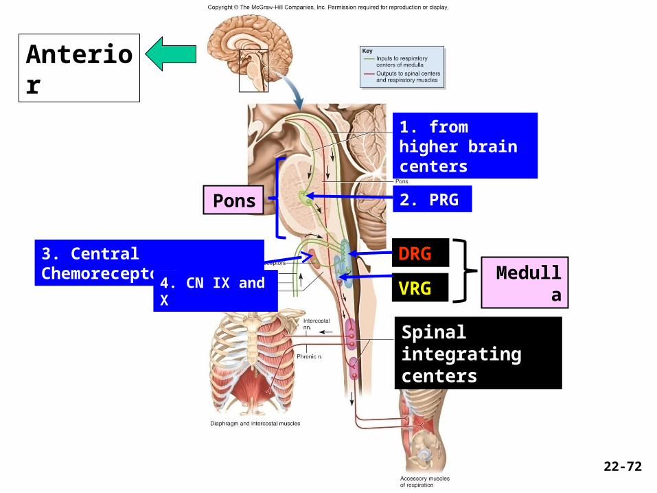
Spinal integrating centers
VRG
DRG
2. PRGPons
3. Central Chemoreceptors
4. CN IX and XMedulla
1. from higher brain centers
22-72
Anterior
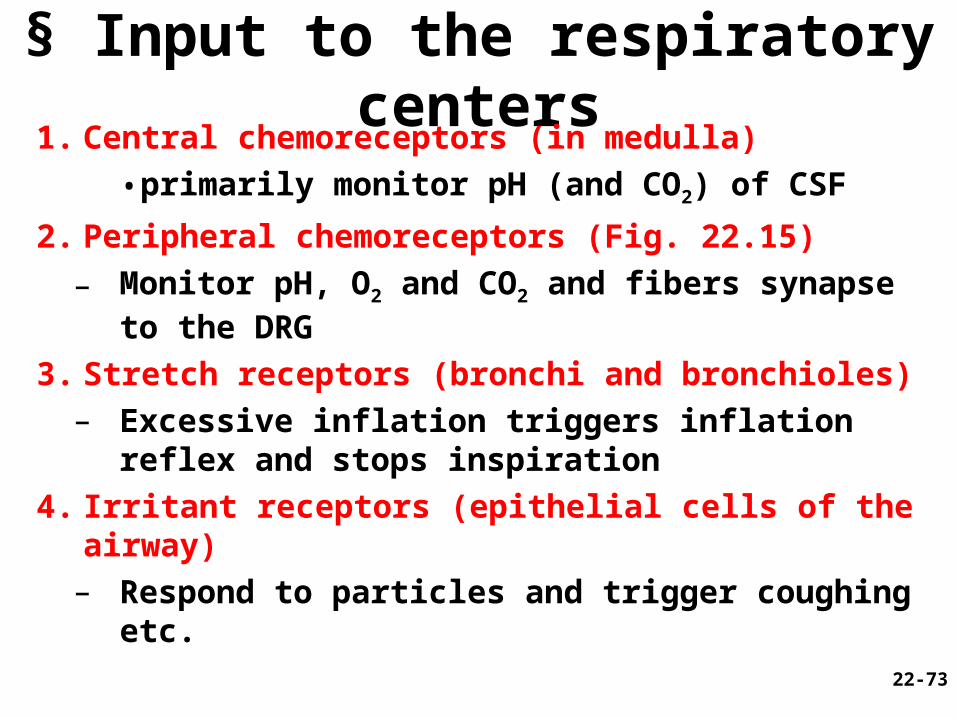
§ Input to the respiratory centers1. Central chemoreceptors (in medulla)
• primarily monitor pH (and CO2) of CSF
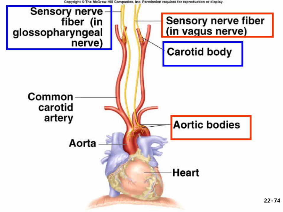
2. Peripheral chemoreceptors (Fig. 22.15)
– Monitor pH, O2 and CO2 and fibers synapse to the DRG
3. Stretch receptors (bronchi and bronchioles)– Excessive inflation triggers inflation reflex
and stops inspiration
4. Irritant receptors (epithelial cells of the airway)– Respond to particles and trigger coughing
etc. 22-73
22-74
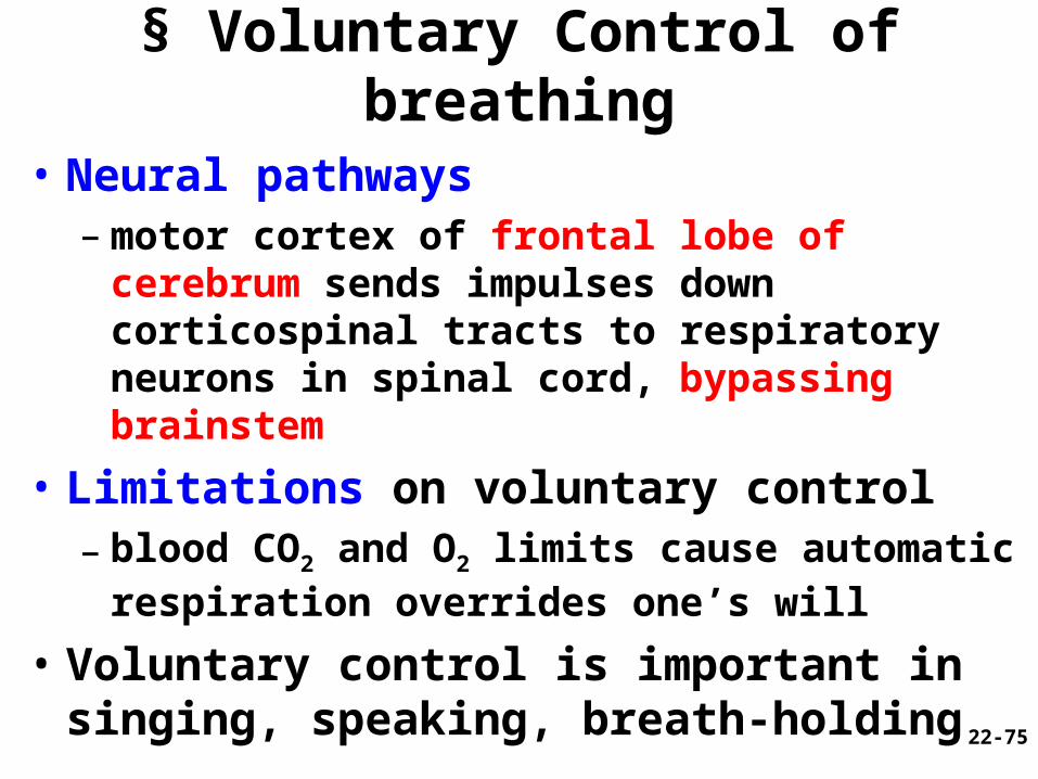
§ Voluntary Control of breathing
• Neural pathways– motor cortex of frontal lobe of cerebrum sends
impulses down corticospinal tracts to respiratory neurons in spinal cord, bypassing brainstem
• Limitations on voluntary control– blood CO2 and O2 limits cause automatic
respiration overrides one’s will
• Voluntary control is important in singing, speaking, breath-holding
22-75
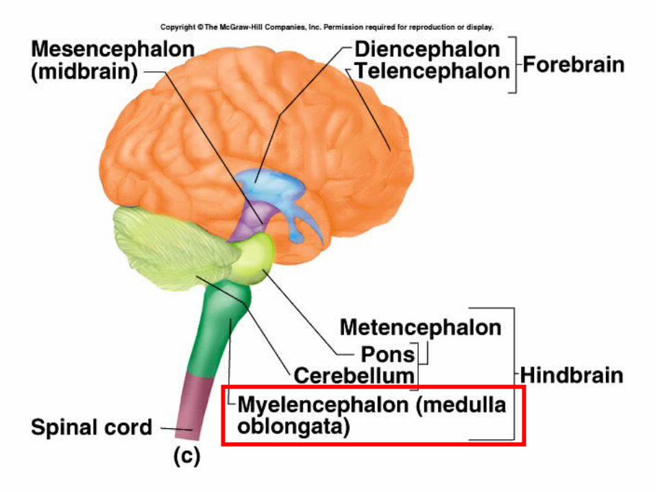
Check Point QuestionsQ--Where exactly are the medulla
oblongata and the pons located, respectively?
Answer: medulla oblongata is the most caudal part of the brainstem (stalklike lower portion of the brain), immediately superior to the spinal cord
• The pons is a part of the brainstem located immediately superior to the medulla oblongata and ventral to the cerebellum
22-76
§ Next section--IV. Pressure, Resistance, and Airflow
22-77
Q-- Is it possible that temperamental children may hold their breath until they die?
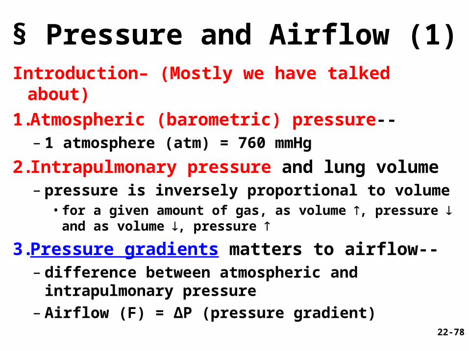
§ Pressure and Airflow (1) Introduction– (Mostly we have talked about)
1.Atmospheric (barometric) pressure--– 1 atmosphere (atm) = 760 mmHg
2.Intrapulmonary pressure and lung volume– pressure is inversely proportional to volume
• for a given amount of gas, as volume , pressure and as volume , pressure
3.Pressure gradients matters to airflow--– difference between atmospheric and
intrapulmonary pressure– Airflow (F) = ΔP (pressure gradient) 22-78
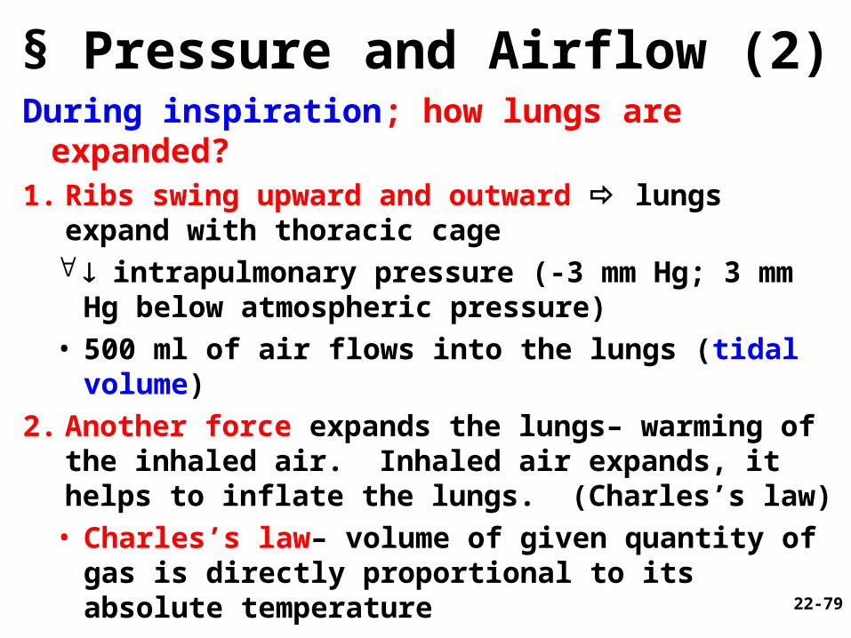
§ Pressure and Airflow (2) During inspiration; how lungs are expanded?1. Ribs swing upward and outward lungs expand
with thoracic cage intrapulmonary pressure (-3 mm Hg; 3 mm Hg
below atmospheric pressure)• 500 ml of air flows into the lungs (tidal volume)
2. Another force expands the lungs– warming of the inhaled air. Inhaled air expands, it helps to inflate the lungs. (Charles’s law)
• Charles’s law– volume of given quantity of gas is directly proportional to its absolute temperature
22-79
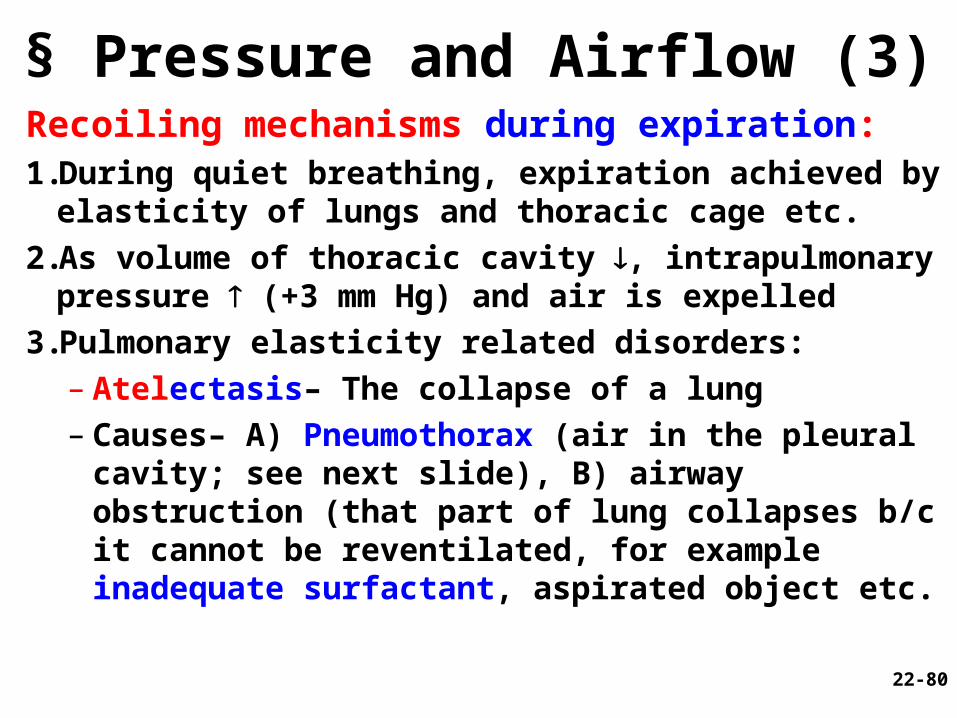
§ Pressure and Airflow (3) Recoiling mechanisms during expiration:1.During quiet breathing, expiration achieved by
elasticity of lungs and thoracic cage etc.
2.As volume of thoracic cavity , intrapulmonary pressure (+3 mm Hg) and air is expelled
3.Pulmonary elasticity related disorders:– Atelectasis– The collapse of a lung– Causes– A) Pneumothorax (air in the pleural
cavity; see next slide), B) airway obstruction (that part of lung collapses b/c it cannot be reventilated, for example inadequate surfactant, aspirated object etc. 22-80
§ Pneumothorax
• Def.—abnormal condition of air entering the pleural sac
• Causes— (see fig. x)
• Consequences— transmural pressure gradient no longer exists and . . .
Figure x
22-81
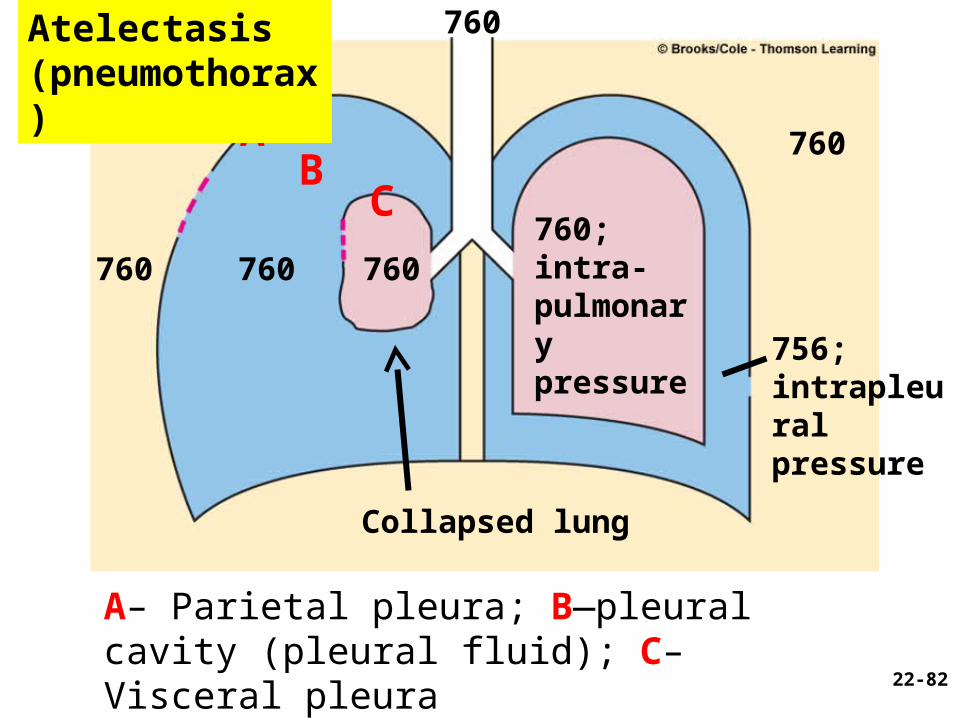
760
760760; intra-pulmonary pressure
Collapsed lung
760
760
756; intrapleural pressure
760
AB
C
A– Parietal pleura; B—pleural cavity (pleural fluid); C– Visceral pleura
22-82
Atelectasis (pneumothorax)
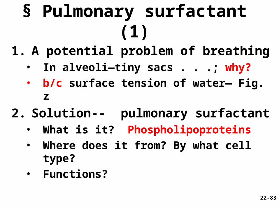
§ Pulmonary surfactant (1)
1. A potential problem of breathing• In alveoli—tiny sacs . . .; why?• b/c surface tension of water— Fig. z
2. Solution-- pulmonary surfactant• What is it? Phospholipoproteins• Where does it from? By what cell type?• Functions?
22-83
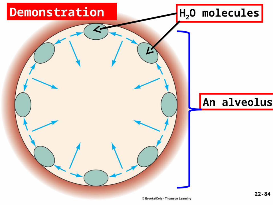
H2O molecules
An alveolus
22-84
Demonstration
85
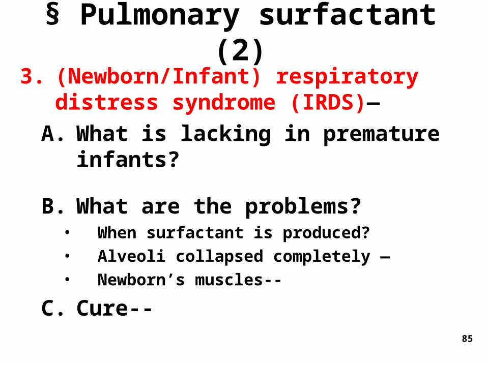
§ Pulmonary surfactant (2)3. (Newborn/Infant) respiratory distress
syndrome (IRDS)—
A. What is lacking in premature infants?
B. What are the problems?• When surfactant is produced?• Alveoli collapsed completely —• Newborn’s muscles--
C. Cure--
Check Point Questions
• What types of cells make up the wall of an alveolus? Function?
• What type of cell in the lungs secrete pulmonary surfactant? Function?
22-86
§ V. Alveolar Ventilation
22-87
§ Alveolar Ventilation (1)Does all inhaled air enter the alveoli?1.Dead air (150 ml per breath)
– fills conducting division of airway, cannot exchange gases with the blood
2.Where is the dead air? In anatomic dead space:– It exists in conducting division of airway– Normally about _______mL
3.Physiological (total) dead space– sum of anatomic dead space and any
pathological alveolar dead space 22-88
89
§ Alveolar ventilation (2)1. Alveolar ventilation rate (AVR): body’s
ability to get oxygen to the tissues per minute
– alveolar ventilation rate (AVR) = (Tidal volume - dead space volume) x respiratory rate
– AVR = (500-150mL) x 12 breaths/min = 4,200 mL/min
§ Measurements of Ventilation (1)
1. Spirometer – measures ventilation; specifically respiratory volumes and capacities
Fig. x
22-90
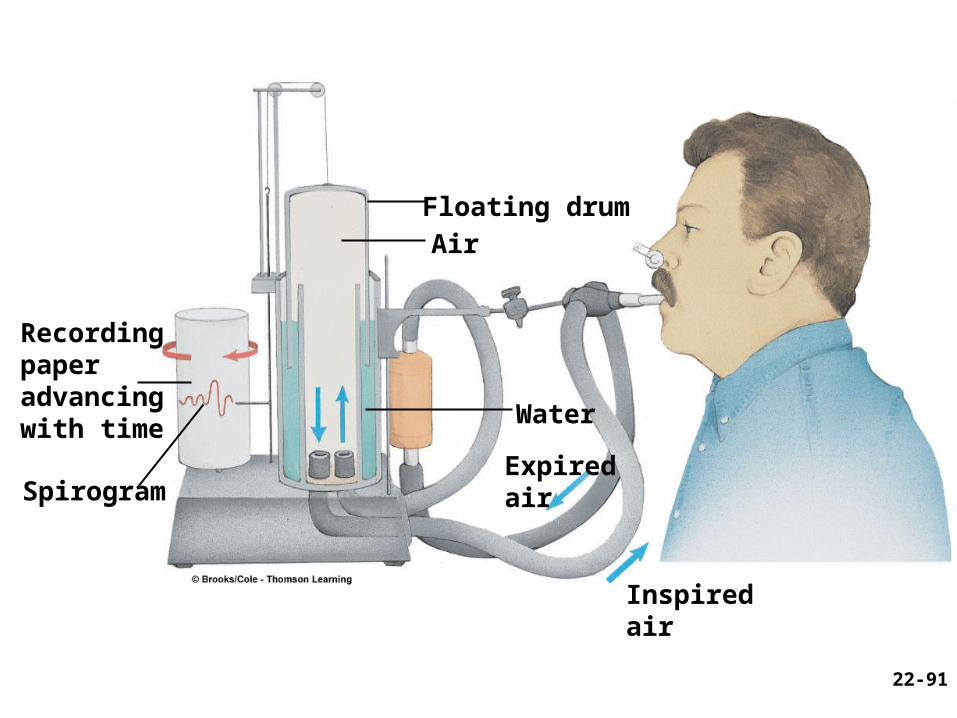
Recordingpaperadvancingwith time
Spirogram
Floating drum
Air
Water
Expiredair
Inspired air
22-91
92
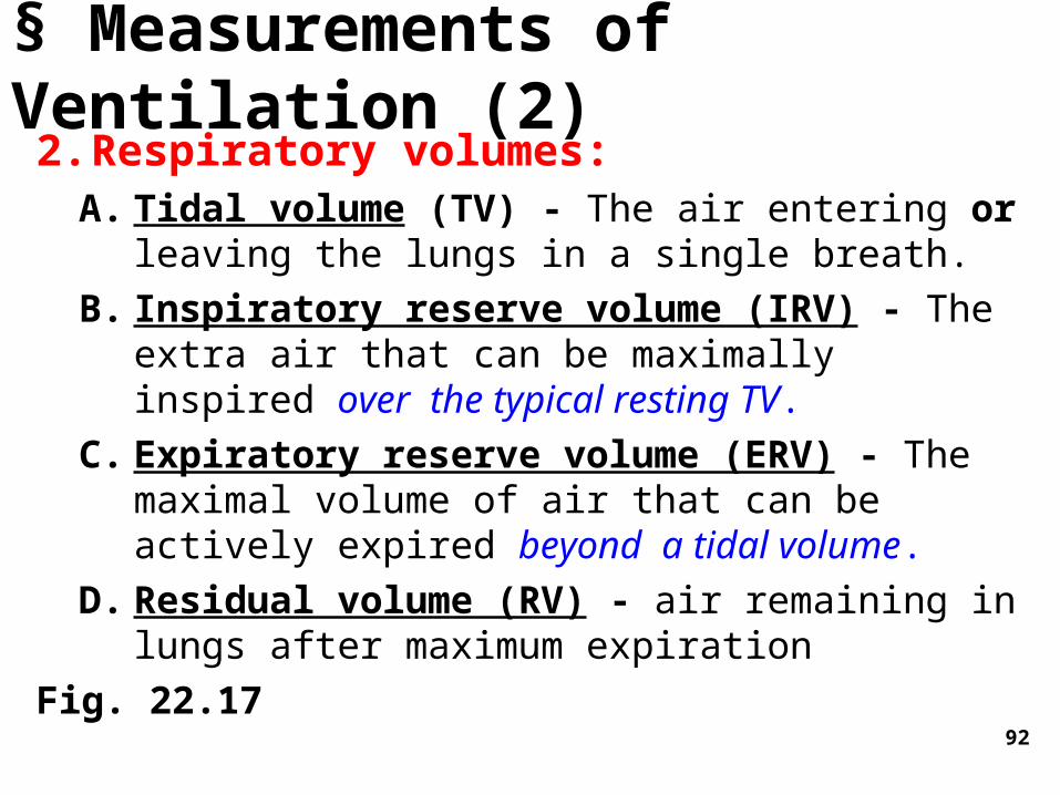
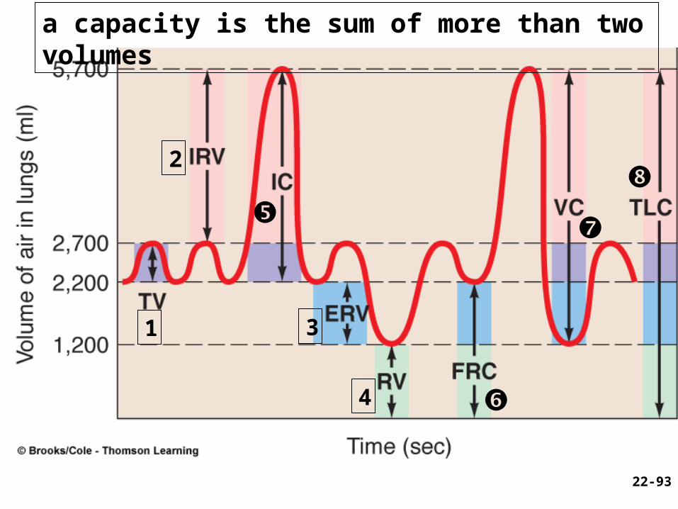
§ Measurements of Ventilation (2)2. Respiratory volumes:
A. Tidal volume (TV) - The air entering or leaving the lungs in a single breath.
B. Inspiratory reserve volume (IRV) - The extra air that can be maximally inspired over the typical resting TV.
C. Expiratory reserve volume (ERV) - The maximal volume of air that can be actively expired beyond a tidal volume.
D. Residual volume (RV) - air remaining in lungs after maximum expiration
Fig. 22.17
1
2
3
4
22-93
a capacity is the sum of more than two volumes
94
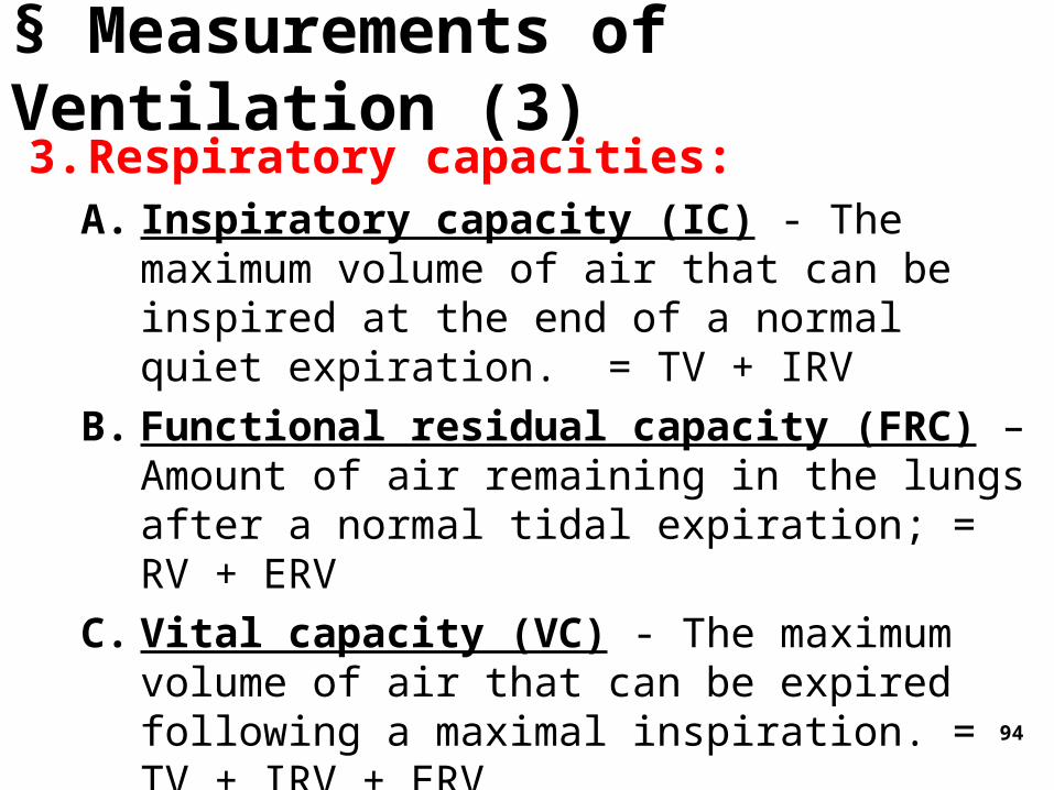
3. Respiratory capacities:A. Inspiratory capacity (IC) - The maximum
volume of air that can be inspired at the end of a normal quiet expiration. = TV + IRV
B. Functional residual capacity (FRC) – Amount of air remaining in the lungs after a normal tidal expiration; = RV + ERV
C. Vital capacity (VC) - The maximum volume of air that can be expired following a maximal inspiration. = TV + IRV + ERV
D. Total lung capacity (TLC) - maximum amount of air lungs can hold; = VC + RV
§ Measurements of Ventilation (3)
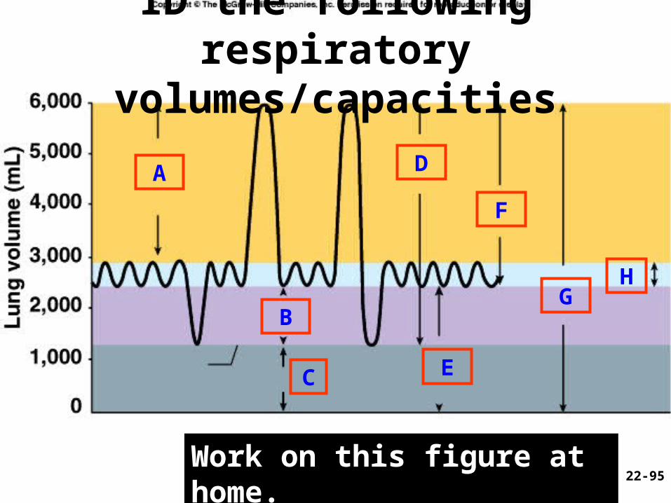
ID the following respiratory volumes/capacities
Work on this figure at home.
A
B
C
D
E
F
GH
22-95
Check Point Question
• If you breathe in as deeply as possible and then exhale as much air as you can, which lung volume or capacity have you demonstrated?
22-96
97
§ Lung disorders and spirometry1. Restrictive disorders– Those having
reduce pulmonary compliance, limiting the amount to which the lungs can be inflated
• Disorders- black lung disease, tuberculosis• Spirometry- reduced IC, VC, TLC
2. Obstructive disorders (COPD; Chronic Obstructive Pulmonary Disease) – those that interfere with airflow by narrowing or blocking the airway
• Disorders– asthma, emphysema etc.• Detection: Forced expiratory volume
(FEV)-- % of vital capacity exhaled/time; healthy adult - ___________% of VC in 1 sec (Fig. Y)
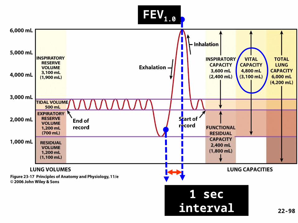
1 sec interval
FEV1.0
22-98
Recommended