Common Adult Fractures
Dr. Abdulrahman Algarni, MD, SSC (Ortho), ABOSAssistant Professor
Consultant Orthopedic and Arthroplasty Surgeon
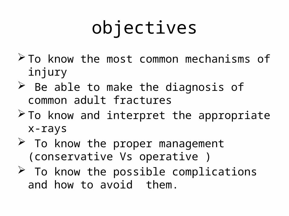
objectives
To know the most common mechanisms of injury Be able to make the diagnosis of common adult
fractures To know and interpret the appropriate x-rays To know the proper management (conservative Vs
operative ) To know the possible complications and how to avoid
them.
Upper limbs fractures
Clavicle
Humeral(Proximal, shaft)
Both Bone forearm(Radius, ulna)
Distal Radius
Mechanism of Injuries of the Upper Limb
• Mostly Indirect
• Commonly described as “ a fall on the outstretched hand “
• Type of injury depends on– position of the upper limb at the time of impact – force of injury– age
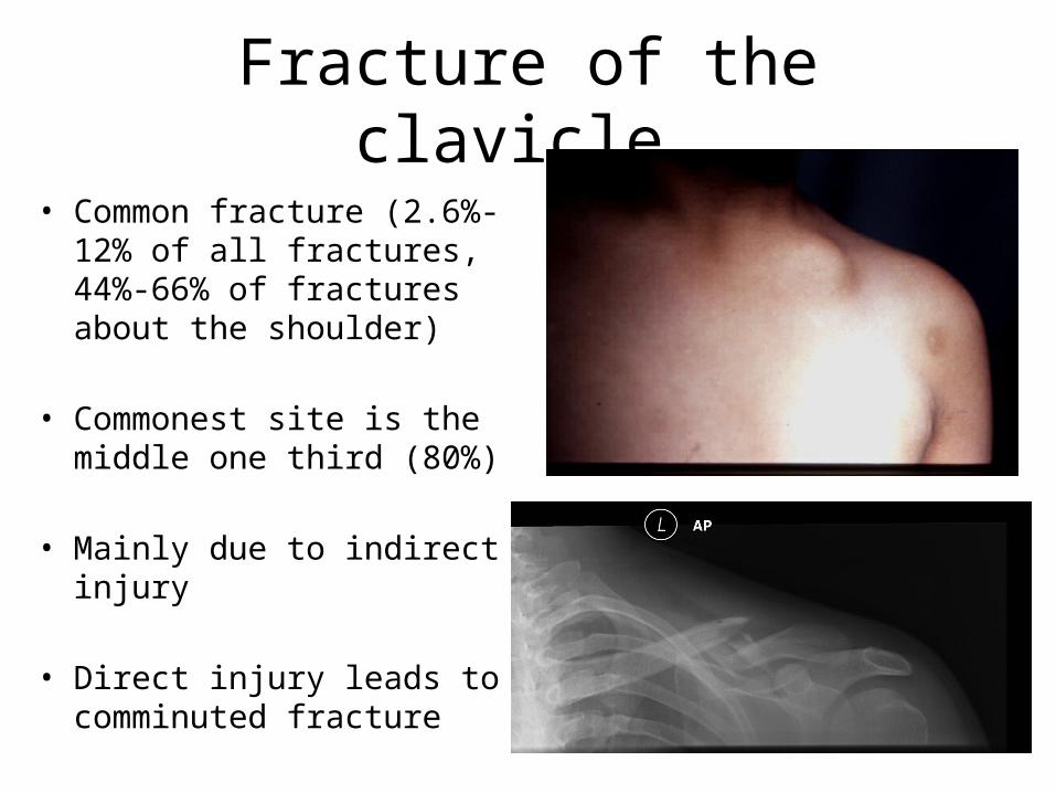
Fracture of the clavicle
• Common fracture (2.6%-12% of all fractures, 44%-66% of fractures about the shoulder)
• Commonest site is the middle one third (80%)
• Mainly due to indirect injury
• Direct injury leads to comminuted fracture
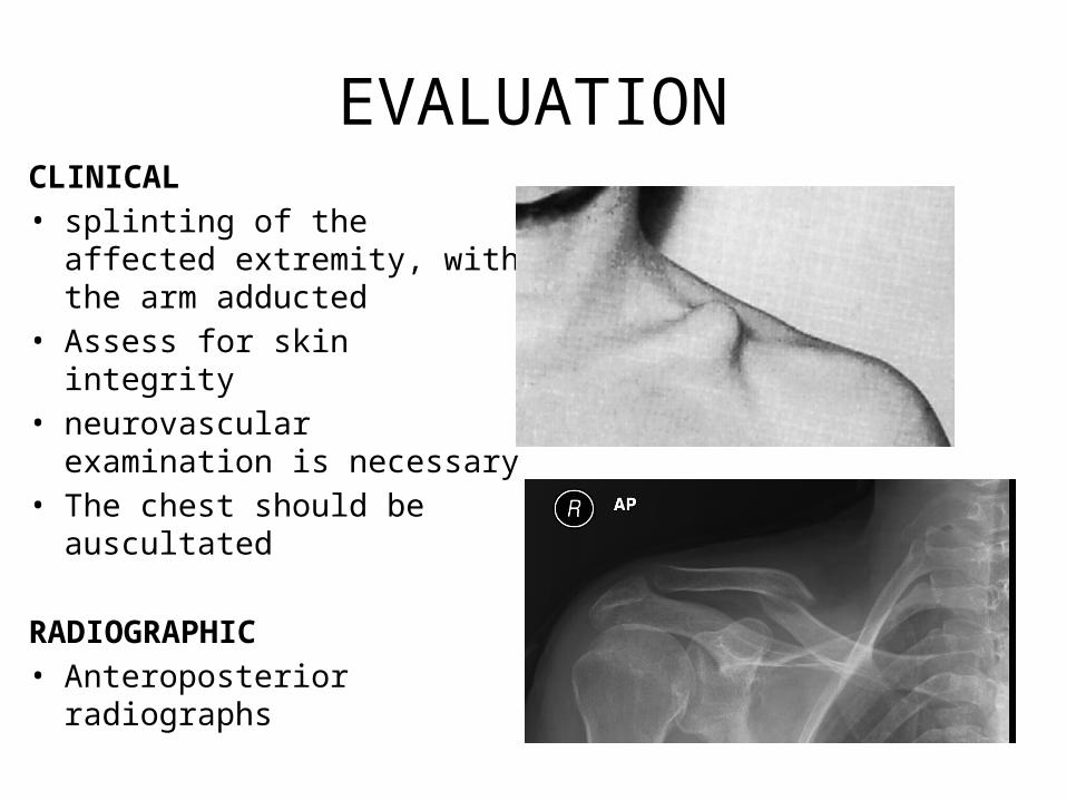
EVALUATIONCLINICAL• splinting of the affected
extremity, with the arm adducted
• Assess for skin integrity• neurovascular examination is
necessary• The chest should be
auscultated
RADIOGRAPHIC• Anteroposterior radiographs
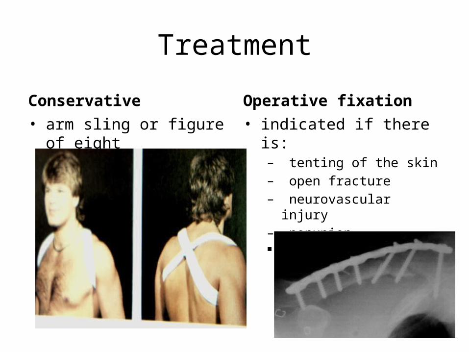
Treatment
Conservative• arm sling or figure of eight
Operative fixation• indicated if there is:
– tenting of the skin– open fracture– neurovascular injury – nonunion Plate and screws
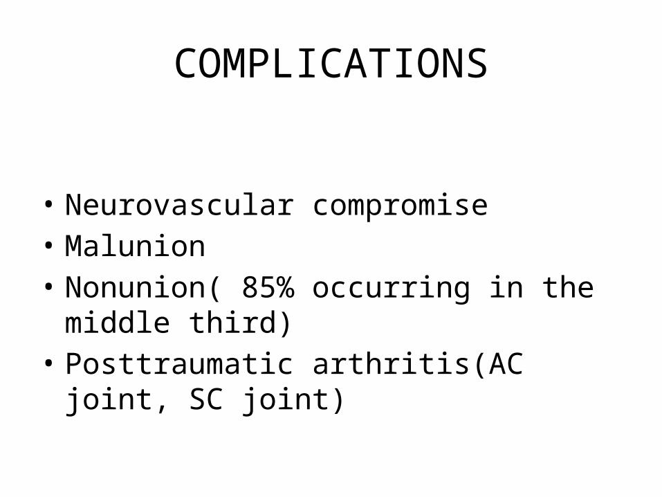
COMPLICATIONS
• Neurovascular compromise• Malunion• Nonunion( 85% occurring in the middle third)• Posttraumatic arthritis(AC joint, SC joint)
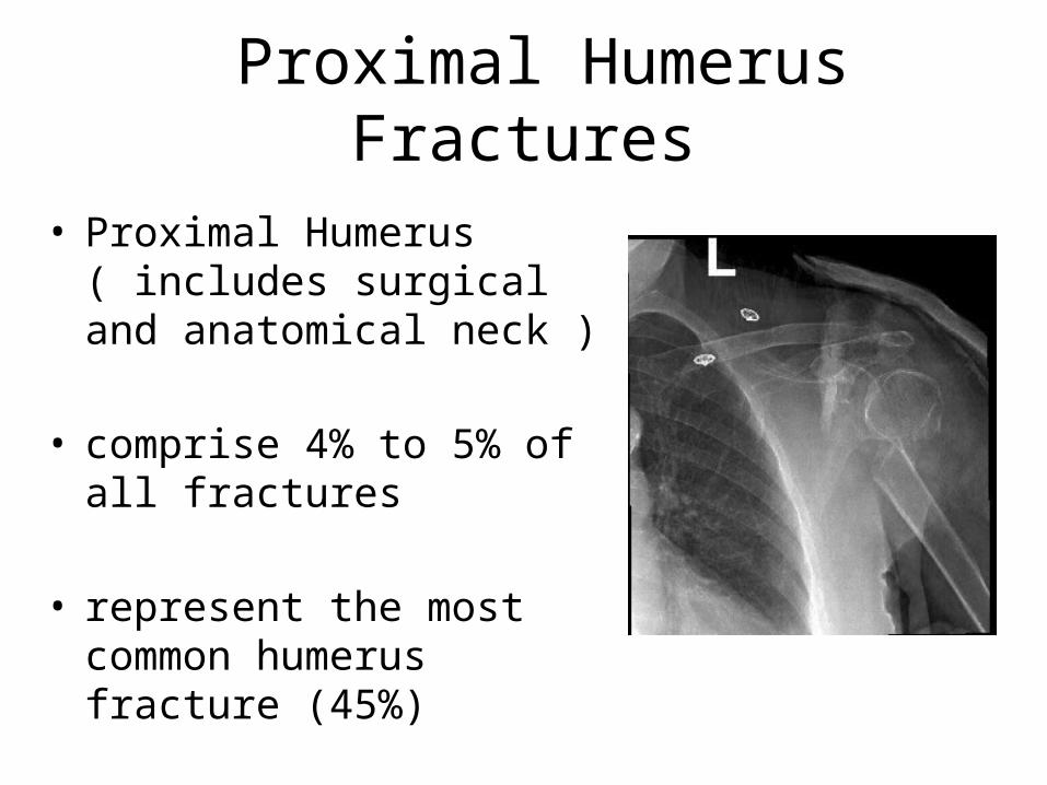
Proximal Humerus Fractures
• Proximal Humerus ( includes surgical and anatomical neck )
• comprise 4% to 5% of all fractures
• represent the most common humerus fracture (45%)
CLINICAL EVALUATION
• pain, swelling, tenderness, painful range of motion, and variable crepitus.
• A careful neurovascular examination is essential, axillary nerve function.
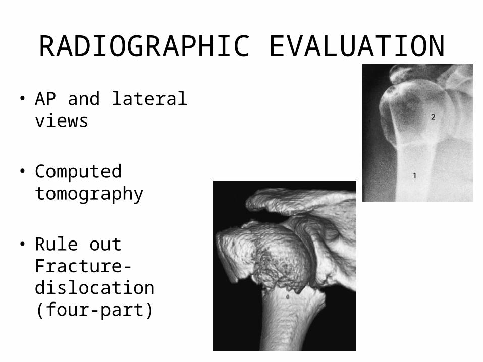
RADIOGRAPHIC EVALUATION
• AP and lateral views
• Computed tomography
• Rule out Fracture-dislocation (four-part)

(Neer’s classification)
Four parts: • humeral shaft • humeral head• Greater tuberosity • Lesser tuberosity
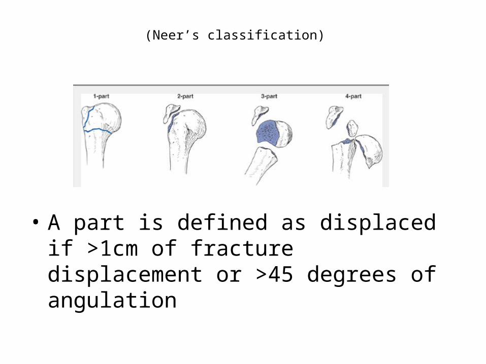
(Neer’s classification)
• A part is defined as displaced if >1cm of fracture displacement or >45 degrees of angulation

Treatment
Conservative
• Non- or minimally displaced fractures ( less than 5 mm)– 85% of fractures are
minimally displaced or nondisplaced.
– Sling immobilization.– Early shoulder motion at
7 to 10 days.
Operative fixation
• displaced more than 10 mm.
• Three- and four-part fractures
• Replacement of humeral head for four-part in elderly
COMPLICATIONS
• Osteonecrosis: four-part (13%-34%), three-part(3% to 14%), anatomic neck fractures.
• Vascular injury (5% to the axillary artery)• Neural injury(Brachial plexus injury, Axillary nerve
injury)• Shoulder stiffness• Nonunion, Malunion, Heterotopic ossification
Fractures Shaft of the Humerus
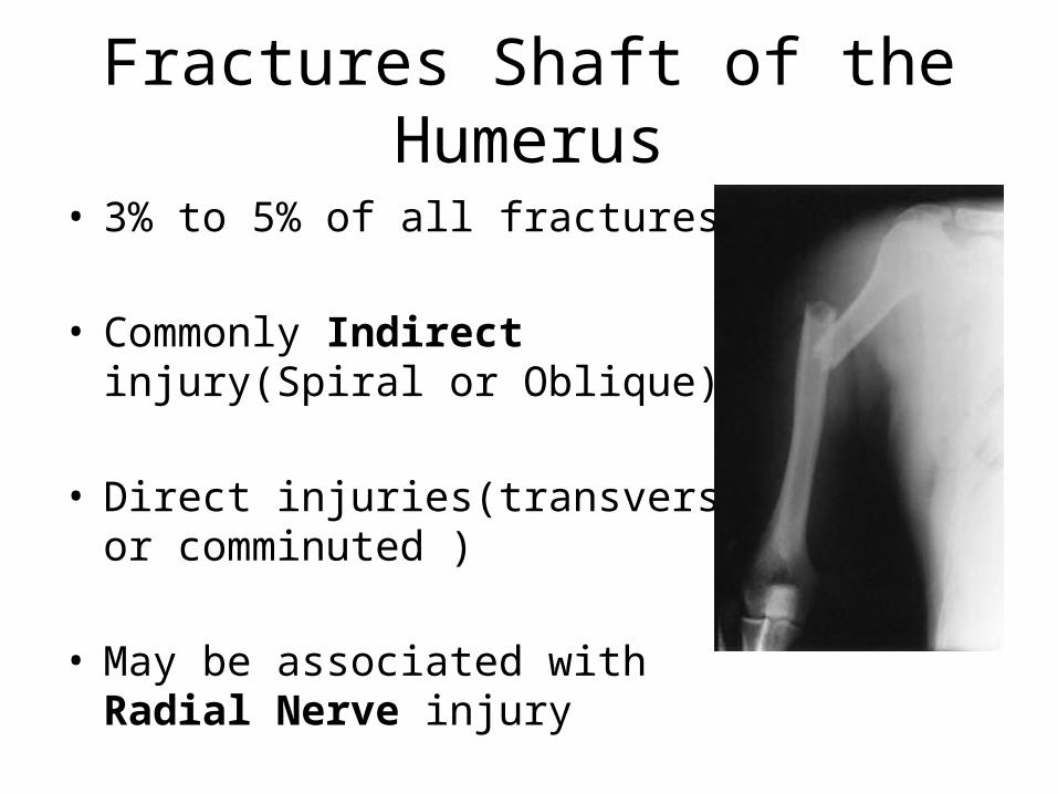
• 3% to 5% of all fractures
• Commonly Indirect injury(Spiral or Oblique)
• Direct injuries(transverse or comminuted )
• May be associated with Radial Nerve injury
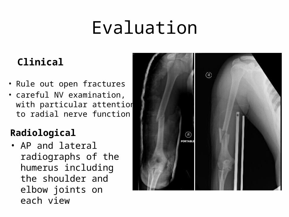
Evaluation
Clinical
• Rule out open fractures• careful NV examination, with
particular attention to radial nerve function
Radiological• AP and lateral radiographs
of the humerus including the shoulder and elbow joints on each view
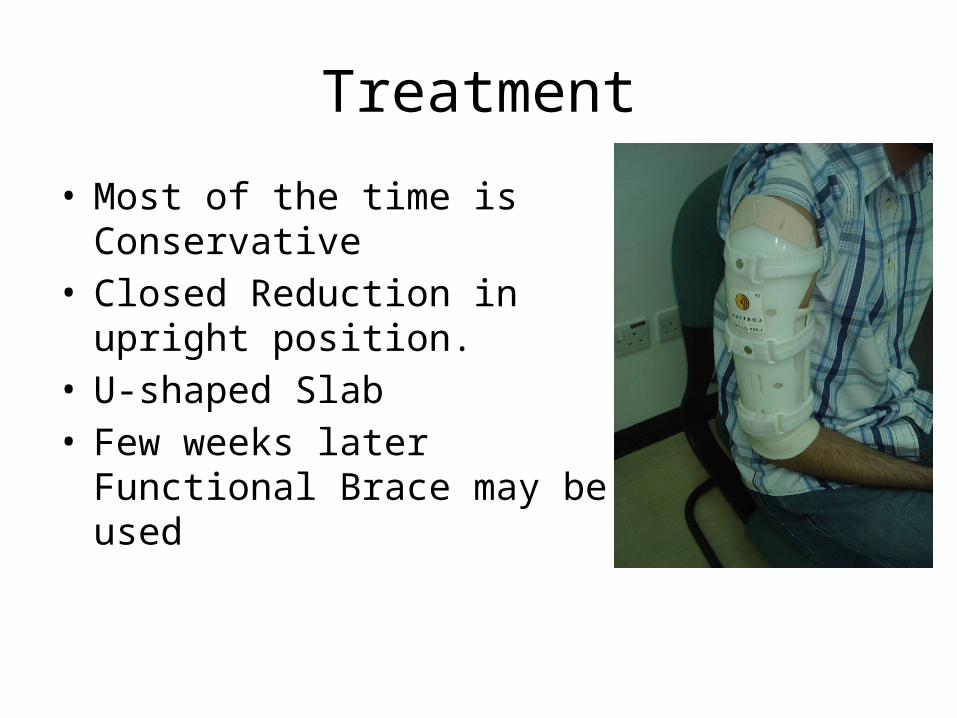
Treatment
• Most of the time is Conservative • Closed Reduction in upright
position.• U-shaped Slab• Few weeks later Functional Brace
may be used
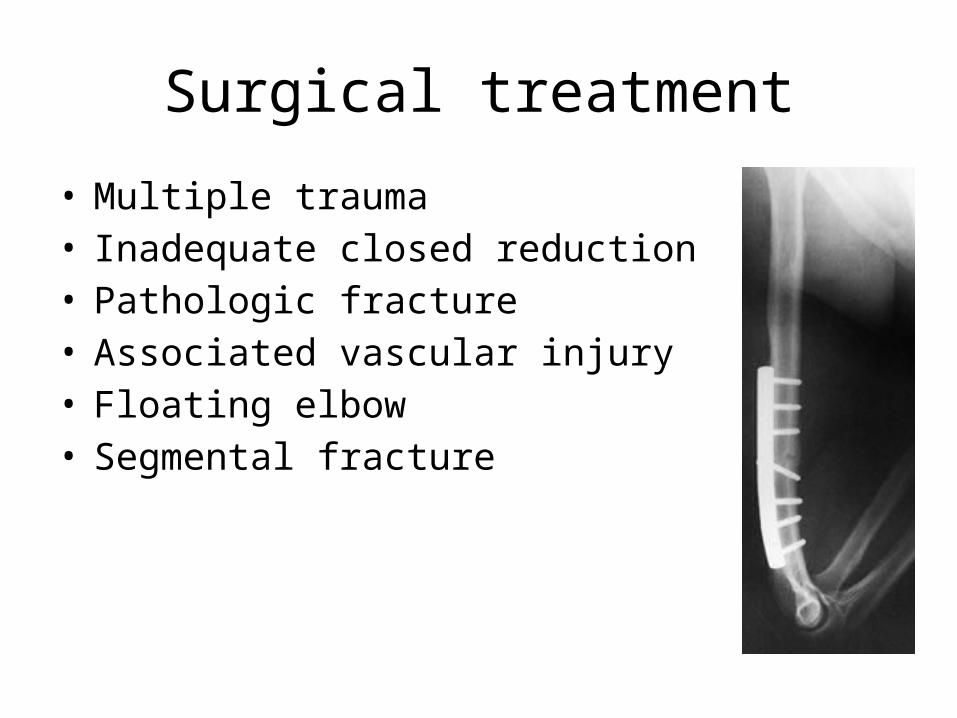
Surgical treatment
• Multiple trauma• Inadequate closed reduction• Pathologic fracture• Associated vascular injury• Floating elbow• Segmental fracture
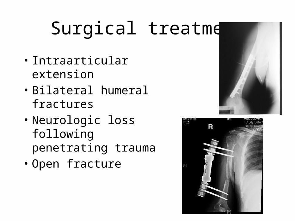
Surgical treatment
• Intraarticular extension• Bilateral humeral fractures• Neurologic loss following
penetrating trauma• Open fracture
COMPLICATIONS
• Radial Nerve Injury (Wrist drop): 12% of fractures2/3( 8%) Neuropraxia1/3 ( 4%) lacerations or transection
In open fractures; immediate exploration and ± repairIn closed injuries treated conservatively
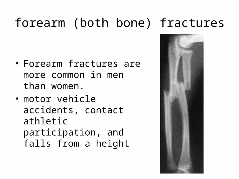
forearm (both bone) fractures
• Forearm fractures are more common in men than women.
• motor vehicle accidents, contact athletic participation, and falls from a height
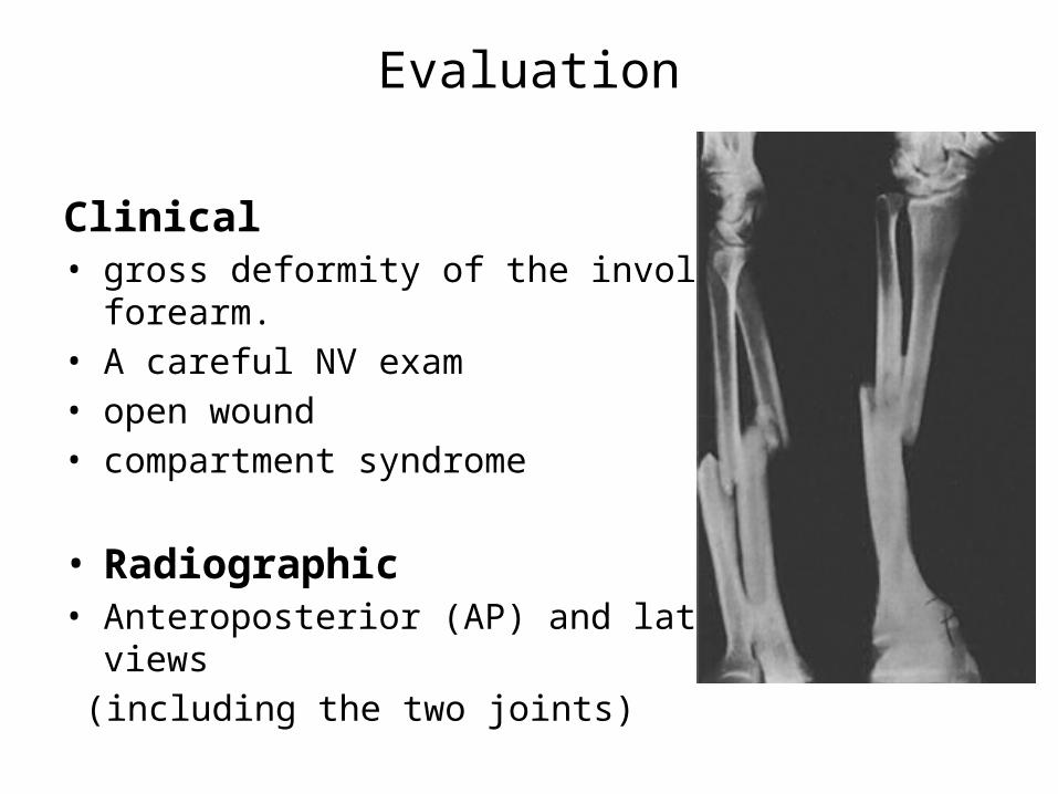
Evaluation
Clinical • gross deformity of the involved forearm.• A careful NV exam • open wound • compartment syndrome
• Radiographic • Anteroposterior (AP) and lateral views (including the two joints)
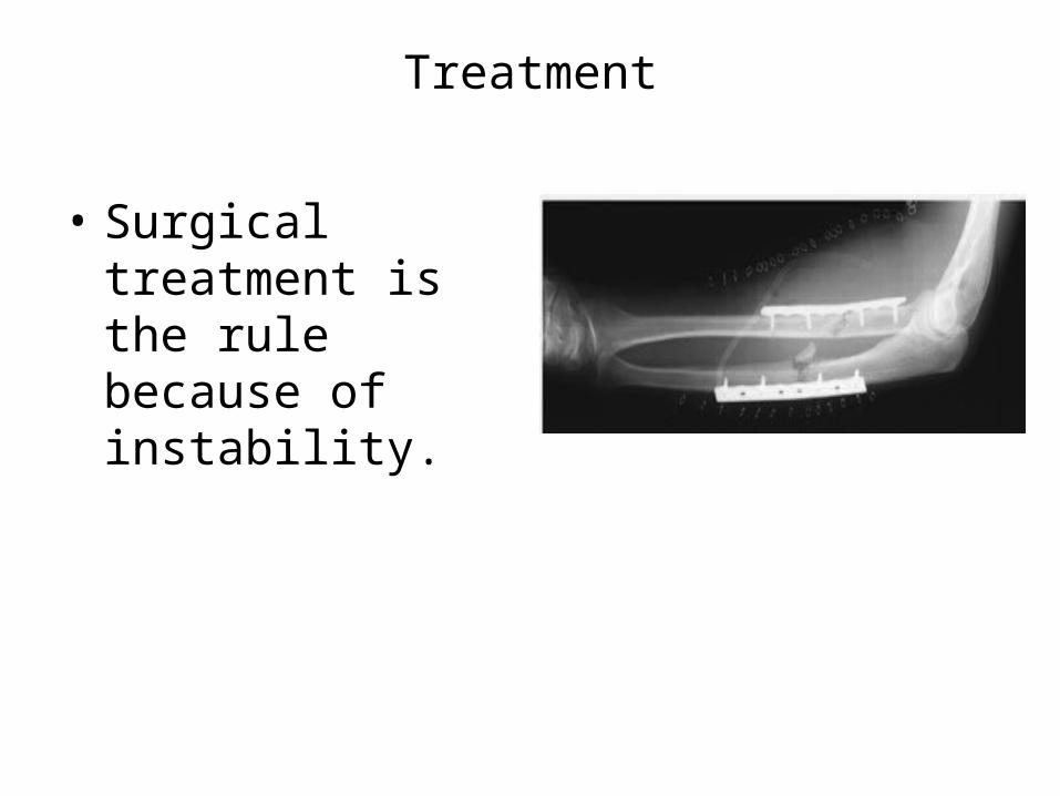
Treatment
• Surgical treatment is the rule because of instability.

Complications
• Nonunion • Compartment Syndrome• Posttraumatic radioulnar
synostosis (3% to 9% )• malunion• Infection• Neurovascular injury
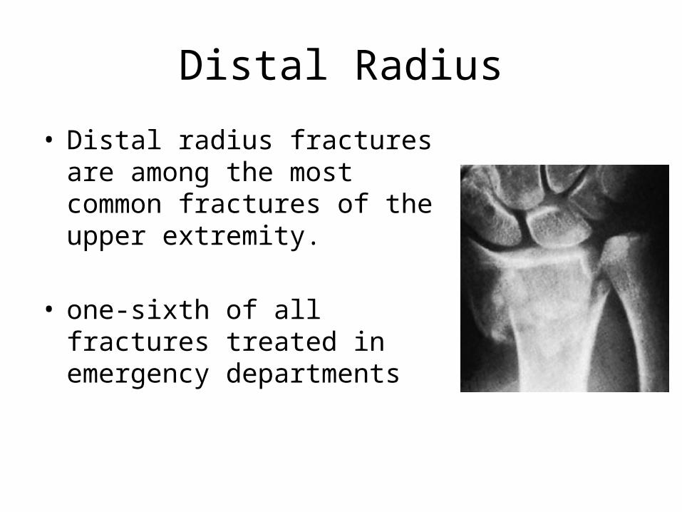
Distal Radius
• Distal radius fractures are among the most common fractures of the upper extremity.
• one-sixth of all fractures treated in emergency departments
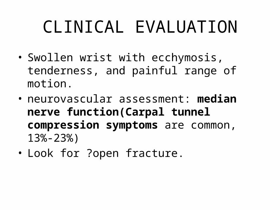
CLINICAL EVALUATION
• Swollen wrist with ecchymosis, tenderness, and painful range of motion.
• neurovascular assessment: median nerve function(Carpal tunnel compression symptoms are common, 13%-23%)
• Look for ?open fracture.
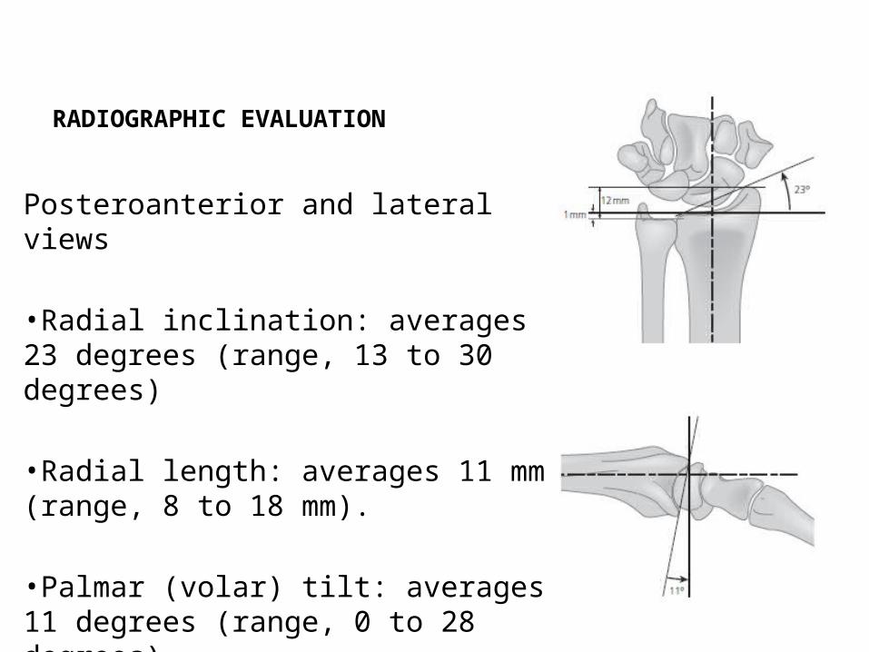
RADIOGRAPHIC EVALUATION
Posteroanterior and lateral views
•Radial inclination: averages 23 degrees (range, 13 to 30 degrees)
•Radial length: averages 11 mm (range, 8 to 18 mm).
•Palmar (volar) tilt: averages 11 degrees (range, 0 to 28 degrees).
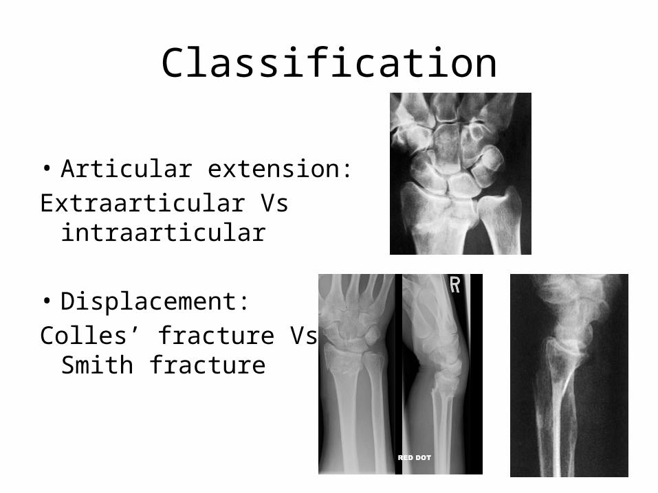
Classification
• Articular extension: Extraarticular Vs
intraarticular
• Displacement:Colles’ fracture Vs Smith
fracture
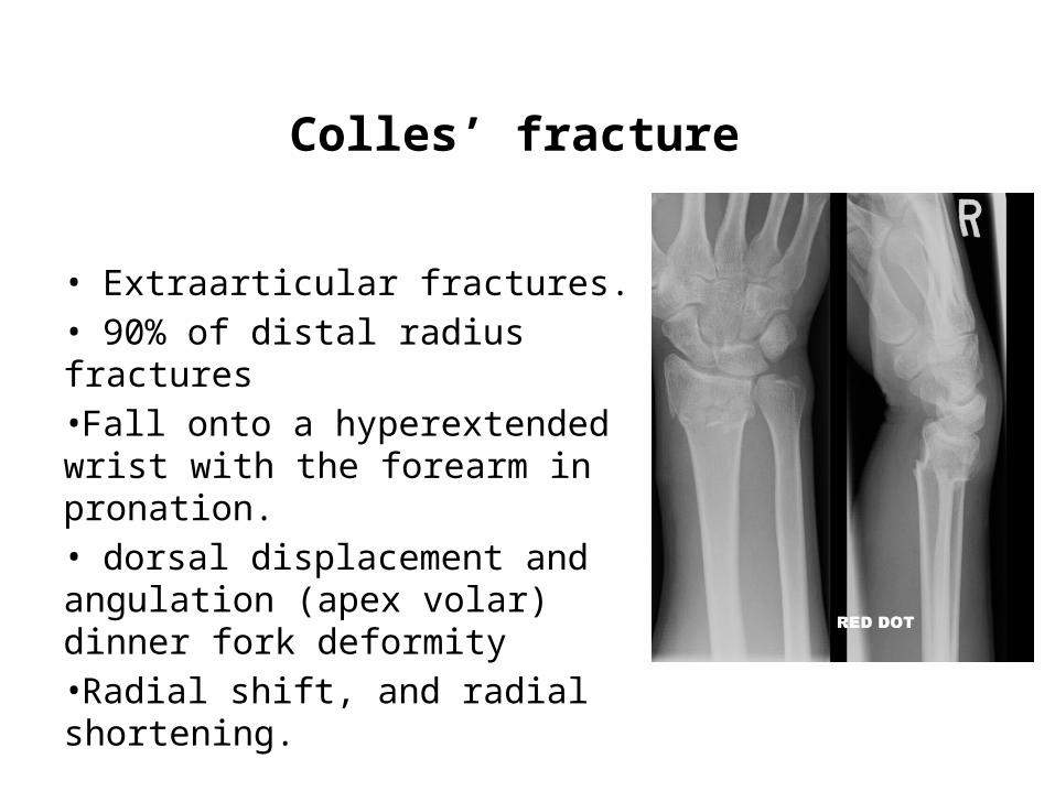
Colles’ fracture
• Extraarticular fractures.• 90% of distal radius fractures•Fall onto a hyperextended wrist with the forearm in pronation.• dorsal displacement and angulation (apex volar) dinner fork deformity•Radial shift, and radial shortening.
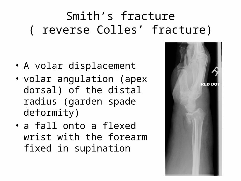
Smith’s fracture( reverse Colles’ fracture)
• A volar displacement • volar angulation (apex dorsal) of
the distal radius (garden spade deformity)
• a fall onto a flexed wrist with the forearm fixed in supination
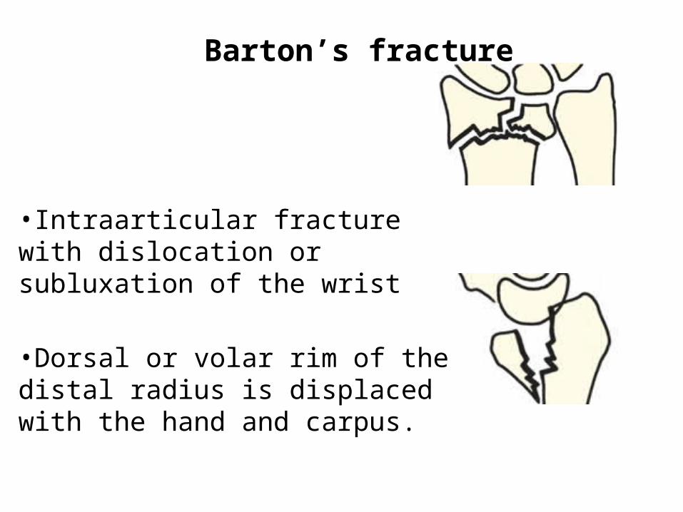
Barton’s fracture
•Intraarticular fracture with dislocation or subluxation of the wrist
•Dorsal or volar rim of the distal radius is displaced with the hand and carpus.
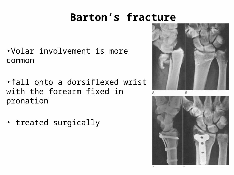
Barton’s fracture
•Volar involvement is more common
•fall onto a dorsiflexed wrist with the forearm fixed in pronation
• treated surgically
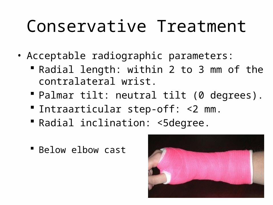
Conservative Treatment
• Acceptable radiographic parameters: Radial length: within 2 to 3 mm of the contralateral
wrist. Palmar tilt: neutral tilt (0 degrees). Intraarticular step-off: <2 mm. Radial inclination: <5degree.
Below elbow cast
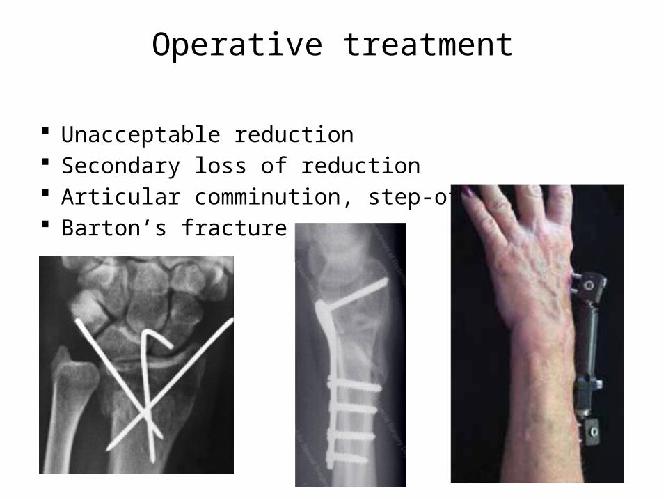
Operative treatment
Unacceptable reduction Secondary loss of reduction Articular comminution, step-off, or gap Barton’s fracture
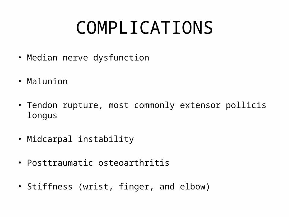
COMPLICATIONS
• Median nerve dysfunction
• Malunion
• Tendon rupture, most commonly extensor pollicis longus
• Midcarpal instability
• Posttraumatic osteoarthritis
• Stiffness (wrist, finger, and elbow)
Lower limbs Fractures
Pelvic Proximal femoral fractures( femoral neck,
intertrochantric ) Femoral shaft Tibial shaft Ankle
Mechanism of fractures
• High energy trauma like MVA, fall , except in elderly people or pathological bones
• Types of fracture are depend on position of limb during impaction and magnitude of forces applied.
• Look at the patient as whole ,not to injured limb alone!
• Save life first, then save limb and finally save limb function.
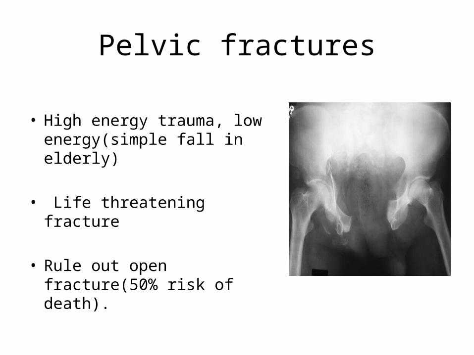
Pelvic fractures
• High energy trauma, low energy(simple fall in elderly)
• Life threatening fracture
• Rule out open fracture(50% risk of death).

Classification
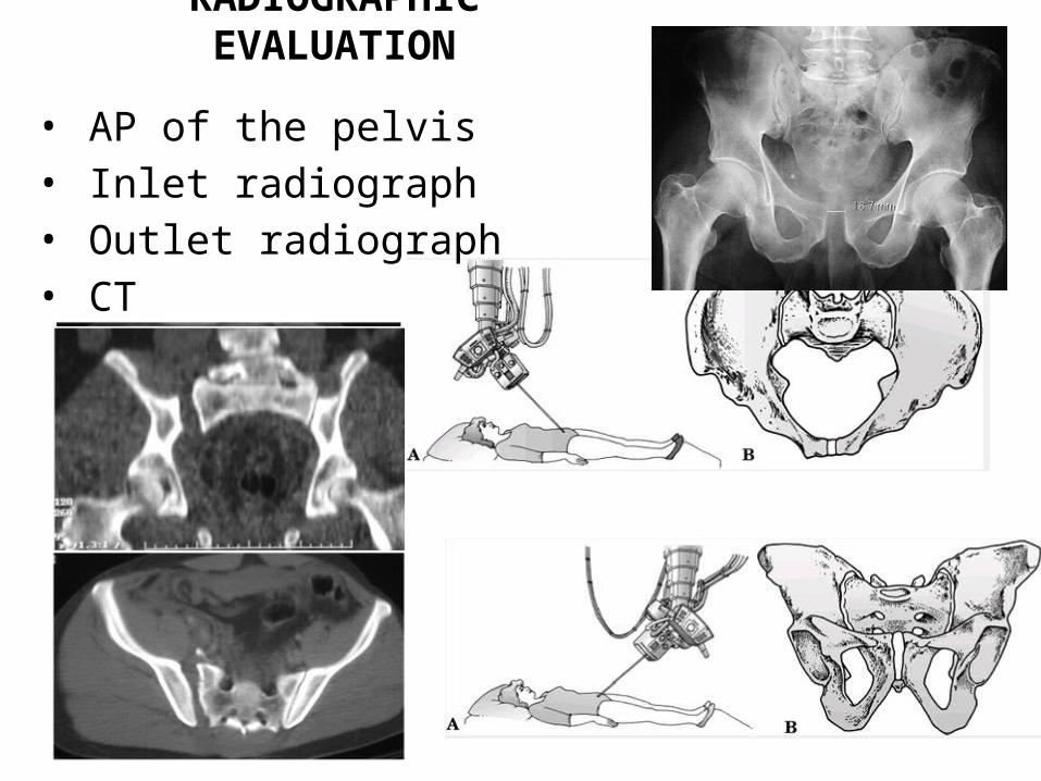
RADIOGRAPHIC EVALUATION
• AP of the pelvis • Inlet radiograph • Outlet radiograph • CT
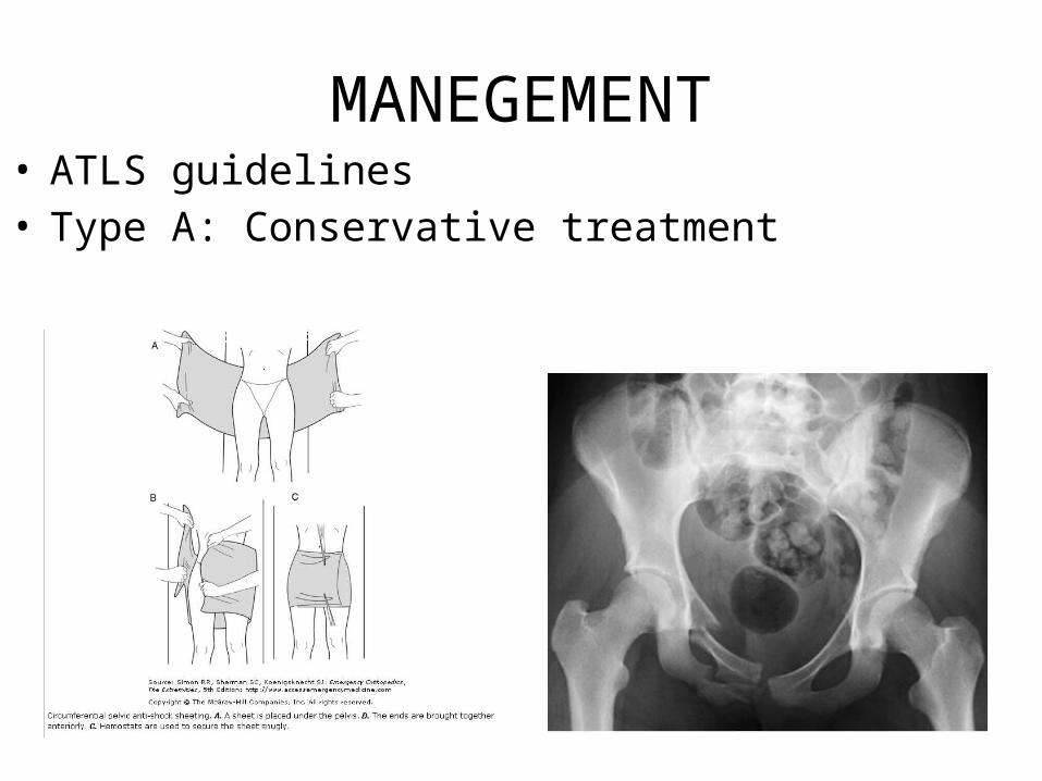
MANEGEMENT• ATLS guidelines• Type A: Conservative treatment
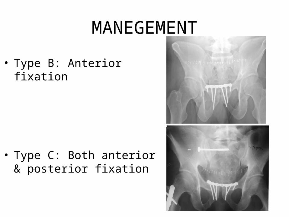
MANEGEMENT
• Type B: Anterior fixation
• Type C: Both anterior & posterior fixation
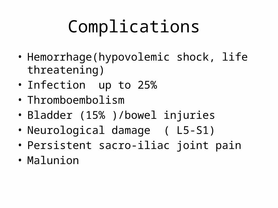
Complications
• Hemorrhage(hypovolemic shock, life threatening)• Infection up to 25%• Thromboembolism• Bladder (15% )/bowel injuries • Neurological damage ( L5-S1)• Persistent sacro-iliac joint pain• Malunion
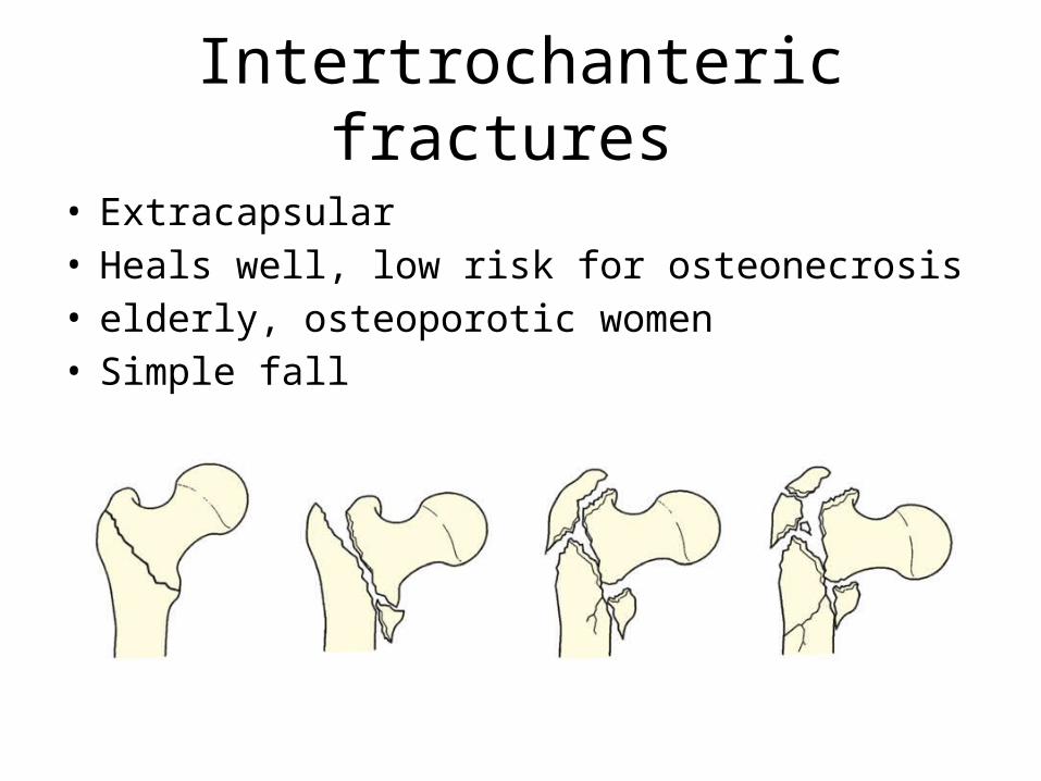
Intertrochanteric fractures
• Extracapsular• Heals well, low risk for osteonecrosis• elderly, osteoporotic women• Simple fall
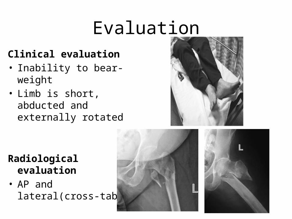
EvaluationClinical evaluation• Inability to bear-weight• Limb is short, abducted
and externally rotated
Radiological evaluation• AP and lateral(cross-
table)
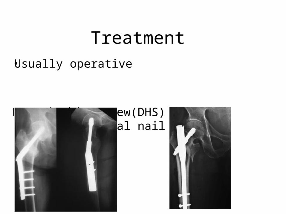
Treatment •Usually operative
Dynamic hip screw(DHS) Proximal femoral nail
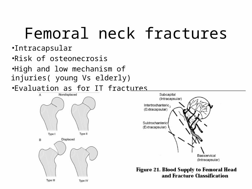
Femoral neck fractures•Intracapsular•Risk of osteonecrosis•High and low mechanism of injuries( young Vs elderly)•Evaluation as for IT fractures
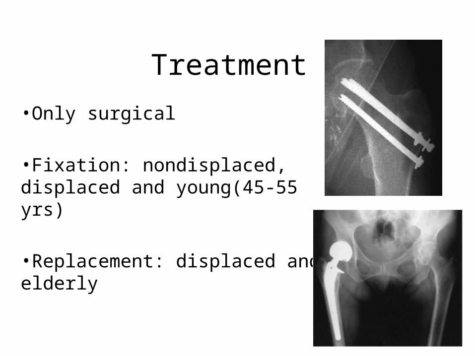
Treatment •Only surgical
•Fixation: nondisplaced, displaced and young(45-55 yrs)
•Replacement: displaced and elderly
COMPLICATIONS
• Nonunion(5% of nondisplaced, 25% of displaced fractures)
• Osteonecrosis(10% of nondisplaced, 27% of displaced fractures)
• Fixation failure(osteoporotic bone or technical problems )
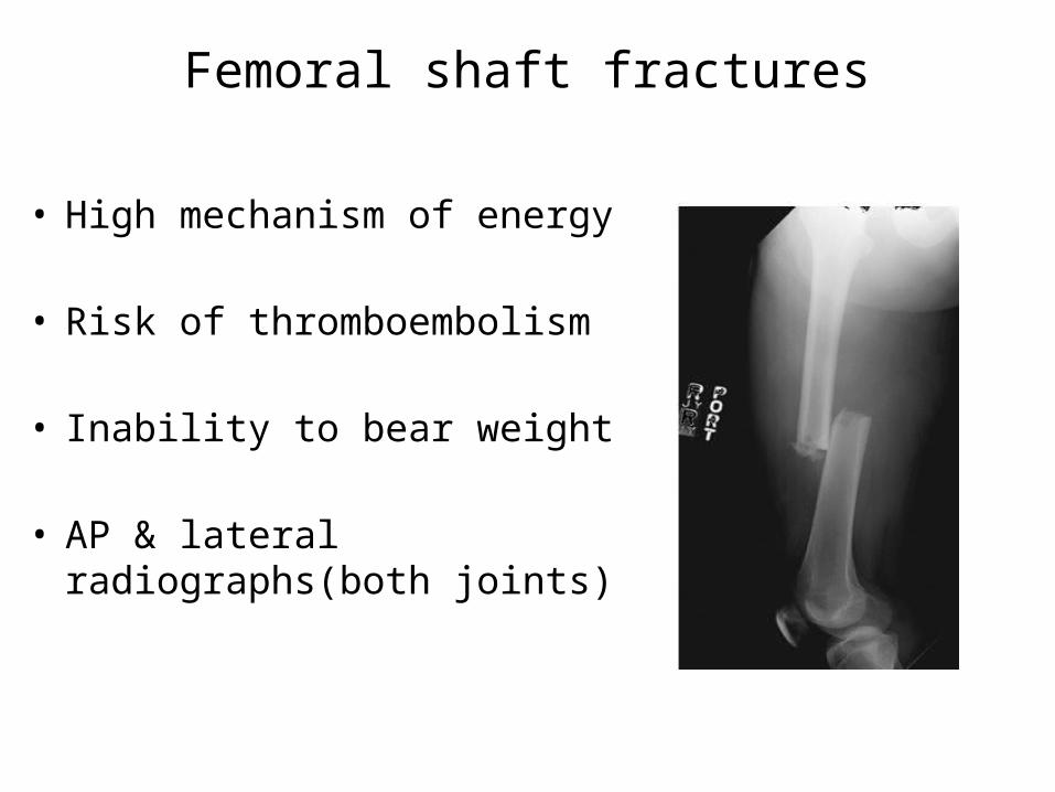
Femoral shaft fractures
• High mechanism of energy
• Risk of thromboembolism
• Inability to bear weight
• AP & lateral radiographs(both joints)
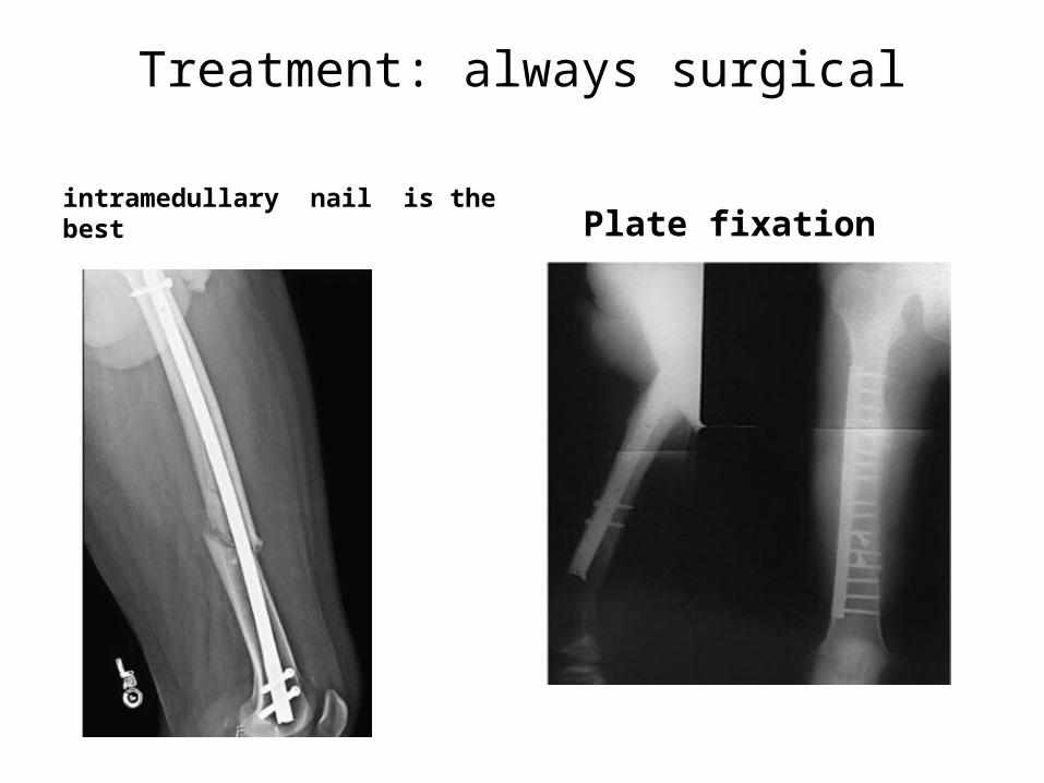
Treatment: always surgical
intramedullary nail is the best Plate fixation
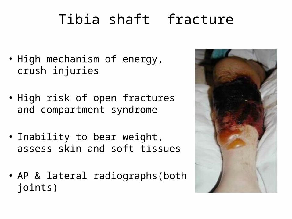
Tibia shaft fracture
• High mechanism of energy, crush injuries
• High risk of open fractures and compartment syndrome
• Inability to bear weight, assess skin and soft tissues
• AP & lateral radiographs(both joints)
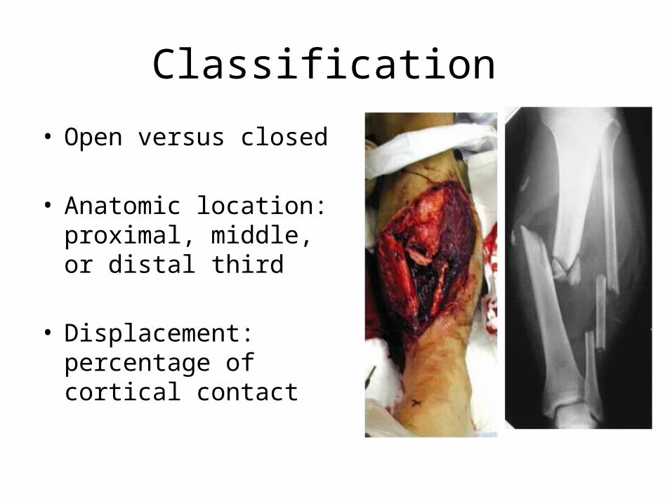
Classification
• Open versus closed
• Anatomic location: proximal, middle, or distal third
• Displacement: percentage of cortical contact
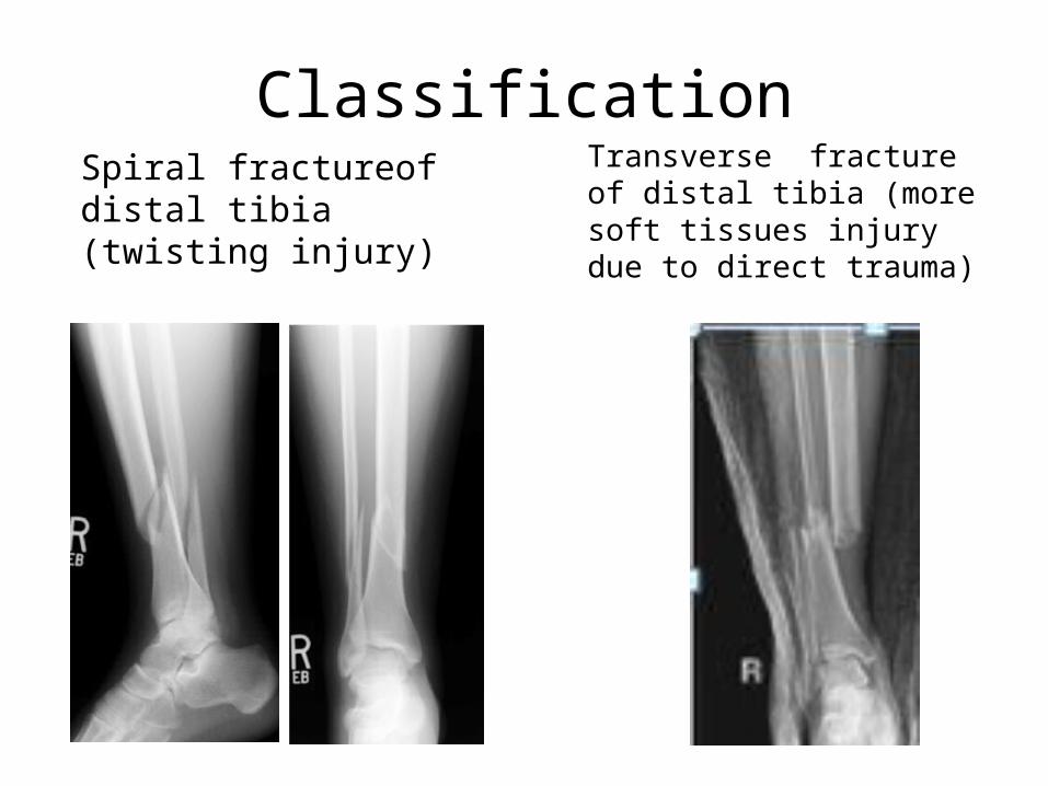
ClassificationTransverse fracture of distal tibia (more soft tissues injury due to direct trauma)
Spiral fractureof distal tibia (twisting injury)
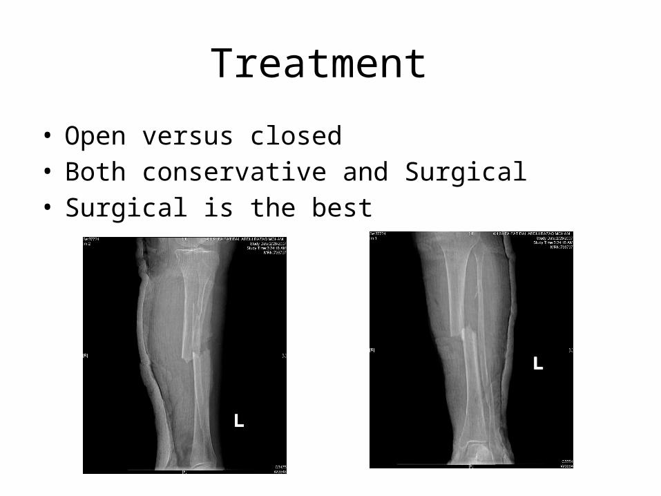
Treatment
• Open versus closed• Both conservative and Surgical• Surgical is the best
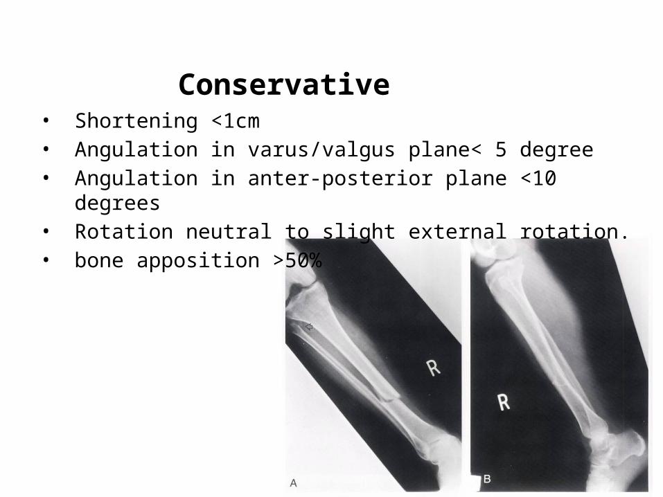
Conservative • Shortening <1cm• Angulation in varus/valgus plane< 5 degree• Angulation in anter-posterior plane <10 degrees• Rotation neutral to slight external rotation.• bone apposition >50%
Conservative
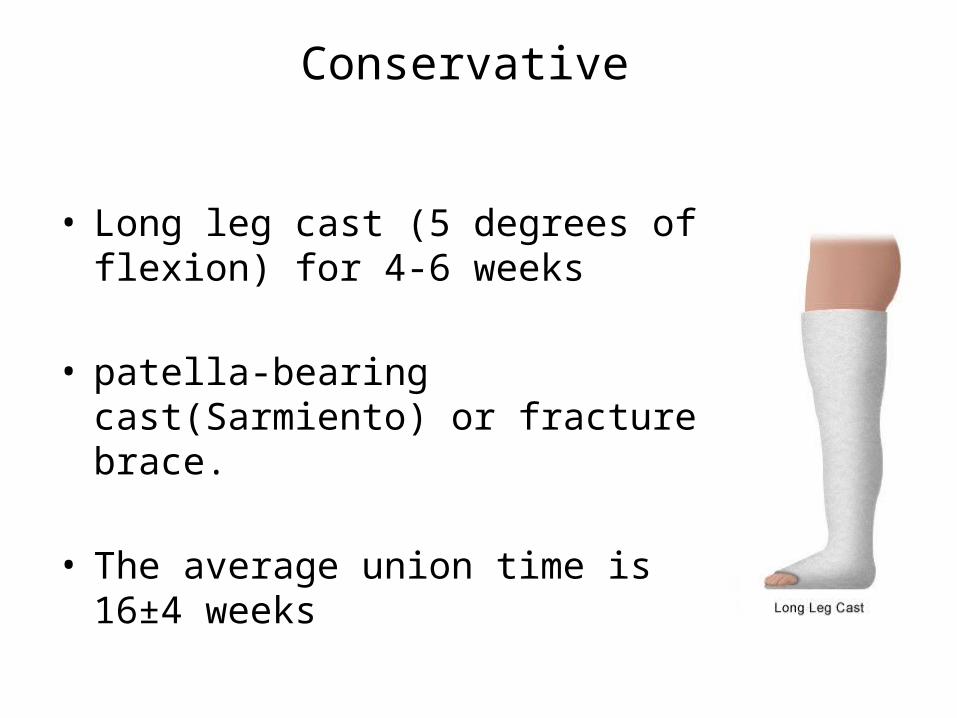
• Long leg cast (5 degrees of flexion) for 4-6 weeks
• patella-bearing cast(Sarmiento) or fracture brace.
• The average union time is 16±4 weeks
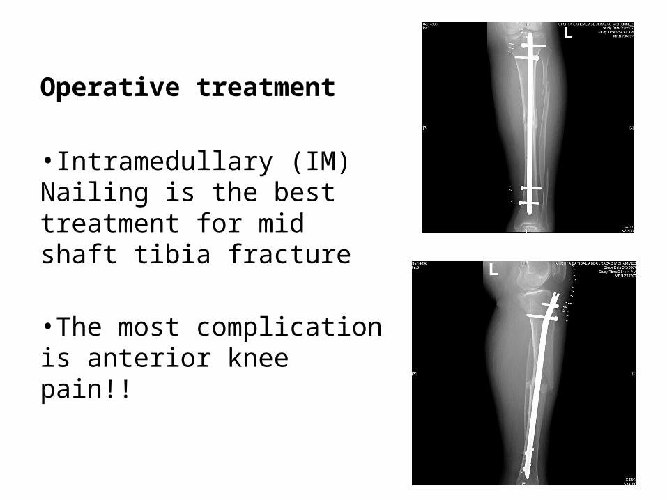
Operative treatment •Intramedullary (IM) Nailing is the best treatment for mid shaft tibia fracture •The most complication is anterior knee pain!!
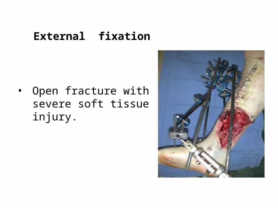
External fixation
• Open fracture with severe soft tissue injury.

Plate fixation
•97% success rates •Complication: infection, wound breakdown, nonunion •increase with higher-energy injury patterns.
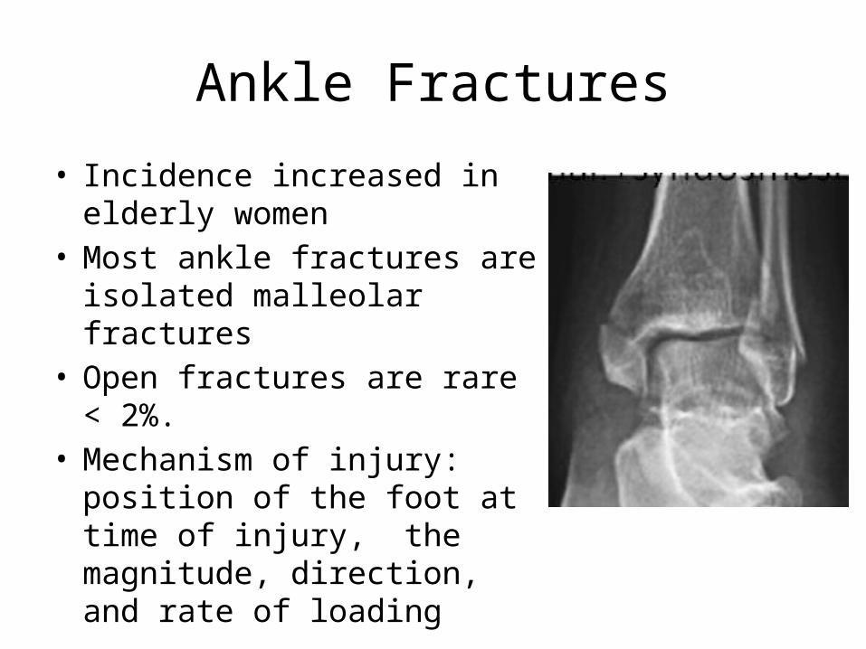
Ankle Fractures
• Incidence increased in elderly women
• Most ankle fractures are isolated malleolar fractures
• Open fractures are rare < 2%.• Mechanism of injury: position of
the foot at time of injury, the magnitude, direction, and rate of loading
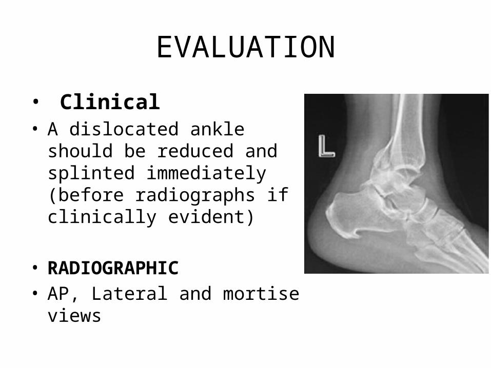
EVALUATION
• Clinical• A dislocated ankle should be
reduced and splinted immediately (before radiographs if clinically evident)
• RADIOGRAPHIC• AP, Lateral and mortise views
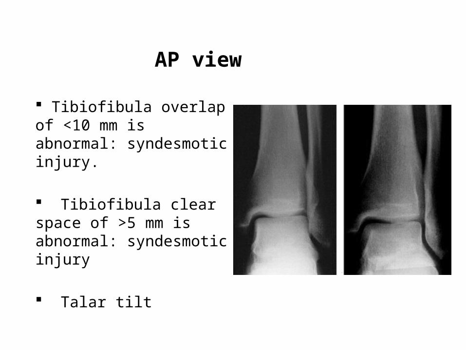
AP view
Tibiofibula overlap of <10 mm is abnormal: syndesmotic injury.
Tibiofibula clear space of >5 mm is abnormal: syndesmotic injury
Talar tilt
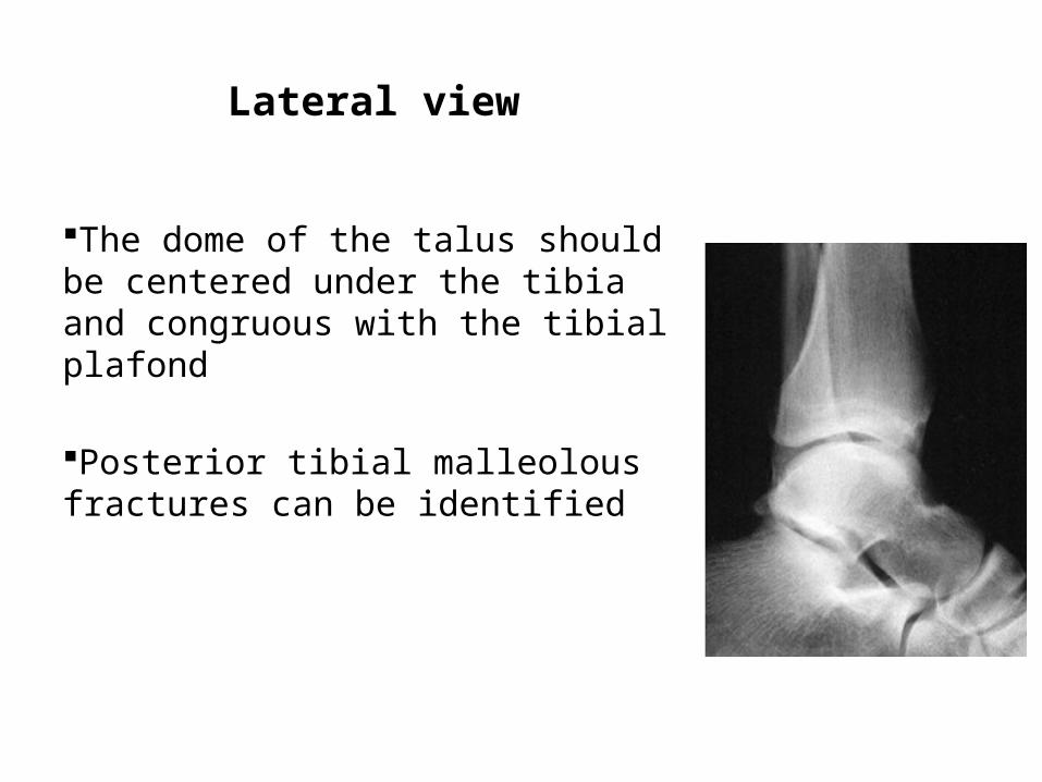
Lateral view
The dome of the talus should be centered under the tibia and congruous with the tibial plafond
Posterior tibial malleolous fractures can be identified
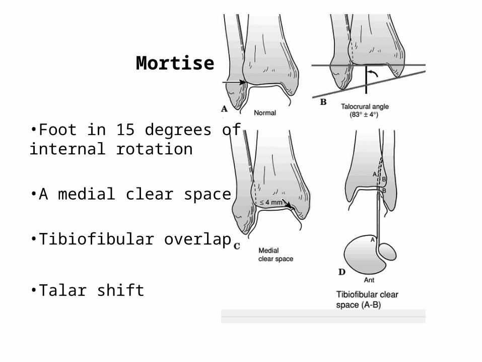
Mortise view
•Foot in 15 degrees of internal rotation
•A medial clear space
•Tibiofibular overlap
•Talar shift
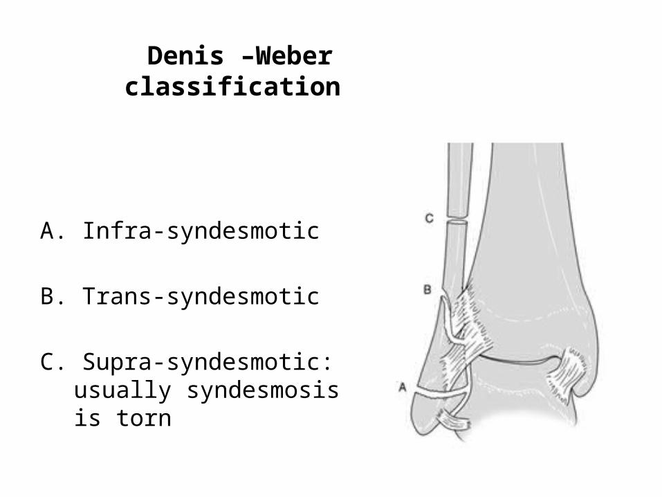
Denis –Weber classification
A. Infra-syndesmotic
B. Trans-syndesmotic
C. Supra-syndesmotic: usually syndesmosis is torn
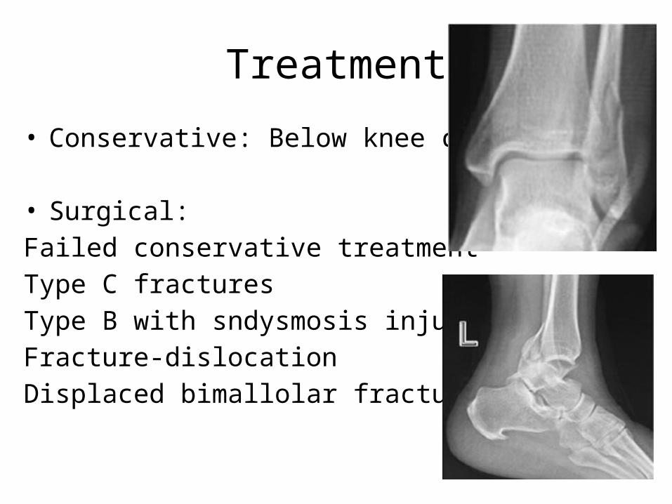
Treatment
• Conservative: Below knee cast
• Surgical:Failed conservative treatmentType C fracturesType B with sndysmosis injuryFracture-dislocationDisplaced bimallolar fracture
complications
• Post traumatic arthritis• Stiffness• Skin necrosis• Malunion or nonunion• Wound infection• Regional complex pain syndrome
Recommended