SERGIO DE OLIVA NASCIF
EFEITOS DA GHRELINA, GHRP-6 E GHRH SOBRE A SECREÇÃO DE GH, ACTH E
CORTISOL NO HIPERTIREOIDISMO
Tese apresentada à Universidade Federal de
São Paulo – Escola Paulista de Medicina para
a obtenção do título de Doutor em Ciências
São Paulo
2008

SERGIO DE OLIVA NASCIF
EFEITOS DA GHRELINA, GHRP-6 E GHRH SOBRE A SECREÇÃO DE GH, ACTH E
CORTISOL NO HIPERTIREOIDISMO
Tese apresentada à Universidade Federal de
São Paulo – Escola Paulista de Medicina para
a obtenção do título de Doutor em Ciências
Orientadora:
Prof. Dra. Ana Maria Judith Lengyel
Coordenador:
Prof. Dr. Sergio Atala Dib
São Paulo
2008

Nascif, Sergio de Oliva Efeitos da Ghrelina, GHRP-6 e GHRH Sobre a Secreção de GH, ACTH e Cortisol no Hipertireoidismo / Sergio de Oliva Nascif – São Paulo, 2008. vii, 99f. Tese (Doutorado) – Universidade Federal de São Paulo. Escola Paulista de Medicina. Programa de pós-graduação em Endocrinologia. Título em inglês: Effects of ghrelin, GHRP-6 and GHRH on GH, ACTH and cortisol secretion in hyperthyroidism. 1. Ghrelina; 2. GHRP-6; 3. GHRH; 4. Hipertireoidismo.

iii
Este trabalho foi realizado com auxílio financeiro da FAPESP
(Fundação de Amparo à Pesquisa do Estado de São Paulo) e CNPq
(Conselho Nacional de Desenvolvimento Científico e Tecnológico), e
com bolsa de pós-graduação concedida pela CAPES (Fundação
Coordenação de Aperfeiçoamento de Pessoal de Nível Superior).
iv
À minha filha Carolina, ainda tão pequenina,
que me tornou uma pessoa mais completa.
À minha amada esposa Karina, companheira
de todas as horas, por me incentivar em todas
as minhas conquistas.
Aos meus pais Celia e Rogério, pelo apoio
permanente e amor incondicional.
v
À Dra. Ana Maria Lengyel, não só pela brilhante
capacidade de orientação e ensinamentos, mas
pelo exemplo de dedicação e amor pela ciência,
toda a minha admiração.

AGRADECIMENTOS
vi
AGRADECIMENTOS
Aos pacientes e voluntários que participaram dos testes, sem os quais
esse estudo não existiria.
Aos Professores da Disciplina de Endocrinologia da UNIFESP pelos
ensinamentos durante o curso de Pós-Graduação, em particular ao grupo de
Tireóide pelo encaminhamento de pacientes. Ao Prof. Julio Abucham pela
convivência agradável e brilhantismo durante a realização dos estudos na
Neuroendocrinologia.
Aos Professores da Disciplina de Endocrinologia da Faculdade de
Medicina de Sorocaba, Maria Helena Senger, Magali Zampieri, Maria Teresa
Quilici e Alexandre Vieira, que proporcionaram um apoio marcante na aquisição
de conhecimentos durante os anos de Residência Médica. Um agradecimento
especial ao João Carlos Ramos Dias pela amizade fraternal e orientação
precisa desde o início da especialização.
Aos colegas de Pós-Graduação da UNIFESP, principalmente do grupo
da Neuroendocrinologia, Teresa, Juliana, Elisa, Mariana, Manoel, Erika,
Rogério, Priscilla, Marcos e Larissa, pelo agradável convívio. Uma menção
especial à Silvia, por todos estes anos de amizade e companheirismo desde o
início da Pós-Graduação, e à Patrícia que desempenhou papel fundamental
para a conclusão deste estudo.
À Amaryllis Salzano pela atenção que sempre me dispensou. À
Filomena, Walkíria e Teresa Kasamatsu pela assistência técnica.
Aos funcionários da Disciplina de Endocrinologia, em especial à Yeda,
Ivonete, João, Fátima, Lourdes e Margareth, pela atenção e ajuda.

ÍNDICE
vii
ÍNDICE página
Introdução.................................................................................................. 01
Objetivos.................................................................................................... 08
Referências bibliográficas....................................................................... 10
Estudo 1 – DECREASED GHRELIN-INDUCED GH RELEASE IN THYROTOXICOSIS: COMPARISON WITH GH-RELEASING PEPTIDE-6 (GHRP-6) AND GHRH Abstract………………………………………………………………………….. 23
Introduction……………………………………………………………………… 23
Subjects and methods................................................................................. 24
Results…………………………………………………………………………… 25
Discussion……………………………………………………………………..... 26
References………………………………………………………………………. 27
Estudo 2 – GHRELIN AND GHRP-6-INDUCED ACTH AND CORTISOL RELEASE IN THYROTOXICOSIS Abstract………………………………………………………………………….. 32
Introduction……………………………………………………………………… 34
Subjects and methods................................................................................. 37
Results…………………………………………………………………………… 40
Discussion……………………………………………………………………..... 44
References………………………………………………………………………. 50
Sumário e Conclusões............................................................................. 58
Anexos – Estudo 1.................................................................................... 60
Anexos – Estudo 2.................................................................................... 79

1
INTRODUÇÃO
2
INTRODUÇÃO
O controle da secreção pulsátil do hormônio de crescimento (GH) pelos
somatotrofos da adenohipófise é resultante de uma complexa interação entre
dois peptídeos hipotalâmicos: o hormônio liberador de GH (GHRH), que
estimula a secreção de GH, e a somatostatina, que tem um efeito inibitório
sobre a liberação deste hormônio (Dieguez et al., 1988). Além do GHRH e da
somatostatina, diversos outros fatores modulam a secreção de GH, atuando
diretamente sobre a hipófise ou sobre a liberação destes dois peptídeos
hipotalâmicos (Lengyel, 1992).
Na década de 70, antes da descoberta do GHRH, Bowers e col.
identificaram pequenos peptídeos sintéticos a partir da molécula de met-
encefalina que eram capazes de liberar GH. Estudos posteriores baseados em
cálculos de energia conformacional, modificações químicas e testes de
atividade biológica resultaram no desenvolvimento de peptídeos mais potentes,
incluindo um hexapeptídeo denominado growth hormone-releasing peptide-6
(GHRP-6) (Bowers et al., 1984). Este peptídeo promove a liberação de GH,
tanto in vitro como in vivo, em todas as espécies de animais testadas e este
efeito é obtido por diferentes vias de administração, incluindo endovenosa e
mesmo por via oral (Ghigo et al., 1997; Isidro & Cordido, 2006). Estudos
realizados com estes secretagogos de GH (GHS) nas últimas décadas
reforçaram a hipótese de existir um papel fisiológico de tais compostos na
regulação da secreção de GH (Dieguez & Casanueva, 2000), uma vez que
exercem sua atividade por mecanismo diferente daquele utilizado pelo GHRH
(Korbonits & Grossman, 1995). A presença de receptores específicos para os
GHS, tanto no hipotálamo quanto na hipófise, sugeria a possível existência de

3
um peptídeo endógeno semelhante ainda não identificado (Codd et al., 1989;
Blake & Smith, 1991; Goth et al., 1992). A clonagem do receptor “órfão” dos
GHS, em 1996, comprovou a hipótese da existência de um terceiro sistema de
controle da secreção de GH (Howard et al., 1996). Finalmente, em 1999,
Kojima e col. clonaram o ligante endógeno dos GHS, que foi isolado a partir de
extrato de estômago, e denominado ghrelina. A estrutura deste peptídeo, que é
acilado, é completamente diferente dos peptídeos conhecidos e também da
estrutura química dos GHS (Kojima et al., 1999). A modificação n-octanoil do
resíduo serina na posição 3 da molécula é fundamental para a atividade
biológica do peptídeo (Kojima et al., 1999; Bednarek et al., 2000). A ghrelina
está presente em altas concentrações no trato gastrointestinal (Kojima et al.,
1999; Date et al., 2000a) e, em menores concentrações, no sistema nervoso
central, principalmente no núcleo arqueado (Kojima et al., 1999; Shuto et al.,
2001).
A ghrelina promove a liberação de GH tanto in vivo como in vitro, em
animais e no homem, de modo dose-dependente (Kojima et al., 1999; Takaya
et al., 2000; Date et al., 2000b; Peino et al., 2000). Este peptídeo é o mais
potente estímulo para a secreção de GH em humanos, promovendo uma maior
liberação de GH quando comparado à hexarelina (um GHS) e ao GHRH em
doses equimolares (Arvat et al., 2001). Da mesma forma que os GHS, a
ghrelina estimula a secreção de GH através de mecanismos hipofisários e
hipotalâmicos (Korbonits et al., 2004). A ghrelina e os GHS ativam os
receptores de GHS (GHS-R) em culturas de células hipofisárias in vitro (Kojima
et al., 1999), porém sua atividade in vivo é maior (Arvat et al., 2001), sugerindo
que seu principal sítio de ação seja hipotalâmico. Além disso, os efeitos destes
4
peptídeos são reduzidos ou abolidos na desconexão hipotálamo-hipofisária
(Popovic et al., 1995; Popovic et al., 2003). Foi demonstrado que a integridade
da via do GHRH é fundamental para a ação da ghrelina e dos GHS (Dickson et
al., 1995; Pandya et al., 1998; Maheshwari et al., 1999; Tannenbaum &
Bowers, 2001; Tannenbaum et al., 2003). Além disso, a ghrelina e os GHS
podem ativar os GHS-R expressos em ¼ dos neurônios produtores de GHRH
no núcleo arqueado (Tannenbaum & Bowers, 2001). Foi proposto um modelo
de ação da ghrelina que envolve, além da liberação hipotalâmica de GHRH, a
amplificação do efeito do GHRH no somatotrofo, e também o antagonismo
funcional da somatostatina (Tannenbaum et al., 2003). A ação da ghrelina e
dos GHS no somatotrofo ocorre através da ativação do sistema da proteína
quinase C, com elevação de diacilglicerol e inositol trifosfato, acarretando
aumento do cálcio intracelular (Howard et al., 1996; Chen et al., 1996), ao
passo que o GHRH estimula o sistema da proteína quinase A após ativação do
AMPc intracelular. (Goth et al., 1992). Mais recentemente, foi demonstrado em
somatotrofos de suínos (Malagón et al., 2003) e de babuínos (Kineman &
Luque, 2007), que a ghrelina também é capaz de ativar o AMPc e estimular os
sistemas de influxo de cálcio intracelular, ação esta mais ampla que a dos GHS
sintéticos, o que poderia explicar sua maior potência. A descoberta da ghrelina
comprovou a existência de uma terceira via de regulação de secreção de GH.
Entretanto, o papel desta via na fisiologia e fisiopatologia da secreção de GH
ainda não é conhecido (Lengyel, 2006).
Os hormônios tireoidianos participam da síntese e secreção de GH.
Apesar do local preciso e mecanismo de ação ainda serem desconhecidos, há
evidências que estes hormônios atuam tanto no hipotálamo quanto na hipófise

5
(Dieguez et al., 1985; Jones et al., 1990; Valcavi et al., 1992; Giustina &
Wehrenberg, 1995).
Distúrbios da função tireoidiana cursam com alterações na secreção de
GH (Valcavi et al., 1992). No hipertireoidismo, a responsividade do GH à
diversos estímulos farmacológicos, incluindo o GHRH, está diminuída (Burgess
et al., 1966; Giustina et al., 1991; Valcavi et al., 1993; Ramos-Dias et al., 1995).
Os mecanismos responsáveis por estas alterações não estão claros. Um
aumento no tônus hipotalâmico de somatostatina é improvável pois foi
demonstrado que compostos que inibem a liberação de somatostatina são
incapazes de normalizar a resposta do GH aos estímulos farmacológicos
(Yeung, 1973; Valcavi et al., 1991; Ramos-Dias et al., 1995). Embora
controverso (Iranmanesh et al., 1991), a diminuição da secreção de GHRH
hipotalâmico poderia estar envolvida (Jones et al., 1990; Kamegai et al., 2004),
ou poderia haver um efeito direto do excesso de hormônios tireoidianos sobre
os somatotrofos (Dieguez et al., 1985; Jones et al., 1990; Valcavi et al., 1992;
Giustina & Wehrenberg, 1995).
A ghrelina e os GHS também são capazes de estimular o eixo
hipotálamo-hipófise-adrenal em indivíduos normais, sendo que a ghrelina tem
um efeito mais potente (Takaya et al., 2000; Arvat et al., 2001). A ação destes
peptídeos sobre a liberação de ACTH e cortisol é exclusivamente hipotalâmica,
já que não promovem a secreção de ACTH em fragmentos de hipófise in vitro
(Elias et al., 1995; Kojima et al., 1999), e os GHS-R não foram encontrados em
corticotrofos normais (Smith et al., 1997). Além disso, na desconexão
hipotálamo-hipofisária o efeito da ghrelina e dos GHS sobre o ACTH e cortisol
é abolido (Popovic et al., 1995; Popovic et al., 2003). Foi demonstrado que o

6
GHRP-6 estimula a liberação de arginina-vasopressina (AVP) em fragmentos
hipotalâmicos in vitro (Korbonits et al., 1999b), ao passo que a ghrelina tem
uma ação mais ampla, promovendo a liberação de AVP, CRH e NPY (Wren et
al., 2002), com um efeito predominante na secreção de AVP (Mozid et al.,
2003). Embora controverso, os GHS e a ghrelina estimulam a secreção de
ACTH em humanos provavelmente através do aumento da liberação
hipotalâmica de AVP (Korbonits et al., 1999a; Coiro et al., 2005).
Estudos prévios sugerem que os hormônios tireoidianos podem
participar da regulação do eixo hipotálamo-hipófise adrenal por mecanismos
que ainda não estão completamente elucidados. Classicamente, sabe-se que
na tireotoxicose há aumento compensatório da taxa de produção de cortisol
secundário a uma maior degradação (Levin & Daughaday, 1955; Peterson,
1958; Gordon & Southren, 1977). Dessa forma, no hipertireoidismo prolongado,
as adrenais estão estimuladas cronicamente e, dependendo da severidade
clínica e da duração da doença, a resposta de cortisol após diferentes
estímulos como ACTH, CRH e teste de tolerância à insulina pode estar
diminuída (Jackson et al., 1966; Goswami & Kochupillai, 2001; Tsatsoulis et al.,
2000; Yamakita et al., 2001). Entretanto, resposta normal de cortisol após
indução de hipoglicemia também tem sido observada (Jackson et al., 1966;
Giustina et al., 1971; Brauman et al., 1973; Moghetti et al., 1994). O
comprometimento da reserva adrenocortical durante o hipertireoidismo, e sua
normalização após obtenção do eutireoidismo, pode estar relacionada à
diminuição dos níveis da globulina ligadora de corticoesteróides (CBG) nos
indivíduos não-tratados, uma vez que o tratamento do hipertireoidismo cursa
com aumento nos níveis de CBG (Dumoulin et al., 1995; Mishra et al., 2007).
7
Entretanto, a elevação da concentração de corticosterona no líquor de ratos
tireotóxicos e também da razão cortisol/CBG no hipertireoidismo (Kamilaris et
al., 1991; Johnson et al., 2005; Mishra et al., 2007) sugere que um aumento de
cortisol circulante está realmente presente por excesso de secreção de cortisol
na vigência de elevação dos hormônios tireoidianos. Essa hipótese é reforçada
pelos poucos estudos que mostram aumento dos níveis de ACTH basal
(Moghetti et al., 1994; Yamakita et al., 2001; Mishra et al., 2007) e após
estímulo com CRH e hipoglicemia em indivíduos portadores de hipertireoidismo
(Moghetti et al., 1994; Lizcano & Salvador, 2008).
A administração de ghrelina também acarreta aumento dos níveis de
glicose plasmática em indivíduos normais (Broglio et al., 2001), provavelmente
por estimular a liberação de glicose hepática (Gauna et al., 2005). Esta ação é
independente do GH, uma vez que está presente mesmo em pacientes com
deficiência de GH (Gauna et al., 2004). Sabe-se que o hipertireoidismo pode
cursar com resistência à insulina e hiperinsulinemia (O’Meara et al., 1993;
Dimitriadis et al., 2008) mas não há relatos sobre os efeitos da ghrelina/GHS
nos níveis circulantes de glicose em pacientes com excesso de hormônios
tireoidianos.

8
OBJETIVOS
9
OBJETIVOS
(1) Avaliar os efeitos da ghrelina e do GHRP-6 sobre a liberação GH e
glicose em pacientes com hipertireoidismo. Os níveis de GH após a
administração de GHRH também foram estudados.
(2) Avaliar os efeitos da ghrelina e do GHRP-6 sobre a liberação de ACTH e
cortisol em pacientes tireotóxicos.

10
REFERÊNCIAS BIBLIOGRÁFICAS
11
REFERÊNCIAS BIBLIOGRÁFICAS
Arvat E, Maccario M, di Vito L, et al. 2001 Endocrine activities of ghrelin, a
natural growth hormone secretagogue (GHS), in humans: comparison and
interactions with hexarelin, a nonnatural peptidyl GHS, and GH-releasing
hormone. J Clin Endocrinol Metab 86:1169-1174.
Bednarek MA, Feighner SD, Pong SS, et al. 2000 Structure-function studies
on the new growth hormone-releasing peptide ghrelin: minimal sequence
necessary for activation of growth hormone secretagogue receptor 1a. J Med
Chem 43:4370-4376.
Blake AD & Smith RG. 1991 Desensitization studies using perifused rat
pituitary cells show that growth hormone-releasing hormone and His-D-Trp-Ala-
Trp-D-Phe-Lys-NH2 stimulate growth hormone release through distinct receptor
sites. J Endocrinol 129:11-19.
Bowers CY, Momany FA, Reynolds GA, et al. 1984 On the in vitro and in vivo
activity of a new synthetic hexapeptide that acts on the pituitary to specifically
release growth hormone. Endocrinology 114:1537-1545.
Brauman H, Smets P, Corvilain J. 1973 Comparative study of growth
response to hypoglycemia in normal subjects and in patients with primary
myxedema or hyperthyroidism before and after treatment. J Clin Endocrinol
Metab 36:1162-1174.
12
Broglio F, Arvat E, Benso A, et al. 2001 Ghrelin, a natural GH secretagogue
produced by the stomach, induces hyperglycemia and reduces insulin secretion
in humans. J Clin Endocrinol Metab 86:5083-5086.
Burgess JA, Smith BR, Merimee TJ. 1966 Growth hormone in thyrotoxicosis:
effect of insulin-induced hypoglycemia. J Clin Endocr 26:1257-1260.
Chen C, Wu D, Clarke IJ. 1996 Signal transduction systems employed by
synthetic GH-releasing peptides in somatotrophs. J Endocrinol 148:381-386.
Codd EE, Shu AY, Walker RF. 1989 Binding of growth hormone releasing
hexapeptide to specific hypothalamic and pituitary binding sites.
Neuropharmacology 28:1139-1144.
Coiro V, Saccani-Jotti G, Minelli R, et al. 2005 Adrenocorticotropin/cortisol
and arginine-vasopressin secretory patterns in response to ghrelin in normal
men. Neuroendocrinology 81:103-106.
Date Y, Kojima M, Hosoda H, et al. 2000a Ghrelin, a novel growth hormone-
releasing acylated peptide, is synthesized in a distinct endocrine cell type in the
gastrointestinal tracts of rats and humans. Endocrinology 141:4255-4261.
13
Date Y, Murakami N, Kojima M, et al. 2000b Central effects of a novel
acylated peptide, ghrelin, on growth hormone release in rats. Biochem Biophys
Res Commun 275:477-480.
Dickson SL, Doutrelant-Viltart O, Leng G. 1995 GH-deficient dw/dw rats and
lit/lit mice show increased Fos expression in the hypothalamic arcuate nucleus
following systemic injection of GH-releasing peptide-6. J Endocrinol 146:519-
526.
Dieguez C & Casanueva FF. 2000 Ghrelin: a step forward in the understanding
of somatotroph cell function and growth regulation. Eur J Endocrinol 142:413-
417.
Dieguez C, Foord SM, Peters JR, Hall R, Scanlon MF. 1985 The effects of
thyroid hormone deprivation in vivo and in vitro on growth hormone (GH)
responses to human pancreatic (tumour) GH-releasing factor (1-40) by
dispersed rat anterior pituitary cells. Endocrinology 116:1066-1070.
Dieguez C, Page MD, Scanlon MF. 1988 Growth hormone neuroregulation
and its alterations in disease states. Clin Endocrinol 28:109-143.
Dimitriadis G, Mitrou P, Lambadiari V, et al. 2008 Insulin-stimulated rates of
glucose uptake in muscle in hyperthyroidism: the importance of blood flow. J
Clin Endocrinol Metab 93:2413-2415.
14
Dumoulin SC, Perret BP, Bennet AP, Caron PJ. 1995 Opposite effects of
thyroid hormones on binding proteins for steroid hormones (sex hormone-
binding globulin and corticosteroid-binding globulin) in humans. Eur J
Endocrinol 132:594-598.
Elias KA, Ingle GS, Burnier JP, et al. 1995 In vitro characterization of four
novel classes of growth hormone-releasing peptide. Endocrinology 136:5694-
5699.
Gauna C, Delhanty PJ, Hofland LJ, et al. 2005 Ghrelin stimulates, whereas
des-octanoyl ghrelin inhibits, glucose output by primary hepatocytes. J Clin
Endocrinol Metab 90:1055-1060.
Gauna C, Meyler FM, Janssen JA, et al. 2004 Administration of acylated
ghrelin reduces insulin sensitivity, whereas the combination of acylated plus
unacylated ghrelin strongly improves insulin sensitivity. J Clin Endocrinol Metab
89:5035-5042.
Ghigo E, Arvat E, Muccioli G, Camanni F. 1997 Growth hormone-releasing
peptides. Eur J Endocrinol 136:445-460.
Giustina A, Buffoli MG, Bussi AR, Wehrenberg WB. 1991 Acute effects of
clonidine and growth-hormone-releasing hormone on growth hormone secretion
in patients with hyperthyroidism. Horm Res 36:192-195.

15
Giustina A & Wehrenberg WB. 1995 Influence of thyroid hormones on the
regulation of growth hormone secretion. Eur J Endocrinol 133:646-653.
Giustina G, Reschini E, Valentini F, Cantalamessa L. 1971 Growth hormone
and cortisol responses to insulin-induced hypoglycemia in thyrotoxicosis. J Clin
Endocr 32:571-574.
Gordon GG & Southren AL. 1977 Thyroid-hormone effects on steroid-
hormone metabolism. Bull N Y Acad Med 53:241-254.
Goswami R & Kochupillai N. 2001 Adrenocortical reserves in patients with
Graves’ disease. Eur J Endocrinol 144:85.
Goth MI, Lyons CE, Canny BJ, et al. 1992 Pituitary adenylate cyclase
activating polypeptide, growth hormone (GH)-releasing peptide and GH-
releasing hormone stimulate GH release through distinct pituitary receptors.
Endocrinology 130:939-944.
Howard AD, Feighner SD, Cully DF, et al. 1996 A receptor in pituitary and
hypothalamus that functions in growth hormone release. Science 273:974-977.
Iranmanesh A, Lizarralde G, Johnson ML, Veldhuis JD. 1991 Nature of
altered growth hormone secretion in hyperthyroidism. J Clin Endocrinol Metab
72:108-115.
16
Isidro ML & Cordido F. 2006 Growth hormone secretagogues. Comb Chem
High Throughput Screen 9:175-180.
Jackson IMD, Hassan THA, Prentice CRM, Browning MCK. 1966 Insulin-
induced hypoglycemia as a test of pituitary-adrenal function in thyrotoxicosis. J
Clin Endocr 26:545-549.
Johnson EO, Kamilaris TC, Calogero AE, Gold PW, Chrousos GP. 2005
Experimentally-induced hyperthyroidism is associated with activation of the rat
hypothalamic-pituitary-adrenal axis. Eur J Endocrinol 153:177-185.
Jones PM, Burrin JM, Ghatei MA, O´halloran DJ, Legon S, Bloom SR. 1990
The influence of thyroid hormone status on the hypothalamo-hypophyseal
growth hormone axis. Endocrinology 126:1374-1379.
Kamegai J, Tamura H, Shimizu T, et al. 2004 The role of pituitary ghrelin in
growth hormone (GH) secretion: GH-releasing hormone-dependent regulation
of pituitary ghrelin gene expression and peptide content. Endocrinology
145:3731-3738.
Kamilaris TC, DeBold CR, Johnson EO, et al. 1991 Effects of short and long
duration hypothyroidism and hyperthyroidism on the plasma adrenocorticotropin
and corticosterone responses to ovine corticotropin-releasing hormone in rats.
Endocrinology 128:2567-2576.
17
Kineman RD & Luque RM. 2007 Evidence that ghrelin is as potent as growth
hormone (GH)-releasing hormone (GHRH) in releasing GH from primary
pituitary cell cultures of a nonhuman primate (Papio anubis), acting through
intracellular signaling pathways distinct from GHRH. Endocrinology 148:4440-
4449.
Kojima M, Hosoda H, Date Y, Nakazato M, Matsuo H, Kangawa K. 1999
Ghrelin is a growth-hormone-releasing acylated peptide from stomach. Nature
402:656-660.
Korbonits M, Goldstone AP, Gueorguiev M, Grossman AB. 2004 Ghrelin – a
hormone with multiple functions. Front Neuroendocrinol 25:27-68.
Korbonits M & Grossman AB. 1995 Growth hormone-releasing peptide and
its analogues. Novel stimuli to growth hormone release. Trends Endocrinol
Metab 6:43-49.
Korbonits M, Kaltsas G, Perry LA, et al. 1999a The growth hormone
secretagogue hexarelin stimulates the hypothalamo-pituitary-adrenal axis via
arginine vasopressin. J Clin Endocrinol Metab 84:2489-2495.
Korbonits M, Little JA, Forsling ML, et al. 1999b The effect of growth
hormone secretagogues and neuropeptide Y on hypothalamic hormone release
from acute rat hypothalamic explants. J Neuroendocrinol 11:521-528.

18
Lengyel AMJ. 1992 GH secretion in obesity. In Regulation of growth hormone
and somatic growth (ed, De La Cruz LF), pp 227-251. Elsevier Science
Publishers, Holland.
Lengyel AM. 2006 Novel mechanisms of growth hormone regulation: growth
hormone-releasing peptides and ghrelin. Braz J Med Biol Res. 39:1003-1011.
Levin ME & Daughaday WH. 1955 The influence of the thyroid on
adrenocortical function. J Clin Endocrinol Metab 15:1494-1510.
Lizcano F & Salvador J. 2008 Effects of different treatments for
hyperthyroidism on the hypothalamic-pituitary-adrenal axis. Clin Exp Pharmacol
Physiol (Epub ahead of print).
Maheshwari HG, Rahim A, Shalet SM, Baumann G. 1999 Selective lack of
growth hormone (GH) response to the GH-releasing peptide hexarelin in
patients with GH-releasing hormone receptor deficiency. J Clin Endocrinol
Metab 84:956-959.
Malagón MM, Luque RM, Ruiz-Guerrero E, et al. 2003 Intracellular signaling
mechanisms mediating ghrelin-stimulated growth hormone release in
somatotropes. Endocrinology 144:5372-5380.
Mishra SK, Gupta N, Goswami R. 2007 Plasma adrenocorticotropin (ACTH)
values and cortisol response to 250 and 1 μg ACTH stimulation in patients with

19
hyperthyroidism before and after carbimazole therapy: case-control comparative
study. J Clin Endocrinol Metab 92:1693-1696.
Moghetti P, Castello R, Tosi F, et al. 1994 Glucose counterregulatory
response to acute hypoglycemia in hyperthyroid human subjects. J Clin
Endocrinol Metab 78:169-173.
Mozid AM, Tringali G, Forsling ML, et al. 2003 Ghrelin is released from rat
hypothalamic explants and stimulates corticotrophin-releasing hormone and
arginine-vasopressin. Horm Metab Res 35:455-459.
O’Meara NM, Blackman JD, Sturis J, Polonsky KS. 1993 Alterations in the
kinetics of C-peptide and insulin secretion in hyperthyroidism. J Clin Endocrinol
Metab 76:79-84.
Pandya N, DeMott-Friberg R, Bowers CY, Barkan AL, Jaffe CA. 1998
Growth hormone (GH)-releasing peptide-6 requires endogenous hypothalamic
GH-releasing hormone for maximal GH stimulation. J Clin Endocrinol Metab
83:1186-1189.
Peino R, Baldelli R, Rodriguez-Garcia J, et al. 2000 Ghrelin-induced growth
hormone secretion in humans Eur J Endocrinol 143:11-14.
Peterson RE. 1958 The influence of the thyroid on adrenal cortical function. J
Clin Invest 37:736-743.
20
Popovic V, Damjanovic S, Micic D, Djurovic M, Dieguez C, Casanueva FF.
1995 Blocked growth hormone-releasing peptide (GHRP-6)-induced GH
secretion and absence of the synergic action of GHRP-6 plus GH-releasing
hormone in patients with hypothalamopituitary disconnection: evidence that
GHRP-6 main action is exerted at the hypothalamic level. J Clin Endocrinol
Metab 80:942-947.
Popovic V, Miljic D, Micic D, et al. 2003 Ghrelin main action on the regulation
of growth hormone release is exerted at hypothalamic level. J Clin Endocrinol
Metab 88:3450-3453.
Ramos-Dias JC, Yateman M, Camacho-Hübner C, Grossman A, Lengyel
AMJ. 1995 Low circulating IGF-I levels in hyperthyroidism are associated with
decreased GH response to GH-releasing hormone. Clin Endocrinol 43:583-589.
Shuto Y, Shibasaki T, Wada K, et al. 2001 Generation of polyclonal antiserum
against the growth hormone secretagogue receptor (GHS-R): evidence that the
GHS-R exists in the hypothalamus, pituitary and stomach of rats. Life Sci
68:991-996.
Smith RG, Van der Ploeg LH, Howard AD, et al. 1997 Peptidomimetic
regulation of growth hormone secretion. Endocr Rev 18:621-645.
21
Takaya K, Ariasu H, Kanamoto N, et al. 2000 Ghrelin strongly stimulates
growth hormone (GH) release in humans. J Clin Endocrinol Metab 85:4908-
4911.
Tannenbaum GS & Bowers CY. 2001 Interactions of growth hormone
secretagogues and growth hormone-releasing hormone/somatostatin.
Endocrine 14:21-27.
Tannenbaum GS, Epelbaum J, Bowers CY 2003 Interrelationship between
the novel peptide ghrelin and somatostatin/growth hormone-releasing hormone
in regulation of pulsatile growth hormone secretion. Endocrinology 144:967-974.
Tsatsoulis A, Johnson EO, Kalogera CH, Seferiadis K, Tsolas O. 2000 The
effect of thyrotoxicosis on adrenocortical reserve. Eur J Endocrinol 142:231-
235.
Valcavi R, Dieguez C, Zini M, et al. 1991 Effect of pyridostigmine and
pirenzepine on GH responses to GHRH in hyperthyroid patients. Clin
Endocrinol 35:141-144.
Valcavi R, Dieguez C, Zini M, Muruais C, Casanueva F, Portioli I. 1993
Influence of hyperthyroidism on growth hormone secretion. Clin Endocrinol
38:515-522.
22
Valcavi R, Zini M, Portioli I. 1992 Thyroid hormones and growth hormone
secretion. J Endocrinol Invest 15:313-330.
Wren AM, Small CJ, Fribbens CV, et al. 2002 The hypothalamic mechanisms
of the hypophysiotropic action of ghrelin. Neuroendocrinology 76:316-324.
Yamakita N, Murai T, Kokubo Y, Hayashi M, Akai A, Yasuda K. 2001
Dehydroepiandrosterone sulphate is increased and dehydroepiandrosterone-
response to corticotrophin-releasing hormone is decreased in the hyperthyroid
state compared with the euthyroid state. Clin Endocrinol 55:797-803.
Yeung RTT. 1973 Effect of propranolol on plasma growth hormone response in
insulin-induced hypoglycemia in thyrotoxic patients. J Clin Endocrinol Metab
37:968-971.

23
DECREASED GHRELIN-INDUCED GH RELEASE IN THYROTOXICOSIS: COMPARISON WITH GH-RELEASING
PEPTIDE-6 (GHRP-6) AND GHRH
Estudo 1
24

25
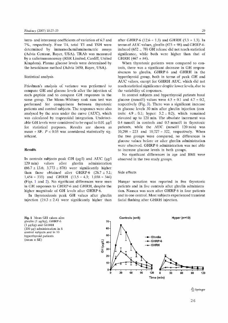
26
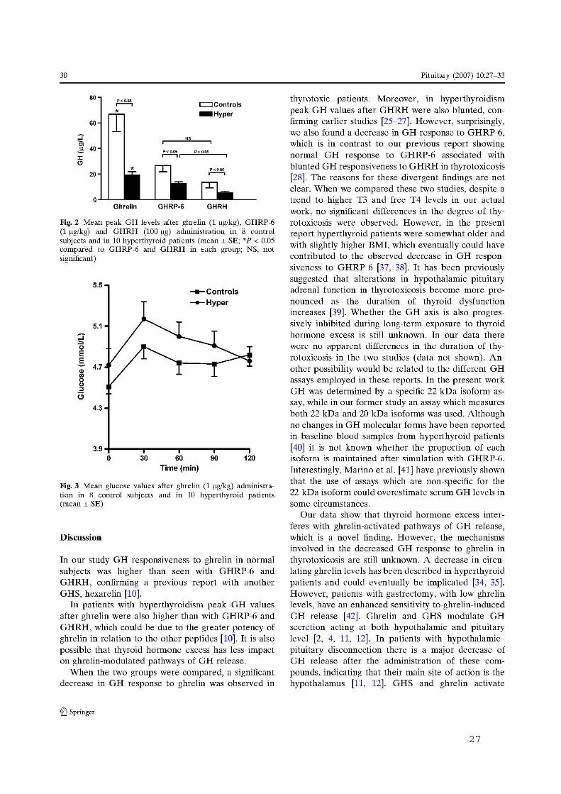
27
28
29
30
31
GHRELIN AND GHRP-6-INDUCED ACTH AND CORTISOL RELEASE IN THYROTOXICOSIS
Estudo 2
32
Ghrelin and GHRP-6-induced ACTH and cortisol release in thyrotoxicosis
Sergio O. Nascif, Patrícia Molica, Silvia R. Correa-Silva, Marcos R. Silva, Ana-
Maria J. Lengyel
Division of Endocrinology, Universidade Federal de São Paulo, UNIFESP/EPM,
São Paulo, Brazil
Abbreviated title: Ghrelin and HPA axis in thyrotoxicosis
Keywords: ghrelin, ACTH, cortisol, thyrotoxicosis
Corresponding author:
Sergio de Oliva Nascif
Rua Pedro de Toledo, 910
04039-002 – São Paulo/SP – Brazil
Phone/fax: 55-11-55748432
E-mail: [email protected]
33
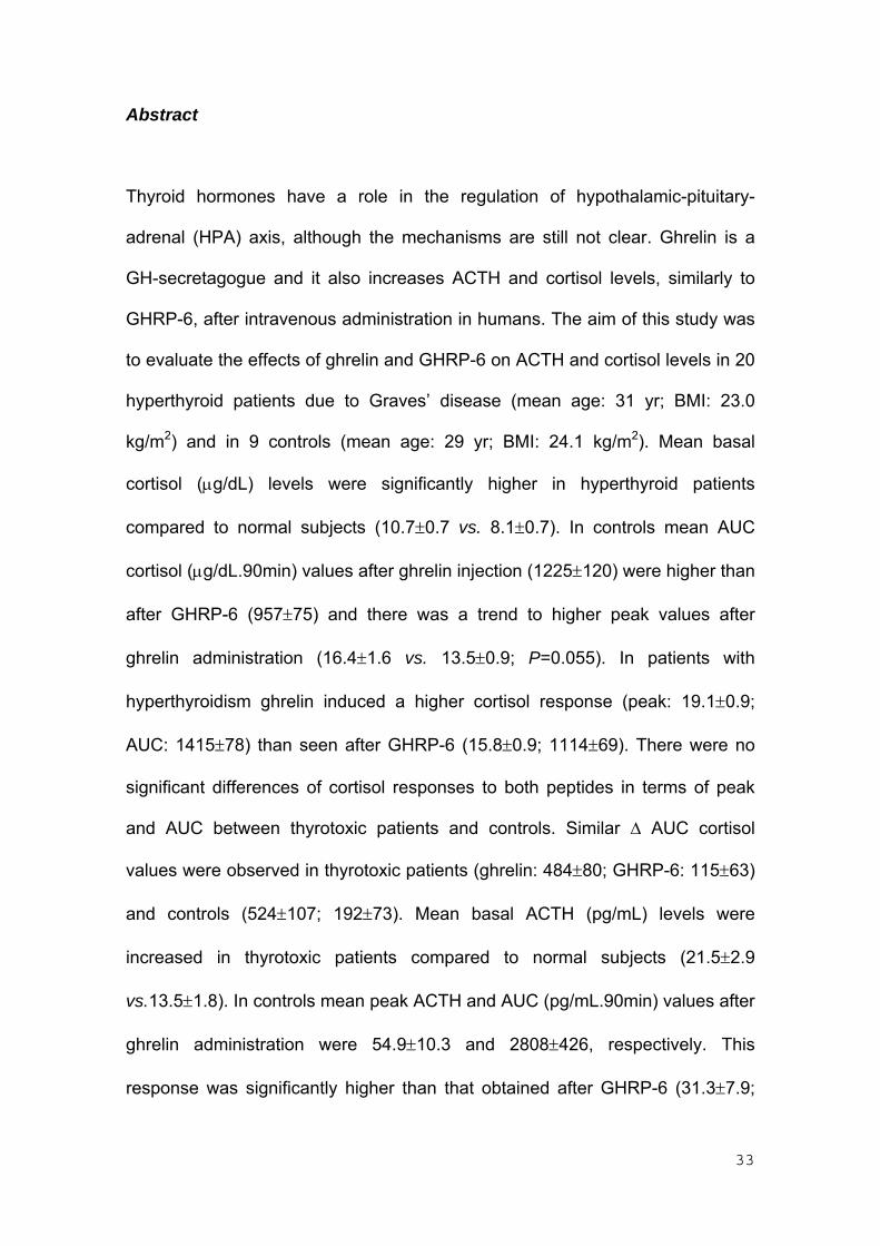
Abstract
Thyroid hormones have a role in the regulation of hypothalamic-pituitary-
adrenal (HPA) axis, although the mechanisms are still not clear. Ghrelin is a
GH-secretagogue and it also increases ACTH and cortisol levels, similarly to
GHRP-6, after intravenous administration in humans. The aim of this study was
to evaluate the effects of ghrelin and GHRP-6 on ACTH and cortisol levels in 20
hyperthyroid patients due to Graves’ disease (mean age: 31 yr; BMI: 23.0
kg/m2) and in 9 controls (mean age: 29 yr; BMI: 24.1 kg/m2). Mean basal
cortisol (μg/dL) levels were significantly higher in hyperthyroid patients
compared to normal subjects (10.7±0.7 vs. 8.1±0.7). In controls mean AUC
cortisol (μg/dL.90min) values after ghrelin injection (1225±120) were higher than
after GHRP-6 (957±75) and there was a trend to higher peak values after
ghrelin administration (16.4±1.6 vs. 13.5±0.9; P=0.055). In patients with
hyperthyroidism ghrelin induced a higher cortisol response (peak: 19.1±0.9;
AUC: 1415±78) than seen after GHRP-6 (15.8±0.9; 1114±69). There were no
significant differences of cortisol responses to both peptides in terms of peak
and AUC between thyrotoxic patients and controls. Similar ∆ AUC cortisol
values were observed in thyrotoxic patients (ghrelin: 484±80; GHRP-6: 115±63)
and controls (524±107; 192±73). Mean basal ACTH (pg/mL) levels were
increased in thyrotoxic patients compared to normal subjects (21.5±2.9
vs.13.5±1.8). In controls mean peak ACTH and AUC (pg/mL.90min) values after
ghrelin administration were 54.9±10.3 and 2808±426, respectively. This
response was significantly higher than that obtained after GHRP-6 (31.3±7.9;

34
1668±265), both in terms of peak ACTH and AUC levels. In thyrotoxicosis mean
peak ACTH and AUC values after ghrelin injection (149.7±39.8; 6209±1556)
were also significantly higher than after GHRP-6 injection (53.9±11.2;
2767±487). Ghrelin-induced peak ACTH release was increased in hyperthyroid
patients when compared to controls and there was a trend in terms of AUC
levels (P=0.063). Peak ACTH and AUC values after GHRP-6 in thyrotoxic
patients did not reach statistical significance compared to normal subjects.
When the ∆ AUC values were analyzed, there was a significant increase in
ACTH levels after ghrelin in the thyrotoxic group (patients: 4189±1202; controls:
1499±338). After GHRP-6 administration ∆ ACTH values in thyrotoxic patients
did not reach statistical significance compared to control subjects (patients:
927±330; controls: 539±237). In summary, our results show that cortisol
responsiveness to ghrelin and GHRP-6 is normal in thyrotoxicosis. ACTH
release after ghrelin is increased, but it does not reach statistical significance
with GHRP-6. Our results suggest that the pathways of ACTH release mediated
by ghrelin might be activated by thyroid hormone excess, but adrenocortical
reserve is maintained.
35
Introduction
The hypothalamic-pituitary-adrenal (HPA) axis is regulated mainly by
corticotropin-releasing hormone (CRH) and arginine vasopressin (AVP), which
stimulate the release of ACTH and cortisol. However, several neurotransmitters
and peptides could have an additional role in the modulation of CRH, AVP and
ACTH secretion (Melmed & Kleinberg, 2008).
Thyroid hormones may have a role in the regulation of HPA axis by
mechanisms that are not fully elucidated. There is an increase in the production
rate of cortisol in thyrotoxicosis because its degradation is accelerated (Levin &
Daughaday, 1955; Peterson, 1958; Gordon & Southren, 1977). Thus, in
prolonged hyperthyroidism the adrenals secrete at their maximal rate and,
depending on the severity/duration of the disease, cortisol responsiveness to
different stimuli such as hypoglycemia, ACTH and CRH might be decreased
(Jackson et al., 1966; Tsatsoulis et al., 2000; Goswami & Kochupillai, 2001;
Yamakita et al., 2001). However, normal cortisol responses to hypoglycemia
have also been observed (Jackson et al., 1966; Giustina et al., 1971; Brauman
et al., 1973; Moghetti et al., 1994). The impairment of adrenocortical reserve in
hyperthyroidism and its normalization after treatment could be due to a
decrease in cortisol-binding globulin (CBG) levels, as this globulin is reduced in
thyrotoxicosis and returns to normal in the euthyroid state (Dumoulin et al.,
1995; Mishra et al., 2007). However, corticosterone in the cerebrospinal fluid
and free cortisol index (cortisol/CBG), which reflect free cortisol, are increased
in hyperthyroidism (Kamilaris et al., 1991; Johnson et al., 2005; Mishra et al.,

36
2007), suggesting that high circulating cortisol levels are really present and are
due to enhanced secretion in this setting.
There are few data in the literature about basal and stimulated ACTH
levels in hyperthyroidism. Experimental studies have suggested that
thyrotoxicosis is associated with hyperactivity of the HPA axis (Kamilaris et al.,
1991; Johnson et al., 2005) and higher basal ACTH levels have been observed
in hyperthyroid subjects (Moghetti et al., 1994; Yamakita et al., 2001; Mishra et
al., 2007). It has also been shown that these patients have normal or higher
ACTH responsiveness to hypoglycemia or CRH stimulation test (Moghetti et al.,
1994; Yamakita et al., 2001; Lizcano & Salvador, 2008).
Ghrelin, the endogenous ligand of growth hormone secretagogue (GHS)
receptor (GHS-R), was discovered in the stomach, but is also present in the
hypothalamus, mainly in the arcuate nucleus (Kojima et al., 1999; Gnanapavan
et al., 2002; van der Lely et al., 2004). The active form of this peptide is
acylated and its chemical structure is different from GHS (Kojima et al., 1999).
Ghrelin and GH-releasing peptide-6 (GHRP-6), a GHS, induce GH, ACTH and
cortisol release (Arvat et al., 2001; Correa-Silva et al., 2006). Their main site of
action is the hypothalamus, as in hypothalamic-pituitary disconnection these
effects are reduced or abolished (Popovic et al., 1995; Popovic et al., 2003). A
direct adrenal action of these peptides on cortisol release has been suggested
as GHS-R mRNA expression was found in this tissue (Gnanapavan et al.,
2002), although in much lower concentrations than in the hypothalamic nuclei
(Bennett et al., 1997; Guan et al., 1997; Mozid et al., 2003; Korbonits et al.,
2004). However, this hypothesis seems unlikely as the effects of ghrelin/GHS
on cortisol release are abolished in hypothalamic-pituitary disconnection.

37
There are no data in the literature about the effect of thyroid hormone
excess on ACTH and cortisol release after the administration of ghrelin and
GHRP-6. Therefore, the aim of our study was to evaluate ACTH and cortisol
responses to ghrelin and GHRP-6 in thyrotoxic patients due to Graves´ disease.
38
Subjects and methods
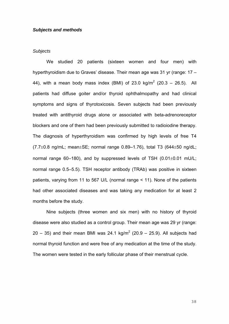
Subjects
We studied 20 patients (sixteen women and four men) with
hyperthyroidism due to Graves’ disease. Their mean age was 31 yr (range: 17 –
44), with a mean body mass index (BMI) of 23.0 kg/m2 (20.3 – 26.5). All
patients had diffuse goiter and/or thyroid ophthalmopathy and had clinical
symptoms and signs of thyrotoxicosis. Seven subjects had been previously
treated with antithyroid drugs alone or associated with beta-adrenoreceptor
blockers and one of them had been previously submitted to radioiodine therapy.
The diagnosis of hyperthyroidism was confirmed by high levels of free T4
(7.7±0.8 ng/mL; mean±SE; normal range 0.89–1.76), total T3 (644±50 ng/dL;
normal range 60–180), and by suppressed levels of TSH (0.01±0.01 mU/L;
normal range 0.5–5.5). TSH receptor antibody (TRAb) was positive in sixteen
patients, varying from 11 to 567 U/L (normal range < 11). None of the patients
had other associated diseases and was taking any medication for at least 2
months before the study.
Nine subjects (three women and six men) with no history of thyroid
disease were also studied as a control group. Their mean age was 29 yr (range:
20 – 35) and their mean BMI was 24.1 kg/m2 (20.9 – 25.9). All subjects had
normal thyroid function and were free of any medication at the time of the study.
The women were tested in the early follicular phase of their menstrual cycle.

39
Study protocol
The experimental protocol was approved by the ethics committee of
Universidade Federal de São Paulo, and all subjects were studied after giving
informed consent. The tests were performed after an overnight fast and the
subjects remained recumbent throughout it. Each subject underwent two tests,
randomly, with an interval of at least 48 h between them. Forty-five minutes
before starting the test, an indwelling catheter was inserted into an antecubital
vein and kept patent by slow 0.9% saline infusion. After the first blood sample
each subject received acylated ghrelin (Neosystem, Strasbourg, France) at a
dose of 1 μg/kg or GHRP-6 (Bachem, San Carlos, USA) at a dose of 1 μg/kg,
i.v., in bolus. Blood samples were collected every 15 minutes until 90 minutes
for subsequent ACTH and cortisol determination. Baseline blood samples were
also obtained for free T4, total T3 and TSH. Control subjects were also
submitted to the tests using the same procedure as above.
Assays
Serum ACTH was measured by an immunochemiluminometric assay
(DPC, Los Angeles, USA). The sensitivity of the method is 5 pg/mL, with mean
inter- and intra-assay coefficients of variation of 3.6% and 2.8%, respectively.
Serum cortisol levels were measured in duplicate by a fluoroimmunoassay
(Wallac Oy, Turku, Finland), with sensitivity of 0.2 μg/dL, and mean inter- and
intra-assay coefficients of variation of 8.2% and 6.2%, respectively. Free T4,
total T3 and TSH were determined by immunochemiluminometric assays (Advia
Centaur, Bayer, USA). TRAb was measured by a radioimmunoassay (RSR
Limited, Cardiff, United Kingdom).
40
Statistical analysis
Friedman’s analysis of variance was performed to compare ACTH and
cortisol levels after the injection of each peptide. Wilcoxon signed rank test was
used for comparisons of ACTH and cortisol values within the same group.
Mann-Whitney rank sum test was performed for comparisons between
thyrotoxic patients and control subjects. Mean basal levels were calculated
using all individual values obtained before the injection of each peptide. The
responses were also analyzed by the area under the curve (AUC), which was
calculated by trapezoidal integration. Delta (Δ) AUC values subtracting baseline
were also calculated when appropriate. The Spearman correlation coefficient
was calculated when appropriate. Results are shown as mean ± SE. P < 0.05
was considered statistically significant.

41
Results
No significant differences in age and BMI were observed in the two study
groups.
Mean basal cortisol (μg/dL) levels were significantly higher in
hyperthyroid patients (10.7±0.7) when compared to normal subjects (8.1±0.7).
In control subjects mean AUC cortisol (μg/dL.90min) values after ghrelin
injection (1225±120) were higher than after GHRP-6 (957±75). In terms of peak
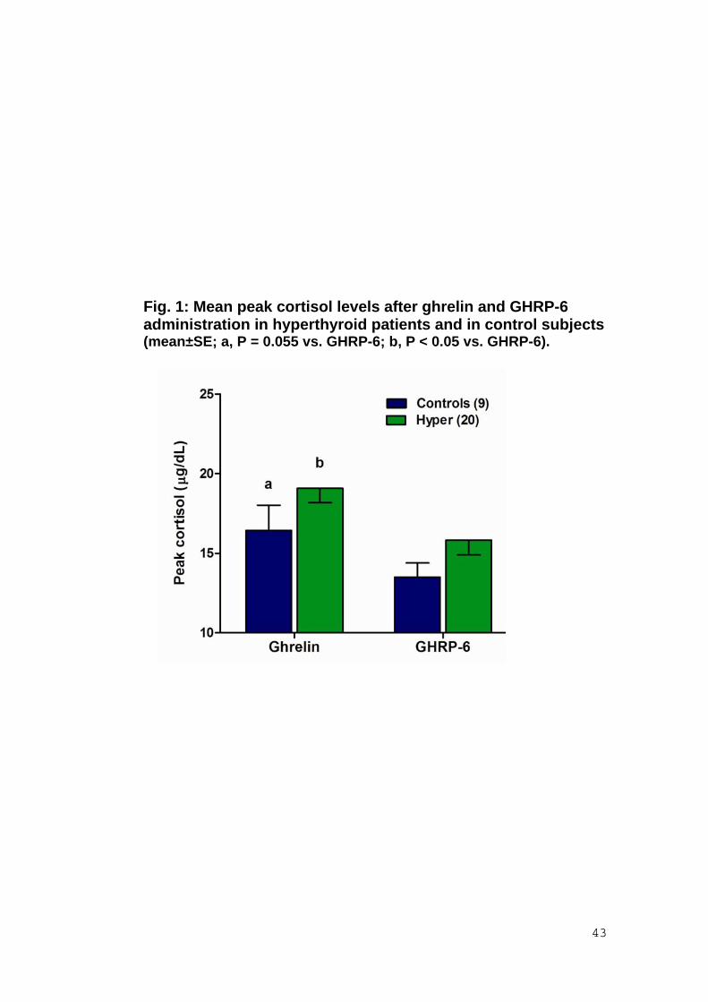
levels (Fig. 1), there was a trend to higher values after ghrelin administration
(16.4±1.6 vs. 13.5±0.9; P=0.055). In patients with hyperthyroidism ghrelin
induced a higher cortisol response (peak: 19.1±0.9; AUC: 1415±78) than seen
after GHRP-6 (15.8±0.9; 1114±69).
When patients with hyperthyroidism were compared to controls, there
were no significant differences of cortisol responses to both peptides in terms of
peak and AUC. Also, similar ∆ AUC cortisol values were observed in thyrotoxic
patients (ghrelin: 484±80; GHRP-6: 115±63) and controls (524±107; 192±73).
Mean basal ACTH (pg/mL) levels were increased in thyrotoxic patients
compared to normal subjects (21.5±2.9 vs.13.5±1.8). In controls mean peak
ACTH and AUC (pg/mL.90min) values after ghrelin administration were
54.9±10.3 and 2808±426, respectively (Figs. 2 and 3). This response was
significantly higher than that obtained after GHRP-6 (31.3±7.9; 1668±265), both
in terms of peak ACTH and AUC levels. In thyrotoxicosis mean peak ACTH and
AUC values after ghrelin injection (149.7±39.8; 6209±1556) were also
significantly higher than after GHRP-6 injection (53.9±11.2; 2767±487).
42
Ghrelin-induced peak ACTH release was increased in hyperthyroid
patients when compared to controls and there was a trend in terms of AUC
levels (P=0.063). Peak ACTH and AUC values after GHRP-6 in thyrotoxic
patients did not reach statistical significance compared to normal subjects.
When the ∆ AUC values were analyzed, there was a significant increase in
ACTH levels after ghrelin in the thyrotoxic group (patients: 4189±1202; controls:
1499±338). After GHRP-6 administration ∆ ACTH values in thyrotoxic patients
did not reach statistical significance compared to control subjects (patients:
927±330; controls: 539±237).
No significant correlations were found between free T4 and other
parameters and basal and stimulated cortisol and ACTH values.
Side effects
Hunger sensation, nausea, sleepiness and facial flushing were reported
occasionally after ghrelin administration. Facial flushing was the most prominent
symptom after GHRP-6 injection.
43
Fig. 1: Mean peak cortisol levels after ghrelin and GHRP-6 administration in hyperthyroid patients and in control subjects (mean±SE; a, P = 0.055 vs. GHRP-6; b, P < 0.05 vs. GHRP-6).

44
Fig. 2: Mean ACTH values after ghrelin and GHRP-6 administration in hyperthyroid patients and in control subjects (mean±SE).
Fig. 3: Mean peak ACTH levels after ghrelin and GHRP-6 administration in hyperthyroid patients and in control subjects (mean±SE; a, P < 0.05 vs. GHRP-6).

45
Discussion
In our study cortisol responsiveness to ghrelin in normal controls and in
thyrotoxic patients was higher than seen with GHRP-6. In normal subjects this
has been previously described (Correa-Silva et al., 2006) and was also
observed with another GHS, hexarelin (Arvat et al., 2001). This could be due to
the greater potency of ghrelin in relation to the synthetic analogues (Arvat et al.,
2001). It is also possible that ghrelin activates additional pathways to stimulate
the HPA axis (Wren et al., 2002; Mozid et al., 2003).
When thyrotoxic patients and controls were compared, no significant
differences in terms of peak and AUC cortisol levels were observed between
the two study groups, although mean basal cortisol levels were slightly higher in
hyperthyroid patients, confirming our previous findings (Molica et al., 2007).
Classically, thyroid hormones increase the conversion of cortisol to cortisone.
Therefore, the disposal of cortisol is accelerated, but as its rate of secretion is
also increased, plasma cortisol concentrations remain normal (Gordon &
Southren, 1977; Davies & Larsen, 2008). However, conflicting results about
fasting cortisol values have been reported in hyperthyroid patients, with slightly
higher (Jackson et al., 1966), normal (Moghetti et al., 1994; Yamakita et al.,
2001; Mishra et al., 2007; Lizcano & Salvador, 2008) or even reduced values
compared to normal subjects (Goswami & Kochupillai, 2001). Our finding of
slightly higher basal cortisol levels in hyperthyroidism is in agreement with
previous reports in humans and also in thyrotoxic rats, which have an increase
in circulating corticosterone values, the main glucocorticoid in rodents (Jackson
et al., 1966; Johnson et al., 2005). In addition, it has been suggested, in
46
experimentally-induced hyperthyroidism, that alterations in hypothalamic-
pituitary-adrenal function become more pronounced as the duration and
severity of thyroid dysfunction increases (Levin & Daughaday, 1955; Johnson et
al., 2005), but this information is lacking in thyrotoxic patients. Although there
are few data in the literature to allow this analysis, normal plasma cortisol
concentrations were found in patients who had lower T4 values (Mishra et al.,
2007) compared to our study group. Therefore, although no correlations were
found between cortisol and T4 values in our patients, it is possible that
differences in duration and/or clinical severity might explain the discrepancies in
mean basal cortisol levels in the literature.
The excessive catabolism of cortisol and continuing hyperactivity of the
HPA axis in thyrotoxicosis may result in exhaustion of adrenocortical reserve
(Gordon & Southren, 1977; Goswami & Kochupillai, 2001). Although
controversial, it has been suggested that severely thyrotoxic patients might
have an impaired cortisol response to insulin-induced hypoglycemia (Jackson et
al., 1966) and to both high and low-dose ACTH stimulation, associated or not
with dexamethasone (Tsatsoulis et al., 2000; Goswami & Kochupillai, 2001;
Mishra et al., 2007). After normalization of thyroid function this impairment
disappears, which suggests that adrenal autoimmune disease is an unlikely
cause for the reduced adrenocortical reserve (Goswami & Kochupillai, 2001;
Mishra et al., 2007). Moreover, it has been previously shown that in
hyperthyroidism there is a significant reduction in CBG (Dumoulin et al., 1995;
Mishra et al., 2007), which could decrease serum total cortisol measurements
and contribute to these findings. However, our results show that cortisol
responsiveness to ghrelin and GHRP-6 is similar in hyperthyroid patients

47
compared to normal subjects, suggesting that adrenocortical reserve is
preserved. This is in agreement with our previous observations (Molica et al.,
2007) and also with reports of normal cortisol responsiveness to hypoglycemia
and CRH in hyperthyroidism (Giustina et al., 1971; Brauman et al., 1973;
Moghetti et al., 1994; Lizcano & Salvador, 2008). It is possible that these
different findings are related to the variable clinical features of thyrotoxic
patients.
In our study, ghrelin and GHRP-6 were able to release ACTH in normal
subjects and in hyperthyroid patients, as previously reported (Frieboes et al.,
1995; Arvat et al., 2001; Correa-Silva et al., 2006, Molica et al., 2007). The
mean peak and AUC ACTH levels after ghrelin administration were significantly
higher than seen after GHRP-6 injection in the two study groups.
Interestingly, we observed an increase in basal ACTH values and also in
the ACTH response to ghrelin in thyrotoxicosis compared to normal subjects,
which did not reach statistical significance with GHRP-6. It has been previously
demonstrated that thyroid hormone excess stimulates the conversion of cortisol
to cortisone, which is biologically inactive and unable to inhibit pituitary function
(Gordon & Southren, 1977; Davies & Larsen, 2008). Therefore, there is an
enhancement in ACTH release, which could explain the increase in basal ACTH
values observed by us and by others (Moghetti et al., 1994; Yamakita et al.,
2001; Mishra et al., 2007). Also, ACTH values decrease after treatment in
hyperthyroid patients (Yamakita et al., 2001; Mishra et al., 2007). There are few
studies in the literature about ACTH responsiveness to stimulation in
hyperthyroidism. Normal and higher ACTH responses to hypoglycemia and
CRH have been observed both in short- and long-term thyrotoxicosis in humans

48
and in rats (Moghetti et al., 1994; Yamakita, 2001 et al.; Johnson et al., 2005;
Lizcano & Salvador, 2008). Some of these studies suggest hyperactivity of HPA
axis in hyperthyroidism, which support our findings of enhanced ACTH
responsiveness to ghrelin in hyperthyroid patients and also of a decrease in
ACTH values after ghrelin, GHRP-6 and CRH with normalization of thyroid
function (Molica et al., 2007; Lizcano & Salvador, 2008).
The central mechanisms modulating the increased ACTH release in
thyrotoxicosis are still unknown. It is possible that hypothalamic pathways,
which are activated by hypoglycemia, and/or pituitary sensitivity to CRH are
enhanced in this condition.
It has been shown that the main action of ghrelin/GHS on ACTH/cortisol
secretion is exerted at hypothalamic level as these effects are abolished after
hypothalamic-pituitary disconnection (Popovic et al., 1995; Popovic et al.,
2003). Moreover, GHS are not able to release ACTH in pituitary cells in vitro
(Elias et al., 1995; Kojima et al., 1999) and normal corticotrophs lack GHS-R
(Smith et al., 1997). It has been suggested that HPA activation induced by
these peptides probably occurs via stimulation of AVP and/or CRH release.
(Thomas et al., 1997; Korbonits et al., 1999a). Reinforcing this hypothesis, it
has been shown that GHS-R are located in the paraventricular and arcuate
nucleus of the hypothalamus, where AVP, CRH and NPY neurons are found
(Bennett et al., 1997; Guan et al., 1997; Korbonits et al., 2004). Experimental
studies have shown that GHRP-6 increases mainly AVP release from
hypothalamic fragments in vitro (Korbonits et al., 1999b), while ghrelin has
apparently a broader effect, enhancing AVP, CRH and NPY secretion, with a
predominant action on AVP (Wren et al., 2002; Mozid et al., 2003). Although
49
controversial (Kaji et al., 2001), thyroid hormones enhance the activity of the
human GHS-R promoter (Petersenn et al., 2001). Therefore, it is possible that
these pathways are activated in thyrotoxicosis, which might explain our findings.
Furthermore, it has been previously suggested that a centrally mediated
stimulation of ACTH release is present in hyperthyroidism, which is reinforced
by the finding of bilateral increase in the adrenal glands found both in thyrotoxic
patients and rats (Goswami & Kochupillai, 2001; Johnson et al., 2005), together
with high ACTH and cortisol/corticosterone levels (Moghetti et al., 1994;
Yamakita et al., 2001; Johnson et al., 2005; Mishra et al., 2007).
In summary, our results show that cortisol responsiveness to ghrelin and
GHRP-6 is normal in thyrotoxicosis. ACTH release after ghrelin is increased,
although not reaching statistical significance with GHRP-6. Our results suggest
that the pathways of ACTH release mediated by ghrelin might be activated by
thyroid hormone excess, but adrenocortical reserve is maintained.
50
Acknowledgements
We would like to thank Dr. Carlos Dieguez for the initial encouragement
and help. We are grateful to Ap. Filomena P. Machado and Walkiria Miranda for
the technical assistance. We would like to thank FAPESP (Fundação de
Amparo à Pesquisa do Estado de São Paulo) and CNPq (Conselho Nacional de
Desenvolvimento Científico e Tecnologia) for financial support.
51
References
Altinova AE, Törüner FB, Aktürk M, et al. 2006 Reduced serum acylated
ghrelin levels in patients with hyperthyroidism. Horm Res 65:295-299.
Arvat E, Maccario M, di Vito L, et al. 2001 Endocrine activities of ghrelin, a
natural growth hormone secretagogue (GHS), in humans: comparison and
interactions with hexarelin, a nonnatural peptidyl GHS, and GH-releasing
hormone. J Clin Endocrinol Metab 86:1169-1174.
Bennett PA, Thomas GB, Howard AD, et al. 1997 Hypothalamic growth
hormone secretagogue-receptor (GHS-R) expression is regulated by growth
hormone in the rat. Endocrinology 138:4552-4557.
Brauman H, Smets P, Corvilain J. 1973 Comparative study of growth
response to hypoglycemia in normal subjects and in patients with primary
myxedema or hyperthyroidism before and after treatment. J Clin Endocrinol
Metab 36:1162-1174.
Correa-Silva SR, Nascif SO, Lengyel AM. 2006 Decreased GH secretion and
enhanced ACTH and cortisol release after ghrelin administration in Cushing’s
disease: comparison with GH-releasing peptide-6 (GHRP-6) and GHRH.
Pituitary 9:101-107.

52
Davies TF & Larsen PR. 2008 Thyrotoxicosis. In Williams Textbook of
Endocrinology (ed. Kronenberg HM), pp. 333-375. Saunders Elsevier,
Philadelphia, PA.
Dumoulin SC, Perret BP, Bennet AP, Caron PJ. 1995 Opposite effects of
thyroid hormones on binding proteins for steroid hormones (sex hormone-
binding globulin and corticosteroid-binding globulin) in humans. Eur J
Endocrinol 132:594-598.
Elias KA, Ingle GS, Burnier JP, et al. 1995 In vitro characterization of four
novel classes of growth hormone-releasing peptide. Endocrinology 136:5694-
5699.
Frieboes RM, Murck H, Maier P, Schier T, Holsboer F, Steiger A. 1995
Growth hormone-releasing peptide-6 stimulates sleep, growth hormone, ACTH
and cortisol release in normal man. Neuroendocrinology 61:584-589.
Giménez-Palop O, Giménez-Péres G, Mauricio D, et al. 2005 Circulating
ghrelin in thyroid dysfunction is related to insulin resistance and not to hunger,
food intake or anthropometric changes. Eur J Endocrinol 153:73-79.
Giustina G, Reschini E, Valentini F, Cantalamessa L. 1971 Growth hormone
and cortisol responses to insulin-induced hypoglycemia in thyrotoxicosis. J Clin
Endocr 32:571-574.
53
Gnanapavan S, Kola B, Bustin SA, et al. 2002 The tissue distribution of the
mRNA of ghrelin and subtypes of its receptor, GHS-R, in humans. J Clin
Endocrinol Metab 87:2988-2991.
Gordon GG & Southren AL. 1977 Thyroid-hormone effects on steroid-
hormone metabolism. Bull N Y Acad Med 53:241-254.
Goswami R & Kochupillai N. 2001 Adrenocortical reserves in patients with
Graves’ disease. Eur J Endocrinol 144:85.
Guan X, Yu H, Palyha OC, et al. 1997 Distribution of mRNA encoding the
growth hormone secretagogues receptor in brain and peripheral tissues. Mol
Brain Res 48:23-29.
Jackson IMD, Hassan THA, Prentice CRM, Browning MCK. 1966 Insulin-
induced hypoglycemia as a test of pituitary-adrenal function in thyrotoxicosis. J
Clin Endocr 26:545-549.
Johnson EO, Kamilaris TC, Calogero AE, Gold PW, Chrousos GP. 2005
Experimentally-induced hyperthyroidism is associated with activation of the rat
hypothalamic-pituitary-adrenal axis. Eur J Endocrinol 153:177-185.
Kaji H, Kishimoto M, Kirimura T, et al. 2001 Hormonal regulation of the
human ghrelin receptor gene transcription. Biochem Biophys Res Commun
284:660-666.

54
Kamilaris TC, DeBold CR, Johnson EO, et al. 1991 Effects of short and long
duration hypothyroidism and hyperthyroidism on the plasma adrenocorticotropin
and corticosterone responses to ovine corticotropin-releasing hormone in rats.
Endocrinology 128:2567-2576.
Kojima M, Hosoda H, Date Y, Nakazato M, Matsuo H, Kangawa K. 1999
Ghrelin is a growth-hormone-releasing acylated peptide from stomach. Nature
402:656-660.
Korbonits M, Goldstone AP, Gueorguiev M, Grossman AB. 2004 Ghrelin – a
hormone with multiple functions. Front Neuroendocrinol 25:27-68.
Korbonits M, Kaltsas G, Perry LA, et al. 1999a The growth hormone
secretagogue hexarelin stimulates the hypothalamo-pituitary-adrenal axis via
arginine vasopressin. J Clin Endocrinol Metab 84:2489-2495.
Korbonits M, Little JA, Forsling ML, et al. 1999b The effect of growth
hormone secretagogues and neuropeptide Y on hypothalamic hormone release
from acute rat hypothalamic explants. J Neuroendocrinol 11:521-528.
Levin ME & Daughaday WH. 1955 The influence of the thyroid on
adrenocortical function. J Clin Endocrinol Metab 15:1494-1510.

55
Lizcano F & Salvador J. 2008 Effects of different treatments for
hyperthyroidism on the hypothalamic-pituitary-adrenal axis. Clin Exp Pharmacol
Physiol (Epub ahead of print).
Melmed S & Kleinberg D. 2008 Anterior Pituitary. In Williams Textbook of
Endocrinology (ed. Kronenberg HM), pp. 155-261. Saunders Elsevier,
Philadelphia, PA.
Mishra SK, Gupta N, Goswami R. 2007 Plasma adrenocorticotropin (ACTH)
values and cortisol response to 250 and 1 μg ACTH stimulation in patients with
hyperthyroidism before and after carbimazole therapy: case-control comparative
study. J Clin Endocrinol Metab 92:1693-1696.
Moghetti P, Castello R, Tosi F, et al. 1994 Glucose counterregulatory
response to acute hypoglycemia in hyperthyroid human subjects. J Clin
Endocrinol Metab 78:169-173.
Molica P, Nascif SO, Correa-Silva SR, Silva MR, Sa LBC, Lengyel AMJ.
2007 Increased GHRP-6 and ghrelin-induced ACTH release in thyrotoxicosis.
Program of the 89th Annual Meeting of The Endocrine Society, Toronto,
Canada, 2007, p348-349 (AbstractP2-89).
Mozid AM, Tringali G, Forsling ML, et al. 2003 Ghrelin is released from rat
hypothalamic explants and stimulates corticotrophin-releasing hormone and
arginine-vasopressin. Horm Metab Res 35:455-459.

56
Petersenn S, Rasch AC, Penshorn M, Beil FU, Schulte HM. 2001 Genomic
structure and transcriptional regulation of the human growth hormone
secretagogue receptor. Endocrinology 142:2649-2659.
Peterson RE. 1958 The influence of the thyroid on adrenal cortical function. J
Clin Invest 37:736-743.
Popovic V, Damjanovic S, Micic D, Djurovic M, Dieguez C, Casanueva FF.
1995 Blocked growth hormone-releasing peptide (GHRP-6)-induced GH
secretion and absence of the synergic action of GHRP-6 plus GH-releasing
hormone in patients with hypothalamopituitary disconnection: evidence that
GHRP-6 main action is exerted at the hypothalamic level. J Clin Endocrinol
Metab 80:942-947.
Popovic V, Miljic D, Micic D, et al. 2003 Ghrelin main action on the regulation
of growth hormone release is exerted at hypothalamic level. J Clin Endocrinol
Metab 88:3450-3453.
Riis ALD, Hansen TK, Moller N, Weeke J, Jorgensen JOL. 2003
Hyperthyroidism is associated with suppressed circulating ghrelin levels. J Clin
Endocrinol Metab 88:853-857.
Rojdmark S, Calissendorff J, Danielsson O, Brismar K. 2005 Hunger-satiety
signals in patients with Graves´ thyrotoxicosis before, during, and after long-
term pharmacological treatment. Endocrine 27:55-61.

57
Smith RG, Van der Ploeg LH, Howard AD, et al. 1997 Peptidomimetic
regulation of growth hormone secretion. Endocr Rev 18:621-645.
Thomas GB, Fairhall KM, Robinson ICAF. 1997 Activation of the
hypothalamic-pituitary-adrenal axis by the growth hormone (GH) secretagogue,
GH-releasing peptide-6, in rats. Endocrinology 138:1585-1591.
Tsatsoulis A, Johnson EO, Kalogera CH, Seferiadis K, Tsolas O. 2000 The
effect of thyrotoxicosis on adrenocortical reserve. Eur J Endocrinol 142:231-
235.
van der Lely AJ, Tschop M, Heiman ML, Ghigo E. 2004 Biological,
physiological, pathophysiological, and pharmacological aspects of ghrelin.
Endocr Rev 25:426-457.
Wren AM, Small CJ, Fribbens CV, et al. 2002 The hypothalamic mechanisms
of the hypophysiotropic action of ghrelin. Neuroendocrinology 76:316-324.
Yamakita N, Murai T, Kokubo Y, Hayashi M, Akai A, Yasuda K. 2001
Dehydroepiandrosterone sulphate is increased and dehydroepiandrosterone-
response to corticotrophin-releasing hormone is decreased in the hyperthyroid
state compared with the euthyroid state. Clin Endocrinol 55:797-803.

58
SUMÁRIO E CONCLUSÕES
59
SUMÁRIO E CONCLUSÕES Estudo 1
A ghrelina demonstrou ser um estímulo mais potente do que o GHRP-6 e o
GHRH para a secreção de GH em controles e em pacientes com
hipertireoidismo. Porém, a resposta do GH após todos os estímulos foi menor
na tireotoxicose quando comparada aos indivíduos normais. Portanto, o
excesso de hormônios tireoidianos interfere com as vias hipotalâmicas e
hipofisárias de liberação de GH ativadas por ghrelina, GHRP-6 e GHRH.
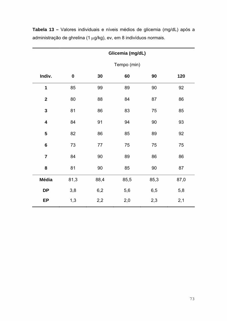
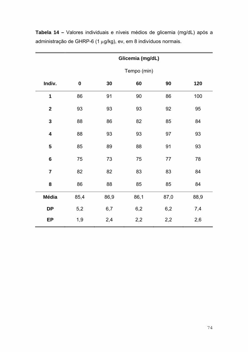
Os valores basais de glicemia dos pacientes com hipertireoidismo não
foram significativamente diferentes dos observados em controles. A ghrelina
promoveu um aumento similar nas concentrações plasmáticas de glicose em
ambos os grupos, enquanto que o GHRP-6 não alterou os níveis circulantes de
glicose. Portanto, o excesso de hormônios tireoidianos não interfere nos
mecanismos de liberação de glicose estimulados pela ghrelina.
Estudo 2 Foi observado um aumento nos valores basais de ACTH e de cortisol
nos pacientes com hipertireoidismo comparado com controles, sugerindo que o
excesso de hormônios tireoidianos interfere com o eixo hipotálamo-hipófise
adrenal.
A administração da ghrelina promoveu uma liberação maior de ACTH e
de cortisol em ambos os grupos, confirmando que a ghrelina é um estímulo
potente para ativação do eixo hipotálamo-hipófise adrenal.
A resposta do cortisol à ghrelina e ao GHRP-6 em pacientes com
hipertireoidismo foi semelhante a dos controles. A liberação de ACTH após
ghrelina na tireotoxicose foi maior que em indivíduos normais, enquanto que
com GHRP-6 os valores não alcançaram significância estatística.
Nossos dados sugerem que as vias de liberação de ACTH estimuladas
pela ghrelina são ativadas pelo excesso de hormônios tireoidianos, porém sem
repercussões significativas na resposta adrenocortical.

60
ANEXOS – Estudo 1
61
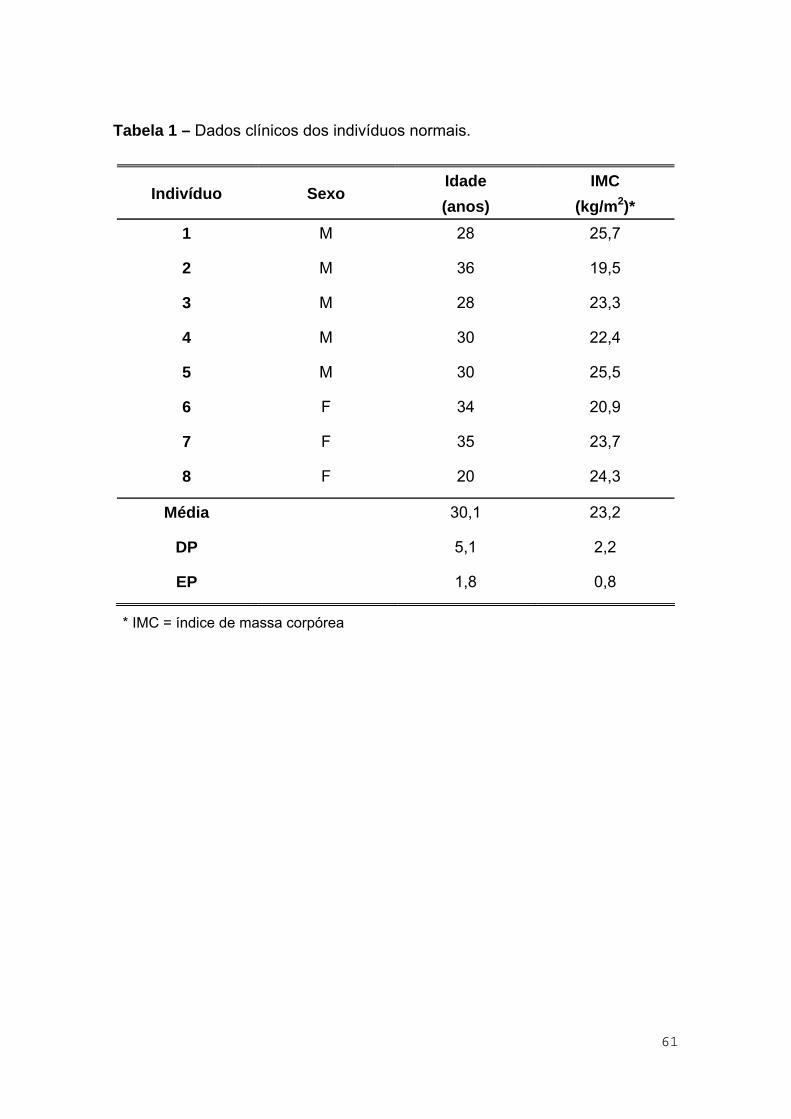
Tabela 1 – Dados clínicos dos indivíduos normais.
Indivíduo Sexo Idade (anos)
IMC (kg/m2)*
1 M 28 25,7
2 M 36 19,5
3 M 28 23,3
4 M 30 22,4
5 M 30 25,5
6 F 34 20,9
7 F 35 23,7
8 F 20 24,3
Média 30,1 23,2
DP 5,1 2,2
EP 1,8 0,8
* IMC = índice de massa corpórea
62
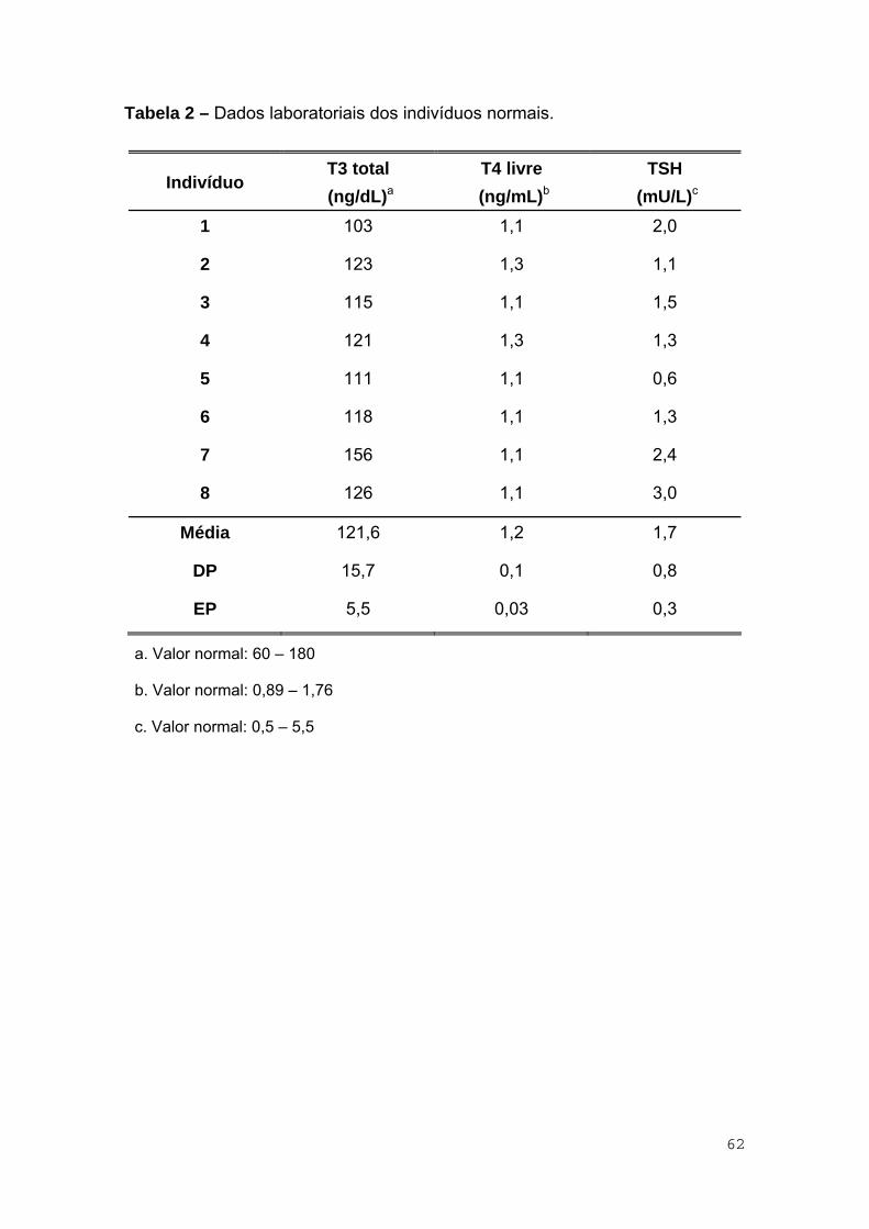
Tabela 2 – Dados laboratoriais dos indivíduos normais.
Indivíduo T3 total (ng/dL)a
T4 livre (ng/mL)b
TSH (mU/L)c
1 103 1,1 2,0
2 123 1,3 1,1
3 115 1,1 1,5
4 121 1,3 1,3
5 111 1,1 0,6
6 118 1,1 1,3
7 156 1,1 2,4
8 126 1,1 3,0
Média 121,6 1,2 1,7
DP 15,7 0,1 0,8
EP 5,5 0,03 0,3
a. Valor normal: 60 – 180
b. Valor normal: 0,89 – 1,76
c. Valor normal: 0,5 – 5,5
63
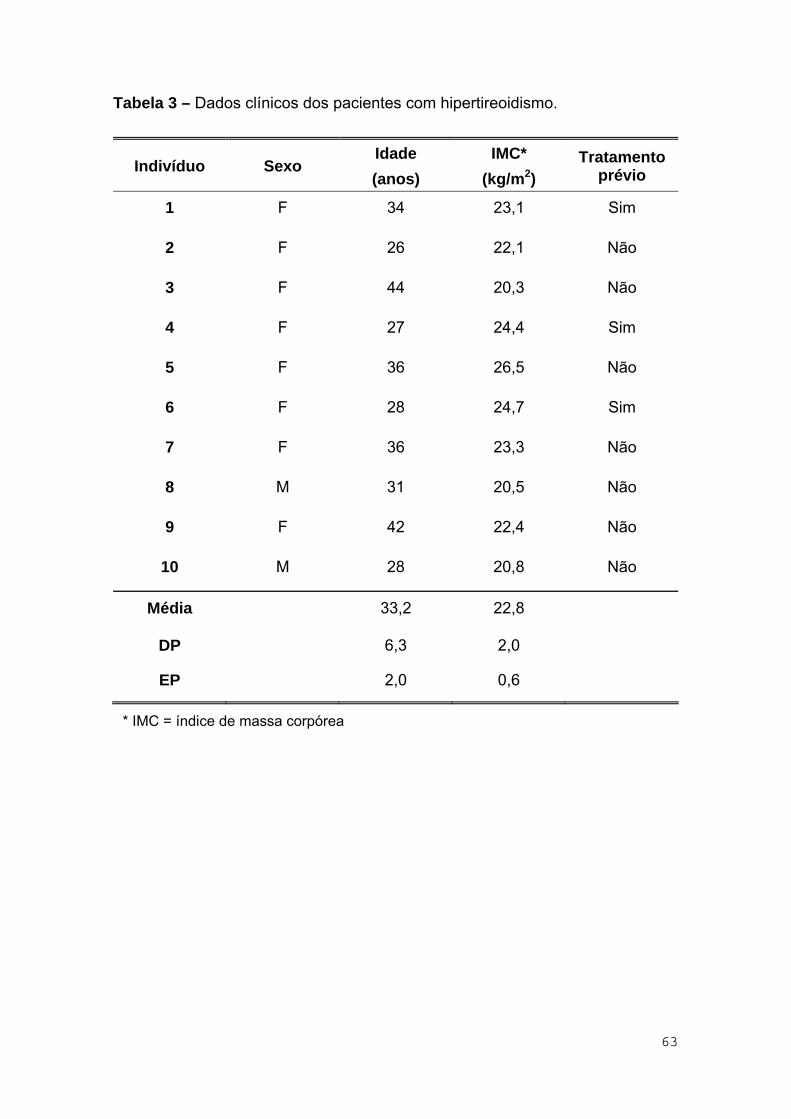
Tabela 3 – Dados clínicos dos pacientes com hipertireoidismo.
Indivíduo Sexo Idade (anos)
IMC* (kg/m2)
Tratamento prévio
1 F 34 23,1 Sim
2 F 26 22,1 Não
3 F 44 20,3 Não
4 F 27 24,4 Sim
5 F 36 26,5 Não
6 F 28 24,7 Sim
7 F 36 23,3 Não
8 M 31 20,5 Não
9 F 42 22,4 Não
10 M 28 20,8 Não
Média 33,2 22,8
DP 6,3 2,0
EP 2,0 0,6
* IMC = índice de massa corpórea
64
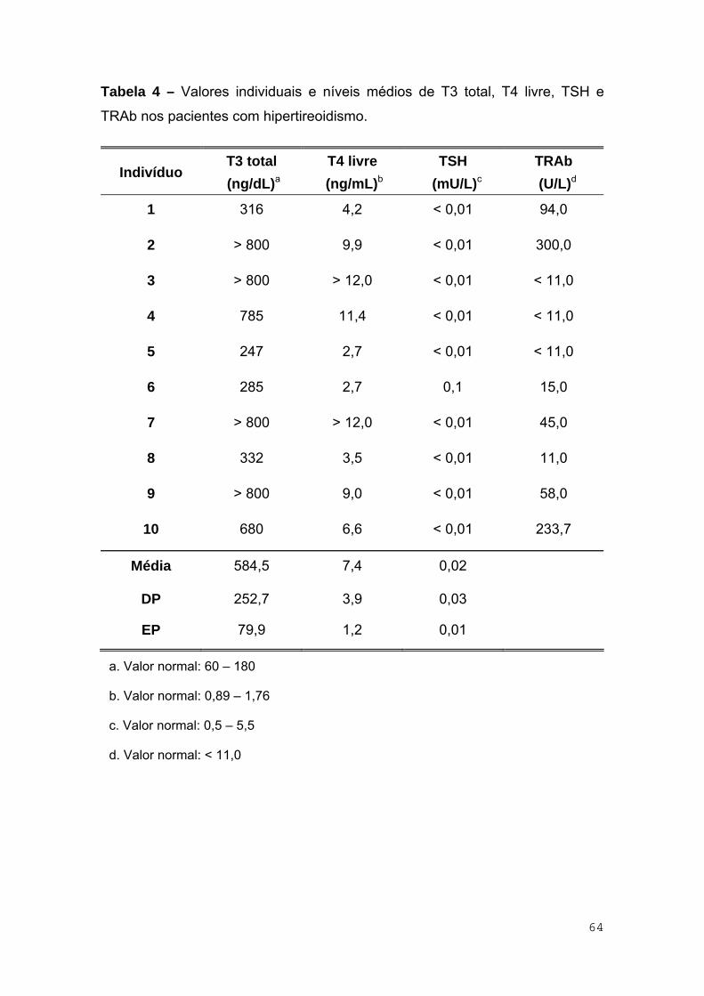
Tabela 4 – Valores individuais e níveis médios de T3 total, T4 livre, TSH e
TRAb nos pacientes com hipertireoidismo.
Indivíduo T3 total (ng/dL)a
T4 livre (ng/mL)b
TSH (mU/L)c
TRAb (U/L)d
1 316 4,2 < 0,01 94,0
2 > 800 9,9 < 0,01 300,0
3 > 800 > 12,0 < 0,01 < 11,0
4 785 11,4 < 0,01 < 11,0
5 247 2,7 < 0,01 < 11,0
6 285 2,7 0,1 15,0
7 > 800 > 12,0 < 0,01 45,0
8 332 3,5 < 0,01 11,0
9 > 800 9,0 < 0,01 58,0
10 680 6,6 < 0,01 233,7
Média 584,5 7,4 0,02
DP 252,7 3,9 0,03
EP 79,9 1,2 0,01
a. Valor normal: 60 – 180
b. Valor normal: 0,89 – 1,76
c. Valor normal: 0,5 – 5,5
d. Valor normal: < 11,0
65
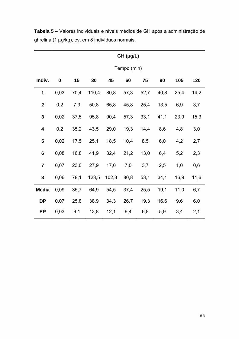
Tabela 5 – Valores individuais e níveis médios de GH após a administração de
ghrelina (1 μg/kg), ev, em 8 indivíduos normais.
GH (μg/L)
Tempo (min)
Indiv. 0 15 30 45 60 75 90 105 120
1 0,03 70,4 110,4 80,8 57,3 52,7 40,8 25,4 14,2
2 0,2 7,3 50,8 65,8 45,8 25,4 13,5 6,9 3,7
3 0,02 37,5 95,8 90,4 57,3 33,1 41,1 23,9 15,3
4 0,2 35,2 43,5 29,0 19,3 14,4 8,6 4,8 3,0
5 0,02 17,5 25,1 18,5 10,4 8,5 6,0 4,2 2,7
6 0,08 16,8 41,9 32,4 21,2 13,0 6,4 5,2 2,3
7 0,07 23,0 27,9 17,0 7,0 3,7 2,5 1,0 0,6
8 0,06 78,1 123,5 102,3 80,8 53,1 34,1 16,9 11,6
Média 0,09 35,7 64,9 54,5 37,4 25,5 19,1 11,0 6,7
DP 0,07 25,8 38,9 34,3 26,7 19,3 16,6 9,6 6,0
EP 0,03 9,1 13,8 12,1 9,4 6,8 5,9 3,4 2,1
66
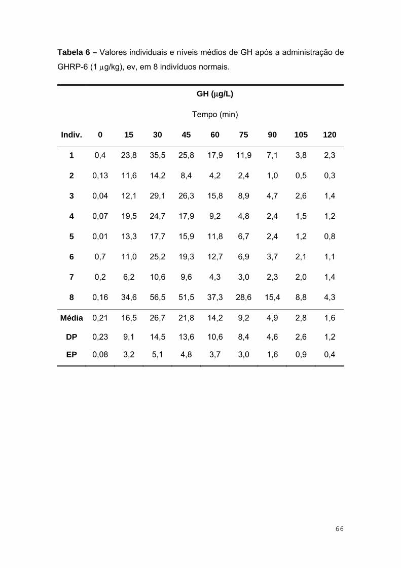
Tabela 6 – Valores individuais e níveis médios de GH após a administração de
GHRP-6 (1 μg/kg), ev, em 8 indivíduos normais.
GH (μg/L)
Tempo (min)
Indiv. 0 15 30 45 60 75 90 105 120
1 0,4 23,8 35,5 25,8 17,9 11,9 7,1 3,8 2,3
2 0,13 11,6 14,2 8,4 4,2 2,4 1,0 0,5 0,3
3 0,04 12,1 29,1 26,3 15,8 8,9 4,7 2,6 1,4
4 0,07 19,5 24,7 17,9 9,2 4,8 2,4 1,5 1,2
5 0,01 13,3 17,7 15,9 11,8 6,7 2,4 1,2 0,8
6 0,7 11,0 25,2 19,3 12,7 6,9 3,7 2,1 1,1
7 0,2 6,2 10,6 9,6 4,3 3,0 2,3 2,0 1,4
8 0,16 34,6 56,5 51,5 37,3 28,6 15,4 8,8 4,3
Média 0,21 16,5 26,7 21,8 14,2 9,2 4,9 2,8 1,6
DP 0,23 9,1 14,5 13,6 10,6 8,4 4,6 2,6 1,2
EP 0,08 3,2 5,1 4,8 3,7 3,0 1,6 0,9 0,4

67
Tabela 7 – Valores individuais e níveis médios de GH após a administração de
GHRH (100 μg), ev, em 8 indivíduos normais.
GH (μg/L)
Tempo (min)
Indiv. 0 15 30 45 60 75 90 105 120
1 0,03 15,8 19,3 28,8 37,6 38,5 30,8 18,7 11,0
2 0,04 3,4 3,6 2,2 2,3 1,8 1,2 0,8 0,8
3 0,03 8,3 9,2 6,8 7,5 7,3 5,7 3,8 2,4
4 0,03 6,2 5,0 4,0 3,5 2,7 2,2 1,4 1,1
5 0,01 3,9 4,8 5,4 4,4 3,2 1,9 1,2 0,6
6 0,06 7,5 8,1 6,6 4,8 2,4 1,4 0,8 0,4
7 0,13 16,0 21,5 24,8 22,6 19,8 13,7 18,2 13,8
8 0,6 8,0 8,1 11,0 8,2 9,4 12,5 7,1 5,3
Média 0,12 8,6 10,0 11,2 11,4 10,6 8,7 6,5 4,4
DP 0,2 4,8 6,8 10,0 12,4 12,7 10,2 7,7 5,2
EP 0,07 1,7 2,4 3,5 4,4 4,5 3,6 2,7 1,8
68
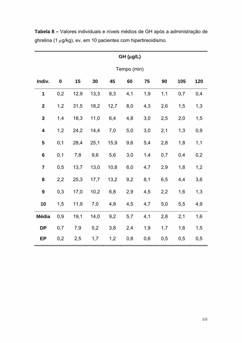
Tabela 8 – Valores individuais e níveis médios de GH após a administração de
ghrelina (1 μg/kg), ev, em 10 pacientes com hipertireoidismo.
GH (μg/L)
Tempo (min)
Indiv. 0 15 30 45 60 75 90 105 120
1 0,2 12,9 13,3 8,3 4,1 1,9 1,1 0,7 0,4
2 1,2 31,5 18,2 12,7 8,0 4,3 2,6 1,5 1,3
3 1,4 18,3 11,0 6,4 4,8 3,0 2,5 2,0 1,5
4 1,2 24,2 14,4 7,0 5,0 3,0 2,1 1,3 0,9
5 0,1 28,4 25,1 15,9 9,6 5,4 2,8 1,8 1,1
6 0,1 7,8 9,6 5,6 3,0 1,4 0,7 0,4 0,2
7 0,5 13,7 13,0 10,8 6,0 4,7 2,9 1,8 1,2
8 2,2 25,3 17,7 13,2 9,2 8,1 6,5 4,4 3,6
9 0,3 17,0 10,2 6,8 2,9 4,5 2,2 1,6 1,3
10 1,5 11,9 7,0 4,8 4,5 4,7 5,0 5,5 4,9
Média 0,9 19,1 14,0 9,2 5,7 4,1 2,8 2,1 1,6
DP 0,7 7,9 5,2 3,8 2,4 1,9 1,7 1,6 1,5
EP 0,2 2,5 1,7 1,2 0,8 0,6 0,5 0,5 0,5
69
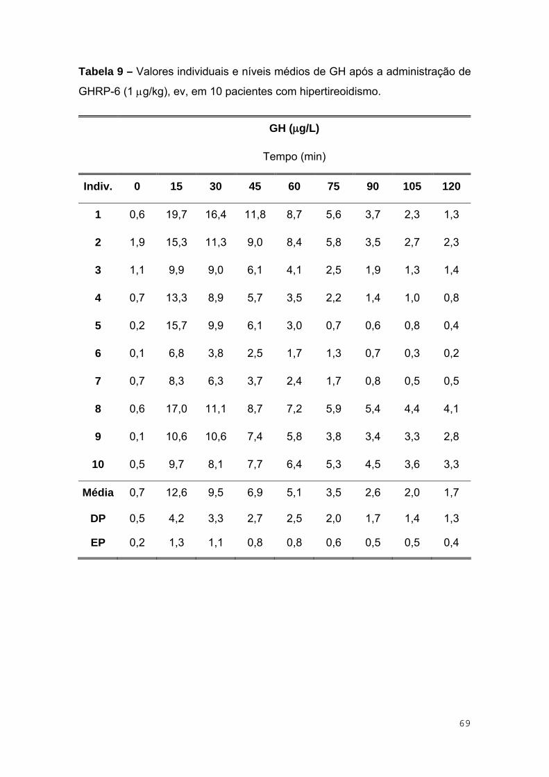
Tabela 9 – Valores individuais e níveis médios de GH após a administração de
GHRP-6 (1 μg/kg), ev, em 10 pacientes com hipertireoidismo.
GH (μg/L)
Tempo (min)
Indiv. 0 15 30 45 60 75 90 105 120
1 0,6 19,7 16,4 11,8 8,7 5,6 3,7 2,3 1,3
2 1,9 15,3 11,3 9,0 8,4 5,8 3,5 2,7 2,3
3 1,1 9,9 9,0 6,1 4,1 2,5 1,9 1,3 1,4
4 0,7 13,3 8,9 5,7 3,5 2,2 1,4 1,0 0,8
5 0,2 15,7 9,9 6,1 3,0 0,7 0,6 0,8 0,4
6 0,1 6,8 3,8 2,5 1,7 1,3 0,7 0,3 0,2
7 0,7 8,3 6,3 3,7 2,4 1,7 0,8 0,5 0,5
8 0,6 17,0 11,1 8,7 7,2 5,9 5,4 4,4 4,1
9 0,1 10,6 10,6 7,4 5,8 3,8 3,4 3,3 2,8
10 0,5 9,7 8,1 7,7 6,4 5,3 4,5 3,6 3,3
Média 0,7 12,6 9,5 6,9 5,1 3,5 2,6 2,0 1,7
DP 0,5 4,2 3,3 2,7 2,5 2,0 1,7 1,4 1,3
EP 0,2 1,3 1,1 0,8 0,8 0,6 0,5 0,5 0,4
70
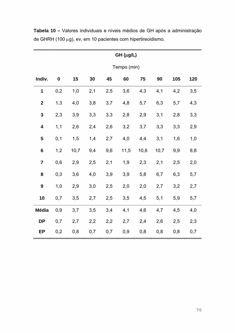
Tabela 10 – Valores individuais e níveis médios de GH após a administração
de GHRH (100 μg), ev, em 10 pacientes com hipertireoidismo.
GH (μg/L)
Tempo (min)
Indiv. 0 15 30 45 60 75 90 105 120
1 0,2 1,0 2,1 2,5 3,6 4,3 4,1 4,2 3,5
2 1,3 4,0 3,8 3,7 4,8 5,7 6,3 5,7 4,3
3 2,3 3,9 3,3 3,3 2,8 2,9 3,1 2,8 3,3
4 1,1 2,6 2,4 2,6 3,2 3,7 3,3 3,3 2,9
5 0,1 1,5 1,4 2,7 4,0 4,4 3,1 1,6 1,0
6 1,2 10,7 9,4 9,6 11,5 10,6 10,7 9,9 8,8
7 0,6 2,9 2,5 2,1 1,9 2,3 2,1 2,5 2,0
8 0,3 3,6 4,0 3,9 3,9 5,8 6,7 6,3 5,7
9 1,0 2,9 3,0 2,5 2,0 2,0 2,7 3,2 2,7
10 0,7 3,5 2,7 2,5 3,5 4,5 5,1 5,9 5,7
Média 0,9 3,7 3,5 3,4 4,1 4,6 4,7 4,5 4,0
DP 0,7 2,7 2,2 2,2 2,7 2,4 2,6 2,5 2,3
EP 0,2 0,8 0,7 0,7 0,9 0,8 0,8 0,8 0,7
71
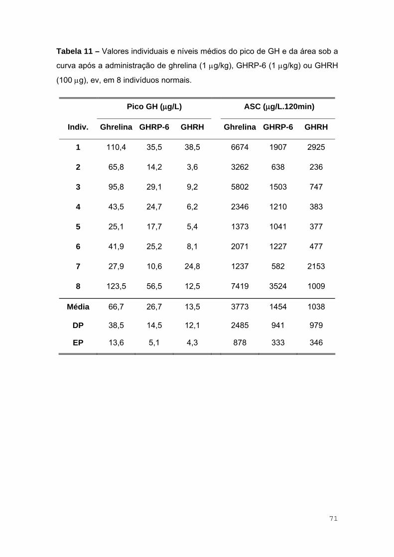
Tabela 11 – Valores individuais e níveis médios do pico de GH e da área sob a
curva após a administração de ghrelina (1 μg/kg), GHRP-6 (1 μg/kg) ou GHRH
(100 μg), ev, em 8 indivíduos normais.
Pico GH (μg/L) ASC (μg/L.120min)
Indiv. Ghrelina GHRP-6 GHRH Ghrelina GHRP-6 GHRH
1 110,4 35,5 38,5 6674 1907 2925
2 65,8 14,2 3,6 3262 638 236
3 95,8 29,1 9,2 5802 1503 747
4 43,5 24,7 6,2 2346 1210 383
5 25,1 17,7 5,4 1373 1041 377
6 41,9 25,2 8,1 2071 1227 477
7 27,9 10,6 24,8 1237 582 2153
8 123,5 56,5 12,5 7419 3524 1009
Média 66,7 26,7 13,5 3773 1454 1038
DP 38,5 14,5 12,1 2485 941 979
EP 13,6 5,1 4,3 878 333 346

72
Tabela 12 – Valores individuais e níveis médios do pico de GH e da área sob a
curva após a administração de ghrelina (1 μg/kg), GHRP-6 (1 μg/kg) ou GHRH
(100 μg), ev, em 10 pacientes com hipertireoidismo.
Pico GH (μg/L) ASC (μg/L.120min)
Indiv. Ghrelina GHRP-6 GHRH Ghrelina GHRP-6 GHRH
1 13,3 19,7 4,3 639 1037 355
2 31,5 15,3 6,3 1201 872 552
3 18,3 9,9 3,9 742 541 374
4 24,2 13,3 3,7 871 551 346
5 28,4 15,7 4,4 1344 557 289
6 9,6 6,8 11,5 430 259 1161
7 13,7 8,3 2,9 806 365 264
8 25,3 17,0 6,7 1310 931 559
9 17,0 10,6 3,2 690 695 302
10 11,9 9,7 5,9 695 708 464
Média 19,3 12,6 5,3 873 652 467
DP 7,6 4,2 2,5 309 247 265
EP 2,4 1,3 1,3 98 78 84
73
Tabela 13 – Valores individuais e níveis médios de glicemia (mg/dL) após a
administração de ghrelina (1 μg/kg), ev, em 8 indivíduos normais.
Glicemia (mg/dL)
Tempo (min)
Indiv. 0 30 60 90 120
1 85 99 89 90 92
2 80 88 84 87 86
3 81 86 83 75 85
4 84 91 94 90 93
5 82 86 85 89 92
6 73 77 75 75 75
7 84 90 89 86 86
8 81 90 85 90 87
Média 81,3 88,4 85,5 85,3 87,0
DP 3,8 6,2 5,6 6,5 5,8
EP 1,3 2,2 2,0 2,3 2,1
74
Tabela 14 – Valores individuais e níveis médios de glicemia (mg/dL) após a
administração de GHRP-6 (1 μg/kg), ev, em 8 indivíduos normais.
Glicemia (mg/dL)
Tempo (min)
Indiv. 0 30 60 90 120
1 86 91 90 86 100
2 93 93 93 92 95
3 88 86 82 85 84
4 88 93 93 97 93
5 85 89 88 91 93
6 75 73 75 77 78
7 82 82 83 83 84
8 86 88 85 85 84
Média 85,4 86,9 86,1 87,0 88,9
DP 5,2 6,7 6,2 6,2 7,4
EP 1,9 2,4 2,2 2,2 2,6

75
Tabela 15 – Valores individuais e níveis médios de glicemia (mg/dL) após a
administração de ghrelina (1 μg/kg), ev, em 10 pacientes com hipertireoidismo.
Glicemia (mg/dL)
Tempo (min)
Indiv. 0 30 60 90 120
1 84 89 91 90 86
2 82 91 89 87 85
3 64 71 71 70 71
4 92 109 100 98 92
5 95 96 95 94 97
6 87 95 94 91 89
7 88 98 93 93 83
8 95 98 96 97 95
9 81 85 83 81 76
10 83 101 90 85 84
Média 85,1 93,3 90,2 88,6 85,8
DP 9,0 10,3 8,1 8,4 8,1
EP 2,9 3,2 2,6 2,7 2,6
76
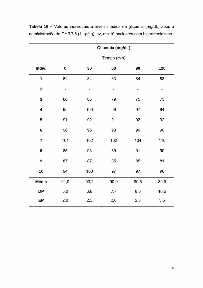
Tabela 16 – Valores individuais e níveis médios de glicemia (mg/dL) após a
administração de GHRP-6 (1 μg/kg), ev, em 10 pacientes com hipertireoidismo.
Glicemia (mg/dL)
Tempo (min)
Indiv. 0 30 60 90 120
1 82 84 83 84 83
2 - - - - -
3 88 85 79 75 73
4 95 100 99 97 94
5 91 92 91 92 92
6 96 96 93 90 90
7 101 102 102 104 110
8 85 93 89 91 90
9 87 87 85 85 81
10 94 100 97 97 96
Média 91,0 93,2 90,9 90,6 89,9
DP 6,0 6,8 7,7 8,5 10,5
EP 2,0 2,3 2,6 2,8 3,5

77
Tabela 17 – Valores individuais e níveis médios do pico de glicemia e da área
sob a curva após a administração de ghrelina (1 μg/kg) ou GHRP-6 (1 μg/kg),
ev, em 8 indivíduos normais.
Pico glicemia (mg/dL) ASC (mg/dL.120min)
Indiv. Ghrelina GHRP-6 Ghrelina GHRP-6
1 99 100 10995 10800
2 88 95 10260 11160
3 86 86 9810 10170
4 94 97 10905 11205
5 92 93 10410 10710
6 77 78 9030 9045
7 90 84 10500 9930
8 90 88 10470 10290
Média 89,5 90,1 10298 10414
DP 10,7 7,4 631 717
EP 3,8 2,6 223 254

78
Tabela 18 – Valores individuais e níveis médios do pico de glicemia e da área
sob a curva após a administração de ghrelina (1 μg/kg) ou GHRP-6 (1 μg/kg),
ev, em 10 pacientes com hipertireoidismo.
Pico glicemia (mg/dL) ASC (mg/dL.120min)
Indiv. Ghrelina GHRP-6 Ghrelina GHRP-6
1 91 84 10650 10005
2 91 - 10515 -
3 71 85 8385 9585
4 109 100 11970 11715
5 97 92 11430 10995
6 95 96 11040 11160
7 98 110 11085 12405
8 98 93 11580 11115
9 85 87 9825 10230
10 101 100 10785 11670
Média 93,6 94,1 10727 10987
DP 10,2 8,4 1019 905
EP 3,2 2,8 322 302

79
ANEXOS – Estudo 2
80
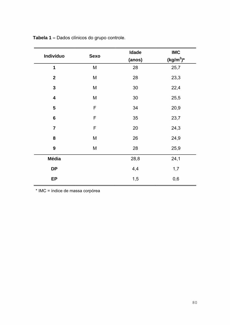
Tabela 1 – Dados clínicos do grupo controle.
Indivíduo Sexo Idade (anos)
IMC (kg/m2)*
1 M 28 25,7
2 M 28 23,3
3 M 30 22,4
4 M 30 25,5
5 F 34 20,9
6 F 35 23,7
7 F 20 24,3
8 M 26 24,9
9 M 28 25,9
Média 28,8 24,1
DP 4,4 1,7
EP 1,5 0,6
* IMC = índice de massa corpórea

81
Tabela 2 – Valores individuais e níveis médios das dosagens basais do grupo
controle.
Indivíduo TSH
(mU/L)a ACTH
(pg/mL)b Cortisol (µg/dL)c
1 2,0 7,3 9,7
2 1,5 27,6 12
3 1,3 7,7 10,1
4 0,6 8,1 8,7
5 1,3 14,4 7,4
6 2,4 8,5 4,7
7 3,0 9,6 7,5
8 1,3 22,2 8,4
9 1,2 16,6 5,1
Média 1,6 13,6 8,2
DP 0,7 7,3 2,3
EP 0,2 2,4 0,8
a. Valor normal: 0,5 – 5,5
b. Valor normal: até 46,0
c. Valor normal: 7,0 – 25,0
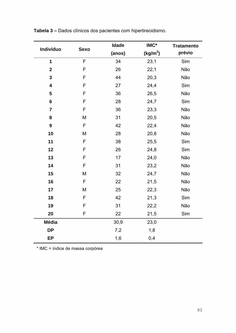
82
Tabela 3 – Dados clínicos dos pacientes com hipertireoidismo.
Indivíduo Sexo Idade (anos)
IMC* (kg/m2)
Tratamento prévio
1 F 34 23,1 Sim
2 F 26 22,1 Não
3 F 44 20,3 Não
4 F 27 24,4 Sim
5 F 36 26,5 Não
6 F 28 24,7 Sim
7 F 36 23,3 Não
8 M 31 20,5 Não
9 F 42 22,4 Não
10 M 28 20,8 Não
11 F 38 25,5 Sim
12 F 26 24,8 Sim
13 F 17 24,0 Não
14 F 31 23,2 Não
15 M 32 24,7 Não
16 F 22 21,5 Não
17 M 25 22,3 Não
18 F 42 21,3 Sim
19 F 31 22,2 Não
20 F 22 21,5 Sim
Média 30,9 23,0
DP 7,2 1,8
EP 1,6 0,4
* IMC = índice de massa corpórea
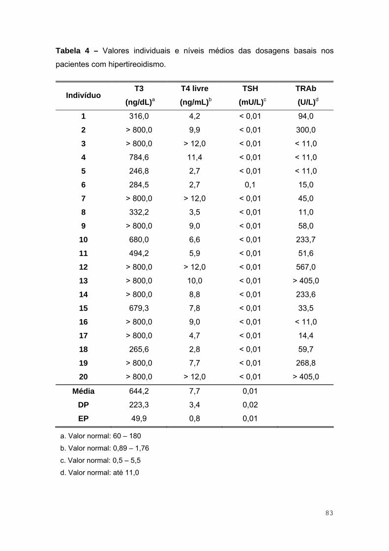
83
Tabela 4 – Valores individuais e níveis médios das dosagens basais nos
pacientes com hipertireoidismo.
Indivíduo T3
(ng/dL)a
T4 livre
(ng/mL)b
TSH
(mU/L)c
TRAb
(U/L)d
1 316,0 4,2 < 0,01 94,0
2 > 800,0 9,9 < 0,01 300,0
3 > 800,0 > 12,0 < 0,01 < 11,0
4 784,6 11,4 < 0,01 < 11,0
5 246,8 2,7 < 0,01 < 11,0
6 284,5 2,7 0,1 15,0
7 > 800,0 > 12,0 < 0,01 45,0
8 332,2 3,5 < 0,01 11,0
9 > 800,0 9,0 < 0,01 58,0
10 680,0 6,6 < 0,01 233,7
11 494,2 5,9 < 0,01 51,6
12 > 800,0 > 12,0 < 0,01 567,0
13 > 800,0 10,0 < 0,01 > 405,0
14 > 800,0 8,8 < 0,01 233,6
15 679,3 7,8 < 0,01 33,5
16 > 800,0 9,0 < 0,01 < 11,0
17 > 800,0 4,7 < 0,01 14,4
18 265,6 2,8 < 0,01 59,7
19 > 800,0 7,7 < 0,01 268,8
20 > 800,0 > 12,0 < 0,01 > 405,0
Média 644,2 7,7 0,01
DP 223,3 3,4 0,02
EP 49,9 0,8 0,01
a. Valor normal: 60 – 180
b. Valor normal: 0,89 – 1,76
c. Valor normal: 0,5 – 5,5
d. Valor normal: até 11,0
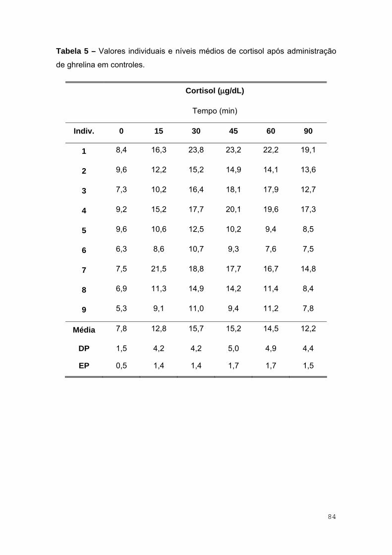
84
Tabela 5 – Valores individuais e níveis médios de cortisol após administração
de ghrelina em controles.
Cortisol (μg/dL)
Tempo (min)
Indiv. 0 15 30 45 60 90
1 8,4 16,3 23,8 23,2 22,2 19,1
2 9,6 12,2 15,2 14,9 14,1 13,6
3 7,3 10,2 16,4 18,1 17,9 12,7
4 9,2 15,2 17,7 20,1 19,6 17,3
5 9,6 10,6 12,5 10,2 9,4 8,5
6 6,3 8,6 10,7 9,3 7,6 7,5
7 7,5 21,5 18,8 17,7 16,7 14,8
8 6,9 11,3 14,9 14,2 11,4 8,4
9 5,3 9,1 11,0 9,4 11,2 7,8
Média 7,8 12,8 15,7 15,2 14,5 12,2
DP 1,5 4,2 4,2 5,0 4,9 4,4
EP 0,5 1,4 1,4 1,7 1,7 1,5

85
Tabela 6 – Valores individuais e níveis médios de cortisol após administração
de GHRP-6 em controles.
Cortisol (μg/dL)
Tempo (min)
Indiv. 0 15 30 45 60 90
1 10,9 13,3 16,2 16,3 12,7 10,6
2 14,3 15,6 15,7 16,8 13,6 13,0
3 12,9 12,5 11,6 10,6 9,6 7,5
4 8,2 12,6 11,7 10,3 9,5 8,0
5 5,1 10,3 8,8 6,6 5,9 6,9
6 3,0 9,0 7,1 6,3 7,7 7,8
7 7,4 11,2 12,9 15,3 12,0 9,1
8 9,8 12,7 11,4 10,4 9,0 7,8
9 4,9 15,6 12,2 11,5 9,0 7,2
Média 8,5 12,5 12,0 11,6 9,9 8,7
DP 3,8 2,2 2,9 3,9 2,5 2,0
EP 1,3 0,7 1,0 1,3 0,8 0,7

86
Tabela 7 – Valores individuais e níveis médios de ACTH após a administração
de ghrelina em controles.
ACTH (pg/mL)
Tempo (min)
Indiv. 0 15 30 45 60 90
1 7,6 66,1 42,4 30,2 25,0 12,4
2 33,4 67,1 58,9 47,4 38,1 25,4
3 5,0 21,4 32,4 25,1 16,5 5,0
4 9,5 60,0 57,5 33,4 26,3 13,5
5 15,7 21,9 19,6 16,5 12,8 11,9
6 10,0 19,2 21,4 16,2 14,0 11,0
7 11,1 30,3 28,3 21,5 16,5 9,6
8 23,6 82,6 68,6 51,6 34,7 20,5
9 15,0 112,0 74,2 48,5 30,5 14,2
Média 14,5 53,4 44,8 32,4 23,8 13,7
DP 8,9 32,4 20,6 13,9 9,4 6,0
EP 3,0 10,8 6,9 4,6 3,1 2,0

87
Tabela 8 – Valores individuais e níveis médios de ACTH após a administração
de GHRP-6 em controles.
ACTH (pg/mL)
Tempo (min)
Indiv. 0 15 30 45 60 90
1 6,9 49,4 29,4 19,9 15,3 9,6
2 21,8 27,3 22,0 18,9 17,1 14,9
3 10,4 10,8 8,5 7,0 7,8 7,6
4 6,7 13,6 12,8 10,4 7,7 6,5
5 13,1 21,0 16,5 14,0 13,2 19,0
6 7,0 20,0 15,0 14,5 16,4 19,0
7 8,1 24,0 18,3 15,0 13,0 11,0
8 20,8 28,9 22,6 19,0 16,8 14,6
9 18,1 87,1 56,1 34,1 21,7 12,5
Média 12,5 31,3 22,4 17,0 14,3 12,7
DP 6,2 23,7 14,0 7,7 4,5 4,5
EP 2,1 7,9 4,7 2,6 1,5 1,5

88
Tabela 9 – Valores individuais e níveis médios de cortisol após a administração
de ghrelina em pacientes com hipertireoidismo.
Cortisol (μg/dL)
Tempo (min)
Indiv. 0 15 30 45 60 90
1 9,9 19,8 20,5 22,1 21,6 17,4
2 2,5 11,3 15,4 18,4 19,0 16,5
3 12,5 21,6 22,7 26,1 28,0 27,9
4 8,4 14,5 14,1 12,1 9,4 4,8
5 8,6 16,1 18,8 20,1 16,8 12,8
6 7,0 12,1 16,1 17,3 14,8 11,7
7 10,1 16,5 19,4 10,3 18,3 12,7
8 7,5 16,5 22,3 21,5 18,6 13,1
9 14,9 12,4 13,9 12,6 10,4 10,8
10 16,7 20,7 21,3 22,8 23,4 21,5
11 7,6 13,9 16,4 17,9 16,6 11,5
12 8,8 13,8 14,9 15,9 13,5 9,1
13 7,4 12,4 13,3 13,5 9,4 5,1
14 13,5 17,1 18,3 15,9 17,3 8,0
15 18,6 20,4 20,9 22,4 21,9 15,9
16 6,4 10,8 14,2 14,9 18,2 9,7
17 6,8 11,5 14,5 14,6 10,9 6,7
18 20,7 22,6 24,9 21,3 17,9 11,0
19 11,9 16,0 19,4 20,1 21,6 15,1
20 7,1 9,9 14,3 14,7 13,2 11,2
Média 10,3 15,5 17,8 17,7 17,0 12,6
DP 4,6 3,9 3,5 4,2 5,0 5,5
EP 1,0 0,9 0,8 1,2 1,1 1,2

89
Tabela 10 – Valores individuais e níveis médios de cortisol após a
administração de GHRP-6 em pacientes com hipertireoidismo.
Cortisol (μg/dL)
Tempo (min)
Indiv. 0 15 30 45 60 90
1 15,7 18,5 15,2 11,7 10,3 7,2
2 5,9 11,7 15,3 16,4 16,0 11,1
3 11,0 17,1 15,0 11,8 9,9 13,0
4 9,3 13,5 12,6 10,7 11,0 6,5
5 9,7 15,4 12,6 10,5 7,9 6,7
6 11,2 10,9 8,9 7,7 6,9 6,7
7 6,3 10,7 10,0 8,6 10,2 10,4
8 9,2 19,9 22,0 17,7 15,7 11,3
9 10,8 12,1 12,1 11,3 10,0 9,8
10 15,6 17,8 20,7 20,7 19,0 14,2
11 12,3 14,7 16,9 14,5 13,2 19,2
12 10,6 15,4 16,0 14,8 16,3 10,1
13 6,2 11,7 13,9 12,2 10,5 6,5
14 13,5 15,1 15,6 13,6 10,9 5,8
15 12,7 14,7 13,8 11,5 9,6 11,5
16 11,6 13,7 13,6 8,8 7,1 7,6
17 15,2 20,1 21,5 18,4 14,6 9,5
18 11,7 16,9 17,7 15,4 11,5 7,2
19 20,2 21,1 20,6 15,2 14,5 8,7
20 3,3 5,7 4,0 2,7 2,7 1,0
Média 11,1 14,8 14,9 12,7 11,4 9,2
DP 3,9 3,8 4,4 4,2 3,9 3,8
EP 0,9 0,8 1,0 0,9 0,9 0,8

90
Tabela 11 – Valores individuais e níveis médios de ACTH após administração
de ghrelina em pacientes com hipertireoidismo.
ACTH (pg/mL)
Tempo (min)
Indiv. 0 15 30 45 60 90
1 19,4 98,6 74,6 57,4 48,7 27,5
2 6,7 181,0 146,0 80,2 45,4 28,7
3 20,0 336,0 205,0 120,0 87,2 61,4
4 13,6 74,7 57,1 25,0 17,7 10,1
5 10,8 44,0 47,1 24,7 18,3 12,3
6 7,0 23,0 28,5 18,9 12,1 5,8
7 13,1 140,0 100,0 64,0 33,2 19,0
8 12,0 130,0 78,3 54,0 32,0 18,0
9 5,2 67,5 52,0 41,0 23,0 16,4
10 94,2 817,0 559,0 349,0 225,0 90,8
11 12,2 103,0 60,1 36,3 23,4 12,0
12 20,4 160,0 105,0 67,8 42,9 26,1
13 9,6 28,4 18,2 15,4 11,0 8,3
14 23,0 62,5 45,0 33,7 24,4 16,5
15 71,3 287,0 152,0 91,7 61,3 28,1
16 11,1 50,6 53,9 40,0 25,2 16,4
17 12,9 52,9 45,8 32,1 22,2 13,9
18 61,8 202,0 118,0 66,7 30,0 15,5
19 12,7 66,4 61,7 39,9 27,1 16,2
20 12,0 58,3 47,9 37,6 29,4 18,9
Média 22,5 149,1 102,8 64,8 42,0 23,1
DP 24,1 178,3 117,1 71,9 46,7 19,8
EP 5,4 39,9 26,2 16,1 10,4 4,4

91
Tabela 12 – Valores individuais e níveis médios de ACTH após administração
de GHRP-6 em pacientes com hipertireoidismo.
ACTH (pg/mL)
Tempo (min)
Indiv. 0 15 30 45 60 90
1 13,3 16,6 12,3 11,3 7,4 8,1
2 12,3 121,0 92,4 64,8 46,3 25,9
3 13,5 41,7 29,3 22,7 17,1 24,9
4 16,8 44,9 27,7 23,2 22,2 12,7
5 14,0 19,5 16,1 12,4 10,2 8,5
6 12,5 12,6 10,3 9,1 10,1 9,0
7 9,0 14,3 14,5 16,0 15,6 16,0
8 18,5 81,5 45,0 35,0 26,0 15,0
9 23,0 55,3 45,0 33,0 29,0 22,0
10 60,2 222,0 155,0 96,0 84,7 57,8
11 23,7 49,6 32,5 25,8 20,1 18,7
12 20,8 60,7 49,5 41,9 39,4 27,8
13 13,1 23,8 25,2 21,5 14,8 11,2
14 23,6 31,6 25,6 23,1 17,4 14,0
15 26,5 32,3 27,5 22,9 20,4 22,2
16 15,7 30,4 19,9 14,1 18,3 14,4
17 29,7 112,0 61,9 40,2 24,8 14,8
18 29,5 59,7 51,8 38,3 29,0 18,6
19 25,8 34,8 22,1 14,4 11,3 10,5
20 7,5 10,4 8,3 7,9 6,2 6,9
Média 20,5 53,7 38,6 28,7 23,5 18,0
DP 11,5 50,1 34,2 21,1 17,6 11,2
EP 2,6 11,2 7,6 4,7 3,9 2,5
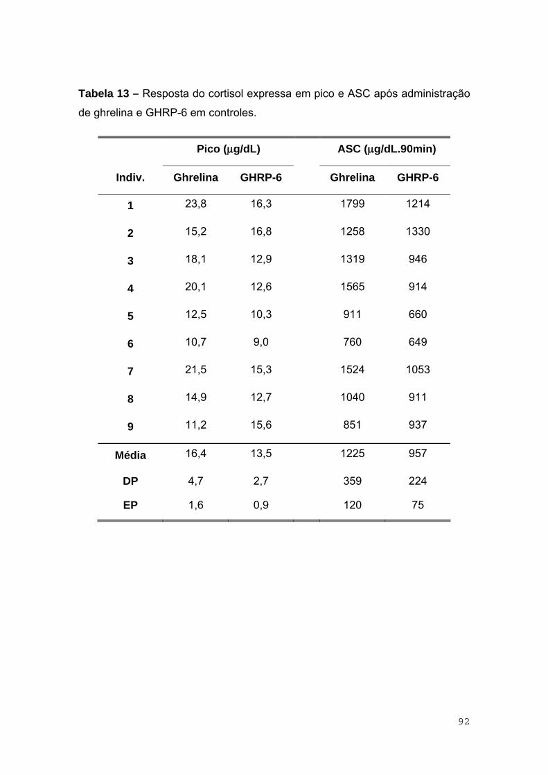
92
Tabela 13 – Resposta do cortisol expressa em pico e ASC após administração
de ghrelina e GHRP-6 em controles.
Pico (μg/dL) ASC (μg/dL.90min)
Indiv. Ghrelina GHRP-6 Ghrelina GHRP-6
1 23,8 16,3 1799 1214
2 15,2 16,8 1258 1330
3 18,1 12,9 1319 946
4 20,1 12,6 1565 914
5 12,5 10,3 911 660
6 10,7 9,0 760 649
7 21,5 15,3 1524 1053
8 14,9 12,7 1040 911
9 11,2 15,6 851 937
Média 16,4 13,5 1225 957
DP 4,7 2,7 359 224
EP 1,6 0,9 120 75
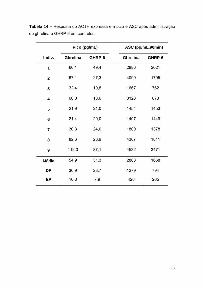
93
Tabela 14 – Resposta do ACTH expressa em pcio e ASC após administração
de ghrelina e GHRP-6 em controles.
Pico (pg/mL) ASC (pg/mL.90min)
Indiv. Ghrelina GHRP-6 Ghrelina GHRP-6
1 66,1 49,4 2886 2021
2 67,1 27,3 4090 1795
3 32,4 10,8 1667 762
4 60,0 13,6 3128 873
5 21,9 21,0 1454 1453
6 21,4 20,0 1407 1449
7 30,3 24,0 1800 1378
8 82,6 28,9 4307 1811
9 112,0 87,1 4532 3471
Média 54,9 31,3 2808 1668
DP 30,9 23,7 1279 794
EP 10,3 7,9 426 265
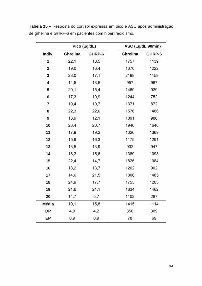
94
Tabela 15 – Resposta do cortisol expressa em pico e ASC após administração
de grhelina e GHRP-6 em pacientes com hipertireoidismo.
Pico (μg/dL) ASC (μg/dL.90min)
Indiv. Ghrelina GHRP-6 Ghrelina GHRP-6
1 22,1 18,5 1757 1139
2 19,0 16,4 1370 1222
3 28,0 17,1 2198 1159
4 14,5 13,5 957 967
5 20,1 15,4 1460 929
6 17,3 10,9 1244 752
7 19,4 10,7 1371 872
8 22,3 22,0 1576 1486
9 13,9 12,1 1091 986
10 23,4 20,7 1946 1646
11 17,9 19,2 1326 1369
12 15,9 16,3 1175 1291
13 13,5 13,9 932 947
14 18,3 15,6 1380 1098
15 22,4 14,7 1826 1084
16 18,2 13,7 1202 902
17 14,6 21,5 1006 1485
18 24,9 17,7 1755 1205
19 21,6 21,1 1634 1462
20 14,7 5,7 1102 287
Média 19,1 15,8 1415 1114
DP 4,0 4,2 350 309
EP 0,9 0,9 78 69

95
Tabela 16 – Resposta do ACTH expressa em pico e ASC após administração
de ghrelina e GHRP-6 em pacientes com hipertireoidismo.
Pico (pg/mL) ASC (pg/mL.90min)
Indiv. Ghrelina GHRP-6 Ghrelina GHRP-6
1 98,6 16,6 5113 991
2 181,0 121,0 7610 5696
3 336,0 41,7 12948 2265
4 74,7 44,9 3004 2253
5 47,1 19,5 2414 1182
6 28,5 12,6 1468 936
7 140,0 16,0 5690 1331
8 130,0 81,5 5015 3371
9 67,5 55,3 3210 3155
10 817,0 222,0 33006 10319
11 103,0 49,6 3789 2529
12 160,0 60,7 6502 3741
13 28,4 25,2 1374 1657
14 62,5 31,6 3087 1983
15 287,0 32,3 10296 2231
16 53,9 30,4 3064 1712
17 52,9 112,0 2767 4214
18 202,0 59,7 7172 3400
19 66,4 34,8 3468 1675
20 58,3 10,4 3192 698
Média 149,7 53,9 6209 2767
DP 178,0 50,0 6957 2176
EP 39,8 11,2 1556 487
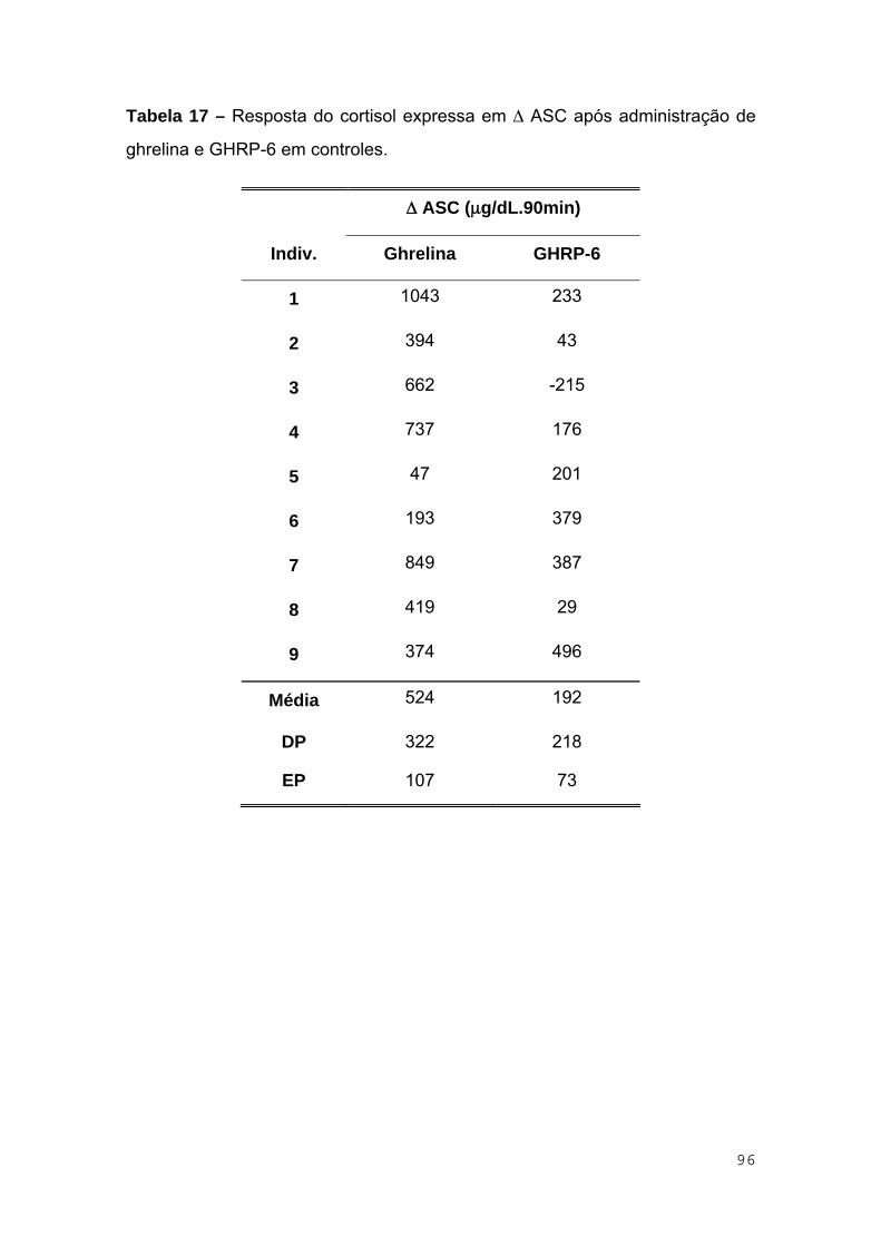
96
Tabela 17 – Resposta do cortisol expressa em ∆ ASC após administração de
ghrelina e GHRP-6 em controles.
∆ ASC (μg/dL.90min)
Indiv. Ghrelina GHRP-6
1 1043 233
2 394 43
3 662 -215
4 737 176
5 47 201
6 193 379
7 849 387
8 419 29
9 374 496
Média 524 192
DP 322 218
EP 107 73
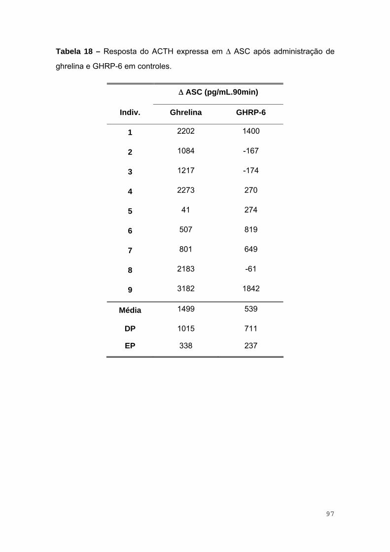
97
Tabela 18 – Resposta do ACTH expressa em ∆ ASC após administração de
ghrelina e GHRP-6 em controles.
∆ ASC (pg/mL.90min)
Indiv. Ghrelina GHRP-6
1 2202 1400
2 1084 -167
3 1217 -174
4 2273 270
5 41 274
6 507 819
7 801 649
8 2183 -61
9 3182 1842
Média 1499 539
DP 1015 711
EP 338 237
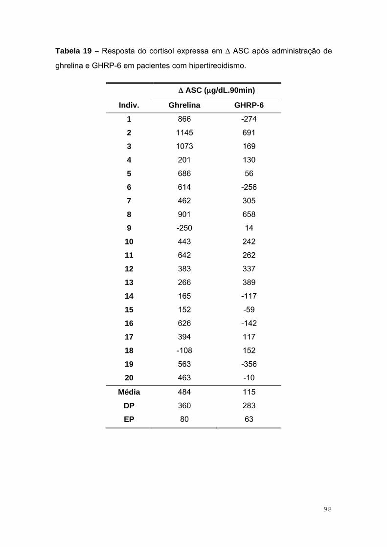
98
Tabela 19 – Resposta do cortisol expressa em ∆ ASC após administração de
ghrelina e GHRP-6 em pacientes com hipertireoidismo.
∆ ASC (μg/dL.90min)
Indiv. Ghrelina GHRP-6
1 866 -274
2 1145 691
3 1073 169
4 201 130
5 686 56
6 614 -256
7 462 305
8 901 658
9 -250 14
10 443 242
11 642 262
12 383 337
13 266 389
14 165 -117
15 152 -59
16 626 -142
17 394 117
18 -108 152
19 563 -356
20 463 -10
Média 484 115
DP 360 283
EP 80 63
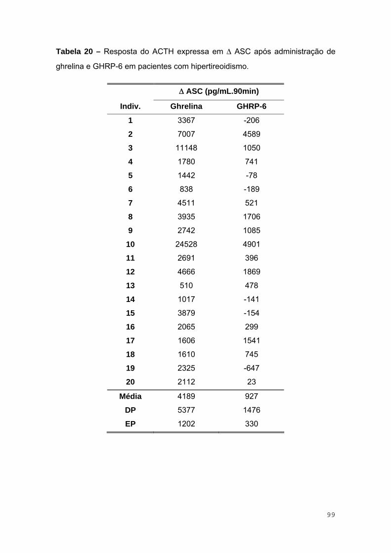
99
Tabela 20 – Resposta do ACTH expressa em ∆ ASC após administração de
ghrelina e GHRP-6 em pacientes com hipertireoidismo.
∆ ASC (pg/mL.90min)
Indiv. Ghrelina GHRP-6
1 3367 -206
2 7007 4589
3 11148 1050
4 1780 741
5 1442 -78
6 838 -189
7 4511 521
8 3935 1706
9 2742 1085
10 24528 4901
11 2691 396
12 4666 1869
13 510 478
14 1017 -141
15 3879 -154
16 2065 299
17 1606 1541
18 1610 745
19 2325 -647
20 2112 23
Média 4189 927
DP 5377 1476
EP 1202 330
Recommended