www.elsevier.com/locate/ynbdiNeurobiology of Disease 27 (2007) 1–10
Review
Functional MAPT haplotypes: Bridging the gap between genotypeand neuropathology
Tara M. Caffrey and Richard Wade-Martins⁎
The Wellcome Trust Centre for Human Genetics, University of Oxford, Roosevelt Drive, Oxford, OX3 7BN UK
Received 23 January 2007; revised 17 April 2007; accepted 27 April 2007Available online 5 May 2007
The microtubule-associated protein tau (MAPT) locus has long beenassociated with sporadic neurodegenerative disease, notably progres-sive supranuclear palsy and corticobasal degeneration, and morerecently with Alzheimer’s disease and Parkinson’s disease. However,the functional biological mechanisms behind the genetic associationhave only now started to emerge. The genomic architecture in theregion spanning MAPT is highly complex, and includes a ∼1.8 Mbblock of linkage disequilibrium (LD). The region is divided into twomajor haplotypes, H1 and H2, defined by numerous single nucleotidepolymorphisms and a 900 kb inversion which suppresses recombina-tion. Fine mapping of the MAPT region has identified sub-clades of theMAPT H1 haplotype which are specifically associated with neurode-generative disease. Here we briefly review the role ofMAPT in sporadicand familial neurodegenerative disease, and then discuss recent workwhich, for the first time, proposes functional mechanisms to linkMAPT haplotypes with the neuropathology seen in patients.© 2007 Elsevier Inc. All rights reserved.
Keywords: MAPT; H1 haplotype; Progressive supranuclear palsy; Tauo-pathy; Splicing; Gene expression; Functional polymorphisms; Suscept-ibility mechanisms
Contentsworldwide and that this number will double to 42 million by 2020(Ferri et al., 2005). Dementias and movement disorders like
Introduction . . . . . . . . . . . . . . . . . . . . . . . . . . 1Cis-elements and trans-acting factors affect splicing. . . . 2
FTDP-17 mutations reveal MAPT regulatory splicingsequences . . . . . . . . . . . . . . . . . . . . . . . . 2Alternative splicing regulation at the 5′ splicesite—Stem–loop theory. . . . . . . . . . . . . . . . . 4Alternative splicing regulation at the 5′ splicesite–Linear sequence theory . . . . . . . . . . . . . . 4
Tauopathy mutations . . . . . . . . . . . . . . . . . . . . 4
⁎ Corresponding author. Current address: Department of Physiology,Anatomy and Genetics, Le Gros Clark Building, University of Oxford,South Parks Road Oxford, OX1 3QX UK. Fax: +44 01865 287501.
E-mail addresses: [email protected],[email protected] (R. Wade-Martins).
0969-9961/$ - see front matter © 2007 Elsevier Inc. All rights reserved.doi:10.1016/j.nbd.2007.04.006
MAPT haplotype association with sporadic tauopathies . . 4The MAPT H1 and H2 haplotypes . . . . . . . . . . . 4MAPT H1 sub-haplotypes . . . . . . . . . . . . . . . . 6
Haplotype promoter strength . . . . . . . . . . . . . . . . 6Promoter strength assayed by reporter gene . . . . . . . 6Whole locus analysis of MAPT expression . . . . . . . 7
Specific tau isoform expression. . . . . . . . . . . . . . . 7Conclusion . . . . . . . . . . . . . . . . . . . . . . . . . . . 7Acknowledgments . . . . . . . . . . . . . . . . . . . . . . . 8References . . . . . . . . . . . . . . . . . . . . . . . . . . . 8
Introduction
Future demographic projections are predicting the number ofpeople aged 60 years or greater to reach nearly 1.2 billion by 2025(http://www.who.int/ageing/en/). Correspondingly, this will in-crease the prevalence of diseases affecting ageing populationssuch as the neurodegenerative dementias and movement disorders.It is estimated that 24 million people currently have dementia
Alzheimer’s disease (AD) and Parkinson’s disease (PD) areexpected to surpass cancer as the second most common cause ofdeath by 2040 (World Health Organization, http://www.who.int/mental_health/neurology/neurogy_atlas_lr.pdf). It is becomingclear that neurodegenerative diseases affecting ageing populationsare an increasingly important public health concern.
Common to many neurodegenerative diseases is the accumula-tion of abnormal protein in cells affected by neurodegeneration.Characteristic neuropathological aggregations are seen in PD inwhich aggregates of α-synuclein form Lewy-bodies and in ADwhere Aβ peptides form neuritic plaques. Intracellular aggregationsof abnormally hyperphosphorylated microtubule-associated proteintau (tau), known as neurofibrillary tangles (NFTs), are the majorpathological feature of tauopathies, a diverse group of neurode-generative dementias and movement disorders. Tauopathies includediseases such as progressive supranuclear palsy (PSP), AD, fronto-
2 T.M. Caffrey, R. Wade-Martins / Neurobiology of Disease 27 (2007) 1–10
temporal dementia with parkinsonism linked to chromosome 17(FTDP-17), corticobasal degeneration (CBD), argyrophilic graindisease (AGD) and Pick’s disease (PiD). Identification of tau as themajor component in neurofibrillary tangles positioned the MAPTlocus as a leading causal candidate gene in these neurodegenerativediseases.
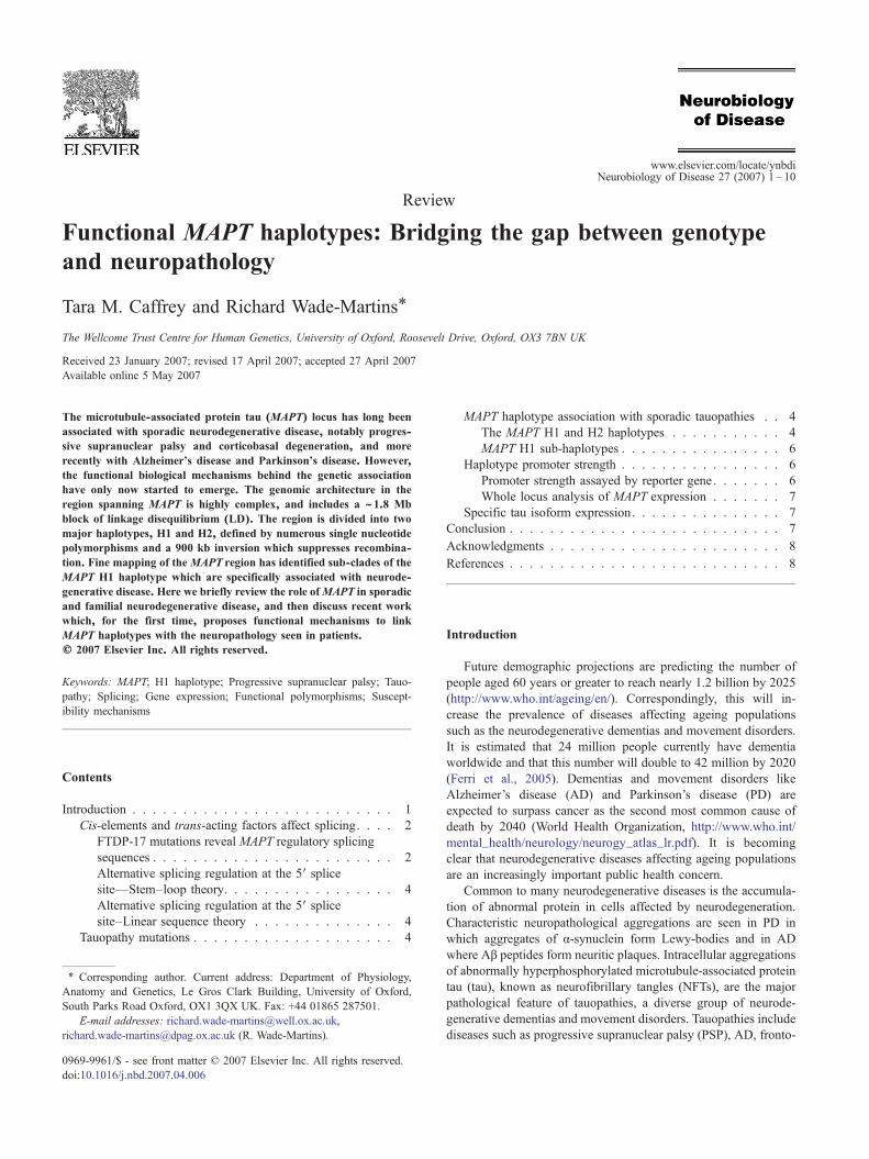
The MAPT gene locus is located on chromosome 17q21 andconsists of 16 exons (Andreadis et al., 1992) (Fig. 1). Tau protein isexpressed predominantly in the neurons of the peripheral andcentral nervous systems where it has a role in building and sta-bilizing microtubules, neuronal polarity and signal transduction (forreview see Shahani and Brandt, 2002). In the adult human centralnervous system, six proteins isoforms are generated by alternativesplicing of exons 2, 3 and 10 (Goedert et al., 1988; Goedert et al.,1989a,b; Andreadis et al., 1992) (Fig. 1). Exons 2 and 3 code forshort amino terminal inserts that which part of the acidic projectiondomain that may interact with the plasma membrane (Brandt et al.,1995) and regulate spacing between microtubules (Chen et al.,1992). The interaction of tau with microtubules is mediated by themicrotubule binding domains formed by imperfect repeats of 31 or32 amino acids at the carboxyl terminus, encoded by exons 9–12(Goedert et al., 1988; Gustke et al., 1994; Trinczek et al., 1995). Thealternative splicing of exon 10 generates proteins with either three(exon 10−; 3R tau) or four (exon 10+; 4R tau) microtubule bindingdomains (Fig. 1).
Neurofibrillary tangles are found in AD, PSP, FTDP-17, CBD,AGD and PiD. The filamentous tau that aggregates to form NFTs inAD consists mainly of paired helical filaments 10–20 mm in dia-meter (Kidd, 1963; Crowther, 1990) whereas the PSP tangles arecomposed of 15- to 18-mm-diameter straight filaments (Tellez-Nagel and Wisniewski, 1973; Powell et al., 1974). Tangle isoform
Fig. 1.MAPT locus consists of 16 exons of which exons 2, 3 and 10 are alternativelythe N-terminal projection domain (black stripes). Exons 9–12 imperfect repeats eaare absent from the CNS, but exon 4A is expressed in the peripheral nervous syssplicing of exons 2 and 3 results in proteins with 0, 1 or 2 N-terminal inserts (ON, 1Nbinding repeats (3R or 4R tau protein).
composition differs between tauopathies, being comprised of both3R tau and 4R tau in AD (Sergeant et al., 1997) and predominately3R tau in PiD (Delacourte et al., 1996). The major component of theinsoluble tangles in PSP, CBD, AGD and FTDP-17 is 4R tau (BueeScherrer et al., 1996; Arai et al., 2001; Togo et al., 2002). Conse-quently, these diseases are referred to as 4R tauopathies.
In 1998 the discovery of multiple MAPT mutations in FTDP-17provided the first evidence that changes in tau alone could causeneurodegenerative disease. In addition, the FTDP-17 splice sitemutations within MAPT that increase the inclusion of exon 10 intranscripts show an imbalance in the ratio of 3R and 4R tau isoformsis sufficient to cause disease (Hutton et al., 1998; Spillantini et al.,1998; D’Souza et al., 1999). In-depth analysis of the region aroundexon 10 containing these rare pathogenic mutations has uncoveredseveral functional cis-elements in the MAPT sequence which maygive insight into how intronic and non-coding mutations orpolymorphisms may cause sporadic tauopathies.
Cis-elements and trans-acting factors affect splicing
FTDP-17 mutations reveal MAPT regulatory splicing sequencesThe alternative splicing of MAPT exon 10 is moderated in part
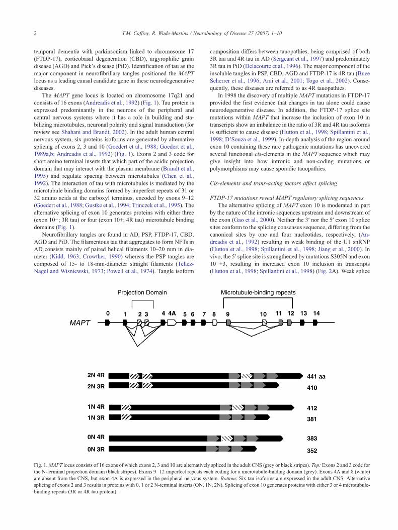
by the nature of the intronic sequences upstream and downstream ofthe exon (Gao et al., 2000). Neither the 3′ nor the 5′ exon 10 splicesites conform to the splicing consensus sequence, differing from thecanonical sites by one and four nucleotides, respectively, (An-dreadis et al., 1992) resulting in weak binding of the U1 snRNP(Hutton et al., 1998; Spillantini et al., 1998; Jiang et al., 2000). Invivo, the 5′ splice site is strengthened by mutations S305N and exon10 +3, resulting in increased exon 10 inclusion in transcripts(Hutton et al., 1998; Spillantini et al., 1998) (Fig. 2A). Weak splice
spliced in the adult CNS (grey or black stripes). Top: Exons 2 and 3 code forch coding for a microtubule-binding domain (grey). Exons 4A and 8 (white)tem. Bottom: Six tau isoforms are expressed in the adult CNS. Alternative, 2N). Splicing of exon 10 generates proteins with either 3 or 4 microtubule-
Fig. 2. Cis-elements affecting splicing ofMAPT exon 10. Silencer (red) and enhancer (green) sequences within and surrounding exon 10 are indicated. Exon 10sequence is designated by capital letters and intronic sequence is indicated by small case letters. (A) Several cis-elements affectMAPTexon 10 splicing, listed 5′ to3′: a SC35-like enhancer, a polypurine enhancer (PPE), an A/C-rich enhancer (ACE), an exonic splicing silencer (ESS), an exonic splicing enhancer (ESE), anintronic splicing silencer (ISS) and an intronic splicingmodulator (ISM). Select FTDP-17 and PSPmutations are also shown. (B) Alternative splicing regulation atthe tau exon 10 5′ splice site. Two of the variant stem–loop structures proposed to regulate splicing at the tau exon 5′ splice site (Hutton et al., 1998; Spillantini etal., 1998). (C) The linear model of tau exon 10 5′ splicing regulation postulates that binding of trans-acting factors to the cis-regulatory elements mediates exon 10alternative splicing. Shown here is a trans-acting factor bound to the ISM. This ISM binding protein sterically hinders a trans-acting silencer (ISS binding protein)from binding the silencing cis-element, thereby allowing access of the U1 snRNP to the 5′ splice site (D'Souza and Schellenberg, 2002, 2005).
3T.M. Caffrey, R. Wade-Martins / Neurobiology of Disease 27 (2007) 1–10
sites around exon 10 may allow subtle spatial and temporal regula-tion of 3R and 4R isoforms, by allowing splicing regulation througha collection of local splicing enhancer and silencer elements.
Systematic deletions across exon 10 have revealed that nearlyall the exon is involved in splicing regulation (D’Souza andSchellenberg, 2000) (Fig. 2A). At the 5′ of exon 10, three exonicsplicing enhancers have been identified: a SC35-like enhancer, apolypurine enhancer (PPE) and an AC-rich element (ACE)(D’Souza et al., 1999; D’Souza and Schellenberg, 2000).
The polypurine enhancer has a high purine content and is theregion affected by the mutations N279K and K280del. N279Kenhances exon 10 splicing by adding an extra AAG-repeat thatstrengthens the PPE (D’Souza et al., 1999; Hasegawa et al., 1999).In contrast, the K280del abolishes exon 10 splicing by removing anAAG-repeat (D’Souza et al., 1999; Rizzu et al., 1999). Trans-actingfactors that bind the PPE include SF2/ASF, Tra2β, SRp30c andSRp54 (Jiang et al., 2003; Kondo et al., 2004; D’Souza andSchellenberg, 2006).
4 T.M. Caffrey, R. Wade-Martins / Neurobiology of Disease 27 (2007) 1–10
Downstream of the PPE is a putative A/C rich enhancer. Thedeletion of nucleotides in this region results mainly in decreasedinclusion of exon 10 in transcripts. This region is affected by thesilent mutation CTTNCTC (L284L) that is thought to exert itseffect by increasing the content of AC nucleotides (D’Souza et al.,1999; D’Souza and Schellenberg, 2000).
Further downstream of these enhancers, three different patho-genic mutations have been found at the same codon. The missenseN296H and silent AATNAAC (N296N) mutations enhance exon 10inclusion, while a deletion at the same codon (N296del) is eitherneutral or enhances exon 10 splicing (Spillantini et al., 2000;Grover et al., 2002; Yoshida et al., 2002). These mutations havebeen proposed to either disrupt an 18-nucleotide silencer or changethe silencer into an enhancer (D’Souza and Schellenberg, 2000).
Alternative splicing regulation at the 5′ splicesite—Stem–loop theory
Mutations in the MAPT exon 10 5′ splice site have highlightedthe silencing function of this region on exon 10 inclusion. Onetheory proposes that these mutations disrupt a stem–loop structurethat blocks U1 snRNP or another factor from binding, thus pre-venting inclusion of exon 10 in transcripts (Hutton et al., 1998;Spillantini et al., 1998) (Fig. 2B). Mutations within the predictedstem increase inclusion of exon 10 in transcripts, while mutationsgenerated in the putative loop have no effect on splicing (Grover etal., 1999). In addition, stabilizing the stem–loop by either insertingextra bases to elongate the stem or by generating more stable pairingalong the stem, leads to a decrease in exon 10 inclusion (Grover etal., 1999; Donahue et al., 2006). Stem–loop secondary structureshave been indicated through in vitro gel migration assays (Grover etal., 1999), by exposure of transcripts to RNase H (Jiang et al., 2000)and by UV melting experiments (Donahue et al., 2006).
While secondary structures can be observed in in vitro ex-periments (Grover et al., 1999; Varani et al., 1999), it is not clear ifthe same structure would form in vivo because in vivo there aremultiple splicing regulatory proteins that coat the pre-mRNApossibly preventing secondary structures from forming (D’Souzaand Schellenberg, 2000). Additionally, the stem–loop structuresproposed are often ambiguous and do not agree between groups(Fig. 2B). Importantly, regulation through the stem–loop structuredoes not necessarily allow for regulation by trans-acting factors thatmay affect differential splicing seen throughout development.
Alternative splicing regulation at the 5′ splicesite–Linear sequence theory
An alternative hypothesis is that the inhibitory sequence inintron 10 is a linear cis-acting element that binds trans-actingsplicing factors (D’Souza et al., 1999; D’Souza and Schellenberg,2000) (Fig. 2C). Some of the mutations that destabilize the stem-loop structure may also function to increase exon 10 inclusion byincreasing affinity for the U1 and U6 snRNPs which have both beenimplicated in exon 10 splicing inclusion. A proposed intronicsilencer at position exon 10+11 to exon 10+18 retains its functioneven after it has been translocated to different positions, including aposition within exon 10 as well as a heterologous setting, where thecomplementary bases are not present to form a stem–loop structure(D’Souza and Schellenberg, 2002). An intronic splicing modulatorlocated at position exon 10+19 to exon 10+26, acts in conjunctionwith the intronic splicing silencer to regulate the splicing of exon 10(D’Souza and Schellenberg, 2002) (Fig. 2C). This splicing modu-lator is disrupted by the exon 10+19 mutation and results in
increased 3R tau isoforms (Stanford et al., 2003). These twoelements seem to act in opposition and it has been suggested thattrans-acting factors that bind the modulator sterically hinder othertrans-acting factors from attaching to the silencer, which in turnallows access to the 5′ splice site (Fig. 2C).
Tauopathy mutations
In contrast to FTDP-17, PSP is overwhelmingly a sporadicdisease. There have been, however, a few individuals identifiedcarrying coding MAPT mutations who present clinically withsymptoms similar to those seen in sporadic PSP. It is thought inthese rare cases that theMAPTmutations do account for the isoformimbalance seen in aggregates, or otherwise affect the aetiology ofthe disease. In one patient with PSP-like clinical symptoms andneuropathology, a dominant silent mutation AGTNAGC (S305S)has been identified (Stanford et al., 2000) (Fig. 2A). S305S fallswithin the putative stem–loop structure and its presence results inover a four-fold increase in exon 10 inclusion and subsequent over-production of 4R tau (Stanford et al., 2000). A recessive mutation, adeletion at N296 (N296del) generates an atypical PSP phenotype(Pastor et al., 2001) and falls in a region defined as a splicingsilencer (D’Souza and Schellenberg, 2000). An exonic splicingenhancer is altered by the G303V mutation, carriers of which showgreater exon 10+ transcripts than controls (Ros et al., 2005).
An interesting mutation generating a PSP-like phenotype is theR5L mutation located in MAPT exon 1 (Poorkaj et al., 2002). Ag-gregated insoluble tau in the R5L case was comprised of predo-minantly 4R tau isoforms similar to sporadic PSP, however, theamount of soluble tau in the frontal and temporal cortices case was1.5–2 times higher than both sporadic PSP and controls. In addition,the mutation altered the interaction of the mutant protein withtubulin and microtubules. It has been suggested that the N-terminalsubstitution exerts its effect on microtubule binding through inter-action of the N-terminal and the microtubule binding domains inpaired helical filaments but not normal tau (Carmel et al., 1996;Poorkaj et al., 2002).
The diverse nature of MAPT mutations indicates the complexinterplay of regulatory sequences in intron 9, exon 10 and intron 10to maintain tau isoform ratios. Much current research is nowfocused on the natural sequence variation in the region of MAPTand how this variation may confer susceptibility over time toneurodegenerative disease.
MAPT haplotype association with sporadic tauopathies
The MAPT H1 and H2 haplotypesThe genetic link between MAPT and PSP started with the
identification of an association between PSP and a polymorphicmarker found in MAPT intron 9 (Conrad et al., 1997) (Fig. 3B).Both the dinucleotide repeat allele A0 and the genotype A0/A0 areover-represented in PSP; for example, one study found A0/A0 iscarried in 57% of controls compared to 95% in PSP patients(Conrad et al., 1997). Further investigation of the polymorphismsover-represented in PSP led to the elucidation of an extendedhaplotype covering the entireMAPT locus (Baker et al., 1999) (Fig.3B). Eight common single nucleotide polymorphisms (SNPs)across MAPT were identified and found to be in complete linkagedisequilibrium (LD) with each other and the A0 allele. The morecommonMAPT H1 haplotype was found to be associated with PSP.A 238-bp deletion was also discovered to be inherited as part of the
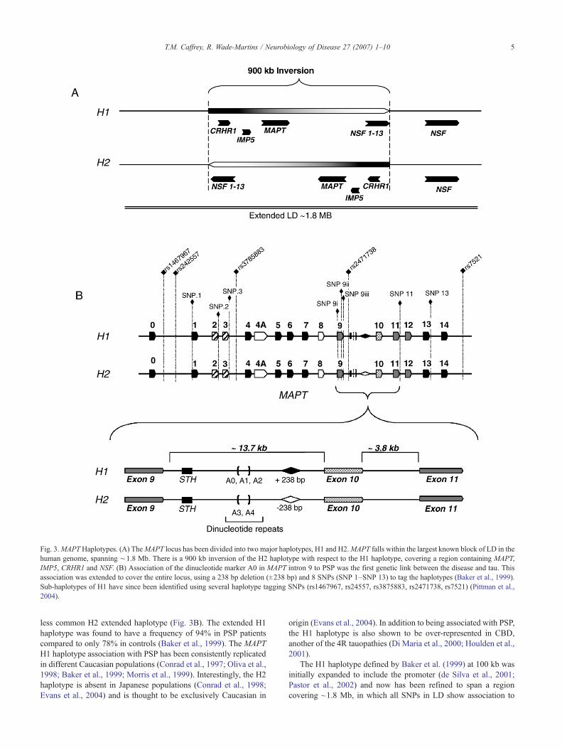
Fig. 3.MAPTHaplotypes. (A) TheMAPT locus has been divided into two major haplotypes, H1 and H2.MAPT falls within the largest known block of LD in thehuman genome, spanning ∼1.8 Mb. There is a 900 kb inversion of the H2 haplotype with respect to the H1 haplotype, covering a region containing MAPT,IMP5, CRHR1 and NSF. (B) Association of the dinucleotide marker A0 in MAPT intron 9 to PSP was the first genetic link between the disease and tau. Thisassociation was extended to cover the entire locus, using a 238 bp deletion (±238 bp) and 8 SNPs (SNP 1–SNP 13) to tag the haplotypes (Baker et al., 1999).Sub-haplotypes of H1 have since been identified using several haplotype tagging SNPs (rs1467967, rs24557, rs3875883, rs2471738, rs7521) (Pittman et al.,2004).
5T.M. Caffrey, R. Wade-Martins / Neurobiology of Disease 27 (2007) 1–10
less common H2 extended haplotype (Fig. 3B). The extended H1haplotype was found to have a frequency of 94% in PSP patientscompared to only 78% in controls (Baker et al., 1999). The MAPTH1 haplotype association with PSP has been consistently replicatedin different Caucasian populations (Conrad et al., 1997; Oliva et al.,1998; Baker et al., 1999; Morris et al., 1999). Interestingly, the H2haplotype is absent in Japanese populations (Conrad et al., 1998;Evans et al., 2004) and is thought to be exclusively Caucasian in
origin (Evans et al., 2004). In addition to being associated with PSP,the H1 haplotype is also shown to be over-represented in CBD,another of the 4R tauopathies (Di Maria et al., 2000; Houlden et al.,2001).
The H1 haplotype defined by Baker et al. (1999) at 100 kb wasinitially expanded to include the promoter (de Silva et al., 2001;Pastor et al., 2002) and now has been refined to span a regioncovering ~1.8 Mb, in which all SNPs in LD show association to
6 T.M. Caffrey, R. Wade-Martins / Neurobiology of Disease 27 (2007) 1–10
PSP (Pittman et al., 2004, 2005). This region is relatively gene rich,with the CRHR1 (corticotrophin) and IMP5 (a presenilin homo-logue) genes at the centromeric end of LD, while WNT3 and NSF(N-ethylmaleimide-sensitive factor) at the telomeric end of the LD(Pittman et al., 2004) (Fig. 3A).
Recently, it has been found that a ~900 kb segment from H2chromosomes is inverted with respect to the H1 haplotype (Ste-fansson et al., 2005) (Fig. 3A). This inversion is thought to haveoccurred between 3 and 3.6 million years ago (Stefansson et al.,2005) and is almost entirely absent outside of European populations(Evans et al., 2004). The relative homogeneity of the H2 haplotypeand the higher frequency in Europeans is consistent with a fewfounder chromosomes, probably expanding due to positive selec-tion (Stefansson et al., 2005).
Further investigations into the genomic architecture of thisregion have identified a number of low-copy repeat (LCR) se-quences (Cruts et al., 2005). There is evidence to suggest that thenon-allelic homologous recombination that generated the inversionat H2 occurred between two low-copy repeat sequences, LCR Aand LCR B, located 250 kb centromeric and 180 kb telomeric toMAPT respectively (Cruts et al., 2005). Non-allelic homologousrecombination between LCRs has also been suggested to be thecause of a recently identified microdeletion encompassingMAPT inpatients with learning disability and developmental delay (Koolenet al., 2006; Shaw-Smith et al., 2006; Varela et al., 2006).
In addition to the previously defined polymorphisms, a complexarrangement of duplicated regions was also discovered along withthe inversion to segregate withMAPT haplotypes (Stefansson et al.,2005). Four H1 surrogate or sub-haplotypes have been defined withrespect to the size, location and dosage of these regions, H1D0–H1D3. The H1D3 variant has a triplication of the first 13 exons ofNSF, while the H1D0 variant has a duplicated region notcontaining NSF. Recently, a study of the global variation of copynumber in the human genome has identified population-specificcopy number polymorphisms in the genomic region surroundingMAPT (Redon et al., 2006). The number of segmental duplica-tions, segmental duplication inversions and copy number poly-morphisms near MAPT highlights the complexity of this genomicregion.
MAPT H1 sub-haplotypesExhaustive sequence analysis in the MAPT genomic region has
also further divided the H1 haplotype into subtypes and sub-sequently allowed the fine mapping of the MAPT association withPSP (Pittman et al., 2005; Rademakers et al., 2005). In addition toH1 and H2 haplotype defining SNPs, there exist SNPs that varyonly on the H1 background, H1-specific SNPs (Pittman et al.,2004). Using 5 haplotype tagging polymorphisms (rs1467967,rs242557, rs3785883, rs2471738, rs7521) and the 238-bp haplo-type defining insertion–deletion to capture 95% of the commonhaplotype diversity, the population has been divided into severalsub-haplotypes for association analysis (Fig. 3B). In both UK andUS cohorts, the H1C haplotype, a sub-clade of H1, is over-represented in PSP patients compared to controls (Pittman et al.,2005). Moreover, strong association of haplotype sub-class IIdefined by three H1-specific haplotype tagging SNPs (rs242557,rs3785883, rs2471738), suggests variability within the H1 cladeitself is a risk factor for PSP (Pittman et al., 2005). The fine mappingof these SNPs indicates the association is conveyed by a regioncovering a minimal distance of ~54 kb from upstream of exon 1 todownstream of exon 9 (Pittman et al., 2005).
Interestingly, another study also shows a strong association tors242557 with an 11.6% increase of the same ‘A’ allele in PSPpatient populations over controls (Rademakers et al., 2005). Thehighest significance in this study was obtained in LD block 2, partof an interval spanning ~22 kb of regulatory sequence upstream ofexon 1 (Rademakers et al., 2005). Both sub-haplotype studiesindicate greatest association of PSP with the regulatory region ofMAPT, making the promoter region a good candidate for functionalstudies.
Haplotype promoter strength
Promoter strength assayed by reporter geneSeveral studies have found strong association between suscept-
ibility to neurological disorders and specific alleles or haplotypes ofcandidate genes (Bray et al., 2003, 2004, 2005; Paracchini et al.,2006; Urak et al., 2006). In the absence of protein coding changes,these investigations have focused on relevant non-coding poly-morphisms and how these polymorphisms regulate gene expres-sion. For example, one study investigating the genetic basis ofdyslexia found association to one risk haplotype that results in~40% reduction in expression of KIAA0319 (Paracchini et al.,2006). Subtle changes in the relative gene expression between riskand non-risk alleles have been suggested as a functional mechanismwhereby a risk haplotype confers susceptibility to disease.
Recent PSP association studies propose that variants in theMAPT promoter region are responsible for susceptibility to PSP.One theory to explain the association of H1 with PSP proposes thatthere are overall expression differences between the two haplotypes.To investigate this one study employed reporter genes downstreamof 1 kb promoter fragments of the H1, H2 or H1′ haplotypes toassay promoter strength (Kwok et al., 2004). H1′ is a promoterhaplotype differing from H1 by one SNP 568 bp upstream of exon-1. In HEK 293 kidney and SKNMC neuroblastoma cell lines the H2promoter showed a 1.2-fold reduction in transcriptional activitycompared to the H1 promoter. In SKNMC cells, the H1′ promoteralso showed a 1.1-fold decrease in transcriptional activity comparedto the H1 promoter (Kwok et al., 2004). While these differences inpromoter strength may be small, it is proposed that over time, thesmall increase in expression from the H1 haplotype may lead todisease.
Another assay analysed a conserved region containing thehaplotype tagging SNP rs242557 (Rademakers et al., 2005), whichhas been shown to be associated with PSP (Pittman et al., 2004,2005; Rademakers et al., 2005). In silico the risk variant waspredicted to abolish binding sites for trans-acting factors, Se-CystRNA gene transcription–activation factor (STAF.01) and CP2-erythrocyte factor related to drosophila Elf1 (CP2). The proposedregulatory region was placed upstream of both a minimal SV40early promoter and a 1.1 kb fragment of the MAPT H1 promoter.Under these conditions, the non-risk allele showed greatertranscriptional activity (Rademakers et al., 2005). In contrast,another study using this same SNP placed downstream of theMAPTpromoter region showed that the H1 haplotype construct expresses4.2-fold greater expression than the non-risk H2 promoter (Myerset al., 2007).
The differing results from these promoter assays are likely be-cause their experimental designs use small fragments of regulatorysequence isolated from their correct genomic context. The studythat found the non-risk regulatory region had greater transcriptionalactivity placed a small 182-bp fragment containing the SNP from

7T.M. Caffrey, R. Wade-Martins / Neurobiology of Disease 27 (2007) 1–10
exon 0, upstream of a promoter (Rademakers et al., 2005). Using amore biologically relevant design, the function of the same SNPwas assayed using a 900-bp fragment in the correct relative linearorder to the MAPT promoter, although with the removal of ~50 kbof intervening sequence (Myers et al., 2007).
Whole locus analysis of MAPT expressionSeveral studies have tried using systems that assay expression
from the whole MAPT genomic locus. Real-time PCR analysis oftau expression in post-mortem human brain tissue showedindividuals carrying the homozygous H1C haplotype have a three-to four-fold greater expression compared to promoters of all othergenotypes (Myers et al., 2007). This data agrees with data obtainedfrom reporter gene assays but carries greater in vivo validity due tothe whole locus analysis that is possible in human tissue.
Powerful tools to investigate the effect of sequence variation ontranscript expression are found in technologies that allow allele-specific or haplotype-specific gene expression studies (Yan et al.,2002; Knight, 2004). The strength of these techniques derives fromtheir ability to analyze allele-specific gene expression within aheterozygous sample eliminating the effects of confounding factorsbetween samples such as sample quality, environmental effects andgenetic background.
Two studies have used allelic-specific gene expression techni-ques to address the question ofMAPT promoter haplotype strength.A haplotype tagging SNP in intron 1 was used to assay haplotypespecific expression in heterozygous samples (Caffrey et al., 2006).Significantly greater expression from the H1 promoter was seen inboth SKNF1 and SKNMC neuroblastoma cell lines, similar to thefindings in reporter gene assays (Kwok et al., 2004; Caffrey et al.,2006; Myers et al., 2007) However, across 14 heterozygous,pathology-free, post-mortem human brain samples, there was noallelic difference observed in expression in either the frontal cortexor globus pallidus (Caffrey et al., 2006). In another study examiningthe effect of sub-haplotype on expression, allele-specific real-timePCR showed that the H1C haplotype has a significant 11–13%greater expression compared to all other MAPT haplotypesexamined (Myers et al., 2007).
Specific tau isoform expression
Another potential mechanism by which tau may confer suscep-tibility to neurodegeneration is through the imbalanced expressionof alternative transcripts, leading to imbalanced protein isoformspecies. The importance of tau isoform balance is highlighted byFTDP-17 mutations which increase the inclusion of exon 10 intranscripts, resulting in a two- to six-fold excess of 4R tau mRNA(Hutton et al., 1998; Spillantini et al., 1998; D’Souza et al., 1999;Connell et al., 2005). The isoform imbalance in FTDP-17 issufficient to cause disease, and suggests the possibility that isoformimbalance may be a contributing factor to sporadic tauopathies.
The first indication of altered tau mRNA isoform expression inPSP was demonstrated using real-time PCR to examine expressionin PSP patient tissue (Chambers et al., 1999). The cerebellar cortex,a region free of NFTs in PSP, was analyzed and showed a significantdecrease in 3R mRNA levels, thus increasing the 4R/3R ratios inthis region. In the frontal cortex, a region with mild NFT pathologyin PSP, there was no significant difference in mRNA levels foreither 4R or 3R tau (Chambers et al., 1999). Finally, in the highlyaffected brainstem region, there was a significant increase in 4R taumRNA in PSP patients compared with controls and AD cases. This
suggests that the ratio of splice products is important for correctfunction.
In support of the a role of isoform imbalance in areas susceptibleto degeneration and in the aetiology of sporadic 4R tauopathies,Takanashi et al. (2002) found the total amounts of 3R and 4R tautranscript did not differ significantly between controls and patients,however the ratio of 4R:3R was significantly higher in the globuspallidus of PSP patients and the 4R:3R ratio is also higher in boththe frontal cortex and globus pallidus in CBD patient tissues(Takanashi et al., 2002). Moreover, a study of pathology-freecontrols showed a significantly higher 4R:3R mRNA transcriptratio in the globus pallidus than in a frontal cortex, a region notaffected by neurodegeneration in PSP (Caffrey et al., 2006). Thiselevated ratio of 4R:3R evidence suggests that the expressionpattern of tau isoforms in brain regions vulnerable to neurodegen-eration in PSP may underlie the susceptibility of these areas todevelop neurodegeneration.
In addition to certain brain regions being more susceptible todisease, functional evidence is emerging to suggest why H1 hap-lotype carriers may also be susceptible. Using allele-specific ex-pression assays in differentiated H1/H2 heterozygous neuroblasto-ma cell line models, significantly greater expression of exon 10+transcripts was shown coming from the H1 chromosome. When theassay was performed in pathology-free post-mortem human braintissue, H1 chromosomes were shown to express significantly moreexon 10+ transcripts than H2 chromosomes, in both the frontalcortex and globus pallidus (Caffrey et al., 2006). This difference inexpression was most pronounced in the globus pallidus where H1expresses up to 40% more 4R mRNA transcripts. In a similar studywhere exon 10+ transcripts were analyzed according to sub-haplotype, a significant 25% greater expression of 4R transcriptswas seen originating from H1C chromosomes (Myers et al., 2007).These are key findings that start bridging the gap between the H1haplotype association of 4R tauopathies such as PSP and CBD andthe neuropathology seen in affected areas.
Conclusion
Recent investigations elucidating the complex genomic archi-tecture of chromosome 17q21 and fine mapping of the linkagedisequilibrium spanning the MAPT gene locus now place us in agood position to start assessing the functional effects of MAPThaplotype. Data from various experimental approaches are conver-ging to reveal subtle differences in haplotype expression, whichover time may lead to neurodegenerative disease. The relativelygreater expression of MAPT exon 10+ transcripts from the PSP-associated H1 haplotype is of particular relevance because 4R tauisoforms predominately form the abnormal tau aggregates in 4Rtauopathies. For the first time, we are now able to link theneuropathology observed in PSP patients with the well-establishedgenetic association to the MAPT H1 haplotype and to begin shed-ding light on how natural variation may confer susceptibility toneurodegeneration.
However, dissecting the precise functional genetic mechanismsand identifying the exact polymorphisms responsible for regulationof MAPT expression and splicing remains a huge challenge. Withinthe MAPT locus alone there are over 400 documented SNPs (http://www.ncbi.nlm.nih.gov/projects/SNP/) indicating the difficulties inlocating the functional polymorphisms. In common with othercomplex genetic diseases we should expect the functional in-fluences of common genetic polymorphisms at the MAPT locus to
8 T.M. Caffrey, R. Wade-Martins / Neurobiology of Disease 27 (2007) 1–10
be subtle and cumulative over many years. The challenge for futurestudies is to design functional experiments to study the biologicalrelevance of genetic polymorphisms within the context of thegenomic DNA locus.
Acknowledgments
This work was supported by a Research Career DevelopmentFellowship awarded to RW-M from the Wellcome Trust and by aClarendon Fund Bursary awarded to TMC.
References
Andreadis, A., Brown, W.M., Kosik, K.S., 1992. Structure and novelexons of the human tau gene. Biochemistry (Mosc.) 31 (43),10626–10633.
Arai, T., Ikeda, K., Akiyama, H., Shikamoto, Y., Tsuchiya, K., Yagishita, S.,Beach, T., Rogers, J., Schwab, C., McGeer, P.L., 2001. Distinct isoformsof tau aggregated in neurons and glial cells in brains of patients withPick’s disease, corticobasal degeneration and progressive supranuclearpalsy. Acta Neuropathol. (Berl.) 101 (2), 167–173.
Baker, M., Litvan, I., Houlden, H., Adamson, J., Dickson, D., Perez-Tur, J.,Hardy, J., Lynch, T., Bigio, E., Hutton, M., 1999. Association of anextended haplotype in the tau gene with progressive supranuclear palsy.Hum. Mol. Genet. 8 (4), 711–715.
Brandt, R., Leger, J., Lee, G., 1995. Interaction of tau with the neural plasmamembrane mediated by tau’s amino-terminal projection domain. J. CellBiol. 131 (5), 1327–1340.
Bray, N.J., Buckland, P.R., Williams, N.M., Williams, H.J., Norton, N.,Owen, M.J., O’Donovan, M.C., 2003. A haplotype implicated in schi-zophrenia susceptibility is associated with reduced COMT expression inhuman brain. Am. J. Hum. Genet. 73 (1), 152–161.
Bray, N.J., Jehu, L., Moskvina, V., Buxbaum, J.D., Dracheva, S.,Haroutunian, V., Williams, J., Buckland, P.R., Owen, M.J., O’Donovan,M.C., 2004. Allelic expression of APOE in human brain: effects ofepsilon status and promoter haplotypes. Hum. Mol. Genet. 13 (22),2885–2892.
Bray, N.J., Preece, A., Williams, N.M., Moskvina, V., Buckland, P.R.,Owen, M.J., O’Donovan, M.C., 2005. Haplotypes at the dystrobrevinbinding protein 1 (DTNBP1) gene locus mediate risk for schizophreniathrough reduced DTNBP1 expression. Hum. Mol. Genet. 14 (14),1947–1954.
Buee Scherrer, V., Hof, P.R., Buee, L., Leveugle, B., Vermersch, P., Perl,D.P., Olanow, C.W., Delacourte, A., 1996. Hyperphosphorylated tauproteins differentiate corticobasal degeneration and Pick’s disease. ActaNeuropathol. (Berl.) 91 (4), 351–359.
Caffrey, T.M., Joachim, C., Paracchini, S., Esiri, M.M., Wade-Martins, R.,2006. Haplotype-specific expression of exon 10 at the human MAPTlocus. Hum. Mol. Genet. 15 (24), 3529–3537.
Carmel, G., Mager, E.M., Binder, L.I., Kuret, J., 1996. The structural basisof monoclonal antibody Alz50’s selectivity for Alzheimer’s diseasepathology. J. Biol. Chem. 271 (51), 32789–32795.
Chambers, C.B., Lee, J.M., Troncoso, J.C., Reich, S., Muma, N.A., 1999.Overexpression of four-repeat tau mRNA isoforms in progressivesupranuclear palsy but not in Alzheimer’s disease. Ann. Neurol. 46 (3),325–332.
Chen, J., Kanai, Y., Cowan, N.J., Hirokawa, N., 1992. Projection domains ofMAP2 and tau determine spacings between microtubules in dendritesand axons. Nature 360 (6405), 674–677.
Connell, J.W., Rodriguez-Martin, T., Gibb, G.M., Kahn, N.M., Grierson,A.J., Hanger, D.P., Revesz, T., Lantos, P.L., Anderton, B.H., Gallo, J.M.,2005. Quantitative analysis of tau isoform transcripts in sporadic tauo-pathies. Brain Res. Mol. Brain Res. 137 (1–2), 104–109.
Conrad, C., Andreadis, A., Trojanowski, J.Q., Dickson, D.W., Kang, D.,Chen, X., Wiederholt, W., Hansen, L., Masliah, E., Thal, L.J., et al.,
1997. Genetic evidence for the involvement of tau in progressive supra-nuclear palsy. Ann. Neurol. 41 (2), 277–281.
Conrad, C., Amano, N., Andreadis, A., Xia, Y., Namekataf, K., Oyama, F.,Ikeda, K., Wakabayashi, K., Takahashi, H., Thal, L.J., et al., 1998.Differences in a dinucleotide repeat polymorphism in the tau genebetween Caucasian and Japanese populations: implication for progres-sive supranuclear palsy. Neurosci. Lett. 250 (2), 135–137.
Crowther, R.A., 1990. Structural aspects of pathology in Alzheimer’sdisease. Biochim. Biophys. Acta 1096 (1), 1–9.
Cruts, M., Rademakers, R., Gijselinck, I., van der Zee, J., Dermaut, B.,de Pooter, T., de Rijk, P., Del-Favero, J., van Broeckhoven, C., 2005.Genomic architecture of human 17q21 linked to frontotemporaldementia uncovers a highly homologous family of low-copy repeatsin the tau region. Hum. Mol. Genet. 14 (13), 1753–1762.
D’Souza, I., Schellenberg, G.D., 2000. Determinants of 4-repeat tauexpression. Coordination between enhancing and inhibitory splicingsequences for exon 10 inclusion. J. Biol. Chem. 275 (23), 17700–17709.
D’Souza, I., Schellenberg, G.D., 2002. tau Exon 10 expression involves abipartite intron 10 regulatory sequence and weak 5′ and 3′ splice sites.J. Biol. Chem. 277 (29), 26587–26599.
D’Souza, I., Schellenberg, G.D., 2005. Regulation of tau isoform expressionand dementia. Biochim. Biophys. Acta 1739 (2–3), 104–115.
D’Souza, I., Schellenberg, G.D., 2006. Arginine/serine-rich protein inter-action domain-dependent modulation of a tau exon 10 splicing en-hancer: altered interactions and mechanisms for functionally antag-onistic FTDP-17 mutations Delta280K and N279K. J. Biol. Chem. 281(5), 2460–2469.
D’Souza, I., Poorkaj, P., Hong, M., Nochlin, D., Lee, V.M., Bird, T.D.,Schellenberg, G.D., 1999. Missense and silent tau gene mutations causefrontotemporal dementia with parkinsonism-chromosome 17 type, byaffecting multiple alternative RNA splicing regulatory elements. Proc.Natl. Acad. Sci. U. S. A. 96 (10), 5598–5603.
Delacourte, A., Robitaille, Y., Sergeant, N., Buee, L., Hof, P.R., Wattez, A.,Laroche-Cholette, A., Mathieu, J., Chagnon, P., Gauvreau, D., 1996.Specific pathological Tau protein variants characterize Pick’s disease.J. Neuropathol. Exp. Neurol. 55 (2), 159–168.
de Silva, R., Weiler, M., Morris, H.R., Martin, E.R., Wood, N.W., Lees, A.J.,2001. Strong association of a novel Tau promoter haplotype inprogressive supranuclear palsy. Neurosci. Lett. 311 (3), 145–148.
Di Maria, E., Tabaton, M., Vigo, T., Abbruzzese, G., Bellone, E., Donati, C.,Frasson, E., Marchese, R., Montagna, P., Munoz, D.G., et al., 2000.Corticobasal degeneration shares a common genetic background withprogressive supranuclear palsy. Ann. Neurol. 47 (3), 374–377.
Donahue, C.P., Muratore, C., Wu, J.Y., Kosik, K.S., Wolfe, M.S., 2006.Stabilization of the tau exon 10 stem loop alters pre-mRNA splicing.J. Biol. Chem. 281 (33), 23302–23306.
Evans, W., Fung, H.C., Steele, J., Eerola, J., Tienari, P., Pittman, A., Silva,R., Myers, A., Vrieze, F.W., Singleton, A., et al., 2004. The tau H2haplotype is almost exclusively Caucasian in origin. Neurosci. Lett. 369(3), 183–185.
Ferri, C.P., Prince, M., Brayne, C., Brodaty, H., Fratiglioni, L., Ganguli, M.,Hall, K., Hasegawa, K., Hendrie, H., Huang, Y., et al., 2005. Globalprevalence of dementia: a Delphi consensus study. Lancet 366 (9503),2112–2117.
Gao, Q.S., Memmott, J., Lafyatis, R., Stamm, S., Screaton, G., Andreadis,A., 2000. Complex regulation of tau exon 10, whose missplicing causesfrontotemporal dementia. J. Neurochem. 74 (2), 490–500.
Goedert, M., Wischik, C.M., Crowther, R.A., Walker, J.E., Klug, A., 1988.Cloning and sequencing of the cDNA encoding a core protein of thepaired helical filament of Alzheimer disease: identification as the micro-tubule-associated protein tau. Proc. Natl. Acad. Sci. U. S. A. 85 (11),4051–4055.
Goedert, M., Spillantini, M.G., Jakes, R., Rutherford, D., Crowther, R.A.,1989a. Multiple isoforms of human microtubule-associated protein tau:sequences and localization in neurofibrillary tangles of Alzheimer’sdisease. Neuron 3 (4), 519–526.
Goedert, M., Spillantini, M.G., Potier, M.C., Ulrich, J., Crowther, R.A.,
9T.M. Caffrey, R. Wade-Martins / Neurobiology of Disease 27 (2007) 1–10
1989b. Cloning and sequencing of the cDNA encoding an isoform ofmicrotubule-associated protein tau containing four tandem repeats:differential expression of tau protein mRNAs in human brain. EMBO J.8 (2), 393–399.
Grover, A., Houlden, H., Baker, M., Adamson, J., Lewis, J., Prihar, G.,Pickering-Brown, S., Duff, K., Hutton, M., 1999. 5′ splice site mutationsin tau associated with the inherited dementia FTDP-17 affect a stem–loop structure that regulates alternative splicing of exon 10. J. Biol.Chem. 274 (21), 15134–15143.
Grover, A., DeTure, M., Yen, S.H., Hutton, M., 2002. Effects on splicingand protein function of three mutations in codon N296 of tau in vitro.Neurosci. Lett. 323 (1), 33–36.
Gustke, N., Trinczek, B., Biernat, J., Mandelkow, E.M., Mandelkow, E.,1994. Domains of tau protein and interactions with microtubules.Biochemistry (Mosc.) 33 (32), 9511–9522.
Hasegawa, M., Smith, M.J., Iijima, M., Tabira, T., Goedert, M., 1999.FTDP-17 mutations N279K and S305N in tau produce increasedsplicing of exon 10. FEBS Lett. 443 (2), 93–96.
Houlden, H., Baker, M., Morris, H.R., MacDonald, N., Pickering-Brown, S.,Adamson, J., Lees, A.J., Rossor, M.N., Quinn, N.P., Kertesz, A., et al.,2001. Corticobasal degeneration and progressive supranuclear palsyshare a common tau haplotype. Neurology 56 (12), 1702–1706.
Hutton, M., Lendon, C.L., Rizzu, P., Baker, M., Froelich, S., Houlden, H.,Pickering-Brown, S., Chakraverty, S., Isaacs, A., Grover, A., et al., 1998.Association of missense and 5′-splice-site mutations in tau with theinherited dementia FTDP-17. Nature 393 (6686), 702–705.
Jiang, Z., Cote, J., Kwon, J.M., Goate, A.M., Wu, J.Y., 2000. Aberrantsplicing of tau pre-mRNA caused by intronic mutations associated withthe inherited dementia frontotemporal dementia with parkinsonismlinked to chromosome 17. Mol. Cell. Biol. 20 (11), 4036–4048.
Jiang, Z., Tang, H., Havlioglu, N., Zhang, X., Stamm, S., Yan, R., Wu, J.Y.,2003. Mutations in tau gene exon 10 associated with FTDP-17 alter theactivity of an exonic splicing enhancer to interact with Tra2 beta. J. Biol.Chem. 278 (21), 18997–19007.
Kidd, M., 1963. Paired helical filaments in electron microscopy ofAlzheimer’s disease. Nature 197, 192–193.
Knight, J.C., 2004. Allele-specific gene expression uncovered. TrendsGenet. 20 (3), 113–116.
Kondo, S., Yamamoto, N., Murakami, T., Okumura, M., Mayeda, A.,Imaizumi, K., 2004. Tra2 beta, SF2/ASF and SRp30c modulate thefunction of an exonic splicing enhancer in exon 10 of tau pre-mRNA.Genes Cells 9 (2), 121–130.
Koolen, D.A., Vissers, L.E., Pfundt, R., de Leeuw, N., Knight, S.J., Regan,R., Kooy, R.F., Reyniers, E., Romano, C., Fichera, M., 2006. A newchromosome 17q21.31 microdeletion syndrome associated with acommon inversion polymorphism. Nat. Genet. 38 (9), 999–1001.
Kwok, J.B., Teber, E.T., Loy, C., Hallupp, M., Nicholson, G., Mellick, G.D.,Buchanan, D.D., Silburn, P.A., Schofield, P.R., 2004. Tau haplotypesregulate transcription and are associated with Parkinson’s disease. Ann.Neurol. 55 (3), 329–334.
Morris, H.R., Janssen, J.C., Bandmann, O., Daniel, S.E., Rossor, M.N.,Lees, A.J., Wood, N.W., 1999. The tau gene A0 polymorphism inprogressive supranuclear palsy and related neurodegenerative diseases.J. Neurol. Neurosurg. Psychiatry 66 (5), 665–667.
Myers, A.J., Pittman, A.M., Zhao, A.S., Rohrer, K., Kaleem, M., Marlowe,L., Lees, A., Leung, D., McKeith, I.G., Perry, R.H., et al., 2007. TheMAPT H1c risk haplotype is associated with increased expression of tauand especially of 4 repeat containing transcripts. Neurobiol. Dis. 25 (3),561–570.
Oliva, R., Tolosa, E., Ezquerra, M., Molinuevo, J.L., Valldeoriola, F.,Burguera, J., Calopa, M., Villa, M., Ballesta, F., 1998. Significantchanges in the tau A0 and A3 alleles in progressive supranuclear palsyand improved genotyping by silver detection. Arch. Neurol. 55 (8),1122–1124.
Paracchini, S., Thomas, A., Castro, S., Lai, C., Paramasivam, M., Wang, Y.,Keating, B.J., Taylor, J.M., Hacking, D.F., Scerri, T., 2006. Thechromosome 6p22 haplotype associated with dyslexia reduces the
expression of KIAA0319, a novel gene involved in neuronal migration.Hum. Mol. Genet. 15 (10), 1659–1666.
Pastor, P., Pastor, E., Carnero, C., Vela, R., Garcia, T., Amer, G., Tolosa, E.,Oliva, R., 2001. Familial atypical progressive supranuclear palsy asso-ciated with homozygosity for the delN296 mutation in the tau gene.Ann. Neurol. 49 (2), 263–267.
Pastor, P., Ezquerra, M., Tolosa, E., Munoz, E., Marti, M.J., Valldeoriola, F.,Molinuevo, J.L., Calopa, M., Oliva, R., 2002. Further extension of theH1 haplotype associated with progressive supranuclear palsy. Mov.Disord. 17 (3), 550–556.
Pittman, A.M., Myers, A.J., Duckworth, J., Bryden, L., Hanson, M., Abou-Sleiman, P., Wood, N.W., Hardy, J., Lees, A., de Silva, R., 2004. Thestructure of the tau haplotype in controls and in progressive supranuclearpalsy. Hum. Mol. Genet. 13 (12), 1267–1274.
Pittman, A.M., Myers, A.J., Abou-Sleiman, P., Fung, H.C., Kaleem, M.,Marlowe, L., Duckworth, J., Leung, D., Williams, D., Kilford, L., et al.,2005. Linkage disequilibrium fine mapping and haplotype associationanalysis of the tau gene in progressive supranuclear palsy andcorticobasal degeneration. J. Med. Genet. 42 (11), 837–846.
Poorkaj, P., Muma, N.A., Zhukareva, V., Cochran, E.J., Shannon, K.M.,Hurtig, H., Koller, W.C., Bird, T.D., Trojanowski, J.Q., Lee, V.M., et al.,2002. An R5L tau mutation in a subject with a progressive supranuclearpalsy phenotype. Ann. Neurol. 52 (4), 511–516.
Powell, H.C., London, G.W., Lampert, P.W., 1974. Neurofibrillary tangles inprogressive supranuclear palsy. Electron microscopic observations.J. Neuropathol. Exp. Neurol. 33 (1), 98–106.
Rademakers, R., Melquist, S., Cruts, M., Theuns, J., Del-Favero, J., Poorkaj,P., Baker, M., Sleegers, K., Crook, R., De Pooter, T., et al., 2005. High-density SNP haplotyping suggests altered regulation of tau gene ex-pression in progressive supranuclear palsy. Hum. Mol. Genet. 14 (21),3281–3292.
Redon, R., Ishikawa, S., Fitch, K.R., Feuk, L., Perry, G.H., Andrews, T.D.,Fiegler, H., Shapero, M.H., Carson, A.R., Chen, W., et al., 2006. Globalvariation in copy number in the human genome. Nature 444 (7118),444–454.
Rizzu, P., Van Swieten, J.C., Joosse, M., Hasegawa, M., Stevens, M.,Tibben, A., Niermeijer, M.F., Hillebrand, M., Ravid, R., Oostra, B.A.,et al., 1999. High prevalence of mutations in the microtubule-associatedprotein tau in a population study of frontotemporal dementia in theNetherlands. Am. J. Hum. Genet. 64 (2), 414–421.
Ros, R., Thobois, S., Streichenberger, N., Kopp, N., Sanchez, M.P., Perez,M., Hoenicka, J., Avila, J., Honnorat, J., de Yebenes, J.G., 2005. A newmutation of the tau gene, G303V, in early-onset familial progressivesupranuclear palsy. Arch. Neurol. 62 (9), 1444–1450.
Sergeant, N., David, J.P., Lefranc, D., Vermersch, P., Wattez, A., Delacourte,A., 1997. Different distribution of phosphorylated tau protein isoformsin Alzheimer’s and Pick’s diseases. FEBS Lett. 412 (3), 578–582.
Shahani, N., Brandt, R., 2002. Functions and malfunctions of the tauproteins. Cell. Mol. Life Sci. 59 (10), 1668–1680.
Shaw-Smith, C., Pittman, A.M., Willatt, L., Martin, H., Rickman, L.,Gribble, S., Curley, R., Cumming, S., Dunn, C., Kalaitzopoulos, D.,et al., 2006. Microdeletion encompassing MAPT at chromosome17q21.3 is associated with developmental delay and learning disability.Nat. Genet. 38 (9), 1032–1037.
Spillantini, M.G., Murrell, J.R., Goedert, M., Farlow, M.R., Klug, A., Ghetti,B., 1998. Mutation in the tau gene in familial multiple system tauo-pathy with presenile dementia. Proc. Natl. Acad. Sci. U. S. A. 95 (13),7737–7741.
Spillantini, M.G., Yoshida, H., Rizzini, C., Lantos, P.L., Khan, N., Rossor,M.N., Goedert, M., Brown, J., 2000. A novel tau mutation (N296N) infamilial dementia with swollen achromatic neurons and corticobasalinclusion bodies. Ann. Neurol. 48 (6), 939–943.
Stanford, P.M., Halliday, G.M., Brooks, W.S., Kwok, J.B., Storey, C.E.,Creasey, H., Morris, J.G., Fulham, M.J., Schofield, P.R., 2000.Progressive supranuclear palsy pathology caused by a novel silentmutation in exon 10 of the tau gene: expansion of the disease phenotypecaused by tau gene mutations. Brain 123 (Pt 5), 880–893.
10 T.M. Caffrey, R. Wade-Martins / Neurobiology of Disease 27 (2007) 1–10
Stanford, P.M., Shepherd, C.E., Halliday, G.M., Brooks, W.S., Schofield,P.W., Brodaty, H., Martins, R.N., Kwok, J.B., Schofield, P.R., 2003.Mutations in the tau gene that cause an increase in three repeat tau andfrontotemporal dementia. Brain 126 (Pt 4), 814–826.
Stefansson, H., Helgason, A., Thorleifsson, G., Steinthorsdottir, V., Masson,G., Barnard, J., Baker, A., Jonasdottir, A., Ingason, A., Gudnadottir,V.G., et al., 2005. A common inversion under selection in Europeans.Nat. Genet. 37 (2), 129–137.
Takanashi, M., Mori, H., Arima, K., Mizuno, Y., Hattori, N., 2002.Expression patterns of tau mRNA isoforms correlate with susceptiblelesions in progressive supranuclear palsy and corticobasal degeneration.Brain Res. Mol. Brain Res. 104 (2), 210–219.
Tellez-Nagel, I., Wisniewski, H.M., 1973. Ultrastructure of neurofibrillarytangles in Steele–Richardson–Olszewski syndrome. Arch. Neurol. 29(5), 324–327.
Togo, T., Sahara, N., Yen, S.H., Cookson, N., Ishizawa, T., Hutton, M., deSilva, R., Lees, A., Dickson, D.W., 2002. Argyrophilic grain disease isa sporadic 4-repeat tauopathy. J. Neuropathol. Exp. Neurol. 61 (6),547–556.
Trinczek, B., Biernat, J., Baumann, K., Mandelkow, E.M., Mandelkow, E.,1995. Domains of tau protein, differential phosphorylation, and dynamicinstability of microtubules. Mol. Biol. Cell 6 (12), 1887–1902.
Urak, L., Feucht, M., Fathi, N., Hornik, K., Fuchs, K., 2006. A GABRB3promoter haplotype associated with childhood absence epilepsy impairstranscriptional activity. Hum. Mol. Genet. 16, 2533–2541.
Varani, L., Hasegawa, M., Spillantini, M.G., Smith, M.J., Murrell, J.R.,Ghetti, B., Klug, A., Goedert, M., Varani, G., 1999. Structure of tau exon10 splicing regulatory element RNA and destabilization by mutations offrontotemporal dementia and parkinsonism linked to chromosome 17.Proc. Natl. Acad. Sci. U. S. A. 96 (14), 8229–8234.
Varela, M.C., Krepischi-Santos, A.C., Paz, J.A., Knijnenburg, J., Szuhai, K.,Rosenberg, C., Koiffmann, C.P., 2006. A 17q21.31 microdeletionencompassing theMAPT gene in a mentally impaired patient. Cytogenet.Genome Res. 114 (1), 89–92.
Yan, H., Yuan, W., Velculescu, V.E., Vogelstein, B., Kinzler, K.W., 2002.Allelic variation in human gene expression. Science 297 (5584), 1143.
Yoshida, H., Crowther, R.A., Goedert, M., 2002. Functional effects of taugene mutations deltaN296 and N296H. J. Neurochem. 80 (3), 548–551.
Recommended