“Growing Pains”Injury and Skeletal
Immaturity
Ken Knecht, PT, MS, SCS, CSCS

Understanding the Population
Children are not “Little Adults”
Understanding the Population
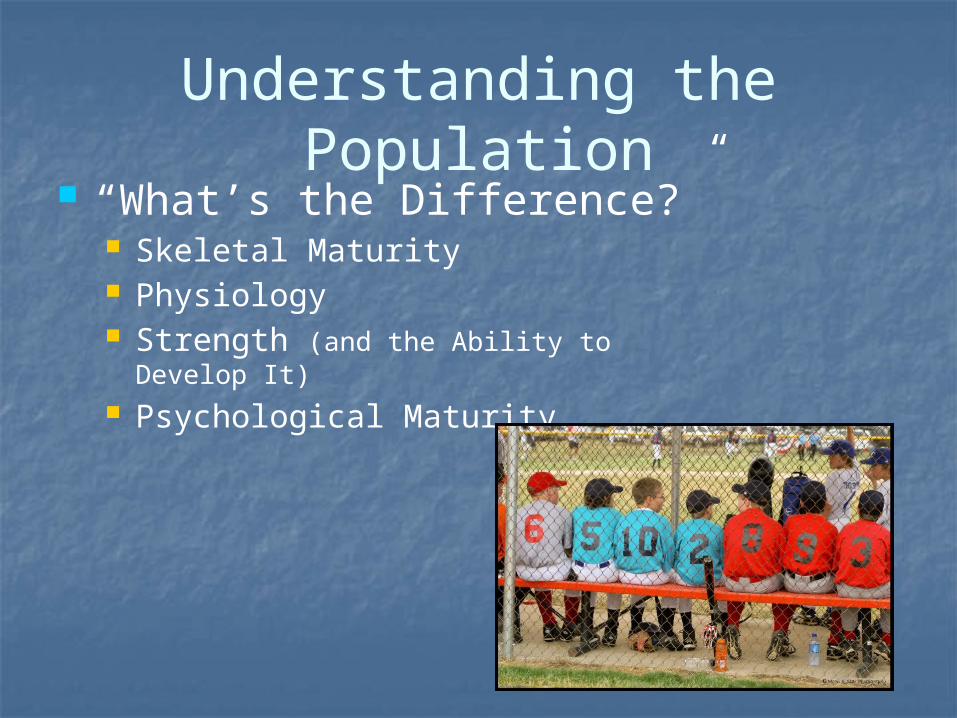
“What’s the Difference?” Skeletal Maturity Physiology Strength (and the Ability to Develop It) Psychological Maturity

Understanding the Population
“What’s the Difference?” Skeletal Maturity Physiology Strength (and the Ability to Develop It) Psychological Maturity
Growth & Development of the Young Athlete
Middle Childhood (6-9 yrs) Maturation of Throwing and Kicking Patterns Entry Level Sports (soccer, baseball/softball) Males and females can still compete with parity
Males slightly Stronger; Girls better Balance Running gait and speed are fairly equal
Late Childhood to Early Adolescence (10-15 yrs) Onset of Puberty “Growth Spurt” – Tanner Stage 3 Differences emerge between sexes Skill Acquisition and Development Easiest

Growth and Development of the Young Athlete
Late Adolescence/Adulthood (16-20 yrs) Increases in Strength & Size become more
gradual “Late Bloomers” may continue to lag behind Skeletal maturity
Growth and Development
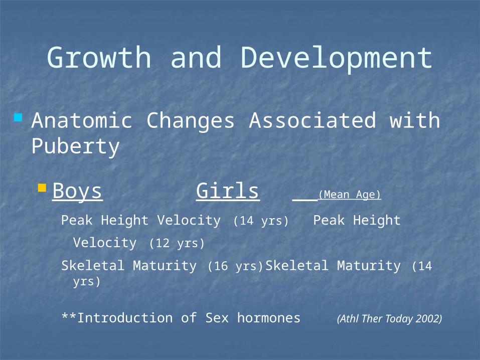
Anatomic Changes Associated with Puberty
Boys Girls (Mean Age)
Peak Height Velocity (14 yrs) Peak Height Velocity (12 yrs)
Skeletal Maturity (16 yrs) Skeletal Maturity (14 yrs)
**Introduction of Sex hormones (Athl Ther Today 2002)
Growth and Development
Significance of Peak Height Velocity The “Growth Spurt”
~ Tanner Stage 3 Bone growth rate can exceed soft tissue
accommodation Hamstrings, Hip flexors, Quadriceps, and
Plantarflexors Decreased Coordination Tightness can affect growth centers

Growth and Development
Significance of Tanner Staging 5 stages of Physical development
Stage 1 = Early Development Stage 5 = Full Maturity
Correlation between Tanner stage and physeal closure. Same Chronologic age ≠ Bone Age Assists with the differential diagnoses
Growth and Development
Tanner Stage 5 Signals end of growth Marked by full development of
secondary sexual characteristics Males will have full facial hair Females will have final breast development

Skeletally Immature Distinctions
Growth “Tissues” Physis Apophysis Articular Cartilage
Issues: Susceptibility to injury
Bone weakest link Surgical Challenges
“Growth Tissue” Physis (Growth Plate)
Responsible for longitudinal growth of bone
Growth centers close distal to proximal Growth centers begin to close in females
approximately 18 – 24 months following menarche
Skeletal Maturity Completed ~18 yrs females; ~21 yrs males
Injury to Physis could create growth disturbance (early closure or bony bridging)
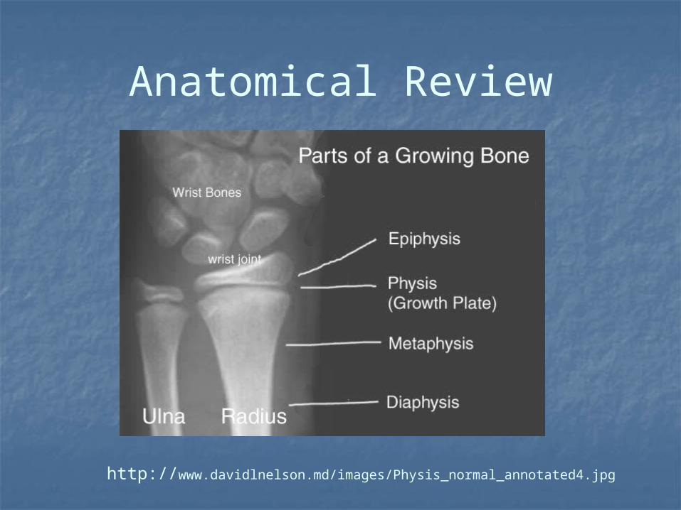
Anatomical Review
http://www.davidlnelson.md/images/Physis_normal_annotated4.jpg
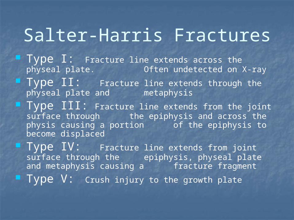
Salter-Harris Fractures Type I: Fracture line extends across the physeal
plate. Often undetected on X-ray Type II: Fracture line extends through the physeal
plate and metaphysis Type III: Fracture line extends from the joint surface
through the epiphysis and across the physis causing a portion of the epiphysis to become displaced
Type IV: Fracture line extends from joint surface through the epiphysis, physeal plate and metaphysis causing a fracture fragment
Type V: Crush injury to the growth plate

Salter Harris Fractures
Separated Above Lower Through E Rammed
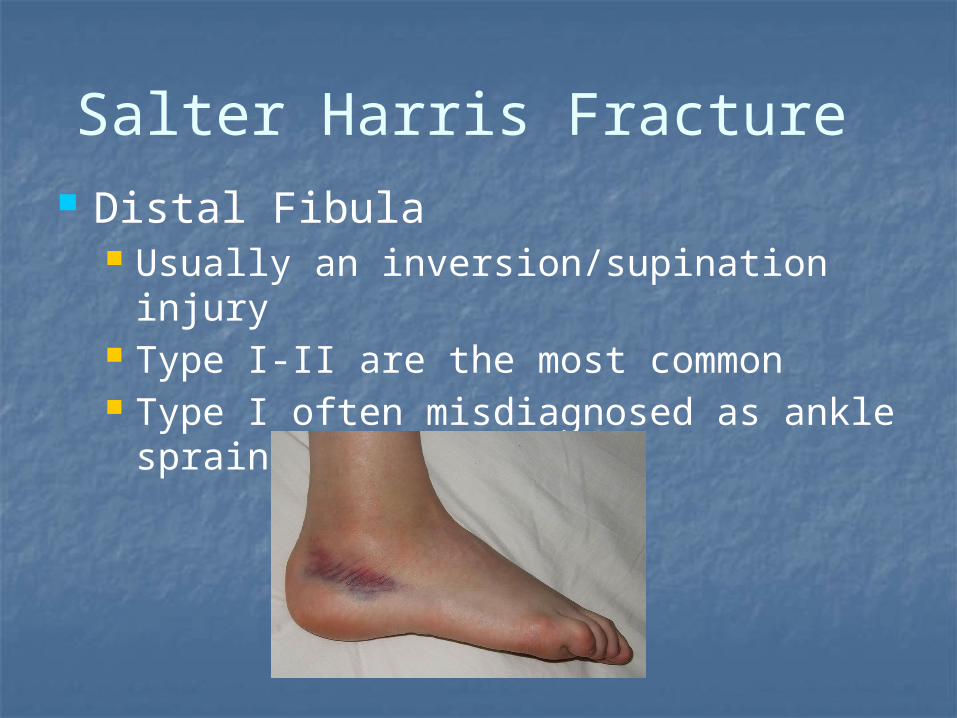
Salter Harris Fracture Distal Fibula
Usually an inversion/supination injury Type I-II are the most common Type I often misdiagnosed as ankle
sprain
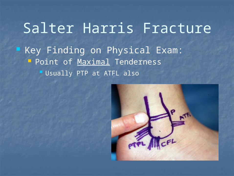
Salter Harris Fracture Key Finding on Physical Exam:
Point of Maximal Tenderness Usually PTP at ATFL also
Salter Harris Fracture
Boot immobilization (casting) Depending on Type; 2-3 weeks + Types III & IV require surgery
Pain free weight bearing status Rehabilitation for post
immobilization ROM, strength, balance & proprioception Sport specific training
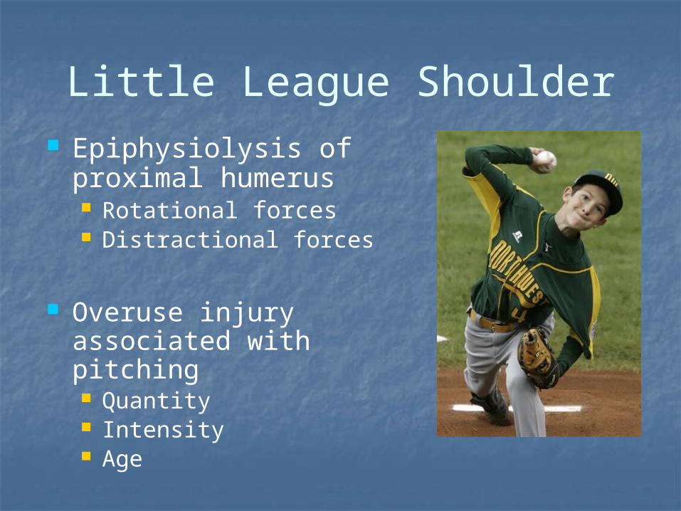
Little League Shoulder Epiphysiolysis of
proximal humerus Rotational forces Distractional forces
Overuse injury associated with pitching Quantity Intensity Age

Phases of Throwing
www.hughston.com/hha/b_16_1_1a.jpg

Little League Shoulder
Clinical Findings Lateral, proximal shoulder pain Weak & painful EROT and Abd Palpable tenderness over
physis Radiographic widening of
physis?
Little League ShoulderTreatment
Aggressive rest to allow physeal healing Address any ROM imbalances & Scapular
dysfunction GIRD, posterior capsule Sick Scapula Scapular stabilization & strengthening
Rotator cuff strengthening Review of throwing mechanics
Return to throwing progression Modification of throwing volume (pitch counts) May need to alter position
Address entire kinetic chain Core strengthening Lower extremity strength/flexibility and proprioception
“Growth Tissue”
Apophysis Cartilaginous structure usually located
at the end of long bones Attachment site for musculotendinous
unit Tensile forces can create inflammation =
Apophysitis Susceptible to Avulsion Fracture

Apophysitis
Overuse injury Often during periods of rapid growth May remain symptomatic until
closure of apophysis Possible to result in an avulsion
fracture

Sever’s Disease aka: Calcaneal
Apophysitis Common During Growth
Spurt Heel pain Tight gastroc/soleus Foot pronation Running/jumping athletes + Squeeze Test
Sever’s Disease Treatment
Activity modification Aggressive rest
Stretching!!! Immobilization may be necessary Can continue to play if pain is mild (no
limp) Typically resolves in several weeks
(months?) Footwear or insert
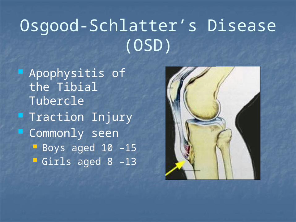
Osgood-Schlatter’s Disease (OSD)
Apophysitis of the Tibial Tubercle
Traction Injury Commonly seen
Boys aged 10 –15 Girls aged 8 –13

Osgood-Schlatter’s Disease (OSD)
Palpable tenderness X-rays may be
positive for displacement
In severe cases tubercle can avulse
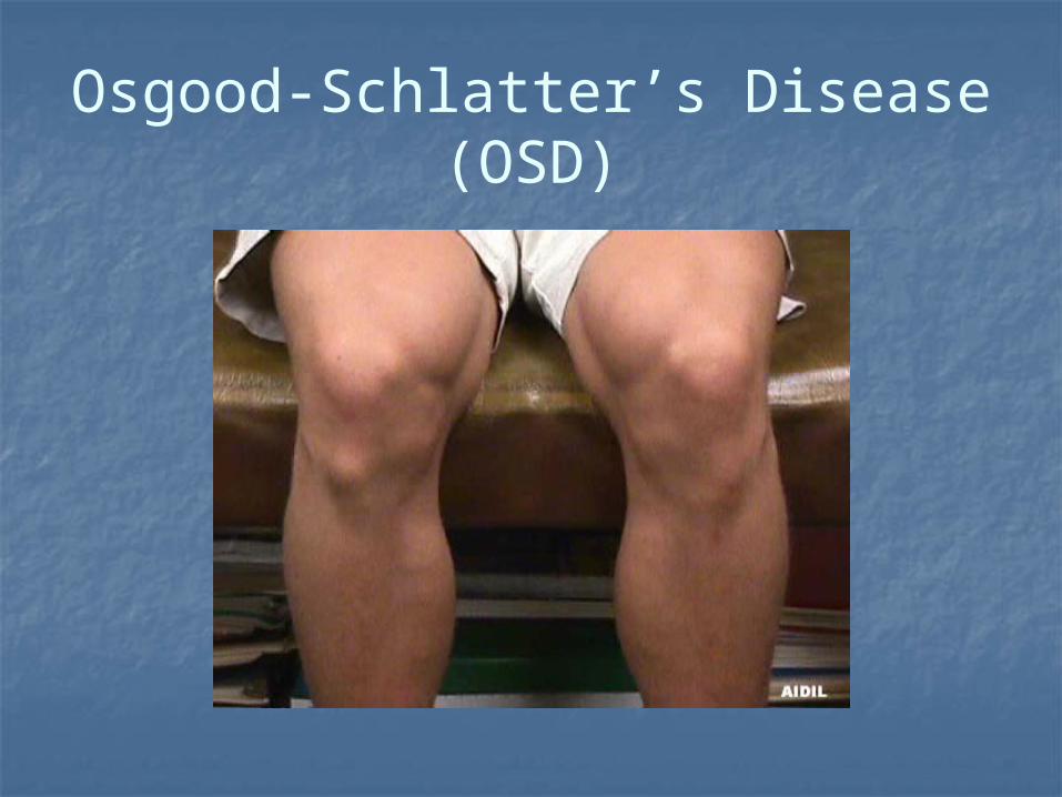
Osgood-Schlatter’s Disease (OSD)
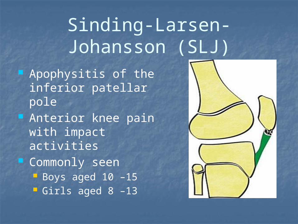
Sinding-Larsen-Johansson (SLJ)
Apophysitis of the inferior patellar pole
Anterior knee pain with impact activities
Commonly seen Boys aged 10 –15 Girls aged 8 –13

Sinding-Larsen-Johansson (SLJ)
Palpable tenderness Inferior pole sometimes
patellar tendon May have quadriceps
lag X-rays may be positive
for displacement Differential diagnosis
Patellar sleeve fracture
Treatment for OSD and SLJ
Activity modification Stretching quads and hams Strengthening progression Plyometric training to work on soft
landings May not have complete resolution of
symptoms In OSD permanent bump is likely
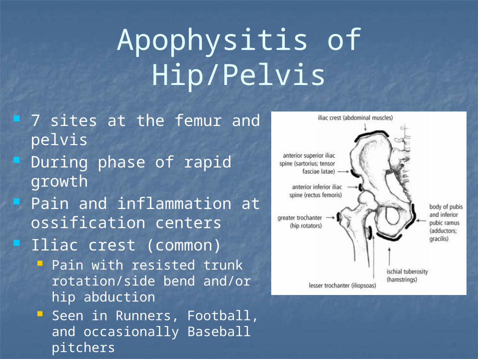
Apophysitis of Hip/Pelvis
7 sites at the femur and pelvis During phase of rapid growth Pain and inflammation at
ossification centers Iliac crest (common)
Pain with resisted trunk rotation/side bend and/or hip abduction
Seen in Runners, Football, and occasionally Baseball pitchers
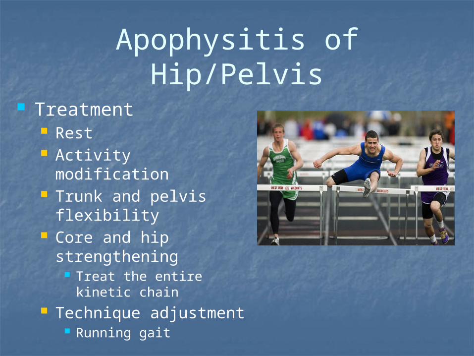
Apophysitis of Hip/Pelvis
Treatment Rest Activity modification Trunk and pelvis
flexibility Core and hip
strengthening Treat the entire kinetic
chain Technique adjustment
Running gait

Avulsion Fractures
Same areas affected as apophysitis Occur with sudden, forceful contraction or stretching
Bone is the weakest link Common sites include ASIS and Ischial tuberosity. Often misdiagnosed as pulled muscle Radiographic evaluation necessary for accurate
diagnosis Surgery if displacement is greater than 2-3cm (???)
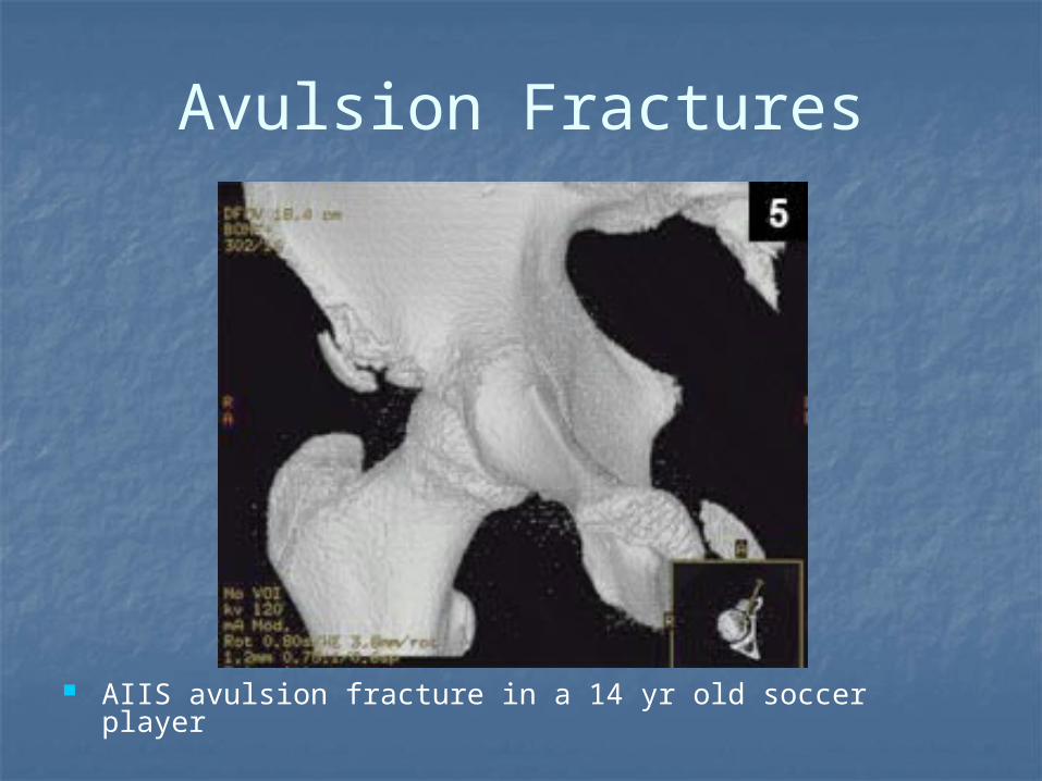
Avulsion Fractures
AIIS avulsion fracture in a 14 yr old soccer player
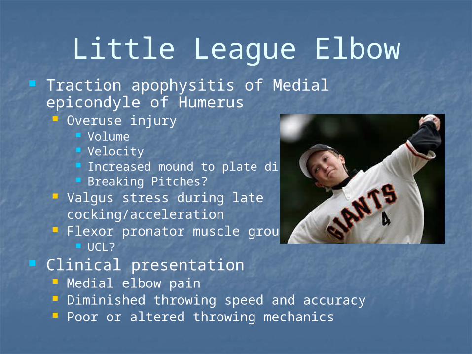
Little League Elbow Traction apophysitis of Medial epicondyle of
Humerus Overuse injury
Volume Velocity Increased mound to plate distance Breaking Pitches?
Valgus stress during late cocking/acceleration
Flexor pronator muscle group UCL?
Clinical presentation Medial elbow pain Diminished throwing speed and accuracy Poor or altered throwing mechanics
Little League Elbow Treatment
RICE: Make rest your friend Activity modification 6-12 weeks
No pitching or overhand throwing Stretching
GIRD is Probable; Assess and address!!! Strengthening
Forearm, posterior cuff, core, contralateral leg Assess throwing mechanics Functional progression to throwing program Identify and correct training errors
“Growth Tissue”
Articular Cartilage Infrastructure similar to Physis
Increased Cellular activity Not yet “Adult” solidity
Repetitive Injury or Excessive shearing forces may result in Osteochondritis Dissecans (OCD)
Osteochondritis Dissecans (OCD)
Impact and shear forces cause bone bruising
Cause is usually repetitive trauma Genetic predisposition?
Subchondral bone death Secondary damage to overlying cartilage “Lesion of dissection” vs dessication May affect any joint
Most frequently seen at knee, elbow, ankle

Osteochondritis Dissecans (OCD)
Risk Factors Age: Occurs most often in people between
the ages of 9 and 18
Sex: Males are 2-3X more likely than females.
Sports participation: Sports that involve rapid changes in direction, jumping or repeated throwing may increase your risk
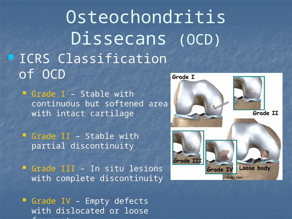
Osteochondritis Dissecans (OCD)
ICRS Classification of OCD Grade I – Stable with continuous
but softened area with intact cartilage
Grade II – Stable with partial discontinuity
Grade III – In situ lesions with complete discontinuity
Grade IV – Empty defects with dislocated or loose fragments

Osteochondritis Dissecans (OCD)
Epiphyseal microtrauma with osteochondral separation
Commonly Lateral aspect of Medial femoral condyle
Etiology is multifactorial Trauma, ischemia, hereditary,
idiopathic (?) Under debate

Osteochondritis Dissecans (OCD)

OCD of Femoral Condyle
Clinical presentation Insidious onset of pain aggravated by
activity Intermittent joint effusion Giving way, catching, or locking Symptoms suggestive of PFPS Confirmed with diagnostic imaging
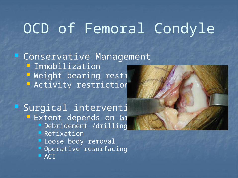
OCD of Femoral Condyle
Conservative Management Immobilization Weight bearing restriction Activity restriction
Surgical intervention Extent depends on Grade
Debridement /drilling Refixation Loose body removal Operative resurfacing ACI

Clinical Summary Bone weakest link in pre pubescent Same Chronological age ≠ Bone Age
Tanner staging helps differential Protect Growth centers

THANK YOU!!!

Ken Knecht PT, MS, SCS, CSCSBoard Certified Sports Clinical SpecialistThe Sports Medicine & Performance Center at CHOP Specialty Care Center at Virtua Health and Wellness Center, 2nd Floor200 Bowman Drive, Suite D260Voorhees, NJ 08043856-719-9932; Fax: 267-425-5416
Recommended