608 AMERICAN JOURNAL OF OPHTHALMOLOGY APRIL, 1980
This unique observation has not received its due consideration in publishedreports on laser safety to date, eventhough it may have significant implications upon the establishment of lasersafety criteria for different races. Muchresearch is required to clarify this issuefurther.
JOSEPH MOISSEIEV, M. D.
AND
ELISHA BARTOV, M.D.
Tel Aviv, Israel
REFERENCES
1. Beatrice E. S., and Velez S.: Laser bioeffectsresearch. Histopathology of retinal effects. SPIE vol.162 Visual and Image Realism, 1978, pp 103-106.
2. Marshal J.: Thermal and mechanical mechanisms in laser damage to the retina. Invest. Ophthalmol. 9:97, 1970.
3. Marshal J., Hamilton A. M., and Bird A. cHistopathology of ruby and argon laser lesions inmonkey and human retina, a comparative study. Br.J. Ophthalmol. 59:610, 1975.
4. Wheeler C. B.: Calculation of retinal temperature distribution resulting from laser irradiation ofthe eye: 1. Continuous lasers. Phys, Med. BioI.21:616, 1976.
5. Lappin P. W.: Assessment of ocular damagethresholds for laser radiation. Am. J. Ophthalmol.48:600, 1971.
6. Gerraets W. J., Williams R. C., Chan G., HamW. T., Guerry D., and Schmidt F. H.: The relativeabsoption of thermal energy in retina and choroid.,Invest. Ophthalmol. 1:340, 1962.
Inexpensive Support for AnteriorSegment Photography
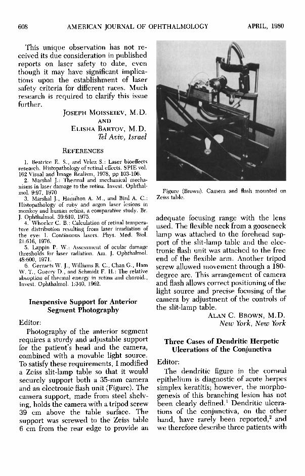
Editor:Photography of the anterior segment
requires a sturdy and adjustable supportfor the patient's head and the camera,combined with a movable light source.To satisfy these requirements, I modifieda Zeiss slit-lamp table so that it wouldsecurely support both a 35-mm cameraand an electronic flash unit (Figure). Thecamera support, made from steel shelving, holds the camera with a tripod screw39 em above the table surface. Thesupport was screwed to the Zeiss table6 em from the rear edge to provide an
Figure (Brown). Camera and flash mounted onZeiss table.
adequate focusing range with the lensused. The flexible neck from a goosenecklamp was attached to the forehead support of the slit-lamp table and the electronic flash unit was attached to the freeend of the flexible arm. Another tripodscrew allowed movement through a 180degree arc. This arrangement of cameraand flash allows correct positioning of thelight source and precise focusing of thecamera by adjustment of the controls ofthe slit-lamp table.
ALAN C. BROWN, M.D.
New York, New York
Three Cases of Dendritic HerpeticUlcerations of the Conjunctiva
Editor:The dendritic figure in the corneal
epithelium is diagnostic of acute herpessimplex keratitis; however, the morphogenesis of this branching lesion has notbeen clearly defined. 1 Dendritic ulcerations of the conjunctiva, On the otherhand, have rarely been reported.! andwe therefore describe three patients with
Recommended