DEPARTMENT OF MICROBIOLOGY, TUMOR AND CELL
BIOLOGY (MTC) Karolinska Institutet, Stockholm, Sweden
Byron Leiva Torres
Stockholm 2009
All previously published papers were reproduced with permission from the publisher. Published by Karolinska Institutet. Printed by Universitetsservice USAB © Byron Leiva Torres, 2009 ISBN 9789174094602
2009
Gårdsvägen 4, 169 70 Solna
Printed by
From Department of Microbiology and Parasitology, University of León, Nicaragua (UNANLeón). Department of Microbiology, Tumor and Cell Biology (MTC), Karolinska Institutet, Stockholm, Sweden.
Byron Leiva Torres
Academic dissertation for the degree of Doctor of Medical Sciences from Karolinska Institutet. External Examiner: Docent Agneta AustKettis. Läkemedelsverket, Uppsala. Examination Board: Prof. Marita TroyeBlomberg. Stockholms Universitet
Prof. Sven Britton. Karolinska Institutet /Institutionen för Infektionssjukdomar. Prof. Anders Örn. Karolinska Institutet/ Institutionen för Mikrobiologi, Tumör och Cellbiologi (MTC).
Stockholm 2009
UNANLeón

1

2
3
About ten yearly cases of liver abscess are presented in University Hospital, León, Nicaragua. Based on serology most of them have been shown to be amoebiasis cases. This raised the question of Entameoba histolytica prevalence in the population. Based on microscopy of stool specimens and serology, about 20% prevalence was found. With the new realization that pathogenic E. histolytica is morphologically indistinguishable from some common apathogenic species such as E. dispar (cysts detected by microscopy are reported as E. histolytica/ E. dispar), the aim was to determine the true prevalence of E. histolytica using various tests designed to differentiate between pathogenic and apathogenic species. In 480 apparently healthy individuals, the prevalence of E. histolytica/ E. dispar was 12% (58/480) as determined by microscopy. Out of these 58 stool samples an E. histolytica specific PCR was positive in 5%; thus the prevalence E. histolytica was 0.6%. In a group of 134 diarrhea patients, the most common finding was E. histolytica/ E. dispar (24%) at the health center laboratory level. In the Microbiology Department E. histolytica/ E. dispar was found only in 4.5%. With the Triage Parasite test, only one case of E. histolytica/ E. dispar was found. By PCR, E. dispar was recognized in 10 (7.5%) and E. histolytica in two cases (1.5%). Over diagnosis was also confirmed in a quality control study where León health centers were examining 10 different stool samples. We found that the health center technicians continue to mix up E. histolytica/ E. dispar with other amoebas. The consequences of the apparent widespread over diagnosis of E. histolytica were studied retrospectively in 100 records of patients with intestinal symptoms. We found that all patients received treatment with metronidazole or related drugs. In 41% these treatments were not based on any laboratory findings at all. In 32 % E. histolytica/E. dispar were found. Other parasites (Entamoeba coli, Giardia intestinalis, Endolimax nana, Enterobius. vermicularis, Iodamoeba bütschlii) were seen in 27%. To explain the high seroprevalence of antiE.histolytica antibodies in seroepidemiological surveys we considered two possibilities: cross reactivity due to the common intestinal apathogenic E. dispar and antibodies to ubiquitous freeliving environmental amoebas. A study was undertaken to identify environmental amoebas and to determine crossreactivity using antibodies from amoebiasis patients. Amoebas isolated from environmental water samples were characterized by morphological and immunohistochemical methods. In fresh water Acanthamoeba spp. were found in 21 %. Fifty three percent of tested wells in the geothermal area contained thermotolerant amoeboflagellates. aegleria spp. was identified in 24 out of 39 (62 %) of isolated amoeboflagellates. Absorption studies did not support the idea that environmental freeliving amoebas induce antibodies crossreacting with E. histolytica. Antigenic cross reactivity between E. dispar and E. histolytica remains a possible explanation for the high seroprevalence in the population. This is supported by the observation that IF antibody titers in sera from healthy individuals are similar with both antigens. Also the ratio of antibody reactivity was similar when measured by a densitometric method. In contrast, sera from patients with invasive amoebiasis reacted preferentially with E. histolytica. We conclude that amoebiasis is not a major problem in the community. Overdiagnosis and overtreatment of diarrhea patients thought to suffer from amoebiasis are serious problems. Thus there is an urgent need for education and quality assessment.
4
This thesis is based on the following publications:
I. Leiva, B., Lebbad, M., Téllez A., WinieckaKrusnell, J. Altamirano, I. and Linder E. Overdiagnosis of and in icaragua: A Microscopic, Triage Parasite Panel and PCR Study. Archives of Medical Research 37 (2006) 529–534.
II. Leiva, B., Clasdotter, E., Linder E. and WinieckaKrusnell J. Freeliving Acanthamoeba and aegleria spp. amebae in water sources of León, icaragua. Rev. Biol. Trop. 56 (2008): 439446.
III. Leiva, B., Lebbad, M., WinieckaKrusnell, J., Argueñal, H. and Linder, E. Infection with intestinal parasites and their relation to poverty in León, icaragua. (Submitted)
IV. Leiva B, WinieckaKrusnell, J. and Linder E. Amoebiasis in León, icaragua: overdiagnosis and overtreatment. (Submitted)
5
1 Introduction...................................................................................................9
1.1 Entamoeba histolytica.........................................................................9 1.1.1 Overview.................................................................................9 1.1.2 History.....................................................................................9 1.1.3 Biology of the parasite..........................................................10
1.2 Epidemiology ....................................................................................11 1.3 Pathogenesis ......................................................................................12
1.3.1 Early invasive lesion with superficial ulceration.................12 1.3.2 Virulence factors...................................................................12
1.4 Immunity ...........................................................................................13 1.4.1 Innate resistance mechanisms ..............................................14 1.4.2 T cell responses.....................................................................14 1.4.3 Humoral response .................................................................14
1.5 Clinical features ................................................................................15 1.5.1 Asymptomatic colonization..................................................15 1.5.2 Dysentery or colitis...............................................................15 1.5.3 Extra intestinal amoebiasis ...................................................15 1.5.4 Clinical diagnosis..................................................................15
1.6 Laboratory diagnosis.........................................................................16 1.6.1 Microscopical examination ..................................................16 1.6.2 Culture and isoenzymes........................................................17 1.6.3 Antibody detection................................................................17 1.6.4 Antigen assays for the diagnosis of amoebiasis...................18 1.6.5 PCRbased methods..............................................................18
1.7 Therapy..............................................................................................19 1.8 Serological diagnostic problems in amoebiasis ...............................19
1.8.1 Free living amoebas: Introduction........................................19 1.8.2 Naegleria spp. .......................................................................20 1.8.3 Acanthamoeba spp................................................................20
2 Aims of the study........................................................................................21 2.1 General aims......................................................................................21 2.2 Specific aims .....................................................................................21
3 Materials and Methods ...............................................................................22 3.1 Prevalence of intestinal parasites......................................................22
3.1.1 Study area..............................................................................22 3.1.2 Study group and Collection material....................................22
3.2 Diagnostic methods...........................................................................23 3.2.1 Microscopical examination of fecal specimens ...................23 3.2.2 Antigen detection..................................................................24 3.2.3 PCR amplification ................................................................24 3.2.4 Antibody detection................................................................25
3.3 Detection of free living amoebas in water sources ..........................26 3.3.1 Sample collection and cultivation of amoebas ....................26 3.3.2 Identification of isolates .......................................................26
3.4 Statistical analysis .............................................................................26
6
4 Results and Discussion...............................................................................27 4.1 True Prevalence of E. histolytica and E. dispar (Papers I and III)..27 4.2 Over diagnosis and Over treatment (Paper IV) ...............................29 4.3 Free living amoebas in water sources (Paper II)..............................29 4.4 Significance of high seroprevalence (Unpublished)........................33 4.5 Future perspectives ...........................................................................35
5 Concluding remarks....................................................................................37 6 Acknowledgements ....................................................................................38 7 Bibliography ...............................................................................................39
7
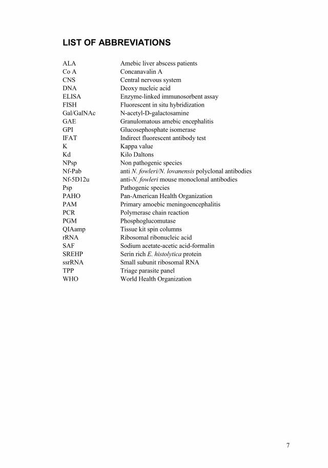
ALA Amebic liver abscess patients Co A Concanavalin A CNS Central nervous system DNA Deoxy nucleic acid ELISA Enzymelinked immunosorbent assay FISH Fluorescent in situ hybridization Gal/GalNAc NacetylDgalactosamine GAE Granulomatous amebic encephalitis GPI Glucosephosphate isomerase IFAT Indirect fluorescent antibody test Κ Kappa value Kd Kilo Daltons NPsp Non pathogenic species NfPab anti . fowleri/. lovanensis polyclonal antibodies Nf5D12u anti. fowleri mouse monoclonal antibodies Psp Pathogenic species PAHO PanAmerican Health Organization PAM Primary amoebic meningoencephalitis PCR Polymerase chain reaction PGM Phosphoglucomutase QIAamp Tissue kit spin columns rRNA Ribosomal ribonucleic acid SAF Sodium acetateacetic acidformalin SREHP Serin rich E. histolytica protein ssrRNA Small subunit ribosomal RNA TPP Triage parasite panel WHO World Health Organization

8
9
The clinical symptoms of amoebiasis caused by the protozoan parasites Entamoeba histolytica ranges from asymptomatic colonization to amoebic dysentery and invasive extra intestinal amoebiasis is most common in the form of liver abscess (Tanyuksel and Petri 2003). Invasive amoebiasis is one of the world’s most prevalent and fatal infectious diseases. Primarily it is a problem of the developing world; around 500 million people are infected world wide while 75,000 die of the disease annually. Behind malaria and schistosomiasis it ranks third on the list of parasitic causes of death worldwide (Walsh 1986; Li and Stanley 1996). E. histolytica and E. dispar are currently recognized as distinct species, mostly based on genetic, biochemical, and immunological studies. It is therefore possible to obtain more reliable and correct epidemiological data using molecular, biochemical, and immunological features, and these allow better diagnosis and treatment (Diamond and Clark 1993).
Although only a minority of E. histolytica infections progress to develop intestinal diseases, such as diarrhea or dysentery, or extra intestinal diseases like amoebic liver abscess (ALA), the basis for this difference in clinical outcome remains unsolved. A recent report suggests that the parasite genotype plays a role in determining outcome of infection by E. histolytica (Ali, Mondal et al. 2007). Therefore, one of the current priorities in a post genomic era is to understand the genomic factors determining the outcome of an E. histolytica infection (Ali, Clark et al. 2008).
An excellent review at history of amoebiasis have been published by Jonathan I Ravdin (Ravdin 2000). In 1875, the Russian physician Fedor Lösch identified what he believed to be the causative agent (motile amoebas containing erythrocytes which he named Amoeba coli) of a case of dysentery, but the doubted its lone role in pathogenesis when it failed to produce disease in three of four dogs experimentally inoculated with it. In 1893 the German pair Quincke and Roos identified an important mode of transmission, when they described 15 diseased patients who all shared the same drinking water source (Guarner 1990).
In 1925, Brumpt proposed that humans can be infected by two morphologically identical species of Entamoeba producing quadrinucleate cysts measuring 10 µm or greater in diameter; the pathogenic organism was identified as E. dysenteriae and Brumpt named the nonpathogenic one E. dispar. It took almost 70 years and the advent of modern biochemical techniques to reestablish Brumpt´s idea (Diamond and Clark 1993).
In 1973, pathogenic (P) and nonpathogenic (NP) species were differentiated by their relative agglutinability with the lectin Con A (MartinezPalomo, GonzalezRobles et al. 1973). This was followed in 1978 by the discovery that pathogenicity could also be
10
correlated the enzymatic profile of the organism. Sargeaunt and colleagues used thinlayer starchgel electrophoresis to separate isoenzyme variants of the glycolytic enzymes glucosephosphate isomerase (GPI), phosphoglucomutase (PGM) and Lmalate:NADP oxidoreductase and classifed 85 stocks of E. histolytica in four groups according to their enzyme profile. Enzyme Group II, which contained a faster migrating band of PGM, was found in all cases of clinical amoebiasis, but it was not found in any isolated obtained from asymptomatic individuals, although Sargeaunt pointed out that in a larger study such persons (in a preclinical state of disease) may be found (Lucas and Upcroft 2001). Diamond and Clark presented a host of data to confirm the existence of E. histolytica and E. dispar, and to honor the hypothesis put forward by Brumpt (Diamond and Clark 1993). Biochemically, they cited thousands of samples which correlated zymodeme class to pathogenicity (Ravdin 2000; Lucas and Upcroft 2001).
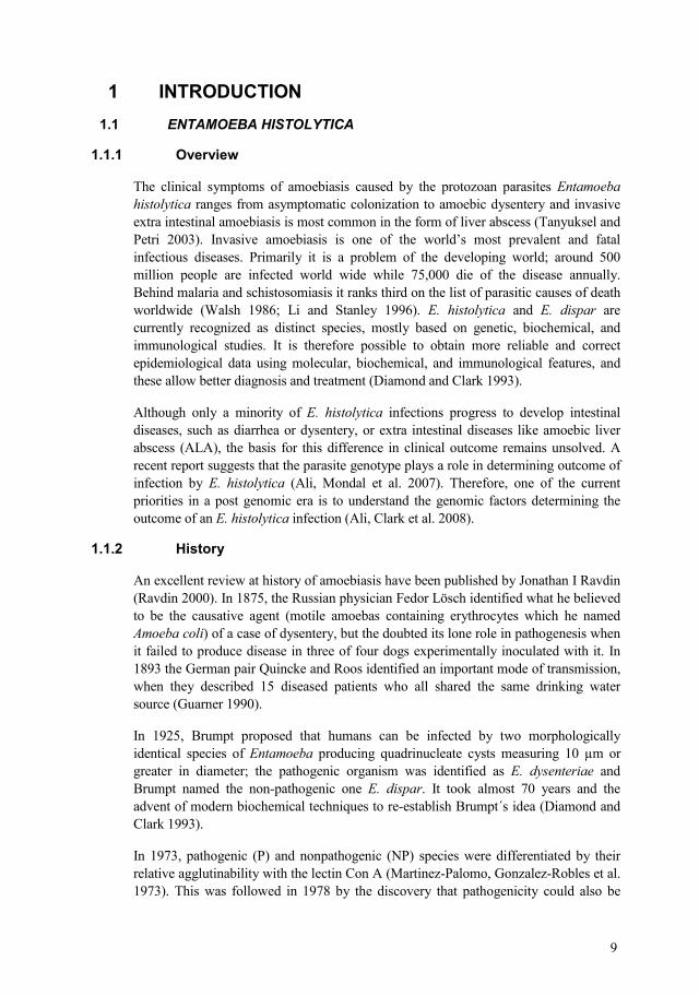
E. histolytica exists in two forms: a motile and invasive trophozoite and an infective cyst.
The trophozoite varies in size; from 12 to 30 m, although occasionally very large forms up to 90 m may be seen. The pseudopods produced by an actively moving trophozoite are broad and fingerlike and the parasite generally moves in one direction at a time. The nucleus is spherical and has a delicate nuclear membrane.
The cyst initially has a single nucleus but finally has 4 nuclei. It is then known as a mature cyst. E. histolytica cysts are round or slightly oval hyaline bodies 10 µm – 16 µm in diameter.
Life cycle. There are four stages in the life cycle of E. histolytica: the trophozoite, precyst, cyst and metacyst. Infection is normally initiated by the ingestion of fecally contaminated water or food containing E. histolytica cysts. The infective cyst form of the parasite survives passage through the stomach and small intestine. Excystation occurs in the bowel lumen, where motile and potentially invasive trophozoites aggregate in the intestinal mucin layer and form new cysts, resulting in a selflimited and asymptomatic infection. The trophozoite of E. histolytica can convert to a precyst form with a nucleus, and this form matures into a tetranucleated cyst as it migrates down and out the colon. In some cases, however, adherence to and lysis of the colonic epithelium, mediated by the galactose and NacetylDgalactosamine (Gal/GalNAc)specific lectin, initiates invasion of the colon by the trophozoites (Petri 2002). Once the intestinal epithelium is invaded, extra intestinal spread to the peritoneum, liver, and other sites may follow. Factors controlling invasion, as opposed to encystations, most likely include parasites “quorum sensing” signaled by the (Gal/GalNAc)specific lectin, interaction of amoebas with the bacterial flora of the intestine, and innate and acquired immune responses of the host (Haque, Mondal et al. 2003; Tanyuksel and Petri 2003).
11
In 1986 Walsh assessed the existing global prevalence data and concluded that in 1981, 480 million people harbored E. histolytica worldwide (Walsh 1986). She went on to extrapolate that 36 million develop clinically overt disease with 40100.000 deaths per year, resulting in amoebiasis ranking third most common cause of death due to parasitic infection after malaria and shistosomiasis. As this data appeared well before the formal redescription of E. histolytica was published (Diamond and Clark 1993), the epidemiological estimations quoted did not distinguish between E. dispar and E. histolytica. Unfortunately, reliable epidemiological data on populations that do not have selection bias and from whom confirmed E. histolytica and E. dispar isolations have been made are rare. Such isolation studies from Durban, South Africa (Gathiram and Jackson 1985), have indicated the overall prevalence of both E. histolytica and E. dispar together to be 10%, with E. histolytica accounting proportionately for 1 % and E. dispar for 9 %. Consequently, global prevalence of E. histolytica could be 50 million and that of E. dispar 450 million. Revisions of the prevalence and incidence estimations are necessary while that of mortality would remain the same. Amoebiasis infections are endemic in most temperate and tropical climates in the developing world. In some tropical countries, antibody prevalence rates exceed 50% (CaballeroSalcedo, ViverosRogel et al. 1994; Tanyuksel and Petri 2003).
Life cycle of Entamoeba histolytica Humans become infected by the oral route by the encysted parasite. Excystation takes place in the intestinal tract and causes local symptoms like diarrhea. The liberated trophozoite form may also invade the intestinal wall and extraintestinal tissues of the host. Severe disease is caused by tissue destruction e.g. in the liver and brain.
Figure 1
12
Epidemiology studies have showed that low socioeconomic status and unsanitary conditions are significant independent risk factor for infection. In addition people living in developing countries have a higher risk earlier age of infection than do those in developed regions. In Mexico, 11 % of the tested population aged 5 to 9 years was infected, with the prevalence of infection being higher in girls (9.34%) (CaballeroSalcedo, ViverosRogel et al. 1994).
The discovery of a third morphologically identical Entamoeba spp. further complicated our understanding of the epidemiology of E. histolytica. Entamoeba moshkovskii is another species of Entamoeba morphologically indistinguishable from E. histolytica and E dispar. This species first described from Moscow sewage by Tshalaia in 1941 and was thereafter reported to occur in many different countries. E. moshkovskii was initially thought to be a freeliving environmental strain. However, in 1961 an E. histolyticalike strain was isolated from a resident of Laredo, Texas. This strain was named the E. histolytica Laredo strain shares many biological features with E. moshkovskii. Both the Laredo strain and E. moshkovskii grow at room temperature and are resistant to emetine. These characteristics distinguished them from E. histolytica and E. dispar. Subsequent molecular studies have confirmed that the E. histolytica Laredo strain is a strain of E. moshkovskii. Although the early isolations of this species have been from sewage, recent studies have reported the recovery of E. moshkovskii from human feces (Fotedar, Stark et al. 2007; Ali, Clark et al. 2008; Pritt and Clark 2008).
The term “pathogenesis”, defined as the mechanisms involved in the initiation, evolution, and ultimate outcome of a disease process, relates to both host and parasite factors.
Three main consecutive events occur at this stage: a focal superficial erosion of the mucosa, small glandular foci of micro invasion, and mild to moderate infiltration of the lamina propria.
Superficial epithelial erosion. Once the mucus barrier has been broken down E. histolytica reaches the luminal surface of enterocytes and initial contact produces a contactdependent “hitandrun” damage to the plasma membrane of effectors cells, phagocytosis, and intracellular degradation of ingested cells (MartinezPalomo, GonzalezRobles et al. 1985).
The adhesion and cytolytic events have been related to three types of molecules: lectins, amoebapores, and proteases.
13
1.3.2.1 GalGalac lectin.
Excystation in the intestinal lumen produces trophozoites that use the galactose and NacetylDgalactosamine (Gal/GalNac)specific lectin to adherence to colonic mucins and thereby colonize the large intestine.
Adhesion of the parasite occurs mainly through a surface Gal/GalNac lectin which binds to exposed terminal Gal/GalNac residues of target cell glycoproteins (Petri, Chapman et al. 1989). Other molecules include a 220kd lectin, a 112kd adhesin and a surface lipophosphoglycan. It has been suggested that the Gal/GalNac lectin could be translocated to the basolateral surface of enterocytes before the parasite excretes its cytolytic effect, but the benefits of this action for the parasite remain unclear (EspinosaCantellano and MartinezPalomo 2000).
Colitis results when the trophozoite penetrates the intestinal mucous layer, which otherwise acts as a barrier to invasion by inhibiting amoebic adherence to the underlying epithelium and by slowing trophozoites, which occurs only after the parasite lectin engages host NacetylDgalactosamine on Olinked cell surface oligosaccharides (Petri 2002).
1.3.2.2 Amoebapores.
Secretion by the ameba of amoebapores, a 5Kd pore forming protein, may contribute to killing. Structural modeling suggests a compact tertiary structure composed of four αhelical structures stabilized by three disulfate bonds. Thus, amoebapores are different from the much larger (65 to 70 kDa) perforins that contain three amphipathic segments, two αhelices and one βsheet. Activation of human caspase 3, a distal effectors molecule in the apoptotic pathway, occurs rapidly after amoebic contact, and caspases are required for cell killing in vitro and for the formation of amoebic liver abscess in vivo (Huston, Houpt et al. 2000; Yan and Stanley 2001).
1.3.2.3 Cysteine proteinase
Interaction of the parasite with intestinal epithelium causes an inflammatory response marked by the activation of nuclear factor κB and the secretion of lymphokines (Stanley 2001). The development of this epithelial response may depend on trophozoite virulence factors such as cysteine proteinase and leads to intestinal abnormalities through neutrophilmediated damage. Neutrophils can also be protective, however, since activation of neutrophils or macrophages by tumor necrosis factor α or interferon γ kills amoebas in vitro and limits the size of amoebic liver abscess (Denis and Chadee 1989). In contrast to the intense inflammatory response typical of early invasive amoebiasis, inflammation surrounding wellestablished colonic ulcers and liver abscesses in minimal, given the degree of tissue damage.
In addition, other amoebic molecules such as lipophosphopeptidoglycan, perioxiredoxin, arginase, and lysine and glutamic acidrich proteins are also implicated in the pathogenicity (Lejeune, Rybicka et al. 2009).
Immunity to infection with E. histolytica is associated with a mucosal IgA response against the carbohydraterecognition domain of the Gal/GalNac lectin. Cellmediated
14
responses have been described in patients with amoebic liver abscess, characterized by lymphocyte proliferation and lymphokine secretion that is amoebicidal in vitro. In patients with liver abscess, the prevalence of the class II MHC haplotype HLADR3 is increased by a factor of more than three, suggesting a role of CD4+ Tcell functions in the outcome of the disease (Arellano, PerezRodriguez et al. 1996).
Initial host resistance to E. histolytica is dependent on natural or innate resistance mechanisms prior to the development of acquired immunity. Amoebas encounter natural “barriers” to infection in both the gut and systematic circulation following tissue invasion. These innate defences may prevent or partially inhibit amoebic tissue invasion, although the level of protection afforded by innate resistance is difficult to quantify precisely. Mucins lining the colonic epithelium provide an important innate defence against tissue invasion by E. histolytica. Therefore, mucins serve as a first line of host defences in the gut by inhibiting amoebic attachment to the underlying mucosal epithelial cells. In the gut, other factors such as competition by intestinal bacteria for attachment to mucins may inhibit amoebic colonization (Ravdin 1989).
In the systematic circulation, E. histolytica is exposed to complement, another innate resistance mechanism that may act to decrease the numbers of invading amoebas. Amoebas activated both the alternative and classical complement pathways in normal serum (Calderon and Schreiber 1985).
A role for antigenspecific T cells in controlling invasive amoebiasis is evident from studies demonstrating that thymectomy or treatment with antiT lymphocyte serum result in exacerbation of infection in animal models of the disease. T cells may participate in an immune response against E. histolytica in three distinct ways: 1 by directly lysing amoebas in a contact dependent manner; 2 by producing cytokines which activate macrophages (and possibly other effectors cells) for amoebicidal activity and; 3 by providing help for B cell antibody production (Ravdin 2000).
Seroepidemiologic studies indicate that 81% to 100% of patients with invasive amoebiasis develop specific circulatory antibodies against E. histolytica (Petri and Ravdin 1986).
Apart from serum and secretory IgA, the dominant class of antibodies produced against E. histolytica appears to be IgG. Both the Gallectin and SREHP molecules, for example, stimulated strong serum IgG responses in immunized animals. Serum IgM has been detected in human cases of intestinal and extraintestinal disease, whereas IgE may be produced during amoebiasis but definitive reports are rare. How the humoral response relates to development of cellular mediated immunity (CMI) is unclear. Th1 and Th2type responses are linked to production of specific antibody subclasses (Mosmann and Coffman 1989). The frequencies of different antibody subclasses in amoebiasis remain to be investigated. As part from a possible contribution to resistance
15
or control of infection, limited reports suggest that antiamoebic antibodies may have deleterious consequences and exacerbate disease. Antibodies to the Gallectin have been shown to enhance of trophozoites to target cells, which may results in increased pathology due to elevated killing of host cells. In one study of amoebiasis patients, immune complexes, which may contribute to pathogenesis, were detected in patients colonic and liver tissue (Ravdin 2000).
Asymptomatic infection with E. histolytica is defined as the presence of E. histolytica in stool in the absence of colitis or extra intestinal infection. Colonization with the morphologically identical parasite, E dispar is three times more common in developing countries and at least 10 times more common in developed countries (Haque, Faruque et al. 1997).
Patients with amoebic colitis typically present with a severalweek history of gradual onset of abdominal pain and tenderness, diarrhea, and bloody stools. In one series, patients with amoebic colitis had an average duration of prehospital illness of 21 days, compared with 4 days for patients with shigellosis. Because of the gradual onset, weight loss is a common finding. Surprisingly, fever is present in only the minority (838%) of patients with amoebic colitis. Colonic lesions can vary from mucosal thickening only to flaskshaped ulcerations to necrosis of the intestinal wall (Tanyuksel and Petri 2003).
The most common extra intestinal manifestation is amoebic liver abscess (ALA), which is associated with significant morbidity and mortality. This was a progressive and almost invariable fatal disease little more than a century ago, but since the introduction of effective medical treatment and rapid diagnosis, mortality has fallen to 1 3 %. Complications of extra intestinal amoebiasis include pleuropulmonary amoebiasis secondary to ALA rupture through the diaphragm, brain abscess, and genitourinary amoebiasis. Diagnosis of liver abscess is confirmed by a positive serological test, as amoebic serology is highly sensitive (> 94%) and highly specific (> 95%) for diagnosis. Abdominal ultrasound or computed tomography scan does not provide specificity for ALA. However, a positive serological test in combination with abdominal images are helpful for diagnosis where PCR is not routinely available (Tanyuksel and Petri 2003).
The differential diagnosis of an illness with diarrhea containing gross or occult blood should include infectious etiologies (including amoebiasis and infections with Shigella sp, Salmonella, Campylobacter, Mycobacterium tuberculosis and enteroinvasive and enterohemorrhagic Escherichia coli) and noninfectious causes (including inflammatory bowel disease, carcinoma, ischemic colitis, arteriovenous malformation, and
16
diverticulitis) (Fotedar, Stark et al. 2007). It can be difficult to make the diagnosis of amoebic colitis, since the presentation of the illness may be insidious or chronic, bleeding may occur without diarrhea, and fever is an unusual finding. Once the diagnosis can also be problematic, because a single stool examination for parasites is insensitive, histopathologic confirmation of infection on biopsy specimens may be difficult, and serological tests for antibodies to amoebas are not always positive. The best initial diagnostic approach at this time is detection of E. histolytica antigen in stool (Petri and Singh 1999). Unusual manifestations of amoebic colitis include acute necrotizing colitis, amoeboma (granulation tissue in colonic lumen mimicking colonic cancer in appearance), cutaneous amoebiasis, and rectovaginal fistulas. Acute fulminant or necrotizing colitis occurs in ≈0.5% of cases, usually requires surgical intervention, and has a mortality of >40%. Abdominal pain and distension and rebound tenderness are present in most patients with fulminant colitis, and identification for surgery includes perforation and persistence of abdominal distention and tenderness while undergoing antiamoebic therapy. Partial or total colostomy with exteriorization of the ends is recommended over primary anastomosis, since anastomosis may fail because of the friable condition of the bowel (Aristizabal, Acevedo et al. 1991; Pritt and Clark 2008).
Laboratory diagnosis of amoebiasis is usually based on microscopy and serological methods including enzymelinked immunosorbent assay (ELISA), indirect haemagglutination assay (IHA) and latex agglutination. During the last decade, there has been remarkable development in molecular biologybased diagnostic procedures to detect E. histolytica, to the point where today they are the preferred approach. Accurate diagnosis is important not just for patients with dysentery but also for the 90% of E. histolytica infections that are asymptomatic, because infection may easily be transmitted from person to person, especially in developing countries which have poor hygienic conditions and inadequate water treatment (Jackson, Gathiram et al. 1985).
Historically, light microscopy has been the method of choice to diagnose amoebiasis. Although the presence of haematophagous amoebic trophozoites (trophozoites that have ingested red blood cells) in a stool sample (feces) strongly suggests E. histolytica infection, such a finding is rarely seen (GonzalezRuiz, Haque et al. 1994). In the absence of haemathophagous trophozoites, the sensitivity and specificity of microscopy is limited by its inability to distinguish between samples infected with E. histolytica and those infected with E. dispar (which is ≈10 times more common). Definitive diagnosis of intestinal amoebiasis requires high levels of skill and experience; inadequate training and diagnostic testing may lead to confusion between E. histolytica, other nonpathogenic amoebas (such as Endolimax nana, Entamoeba coli, Entamoeba hartmanni and €Iodamoeba bütschlii), which with blood cells (leucocytes) in the feces frequently ¨¨¨¨€results in over diagnosis of amoebiasis (Kebede, Verweij et al. 2004). Delays in the processing of stool samples affect the sensitivity of light microscopy, which under the best of circumstances is only 60% of that of the stool culture methods (Haque, Neville et al. 1995; Fotedar, Stark et al. 2007).
17
Stool specimens can be examined either unstained or stained with Lugol´s or D´Antoni´s iodine. Iodine stains make the nucleus perfectly visible. Although several other stains, including Giemsa, methylene blue, Chorazole black E. Wright´s and iodinetrichrome, may be used successfully, Wheatley´s thrichrome staining or one of the modified iron hematoxylin stains for permanent smears has been suggested for routine use in the diagnosis of E. histolytica/E. dispar (Tanyuksel and Petri 2003).
Boeck and Drbohlav first cultivated E. histolytica in a diphasic egg slant medium. Today, the National Institute of Health modification of Lockeegg medium has been used in some research laboratories. However, Robinson medium (Robinson 1968) and TYSGM9 of Diamond (Diamond 1982) are more often used for xenic cultivation of E. histolytica. After being used successful axenic cultivation by Diamond, TYIS33 (Diamond, Harlow et al. 1978) is one of the most widely used axenic media. A method that involves the culture of stools samples followed by isoenzyme analysis can accurately distinguish E. histolytica and E. dispar, and is considered to be the “gold standard” for diagnosis. However, this method takes between one and several weeks to carry out and requires special laboratories facilities, making it impractical for use in the developing world, where most cases of amoebiasis occur. Classically, to differentiate “pathogenic and nonpathogenic” forms, isoenzyme patters obtained from amoebic culture lysates were widely used (Tanyuksel and Petri 2003). A total of 24 different zymodemes, composed of 21 zymodemes from human isolates (9 E. histolytica and 12 E. dispar) and 3 zymodemes from experimental culture amoebic strains (Sargeaunt and Jackson 1985; Sargeaunt 1987), have been recognized.
Most people with intestinal amoebic infection in areas of endemicity have been exposed to E. histolytica many times. Symptoms commonly attributed to E. histolytica may be absent in the majority of cases. This situation makes definitive diagnosis by antibody detection difficult because of the inability to distinguish past from current infection (Gathiram and Jackson 1985). Serological test are more helpful for the identifications, where E. histolytica infection is not common (Ohnishi and Murata 1997). Serum antibodies to E. histolytica can be detected in 75 to 85% of patients with symptomatic E. histolytica infection. Assays that have been used so far involve IHA (indirect haemagglutination assay), counterimmunoelectrophoresis (CIE), amoebic gel diffusion test, complement fixation, indirect fluorescent assay (IFA), latex agglutination and ELISA. The sensitivity of detection of specific antibodies to E. histolytica in serum is reported to be near 100%, which is promising for diagnosis of ALA. Serum antilectin immunoglobulin G (IgG) antibodies could be present within 1 week after the onset of symptoms of patients with amoebic colitis and ALA, with a value over 95%. Serological test results are some times false positive, and the test should be repeated if the result is doubtful. An additional problem is that the specificity of the antibodies detected against E. histolytica found in the population is unknown since the methods commonly used may at lest partly have been induced by cross reacting immunogens present in other amoebas, such as E. dispar or freeliving amoebas (Tanyuksel and Petri 2003).
18
Two new test methods that involve ELISAs to detect antigens in stools samples are able to distinguish accurately between infection with E. histolytica and E. dispar. They are replacing microscopy and stool culture followed by isoenzyme analysis for both clinical and research purpose. Serological test methods that detect the presence of antiamoebic antibodies also remain useful (Huston, Haque et al. 1999).
1.6.4.1 ELISA methods
The ELISA method might also prove useful for the detection of E. histolytica lectin antigen in the sera of patients with amoebic colitis and liver abscess. This test uses a monoclonal antibody against an amoebic adherence lectin that is inhibitable by Gal or GalNac. The lectin is conserved and highly immunogenic, and because of antigenic differences between the lectins of E. histolytica and E. dispar, it can be used to identify the pathogenic species. The E. histolytica TechLab kit I was designed in 1993 to detect specifically E. histolytica in feces (Haque, Neville et al. 1995). This kit was unable to specifically identify disease–causing E. histolytica. The level of detection of amoebic antigens is quite high, requiring approximately 1000 trophozoites well, and is limit testing of fresh or frozen samples. In Nicaragua, a study done in patients with diarrhea showed high sensitivity but low specificity. This kit has been discontinued by the manufacturer and has been replaced by secondgeneration TechLab E. histolytica II kit, which has been found to be sensitive (86 to 96 %) and specific (93 to 100%) comparing with microscopy (Haque, Ali et al. 1998). Other ELISA kits for antigen detection include the Entamoeba CELISA PATH kit (Ceballos, Brookvale, Australia), which uses a monoclonal antibody specific for lectin of E. histolytica, and ProSpectTEIA (Remel Inc.; Sunnyvale, CA), which detects E. histolyticaspecific antigen in fecal specimens (Mirelman, Nuchamowitz et al. 1997; Fotedar, Stark et al. 2007).
1.6.4.2 Immunochromatographic assays
Triage parasite panel (TPP) (Biosite Diagnosis Inc., Sandiego, CA) is the first immunochromatographic assay for simultaneous detection of antigen specific for Giardia intestinalis, E. histolytica/E. dispar, Cryptosporidium parvum. The immunochromatographic strip used in this assay is coated with monoclonal antibodies specific for the 29kDa surface antigen (E. histolytica/E. dispar), alpha1giardin (G. intestinalis), and protein disulfide isomerase (C. parvum). A high sensitivity (96% to 100%) and specificity (99.1% to 100%) of the TPP kit compared to microscopy for E. histolytica/E. dispar were reported (Sharp, Suarez et al. 2001).
Several PCRbased methods that amplify and detect E. histolytica DNA in stool samples have also been developed (Tannich and Burchard 1991). The sensitivity and specificity of PCRbased methods for the diagnosis of E. histolytica infection both approach those of stool culture followed by isoenzyme analysis. When used to detect the presence of trophozoites in culture, PCR amplification and detection of small, ribosomal RNA (rRNA) genes is ≈100 times more sensitive than the best currently available ELISA kit for the detection of E. histolytica antigens (Tanyuksel and Petri
19
2003). There is now a wide variety of PCR methods, targeting different genes, which have been described for detection and differentiation of the three Entamoeba species. The consistent genetic diversity detected between the 18S rDNAs of E. histolytica and E. dispar initiated the use of 18S rDNA as a target for differentiation of the two species (Fotedar, Stark et al. 2007).
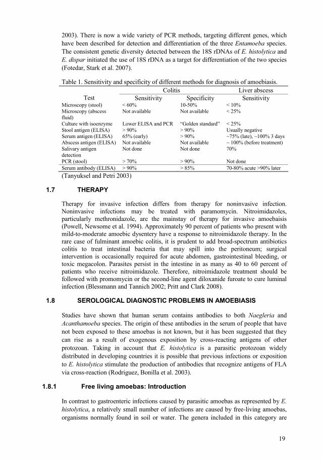
Table 1. Sensitivity and specificity of different methods for diagnosis of amoebiasis. Colitis Liver abscess
Test Sensitivity Specificity Sensitivity Microscopy (stool) < 60% 1050% < 10% Microscopy (abscess fluid)
Not available Not available < 25%
Culture with isoenzyme Lower ELISA and PCR “Golden standard” < 25% Stool antigen (ELISA) > 90% > 90% Usually negative Serum antigen (ELISA) 65% (early) > 90% ~75% (late), ~100% 3 days Abscess antigen (ELISA) Not available Not available ~ 100% (before treatment) Salivary antigen detection
Not done Not done 70%
PCR (stool) > 70% > 90% Not done Serum antibody (ELISA) > 90% > 85% 7080% acute >90% later (Tanyuksel and Petri 2003)
Therapy for invasive infection differs from therapy for noninvasive infection. Noninvasive infections may be treated with paramomycin. Nitroimidazoles, particularly methronidazole, are the mainstay of therapy for invasive amoebaisis (Powell, Newsome et al. 1994). Approximately 90 percent of patients who present with mildtomoderate amoebic dysentery have a response to nitroimidazole therapy. In the rare case of fulminant amoebic colitis, it is prudent to add broadspectrum antibiotics colitis to treat intestinal bacteria that may spill into the peritoneum; surgical intervention is occasionally required for acute abdomen, gastrointestinal bleeding, or toxic megacolon. Parasites persist in the intestine in as many as 40 to 60 percent of patients who receive nitroimidazole. Therefore, nitroimidazole treatment should be followed with promomycin or the secondline agent diloxanide furoate to cure luminal infection (Blessmann and Tannich 2002; Pritt and Clark 2008).
Studies have shown that human serum contains antibodies to both aegleria and Acanthamoeba species. The origin of these antibodies in the serum of people that have not been exposed to these amoebas is not known, but it has been suggested that they can rise as a result of exogenous exposition by crossreacting antigens of other protozoan. Taking in account that E. histolytica is a parasitic protozoan widely distributed in developing countries it is possible that previous infections or exposition to E. histolytica stimulate the production of antibodies that recognize antigens of FLA via crossreaction (Rodriguez, Bonilla et al. 2003).
In contrast to gastroenteric infections caused by parasitic amoebas as represented by E. histolytica, a relatively small number of infections are caused by freeliving amoebas, organisms normally found in soil or water. The genera included in this category are
20
aegleria, Acanthamoeba, and more recently, Balamuthia. Representatives of these genera are pathogenic facultative parasites. They cause disease by being in the right place at the right time or by taking advantage of a host with impaired immune defenses (Martinez and Visvesvara 1997).
aegleria is associated with primary amoebic meningoencephalitis (PAM), a fulminating, rapidly fatal, infection of the central nervous system (CNS). aegleria fowleri is the causal agent of most PMA infections, but other species of eagleria having pathogenic potential have been described (aegleria australiensis and aegleria italica). The type habit for . fowleri is a natural or manmade lake, a thermal polluted body of water, or an inadequately chlorinated swimming pool where the amoebas can feed upon bacteria and proliferate. With respect to humans, mostly children, teenagers, and young adults in good health are infected by swimming or washing in such waters, where amoebas enter the nostril, migrate along the olfactory nerves to the cribriform plate, and gain access to the CNS. Amoebas proliferate rapidly and cause extensive damage to neural tissue.
Diagnosis is difficult and is dependent upon recognition of the amoebas in the microscope and they may not be dismissed as leukocytes. Because of the rapid onset, delayed diagnosis, destructive nature of the disease and lack of effective antimicrobial agents, death is an almost invariable consequence of infection when it takes place at the right time or has the advantage of a host with impaired immune defenses (Schuster and Visvesvara 2004). However, the common presence of thermotolerant aegleria in water, especially . lovanensis, which is an indicator species for . fowleri, suggests that also this pathogenic amoeba may pose a risk to public health in the area (Visvesvara, Moura et al. 2007).
Acanthamoeba spp. also infects the CNS, causing granulomatous amoebic encephalitis (GAE). Much more so than aegleria, Acanthamoeba is ubiquitous in the environment, with amoebas being widely disseminated in soil and water. Unlike the healthy individuals acquiring aegleria infections, persons contracting Acanthamoeba infections of the CNS are compromised host, suffering from concurrent diseases such as AIDS or other conditions such as alcoholism that predispose them to opportunistic infections. The portal of entry of amoebas can vary. It may be intranasal, allowing amoebas to migrate directly to CNS, or entry can be via a break in the skin or through the respiratory tract, with subsequent spread of amoebas to the CNS by hematogenous route. The disease assumes a chronic status, leading to slow deterioration.
Diagnosis is most often made by postmortem examination of brain tissue. Another major class of infection caused by Acanthamoeba spp. is amoebic keratitis. This condition was first noted in individuals suffering corneal trauma due to injury to the corneal surface that became infected with amoebas (Schuster and Visvesvara 2004; Visvesvara, Moura et al. 2007).
21
To determine the true prevalence of E. histolytica and other Amoebas spp. using various tests designed to differentiate between pathogenic and apathogenic species.
1. To determine the true prevalence of E. histolytica in patients with diarrhea and normal population in León Nicaragua.
2. To assess the overdiagnosis and overtreatments of E. histolytica/E. dispar at primary attention level.
3. To evaluate the high seroprevalence.
4. To identify different freeliving amoebas in different sources of environmental water in Nicaragua.
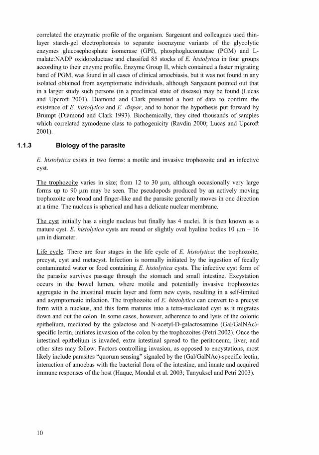
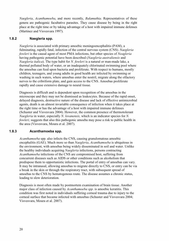
Central America and Nicaragua
22
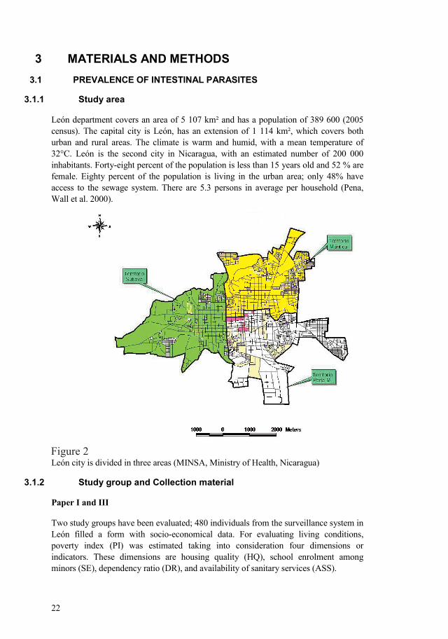
León department covers an area of 5 107 km² and has a population of 389 600 (2005 census). The capital city is León, has an extension of 1 114 km², which covers both urban and rural areas. The climate is warm and humid, with a mean temperature of 32°C. León is the second city in Nicaragua, with an estimated number of 200 000 inhabitants. Fortyeight percent of the population is less than 15 years old and 52 % are female. Eighty percent of the population is living in the urban area; only 48% have access to the sewage system. There are 5.3 persons in average per household (Pena, Wall et al. 2000).
León city is divided in three areas (MINSA, Ministry of Health, Nicaragua)
Paper I and III
Two study groups have been evaluated; 480 individuals from the surveillance system in León filled a form with socioeconomical data. For evaluating living conditions, poverty index (PI) was estimated taking into consideration four dimensions or indicators. These dimensions are housing quality (HQ), school enrolment among minors (SE), dependency ratio (DR), and availability of sanitary services (ASS).
Figure 2
23
The second group 134 individuals, over two years old, presenting with diarrhea at three different health centers in León; this study was carried out from 2002 to 2003. In both study, patients had not used any antiparasitic drugs one week before the sampling. The individuals were invited to participate in this study, and in case of children signed informed consent was provided by the children’s parents before the study started. The study was approved by Ethical committee of Medicine Faculty.
Samples collected in the community and at the health centers were transported within 1 – 2 hrs to the Microbiology Department at the University of León. Immediately after delivery, samples were divided into different containers. One part of the specimen was fixed with 70% ethanol for DNA extraction, one part with SAF (Sodium acetateacetic acidformalin) fixative for staining methods, and the remaining unfixed fresh specimens were examined by direct microscopy of saline and iodine. In addition, formalinethyl acetate sedimentation technique was used for detection of cysts and eggs (Young, Bullock et al. 1979), iron haematoxylin staining for amoebas and flagellates and modified Ziehl Neelsen staining for detection of enteric coccidian (Garcia and Bruckner 1997).
Paper IV
A retrospective study was carried out in two health centers. In this study, we got permission from the head of the health center, and two person collected the information, they visited the health center, between June to November from 2004, the record of patients were given by nurses, the record were chosen at random in patients who showed intestinal symptoms, and over two years old. 100 patients were recorded, and a form from each patient was filled, with following date: abdominal pain, diarrhea, constipation, other symptoms, stools examination, sex, age.
An external quality assessment in coproparasitology was carried out from February to July of 2006 in fifteen laboratories at health centers, in León department, a questionnaire and a panel with ten plastic vials was previously prepared with different intestinal parasites such as: E. histolytica/ E. dispar, Entamoeba coli. Endolimax nana, Giardia intestinalis, Entamoeba hartmanni and helminths eggs such as: Trichuris trichiura, Ascaris lumbricoides, Taenia sp., and one stool sample as negative control. Plastic bags, with ten vials were sealed and sent to each laboratory, and a form was filled in order to know the level of laboratory technicians and the laboratory condition.
Serum samples
Twentytwo serum samples from suspected of liver abscess patients in the León University Hospitals, and 25 samples from individuals in the community were used to evaluate the significance of high prevalence of antiamoebic antibodies. Immunofluorescence and western blotting was used for detecting antibodies against different amoeba spp.
24
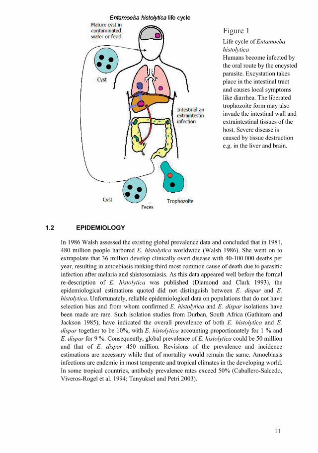
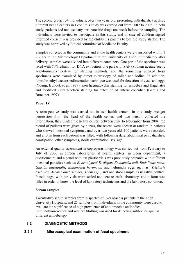
Figure 3. Three different Entamoebas spp. at the microscopic examination.
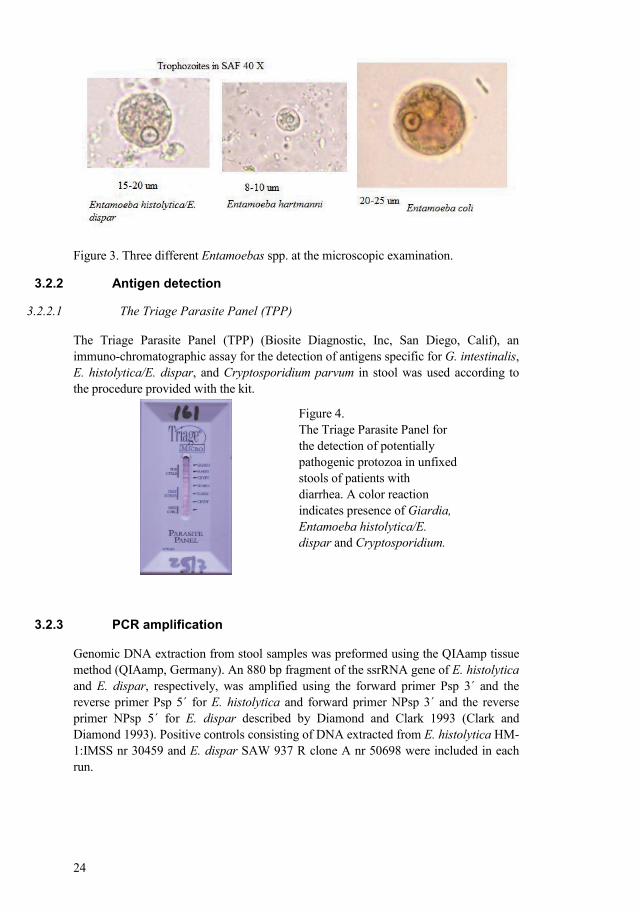
3.2.2.1 The Triage Parasite Panel (TPP)
The Triage Parasite Panel (TPP) (Biosite Diagnostic, Inc, San Diego, Calif), an immunochromatographic assay for the detection of antigens specific for G. intestinalis, E. histolytica/E. dispar, and Cryptosporidium parvum in stool was used according to the procedure provided with the kit.
Genomic DNA extraction from stool samples was preformed using the QIAamp tissue method (QIAamp, Germany). An 880 bp fragment of the ssrRNA gene of E. histolytica and E. dispar, respectively, was amplified using the forward primer Psp 3´ and the reverse primer Psp 5´ for E. histolytica and forward primer NPsp 3´ and the reverse primer NPsp 5´ for E. dispar described by Diamond and Clark 1993 (Clark and Diamond 1993). Positive controls consisting of DNA extracted from E. histolytica HM1:IMSS nr 30459 and E. dispar SAW 937 R clone A nr 50698 were included in each run.
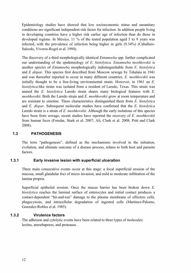
Figure 4. The Triage Parasite Panel for the detection of potentially pathogenic protozoa in unfixed stools of patients with diarrhea. A color reaction indicates presence of Giardia, Entamoeba histolytica/E. dispar and Cryptosporidium.
25
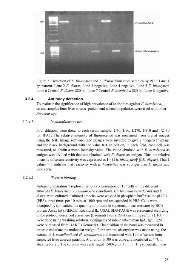
Figure 5. Detection of E. histolytica and E. dispar from stool samples by PCR. Lane 1 bp pattern. Lane 2 E. dispar, Lane 3 negative, Lane 4 negative, Lane 5 E. histolytica, Lane 6 Control E. dispar 880 bp. Lane 7 Control E. histolytica 880 bp, Lane 8 negative.
To evaluate the significance of high prevalence of antibodies against E. histolytica, serum samples form liver abscess patient and normal population were used with other Amoebas spp.
3.2.4.1 Immunofluorescence.
Four dilutions were done, to each serum sample: 1/30, 1/90, 1/270, 1/810 and 1/2430 for IFAT. The relative intensity of fluorescence was measured from digital images using the NIH Image software. The images were inverted to give a “negative” image and the black background with the value 0.0. In edition, in each field, each cell was measured, to obtain a mean intensity value. The value obtained with E. histolytica as antigen was divided with that one obtained with E. dispar as antigen. Thus the relative intensity of serum reactivity was expressed as I = [I E. histolytica]/ [I E. dispar]. Thus I values > 1 indicate that reactivity with E. histolytica was stronger than E. dispar and vice versa.
3.2.4.2 Western blotting
Antigen preparation. Trophozoites to a concentration of 106 cells of the different amoebas E. histolytica, Acanthamoeba castellanii, Hartmanella vermiformis and E. dispar were cultured. Cultured amoeba were washed in phosphate buffer saline pH 7,4 (PBS), three times per 10 min. at 1000 rpm and resuspended in PBS. Cells were disrupted by sonication, the quantity of protein in supernatant was measure by BCA protein Assay kit (PIERCE, Rockford IL, USA). SDSPAGE was preformed according to the protocol described elsewhere (Laemmli 1970). Dilutions of the serum (1/100) were done using washing solution. Conjugates of rabbit antihuman IgA, IgG, IgM were purchased from DAKO (Denmark). The position of the band was measured in order to calculate the molecular weight. Furthermore, absorption was made using; the extract of A. castellanii and H. vermiformis and incubated with 1 ml of serum from suspected liver abscess patients. A dilution 1/100 was done and incubated at 4 °C in shaking for 2h. The solution was centrifuged 1500xg for 15 min. The supernatant was
26
utilized in the immunoblotting assays as first antibody, against E. histolytica, A. castellanii and H. vermiformis.
Water samples were collected from León area in Nicaragua: 88 samples were from rivers and springs, 111 from wells, 74 from water taps and 21 from water tanks in urban and suburban León and from 3 nearby geothermal areas of San Jacinto, Posoltega and Tipitapa. Water samples were collected into sterile 50 ml tubes and transported to the laboratory at Microbiology Department, University of León. Amoebas were isolated from specimens filtered through 0.45 µm filters attached to sterile syringes. Filters were placed on nonnutrient agar plates covered with viable Escherichia coli K12 strain. Agar plates were incubated at 40 °C for the isolation of thermotolerant strains. Growing amoebas were repeatedly subcultured and cloned by limiting dilution.
Initial identification of amoebas was based on microscopical observation of living and fixed cysts and trophozoites. The presence of cellulose in cyst wall of Acanthamoeba was demonstrated using the cellulosebinding domain of cellulose essentially as described (Linder, WinieckaKrusnell et al. 2002). Fluorescent in situ hybridization (FISH) was performed using 18S rRNA targeted fluorescent oligonucleotide probes according to the protocols described before (Stothard, Hay et al. 1999; Grimm, Ludwig et al. 2001). Probes: GSP (genusspecific probe) for the detection of Acanthamoeba and NAEG1088 specific 18S rRNA probe for the detection of aegleria were synthesized by Scandinavian Gene Synthesis AB, 73122 Köping, Sweden.
The statistical analyses were performed using Epi Info software version 6.2 to calculate percentage and frequency of parasites and to determine the agreement between two methods: concordance was calculated using Cohen Kappa indices (κ), sensitivity and specificity using Youden´s index in the Win Episcope 2.0 program and SSPS program v 10 for X2 and marginal odds ratio (ORs) and 95% confidence intervals (95% CIs) were used to measure the strength of association between risk factor and intestinal parasitic infection.
27
PAPER I (Diarrhea patients)
Among 134 diarrhea patients, the most common finding at the health center laboratory level, was E. histolytica/E. dispar (24%). At the Microbiology Department E. histolytica/E. dispar was found in 4.5 %. With the Triage Parasite test, only one case of E. histolytica/E. dispar was found. Upon confirmatory diagnostic analysis by PCR, E. dispar was recognized in 10 (7.5%) and E. histolytica in two cases (1.5%). PCR was capable to identify 2 E. histolytica and 10 E. dispar cases. The correlation between PCR results and microscopical examination was low (κ= 0.4). This finding suggests that the true prevalence of E. histolytica in León is very low. This finding made us reassess the extent of the amoebiasis examination. There is a need to eliminate over diagnosis based on microscopy and to introduce a specific method for identification of E. histolytica, but in a poor country as Nicaragua it is difficult in this setting. Both education and training will be necessary for a good clinical manage and treatment of the patients. In this study, TPP test showed good sensitivity and specificity for G. intestinalis and high concordance with microscopy. However, the sensitivity for E. histolytica/E. dispar and Cryptosporidium spp. was lower than reported previously (Garcia, Shimizu et al. 2000; Sharp, Suarez et al. 2001). The observed lower sensitivity of the TPP could be explained by the relatively high sensitivity of PCR and the problems with specificity of microscopy discussed above. A renewed look at the local epidemiology of amoebiasis in León, Nicaragua demonstrates that the true prevalence of E. histolytica by PCR in individuals with and without diarrhea was relatively low.
PAPER III (ormal population)
In this study of prevalence intestinal parasites using different diagnostic methods, it was determined that one or more intestinal potential pathogenic parasites were found in 40% and 22% of people living in “poor” and “good” condition respectively. In contrast to the previous study 11 years ago, the prevalence of helminths has decreased. This could be due to deworming campaigns, where the children between 2 to 12 year old were given albendazole, an antiparasitic drug which is effective mainly for helminths (Keiser and Utzinger 2008). Correlation between protozoan parasites E. histolytica/E. dispar and G. intestinalis showed a different age distribution, similar to a study in León and reported by other authors (Ostan, Kilimcioglu et al. 2007). The prevalence of Giardia was found to be higher in younger children than in older people (Tellez, Morales et al. 1997).
28
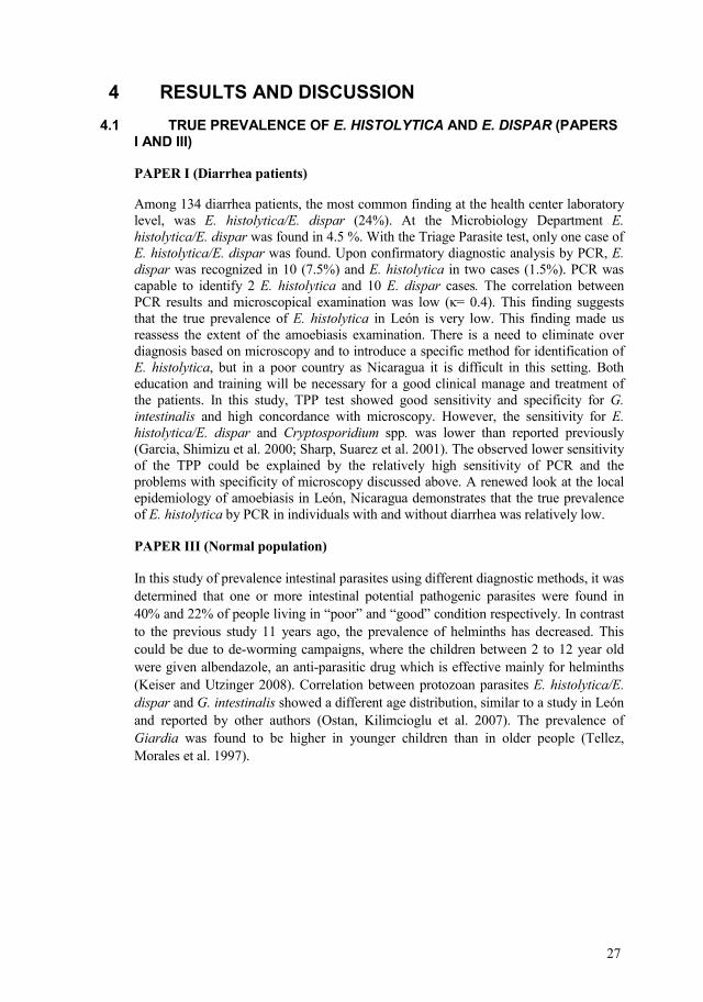
Table 2. Prevalence of intestinal parasites, in patients with diarrhea (Paper I) and in normal population (Paper III) in León, Nicaragua 2003. Paper III Paper I Parasites detected n=480 (%) n=134 (%) Protozoan Blastocystis hominis Entamoeba coli Giardia intestinalis Endolimax nanaEntamoeba histolytica/E. dispar Cryptosporidium sp. Cyclospora cayetanensis Isospora belli Helminthes Trichuris trichiura Hymenolepis nana Ascaris lumbricoides Hymenolepis diminuta Hookworm Taenia sp.
200 126 100 91 58 0 0 0
11 5 3 2 1 1
(42)(26) (21) (19)(12) (0) (0)(0)
(2.3) (1.0) (0.6) (0.4) (0.2) (0.2)
44 14 22 23 8 1 3 1
7 8 5 0 0 0
(33) (10) (16) (17) (6)
(0.7) (2.2) (0.7)
(5.2) (6.7) (3.7)
(0) (0) (0)
Furthermore, this study shows that people living in poor conditions are directly related to potential pathogenic parasite infections (P< 0.05) as other authors have reported (Nematian, Nematian et al. 2004; Okyay, Ertug et al. 2004; Ugbomoiko, Dalumo et al. 2009). In the early study 11 years ago, there was not a good correlation.
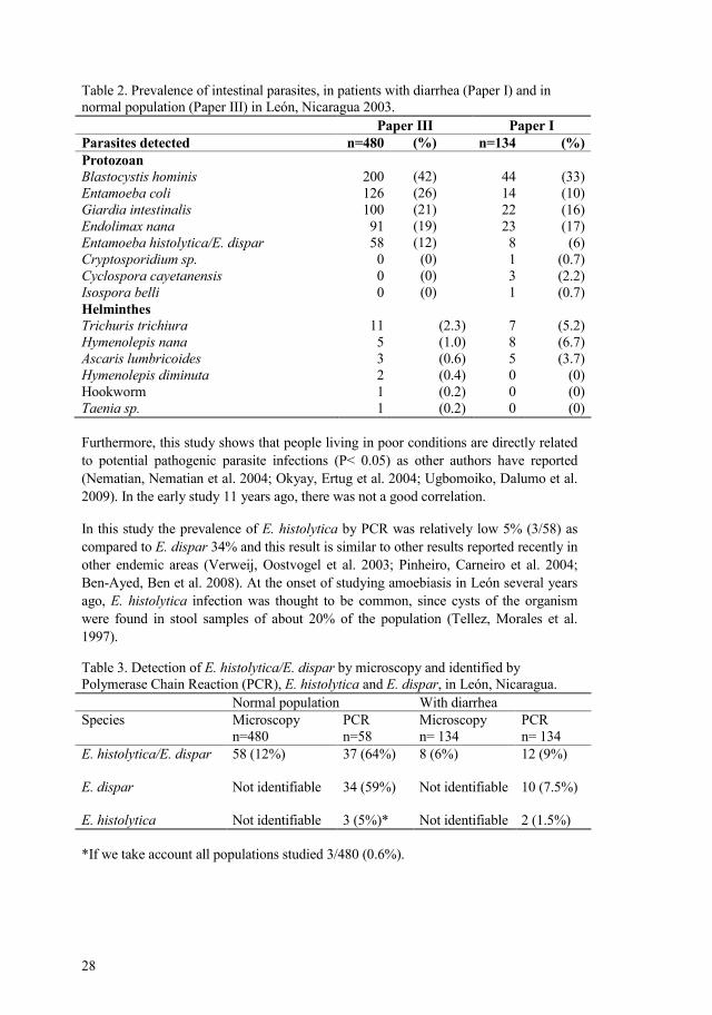
In this study the prevalence of E. histolytica by PCR was relatively low 5% (3/58) as compared to E. dispar 34% and this result is similar to other results reported recently in other endemic areas (Verweij, Oostvogel et al. 2003; Pinheiro, Carneiro et al. 2004; BenAyed, Ben et al. 2008). At the onset of studying amoebiasis in León several years ago, E. histolytica infection was thought to be common, since cysts of the organism were found in stool samples of about 20% of the population (Tellez, Morales et al. 1997).
Table 3. Detection of E. histolytica/E. dispar by microscopy and identified by Polymerase Chain Reaction (PCR), E. histolytica and E. dispar, in León, Nicaragua. Normal population With diarrhea Species Microscopy
n=480 PCR n=58
Microscopy n= 134
PCR n= 134
E. histolytica/E. dispar E. dispar E. histolytica
58 (12%) Not identifiable Not identifiable
37 (64%) 34 (59%) 3 (5%)*
8 (6%) Not identifiable Not identifiable
12 (9%) 10 (7.5%) 2 (1.5%)
*If we take account all populations studied 3/480 (0.6%).
29
In the quality control assessment, 80 % of the technicians successfully identified the main parasites in the panel. The parasites identified correctly were H. nana, A. lumbricoides and the hardest to recognize were E. hartmanni, Taenia sp. and E. nana. All 15 laboratories found E. histolytica /E. dispar in the single sample where it was present. However, some laboratories falsely identified this protozon in more than one sample, in total there were 20 cases. We found that the health center technicians continue to mix up E. histolytica /E. dispar with other amoebas causing false positive. The other reason for over diagnosis is that they were confusing vegetable matter with cysts (Kettelhut, Chiodini et al. 2003).
Furthermore, this report coincides with a previous study, where E. histolytica/E. dispar was overdiagnosed at the health center in León (Leiva, Lebbad et al. 2006). Also a fundamental problem, not restricted to the particular case of parasitological diagnostics, is that this type of routine diagnostics lacks the status of more sophisticated diagnostic procedures, an attitude which is possible to alter by paying attention to education, maintenance of equipment and laboratory routines. Education and training provided by e.g. university institutions, via its teaching arm, has played an important part in the solution of this problem (Kettelhut, Chiodini et al. 2003; Libman, Gyorkos et al. 2008). Novel tools for education and quality control, such as the Web Microscope for Parasitology (WMP) should contribute to an elimination of the problems of over diagnosis (Linder, Lundin et al. 2008).
Regarding the use of antiamoebic drugs in symptomatic patients, 32% of patients were found with E. histolytica/E. dispar and the majority with cysts. On the other hand, no stool examination was not performed on 41 % of the patients. In summary 79% of those patients were treated with methronidazole. A rough calculation on sales of methronidazole and related drugs can be made based on the 100 individuals included in this study. The conclusion is that thousand of dollars are wasted due to overtreatment in a country with limited resources has been pointed out previously (Fotedar, Stark et al. 2007). Apart from the misuse of public and personal economic resources, the practice of overtreatment may hasten the development of drug resistance. Thus, overtreatment appears to be a widespread problem, which needs to be explored in detail (Bansal, Malla et al. 2006).
We conclude that over diagnosis resulting in overtreatment of amoebic dysentery is widespread in the León region of Nicaragua, and that it reflects a global problem affecting most seriously the poorest in developing countries.
We thought that antibodies against E. histolytica may be induced by other amoebas in the environment. Thus we wanted to test this possibility of crossreaction by absorption and immunobloting (WB) experiments and to look for the presence of freeliving amoebas in the environment.
30
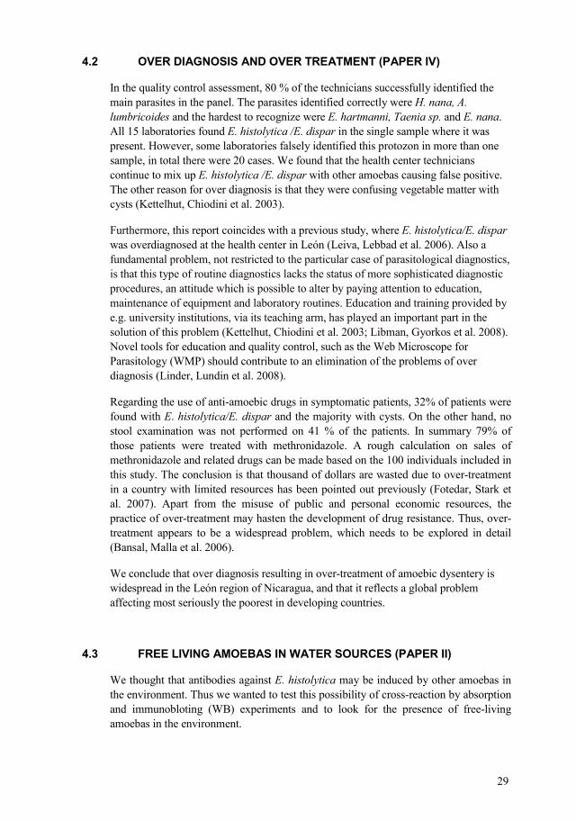
Free living amoebas isolated from the water samples were characterized by morphological and immunohistochemical methods. One hundred twenty five out of the 294 samples (42.5%) collected from different water sources yielded growth of FLA. Amoebas could be recovered from 75 out of 178 (42 %) water samples collected in the León municipality and an almost identical proportion was recovered from samples from the geothermal areas (50 out of 116, 43%). However, a detailed analysis showed some differences; the lowest prevalence of amoebas, 17 out of 74 (23%) was found in tap water in León municipality and the highest prevalence in wells from geothermal areas, 25 out of 36 (69%).
Table 4. Identification of aegleria isolates by different methods.
Enflagellation
FISH1
≥42 °C IFAT2
NfPab3 Nf5D12u4
León Municipality 17 8 8 3 0 Geothermal areas 22 16 16 9 0 Total 39 24 24 12 0 1FISH = Fluorescent in situ hybridization, 2IFAT = Immunofluorescence antibody test, 3NfPab = anti . fowleri /. lovanensis polyclonal antibodies, 4Nf5D12u = anti . fowleri mouse monoclonal antibodies. There were significantly higher levels of amoeboflagellates from wells in geothermal areas as compared to wells in the León municipality, 53% as opposed to 15%. The common presence of thermotolerant, potentially pathogenic amoebas, may pose a risk to public health in the community.
In thermal areas we found significantly more aegleria than Acanthamoeba. Such environmental conditions occur in geothermal areas globally, but may occur also in the environment, which has been heated by other means. Raised temperatures during the hot summer months or warm water from power plants facilitate the growth of . fowleri (Martinez and Visvesvara 1997). However, in the cyst form these protozoa are extremely resistant and may occur in dust (da Silva and da Rosa 2003). Thus the information provided in this study may serve as baseline for future studies on the role of freeliving amoebas e.g. in waterbornedisease outbreaks in the region. Among such potentially important enteropathogens are Vibrio cholerae, E. coli 0157, and Helicobacter pylori (WinieckaKrusnell and Linder 2001; Brown, Smith et al. 2002).
31
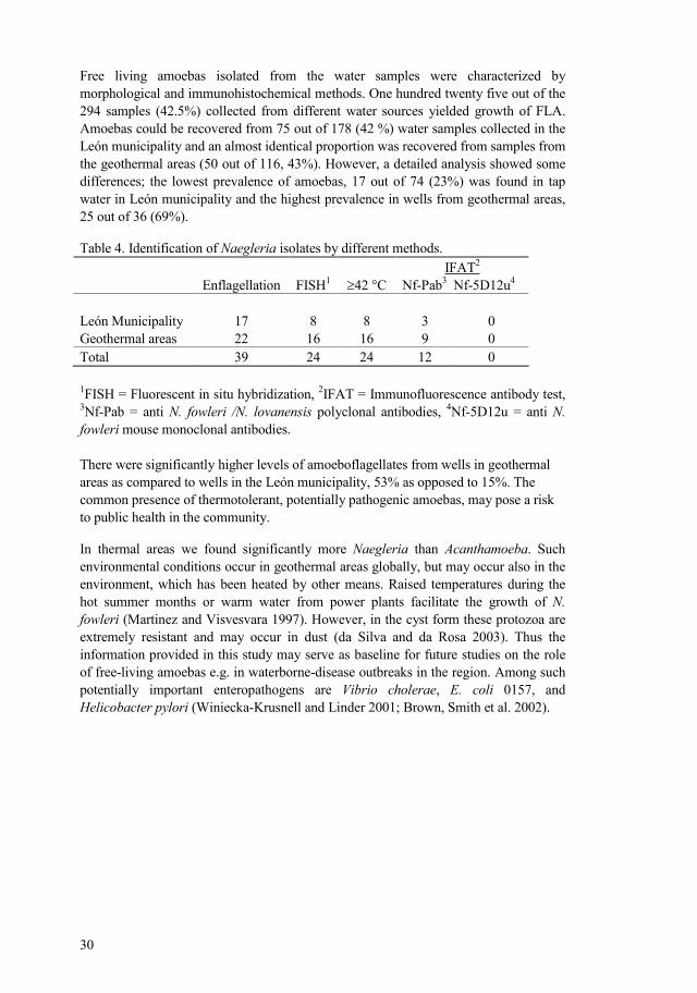
Figure 6. Diagnostic methods for free living amoebas: Phase contrast image seen in culture (Culture), the appearance of amoebas in fixed monolayers stained by the Giemsa stain and in fluorescence microscopy using Fluorescent in situ hybridization (FISH).
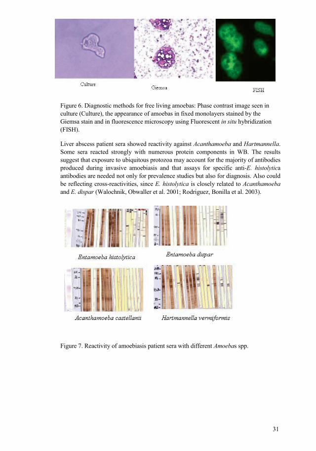
Liver abscess patient sera showed reactivity against Acanthamoeba and Hartmannella. Some sera reacted strongly with numerous protein components in WB. The results suggest that exposure to ubiquitous protozoa may account for the majority of antibodies produced during invasive amoebiasis and that assays for specific antiE. histolytica antibodies are needed not only for prevalence studies but also for diagnosis. Also could be reflecting crossreactivities, since E. histolytica is closely related to Acanthamoeba and E. dispar (Walochnik, Obwaller et al. 2001; Rodriguez, Bonilla et al. 2003).
Figure 7. Reactivity of amoebiasis patient sera with different Amoebas spp.
32
The hypothesis was that common antigenic components could be present in E. histolytica and freeliving amoebas. Contact with freeliving amoebas could therefore give rise to cross reactive antibodies seen in the healthy population as anti E. histolytica antibodies. The purpose was to study if antibody reactivity with freeliving amoebas could be identified in amoebiasis patient sera. We also wanted to see if antibodies in amoebiasis patient sera could be absorbed with freeliving amoebas.
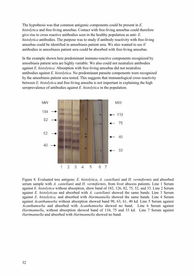
In the example shown here predominant immunoreactive components recognized by amoebiasis patient sera are highly variable. We also could not neutralize antibodies against E. histolytica: Absorption with freeliving amoebas did not neutralize antibodies against E. histolytica. No predominant parasite components were recognized by the amoebiasis patient sera tested. This suggests that immunological cross reactivity between E. histolytica and freeliving amoeba is not important in explaining the high seroprevalence of antibodies against E. histolytica in the population.
Figure 8. Evaluated tree antigens: E. histolytica, A. castellanii and H. vermiformis and absorbed serum sample with A. castellanii and H. vermiformis, from liver abscess patients. Lane 1 Serum against E. histolytica without absorption, show band of 182, 126, 82, 75, 52, and 33. Lane 2 Serum against E. histolytica and absorbed with A. castellanii showed the same bands. Lane 3 Serum against E. histolytica, and absorbed with Hartmannella showed the same bands. Lane 4 Serum against Acanthamoeba without absorption showed band 98, 63, 61, 40 kd. Lane 5 Serum against Acanthamoeba and absorbed with Hartmannella showed no band. Lane 6 Serum against Hartmannella, without absorption showed band of 110, 75 and 33 kd. Lane 7 Serum against Hartmannella and absorbed with Acanthamoeba showed no band.
Figure 8. Evaluated tree antigens: E. histolytica, A. castellanii and H. vermiformis and absorbed serum sample with A. castellanii and H. vermiformis, from liver abscess patients. Line 1 Serum against E. histolytica without absorption, show band of 182, 126, 82, 75, 52, and 33. Line 2 Serum against E. histolyticaa and absorbed with A. castellanii showed the same bands. Line 3 Serum against E. histolytica, and absorbed with Hartmannella showed the same bands. Line 4 Serum against Acanthamoeba without absorption showed band 98, 63, 61, 40 kd. Line 5 Serum againstAcanthamoeba and absorbed with Acanthamoeba showed no band. Line 6 Serum againstHartmannella, without absorption showed band of 110, 75 and 33 kd. Line 7 Serum againstHartmannella and absorbed with Hartmannella showed no band.
33
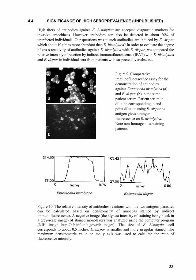
High titers of antibodies against E. histolytica are accepted diagnostic markers for invasive amoebiasis. However antibodies can also be detected in about 20% of uninfected individuals. Our questions was it such antibodies are induced by E. dispar which about 10 times more abundant than E. histolytica? In order to evaluate the degree of cross reactivity of antibodies against E. histolytica with E. dispar, we compared the relative intensity of reaction by indirect immunofluorescence (IFAT) with E. histolytica and E. dispar in individual sera from patients with suspected liver abscess.
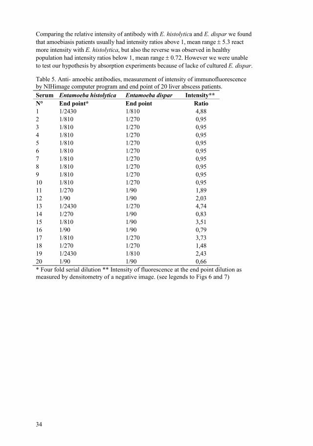
Figure 10. The relative intensity of antibodies reactions with the two antigens parasites can be calculated based on densitometry of amoebas stained by indirect immunofluorescence. A negative image (the highest intensity of staining being black in a greyscale image) of stained monolayers was analyzed using the computer program (NIH image http://rsb.info.nih.gov/nihimage/). The size of E. histolytica cell corresponds to about 0.5 inches. E. dispar is smaller and more irregular stained. The maximum densitometric value on the y axis was used to calculate the ratio of fluorescence intensity.
Figure 9. Comparative immunofluorescence assay for the demonstration of antibodies against Entamoeba histolytica (a) and E. dispar (b) in the same patient serum. Patient serum in dilution corresponding to endpoint dilution using E. dispar as antigen gives stronger fluorescence on E. histolytica. Note nonhomogenous staining patterns.
34
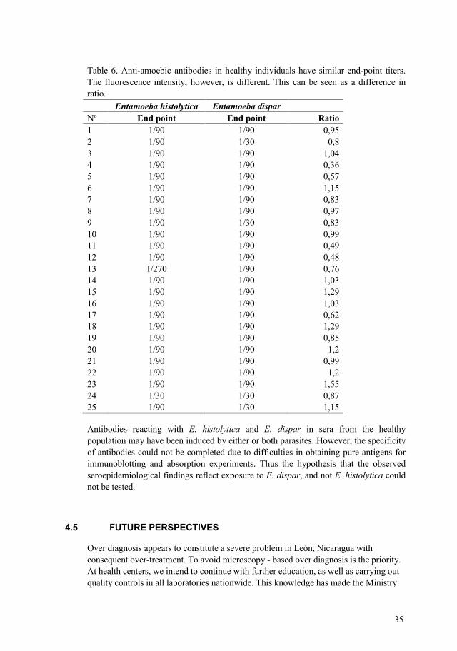
Comparing the relative intensity of antibody with E. histolytica and E. dispar we found that amoebiasis patients usually had intensity ratios above 1, mean range ± 5.3 react more intensity with E. histolytica, but also the reverse was observed in healthy population had intensity ratios below 1, mean range ± 0.72. However we were unable to test our hypothesis by absorption experiments because of lacke of cultured E. dispar.
Table 5. Anti amoebic antibodies, measurement of intensity of immunofluorescence by NIHimage computer program and end point of 20 liver abscess patients. Serum Intensity** ° End point* End point Ratio 1 1/2430 1/810 4,88 2 1/810 1/270 0,95 3 1/810 1/270 0,95 4 1/810 1/270 0,95 5 1/810 1/270 0,95 6 1/810 1/270 0,95 7 1/810 1/270 0,95 8 1/810 1/270 0,95 9 1/810 1/270 0,95 10 1/810 1/270 0,95 11 1/270 1/90 1,89 12 1/90 1/90 2,03 13 1/2430 1/270 4,74 14 1/270 1/90 0,83 15 1/810 1/90 3,51 16 1/90 1/90 0,79 17 1/810 1/270 3,73 18 1/270 1/270 1,48 19 1/2430 1/810 2,43 20 1/90 1/90 0,66 * Four fold serial dilution ** Intensity of fluorescence at the end point dilution as measured by densitometry of a negative image. (see legends to Figs 6 and 7)
35
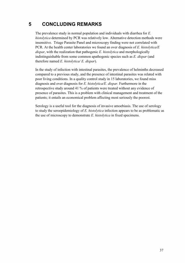
Table 6. Antiamoebic antibodies in healthy individuals have similar endpoint titers. The fluorescence intensity, however, is different. This can be seen as a difference in ratio. Nº End point End point Ratio 1 1/90 1/90 0,95 2 1/90 1/30 0,8 3 1/90 1/90 1,04 4 1/90 1/90 0,36 5 1/90 1/90 0,57 6 1/90 1/90 1,15 7 1/90 1/90 0,83 8 1/90 1/90 0,97 9 1/90 1/30 0,83 10 1/90 1/90 0,99 11 1/90 1/90 0,49 12 1/90 1/90 0,48 13 1/270 1/90 0,76 14 1/90 1/90 1,03 15 1/90 1/90 1,29 16 1/90 1/90 1,03 17 1/90 1/90 0,62 18 1/90 1/90 1,29 19 1/90 1/90 0,85 20 1/90 1/90 1,2 21 1/90 1/90 0,99 22 1/90 1/90 1,2 23 1/90 1/90 1,55 24 1/30 1/30 0,87 25 1/90 1/30 1,15
Antibodies reacting with E. histolytica and E. dispar in sera from the healthy population may have been induced by either or both parasites. However, the specificity of antibodies could not be completed due to difficulties in obtaining pure antigens for immunoblotting and absorption experiments. Thus the hypothesis that the observed seroepidemiological findings reflect exposure to E. dispar, and not E. histolytica could not be tested.
Over diagnosis appears to constitute a severe problem in León, Nicaragua with consequent overtreatment. To avoid microscopy based over diagnosis is the priority. At health centers, we intend to continue with further education, as well as carrying out quality controls in all laboratories nationwide. This knowledge has made the Ministry

36
of Health aware of the problem. We also intend to conduct educational work with primary health care physicians, to update their knowledge about amoebiasis and the problem of over treatment.
There is a novel method allowing an internetbased distribution and viewing of identical parasite containing specimens to an unlimited number of viewers. To determine the feasibility in an area with varying internet access speeds was registered the connection speed from educational institutions in Central America.
Freeliving amoebas are potential problem in environments such as hospitals and agricultural areas could be a public health problem, both as pathogens and as reservoirs carrying pathogenic bacteria.
37
The prevalence study in normal population and individuals with diarrhea for E. histolytica determined by PCR was relatively low. Alternative detection methods were insensitive. Triage Parasite Panel and microscopy finding were not correlated with PCR. At the health center laboratories we found an over diagnosis of E. histolytica/E dispar, with the realization that pathogenic E. histolytica and morphologically indistinguishable from some common apathogenic species such as E. dispar (and therefore named E. histolytica/ E. dispar).
In the study of infection with intestinal parasites, the prevalence of helminths decreased compared to a previous study, and the presence of intestinal parasites was related with poor living conditions. In a quality control study in 15 laboratories, we found miss diagnosis and over diagnosis for E. histolytica/E. dispar. Furthermore in the retrospective study around 41 % of patients were treated without any evidence of presence of parasites. This is a problem with clinical management and treatment of the patients; it entails an economical problem affecting most seriously the poorest.
Serology is a useful tool for the diagnosis of invasive amoebiasis. The use of serology to study the seroepidemiology of E. histolytica infection appears to be as problematic as the use of microscopy to demonstrate E. histolytica in fixed specimens.
38
First, I wish to express my gratitude to all people in Sweden and in Nicaragua who contributed to this thesis.
I express my gratitude, to my tutor Ewert Linder for having led on the world of research, for being very patient and tolerant. Furthermore to my cosupervisor Jadwiga, I have a special appreciation for having given their full cooperation at all times.
I want to thank Marianne, for conveying her knowledge in the diagnosis of parasites, and for her help in manuscript correction. Also in Parasitology Department at SMI to: Cecilia Thors, Lena, Silvia, Inger, Elizabeth, Pet, Johan and Victor, thank all of them for their Cooperation and friendship. Also, especially to Staffan Svard and Mats Wahlgren for theirs collaboration and friendship.
In addition, especial appreciation to Roland and Patricia for their help and giving me their friendship at this final stage. Many thank also Dae Ho, Margareta, Inger.
To Hans Hallander for his friendship and support in Sweden from the beginning.
In icaragua:
Dr. Ernesto Medina for carrying out this project.
I want to express my special gratitude to Teresita Rivera (q.p.d.c), who gave me the opportunity to engage in the Microbiology Department and made my first steps in the research field.
To University authorities: Rector Dr. Rigoberto Sampson, Vicerector (VIP) Leonardo Mendoza. Faculty authorities of Medical Sciences to Dr. Rodolfo Pena by their management to finish this project. Furthermore, before Dean Dr. Rene Altamirano.
To all my colleagues in the Department of Microbiology and Parasitology: Silda, Kenia, Rosario, Elizabeth, Eugenia, Edelma, Ana Ester, especially to Mercedes, Orlando, Isabel, Margarita, Felix and Aleyda for sharing moments abroad. Thank to William Morales for his comments and advise in the writing of the papers and Brenda for her cooperation in laboratory assistantce. Also the young colleagues in Sweden, Filemon, Daniel, Erick, Samuel and Fernando.
To Doña Esperanza thanks for giving me your house in Sweden like my own home.
Finally, to all my family, especially to those who most deserve this effort, my wife Rosa Argentina, Byron Jr. and Heykel. Also especially to my parents Francisco (q.p.d.c.) and Segunda, and my mother in low Gillermina, for giving me theirs support.
This work was supported by Swedish Agency for Research Cooperation with Developing Countries (SAREC) and the National Autonomous University of NicaraguaLeón.
39
Ali, I. K., C. G. Clark, et al. (2008). "Molecular epidemiology of amebiasis." Infect
Genet Evol 8(5): 698707. Ali, I. K., U. Mondal, et al. (2007). "Evidence for a link between parasite genotype
and outcome of infection with Entamoeba histolytica." J Clin Microbiol 45(2): 2859.
Arellano, J., M. PerezRodriguez, et al. (1996). "Increased frequency of HLADR3 and complotype SCO1 in Mexican mestizo children with amoebic abscess of the liver." Parasite Immunol 18(10): 4918.
Aristizabal, H., J. Acevedo, et al. (1991). "Fulminant amebic colitis." World J Surg 15(2): 21621.
Bansal, D., N. Malla, et al. (2006). "Drug resistance in amoebiasis." Indian J Med Res 123(2): 1158.
BenAyed, S., A. R. Ben, et al. (2008). "Molecular differentiation of Entamoeba histolytica and Entamoeba dispar from Tunisian food handlers with amoeba infection initially diagnosed by microscopy." Parasite 15(1): 658.
Blessmann, J. and E. Tannich (2002). "Treatment of asymptomatic intestinal Entamoeba histolytica infection." N Engl J Med 347(17): 1384.
Brown, M. R., A. W. Smith, et al. (2002). "E. coli O157 persistence in the environment." Microbiology 148(Pt 1): 12.
CaballeroSalcedo, A., M. ViverosRogel, et al. (1994). "Seroepidemiology of amebiasis in Mexico." Am J Trop Med Hyg 50(4): 4129.
Calderon, J. and R. D. Schreiber (1985). "Activation of the alternative and classical complement pathways by Entamoeba histolytica." Infect Immun 50(2): 5605.
Clark, C. G. and L. S. Diamond (1993). "Entamoeba histolytica: a method for isolate identification." Exp Parasitol 77(4): 4505.
da Silva, M. A. and J. A. da Rosa (2003). "[Isolation of potencially pathogenic freeliving amoebas in hospital dust]." Rev Saude Publica 37(2): 2426.
Denis, M. and K. Chadee (1989). "Human neutrophils activated by interferongamma and tumour necrosis factoralpha kill Entamoeba histolytica trophozoites in vitro." J Leukoc Biol 46(3): 2704.
Diamond, L. S. (1982). "A new liquid medium for xenic cultivation of Entamoeba histolytica and other lumendwelling protozoa." J Parasitol 68(5): 9589.
Diamond, L. S. and C. G. Clark (1993). "A redescription of Entamoeba histolytica Schaudinn, 1903 (Emended Walker, 1911) separating it from Entamoeba dispar Brumpt, 1925." J Eukaryot Microbiol 40(3): 3404.
Diamond, L. S., D. R. Harlow, et al. (1978). "A new medium for the axenic cultivation of Entamoeba histolytica and other Entamoeba." Trans R Soc Trop Med Hyg 72(4): 4312.
EspinosaCantellano, M. and A. MartinezPalomo (2000). "Pathogenesis of intestinal amebiasis: from molecules to disease." Clin Microbiol Rev 13(2): 31831.
Fotedar, R., D. Stark, et al. (2007). "Laboratory diagnostic techniques for Entamoeba species." Clin Microbiol Rev 20(3): 51132, table of contents.
Garcia, L. S. and D. A. Bruckner (1997). Macroscopic and microscopic examination of fecal specimens. Washington, DC, American Society for microbiology
Garcia, L. S., R. Y. Shimizu, et al. (2000). "Detection of Giardia lamblia, Entamoeba histolytica/Entamoeba dispar, and Cryptosporidium parvum antigens in human fecal specimens using the triage parasite panel enzyme immunoassay." J Clin Microbiol 38(9): 333740.
40
Gathiram, V. and T. F. Jackson (1985). "Frequency distribution of Entamoeba histolytica zymodemes in a rural South African population." Lancet 1(8431): 71921.
GonzalezRuiz, A., R. Haque, et al. (1994). "Value of microscopy in the diagnosis of dysentery associated with invasive Entamoeba histolytica." J Clin Pathol 47(3): 2369.
Grimm, D., W. F. Ludwig, et al. (2001). "Development of 18S rRNAtargeted oligonucleotide probes for specific detection of Hartmannella and Naegleria in Legionellapositive environmental samples." Syst Appl Microbiol 24(1): 7682.
Guarner, V. (1990). History of amebiasis. Boca Rayton, USA, CRC Press Inc. Haque, R., I. K. Ali, et al. (1998). "Comparison of PCR, isoenzyme analysis, and
antigen detection for diagnosis of Entamoeba histolytica infection." J Clin Microbiol 36(2): 44952.
Haque, R., A. S. Faruque, et al. (1997). "Entamoeba histolytica and Entamoeba dispar infection in children in Bangladesh." J Infect Dis 175(3): 7346.
Haque, R., D. Mondal, et al. (2003). "Epidemiologic and clinical characteristics of acute diarrhea with emphasis on Entamoeba histolytica infections in preschool children in an urban slum of Dhaka, Bangladesh." Am J Trop Med Hyg 69(4): 398405.
Haque, R., L. M. Neville, et al. (1995). "Rapid diagnosis of Entamoeba infection by using Entamoeba and Entamoeba histolytica stool antigen detection kits." J Clin Microbiol 33(10): 255861.
Huston, C. D., R. Haque, et al. (1999). "Molecularbased diagnosis of Entamoeba histolytica infection." Expert Rev Mol Med 1999: 111.
Huston, C. D., E. R. Houpt, et al. (2000). "Caspase 3dependent killing of host cells by the parasite Entamoeba histolytica." Cell Microbiol 2(6): 61725.
Jackson, T. F., V. Gathiram, et al. (1985). "Seroepidemiological study of antibody responses to the zymodemes of Entamoeba histolytica." Lancet 1(8431): 7169.
Kebede, A., J. J. Verweij, et al. (2004). "The use of realtime PCR to identify Entamoeba histolytica and E. dispar infections in prisoners and primaryschool children in Ethiopia." Ann Trop Med Parasitol 98(1): 438.
Keiser, J. and J. Utzinger (2008). "Efficacy of current drugs against soiltransmitted helminth infections: systematic review and metaanalysis." Jama 299(16): 193748.
Kettelhut, M. M., P. L. Chiodini, et al. (2003). "External quality assessment schemes raise standards: evidence from the UKNEQAS parasitology subschemes." J Clin Pathol 56(12): 92732.
Laemmli, U. K. (1970). "Cleavage of structural proteins during the assembly of the head of bacteriophage T4." Nature 227(5259): 6805.
Leiva, B., M. Lebbad, et al. (2006). "Overdiagnosis of Entamoeba histolytica and Entamoeba dispar in Nicaragua: a microscopic, triage parasite panel and PCR study." Arch Med Res 37(4): 52934.
Lejeune, M., J. M. Rybicka, et al. (2009). "Recent discoveries in the pathogenesis and immune response toward Entamoeba histolytica." Future Microbiol 4: 10518.
Li, E. and S. L. Stanley, Jr. (1996). "Protozoa. Amebiasis." Gastroenterol Clin North Am 25(3): 47192.
Libman, M. D., T. W. Gyorkos, et al. (2008). "Detection of pathogenic protozoa in the diagnostic laboratory: result reproducibility, specimen pooling, and competency assessment." J Clin Microbiol 46(7): 22005.
41
Linder, E., M. Lundin, et al. (2008). "Webbased virtual microscopy for parasitology: a novel tool for education and quality assurance." PLoS Negl Trop Dis 2(10): e315.
Linder, M., J. WinieckaKrusnell, et al. (2002). "Use of recombinant cellulosebinding domains of Trichoderma reesei cellulase as a selective immunocytochemical marker for cellulose in protozoa." Appl Environ Microbiol 68(5): 25038.
Lucas, R. and J. A. Upcroft (2001). "Clinical significance of the redefinition of the agent of amoebiasis." Rev Latinoam Microbiol 43(4): 1837.
MartinezPalomo, A., A. GonzalezRobles, et al. (1985). "Structural bases of the cytolytic mechanisms of Entamoeba histolytica." J Protozool 32(1): 16675.
MartinezPalomo, A., A. GonzalezRobles, et al. (1973). "Selective agglutination of pathogenic strains of Entamoeba histolytica induced con A." Nat New Biol 245(145): 1867.
Martinez, A. J. and G. S. Visvesvara (1997). "Freeliving, amphizoic and opportunistic amebas." Brain Pathol 7(1): 58398.
Mirelman, D., Y. Nuchamowitz, et al. (1997). "Comparison of use of enzymelinked immunosorbent assaybased kits and PCR amplification of rRNA genes for simultaneous detection of Entamoeba histolytica and E. dispar." J Clin Microbiol 35(9): 24057.
Mosmann, T. R. and R. L. Coffman (1989). "TH1 and TH2 cells: different patterns of lymphokine secretion lead to different functional properties." Annu Rev Immunol 7: 14573.
Nematian, J., E. Nematian, et al. (2004). "Prevalence of intestinal parasitic infections and their relation with socioeconomic factors and hygienic habits in Tehran primary school students." Acta Trop 92(3): 17986.
Ohnishi, K. and M. Murata (1997). "Present characteristics of symptomatic amebiasis due to Entamoeba histolytica in the eastsoutheast area of Tokyo." Epidemiol Infect 119(3): 3637.
Okyay, P., S. Ertug, et al. (2004). "Intestinal parasites prevalence and related factors in school children, a western city sampleTurkey." BMC Public Health 4: 64.
Ostan, I., A. A. Kilimcioglu, et al. (2007). "Health inequities: lower socioeconomic conditions and higher incidences of intestinal parasites." BMC Public Health 7: 342.
Pena, R., S. Wall, et al. (2000). "The effect of poverty, social inequity, and maternal education on infant mortality in Nicaragua, 19881993." Am J Public Health 90(1): 649.
Petri, W. A., Jr. (2002). "Pathogenesis of amebiasis." Curr Opin Microbiol 5(4): 4437.
Petri, W. A., Jr., M. D. Chapman, et al. (1989). "Subunit structure of the galactose and NacetylDgalactosamineinhibitable adherence lectin of Entamoeba histolytica." J Biol Chem 264(5): 300712.
Petri, W. A., Jr. and J. I. Ravdin (1986). "Treatment of homosexual men infected with Entamoeba histolytica." N Engl J Med 315(6): 393.
Petri, W. A., Jr. and U. Singh (1999). "Diagnosis and management of amebiasis." Clin Infect Dis 29(5): 111725.
Pinheiro, S. M., R. M. Carneiro, et al. (2004). "Determination of the prevalence of Entamoeba histolytica and E. dispar in the pernambuco state of northeastern Brazil by a polymerase chain reaction." Am J Trop Med Hyg 70(2): 2214.
Powell, E. L., A. L. Newsome, et al. (1994). "Identification of antigens of pathogenic freeliving amoebae by protein immunoblotting with rabbit immune and human sera." Clin Diagn Lab Immunol 1(5): 4939.
42
Pritt, B. S. and C. G. Clark (2008). "Amebiasis." Mayo Clin Proc 83(10): 11549; quiz 115960.
Ravdin, J. I. (1989). "Immunobiology of human infection by Entamoeba histolytica." Pathol Immunopathol Res 8(34): 179205.
Ravdin, J. I. (2000). Amebiasis. Tropical medicine: Science and practice. London, Imperial College Press.
Robinson, G. L. (1968). "Laboratory cultivation of some human parasitic amoebae." J Gen Microbiol 53(1): 6979.
Rodriguez, M. A., P. Bonilla, et al. (2003). Crossreaction between Naegleria fowleri, Acanthamoeba culberstoni and Entameoba histolytica. The Xth International Meeting on the. Biology and Pathogenicity of FreeLiving. Amoebae. Ciudad. Obregón, Mexico, ITSONDIEP.
Sargeaunt, P. G. (1987). "Zymodemes of Entamoeba histolytica." Parasitol Today 3(5): 158.
Sargeaunt, P. G. and T. F. Jackson (1985). "Amoebic isoenzymes." Lancet 1(8436): 1050.
Schuster, F. L. and G. S. Visvesvara (2004). "Freeliving amoebae as opportunistic and nonopportunistic pathogens of humans and animals." Int J Parasitol 34(9): 10011027.
Sharp, S. E., C. A. Suarez, et al. (2001). "Evaluation of the Triage Micro Parasite Panel for detection of Giardia lamblia, Entamoeba histolytica/Entamoeba dispar, and Cryptosporidium parvum in patient stool specimens." J Clin Microbiol 39(1): 3324.
Stanley, S. L. (2001). "Pathophysiology of amoebiasis." Trends Parasitol 17(6): 2805.
Stothard, D. R., J. Hay, et al. (1999). "Fluorescent oligonucleotide probes for clinical and environmental detection of Acanthamoeba and the T4 18S rRNA gene sequence type." J Clin Microbiol 37(8): 268793.
Tannich, E. and G. D. Burchard (1991). "Differentiation of pathogenic from nonpathogenic Entamoeba histolytica by restriction fragment analysis of a single gene amplified in vitro." J Clin Microbiol 29(2): 2505.
Tanyuksel, M. and W. A. Petri, Jr. (2003). "Laboratory diagnosis of amebiasis." Clin Microbiol Rev 16(4): 71329.
Tellez, A., W. Morales, et al. (1997). "Prevalence of intestinal parasites in the human population of Leon, Nicaragua." Acta Trop 66(3): 11925.
Ugbomoiko, U. S., V. Dalumo, et al. (2009). "Socioenvironmental factors and ascariasis infection among schoolaged children in Ilobu, Osun State, Nigeria." Trans R Soc Trop Med Hyg 103(3): 223228.
Walochnik, J., A. Obwaller, et al. (2001). "AntiAcanthamoeba IgG, IgM, and IgA immunoreactivities in correlation to strain pathogenicity." Parasitol Res 87(8): 6516.
Walsh, J. A. (1986). "Problems in recognition and diagnosis of amebiasis: estimation of the global magnitude of morbidity and mortality." Rev Infect Dis 8(2): 22838.
Verweij, J. J., F. Oostvogel, et al. (2003). "Short communication: Prevalence of Entamoeba histolytica and Entamoeba dispar in northern Ghana." Trop Med Int Health 8(12): 11531156.
WinieckaKrusnell, J. and E. Linder (2001). "Bacterial infections of freeliving amoebae." Res Microbiol 152(7): 6139.
Visvesvara, G. S., H. Moura, et al. (2007). "Pathogenic and opportunistic freeliving amoebae: Acanthamoeba spp., Balamuthia mandrillaris, Naegleria fowleri, and Sappinia diploidea." FEMS Immunol Med Microbiol 50(1): 126.
43
Yan, L. and S. L. Stanley, Jr. (2001). "Blockade of caspases inhibits amebic liver abscess formation in a mouse model of disease." Infect Immun 69(12): 79114.
Young, K. H., S. L. Bullock, et al. (1979). "Ethyl acetate as a substitute for diethyl ether in the formalinether sedimentation technique." J Clin Microbiol 10(6): 8523.

Recommended