Ben Cornell PT, Joe Godges PT Loma Linda U DPT Program KPSoCal Ortho PT Residency
1
Knee Muscle Power Deficits
“Patellofemoral Pain Syndrome” ICD-9-CM: 719.46 Pain in joint - lower leg Diagnostic Criteria History: Anterior knee pain Precipitated by trauma (subluxation), unaccustomed weight bearing
activities, or prolonged sitting Worsens with bent knee sitting and activities – especially squatting,
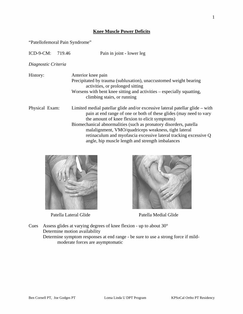
climbing stairs, or running Physical Exam: Limited medial patellar glide and/or excessive lateral patellar glide – with
pain at end range of one or both of these glides (may need to vary the amount of knee flexion to elicit symptoms)
Biomechanical abnormalities (such as pronatory disorders, patella malalignment, VMO/quadriceps weakness, tight lateral retinaculum and myofascia excessive lateral tracking excessive Q angle, hip muscle length and strength imbalances
Patella Lateral Glide Patella Medial Glide Cues Assess glides at varying degrees of knee flexion - up to about 30° Determine motion availability Determine symptom responses at end range - be sure to use a strong force if mild-
moderate forces are asymptomatic

Ben Cornell PT, Joe Godges PT Loma Linda U DPT Program KPSoCal Ortho PT Residency
2
"Patellar Tendinitis" ICD-9-CM: 726.64 Patellar tendinitis Diagnostic Criteria History: Anterior knee pain. Pain associated with repetitive use of extensor mechanism (e.g., jumping,
kicking) Physical Exam: Symptoms reproduced with palpation to inferior pole of patella, or patella
tendon insertion at the tibial tuberosity
Patellar Tendon Palpation/Provocation
Cues: P= Patella 1= Inferior Pole 2= Superior Pole 3= Tibial Tuberosity
Ben Cornell PT, Joe Godges PT Loma Linda U DPT Program KPSoCal Ortho PT Residency
3
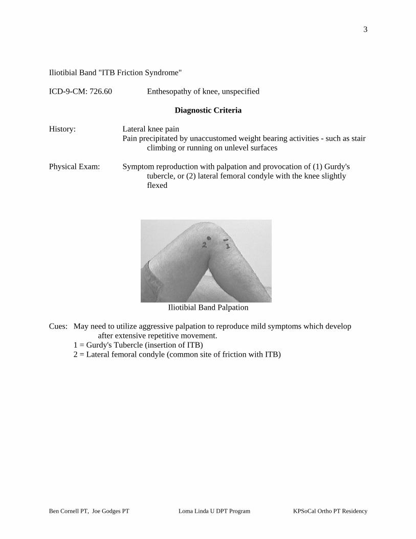
Iliotibial Band "ITB Friction Syndrome" ICD-9-CM: 726.60 Enthesopathy of knee, unspecified
Diagnostic Criteria History: Lateral knee pain Pain precipitated by unaccustomed weight bearing activities - such as stair
climbing or running on unlevel surfaces Physical Exam: Symptom reproduction with palpation and provocation of (1) Gurdy's
tubercle, or (2) lateral femoral condyle with the knee slightly flexed
Iliotibial Band Palpation Cues: May need to utilize aggressive palpation to reproduce mild symptoms which develop
after extensive repetitive movement. 1 = Gurdy's Tubercle (insertion of ITB) 2 = Lateral femoral condyle (common site of friction with ITB)

Ben Cornell PT, Joe Godges PT Loma Linda U DPT Program KPSoCal Ortho PT Residency
4
"Pes Anserinus Bursitis" ICD-9-CM: 726.61 Pes anserinus bursitis Diagnostic Criteria History: Symptom precipitated by recent repetitive activity (e.g., long distance
running) usually in the presence of some biomechanical abnormality (e.g., abnormal pronation)
Physical Exam: Medial knee pain
Symptoms reproduced with palpation of pes anserine bursa
Pes Anserine Palpation Cues: Bursa is located on the medial tibia flare adjacent to the insertion of the semitendinosis
(follow tendon distally to locate bursa) 3 = Pes Anserine Bursa

Ben Cornell PT, Joe Godges PT Loma Linda U DPT Program KPSoCal Ortho PT Residency
5
Patellofemoral Pain Syndrome
ICD-9: 719.46 pain in joint - lower leg
Description: Patellofemoral pain syndrome (PFPS) is described as anterior knee pain during squatting, kneeling, stairs, walking and sitting (especially prolonged sitting) with the knee flexed. It is typically caused by poor mechanics of the patella as it travels in the femoral groove during flexion and extension of the knee. This poor tracking which typically causes the patella to deviate laterally, resulting in excessive stress on the medial patello-femoral compartment due to stretching and irritation, as well as increased lateral compartment compression. Etiology: The specific causes of this disorder can vary in individuals and typically lacks a mechanism of injury. Tight lateral structures including the iliotibial band and the lateral retinaculum are thought to be the primary causes. There are several biomechanical factors that contribute to poor tracking of the patella. These include excessive femoral anteversion and increased midfoot pronation with resultant tibial lateral rotation. **The depth, of the femoral trochlear groove also has direct bearing on the tracking of the patella. Another factor is the motor control/strength of the hip abductors and external rotators during weight loading activities. Intra-articular effusion has been shown to lead to vastus medialis inhibition as well. With inhibition of this muscle, the oblique fibers of the vastus medialis are not effective in tracking the patella medially during extension causing the patient to experience PFPS. This disorder is common in adolescent females due to the biomechanical changes occurring as their bodies develop, though is not limited to this population.
Physical Examinations Findings (Key Impairments) Acute Stage / Severe Condition
• The patient’s reported symptoms are elicited typically with compressive forces about the involved knee during activities such as squatting and sitting for long periods of time
• The patella typically has limited medial gliding of the patella secondary to taut peripatellar structures
• The patient may present with any or all of the following biomechanical abnormalities: an increased Q-angle, femoral anteversion, lateral tibial torsion, and increased midfoot pronation, limited external rotation of the hip, limited tibiofemoral extension, decreased strength in the supinators of the foot during gait, and medial quadriceps weakness.
Ben Cornell PT, Joe Godges PT Loma Linda U DPT Program KPSoCal Ortho PT Residency
6
Sub Acute Stage / Moderate Condition As Above – except:
• The patient’s reported symptoms are elicited intermittently with the activities noted above.
Settled Stage / Mild Condition
• Pain may be elicited only in certain positions of excessive patellofemoral compression maintained over prolonged periods of time such as sitting, sustained stair climbing, running or biking with seat too low.

Ben Cornell PT, Joe Godges PT Loma Linda U DPT Program KPSoCal Ortho PT Residency
7
Intervention Approaches / Strategies Acute Stage / Severe Condition Goals: Decrease inflammation
Decrease pain
• Physical Agents Ice Ultrasound (in conjunction with mobilization/manipulation) Acupuncture, acupressure or electroacupuncture for pain control
• Manual Therapy
Soft tissue mobilization to tight lateral peripatellar structures Joint mobilization to the patella – medial patellar glides, sustained stretch and high velocity low amplitude manipulation Joint mobilization to the tibiofemoral joint – restoring normal knee extension Sacroiliac evaluation and manipulation
• External Devices (Taping/Splinting/Orthotics)
Patellar taping procedures in conjunction with biofeedback and exercise program to promote proper patellar tracking Foot orthotics to correct excessive pronation if present Resistive brace (such as Protonics) to improve hamstring use and restore proper biomechanics
• Neuromuscular Reeducation
Facilitory techniques to improve the contraction of the hip abductors and lateral rotators, foot supinators, and, the quadriceps muscle group, focusing, if possible, on the oblique fibers of the vastus medialis muscle
• Therapeutic Exercises
Stretching exercises for the iliotibial band and hamstrings Initial exercises should be largely closed kinetic chain activities in the pain-free range only
• Re-injury Prevention/Instruction:
Temporarily limit any deep squatting, heavy lifting, or through-range resistive training of the quadriceps
Ben Cornell PT, Joe Godges PT Loma Linda U DPT Program KPSoCal Ortho PT Residency
8
Sub Acute Stage / Moderate Condition Goals: Normalize lower extremity flexibility muscle strength, motor control, and patellofemoral
tracking
• Approaches / Strategies listed above
• Therapeutic Exercises Progress exercises to include training for return to a specific work, recreational, or sport activity Assess the biomechanics of aggravating activity (e.g. cycling with a seat too low can increase pain and cause pressure) Promote painfree, low resistance, repetitive exercises (e.g., cycling) that provide non-injurious compressive loads to the patellofemoral cartilage
Settled Stage / Mild Condition Goal: Return to desired activities
• Approaches / Strategies listed above
• Therapeutic Exercises Progress stretching, strengthening and coordination exercises – which includes training for return to a specific work, recreational, or sport activity
Intervention for High Performance /High Demand Functioning in Workers or Athletes
Goal: Return to desired occupation or sport
• Approaches / Strategies listed above • Further biomechanical assessment during aggravating activity
Ben Cornell PT, Joe Godges PT Loma Linda U DPT Program KPSoCal Ortho PT Residency
9
Selected References Bizzini M, Childs JD, Piva SR, Delitto A. Systematic review of the quality of randomized controlled trials for patellofemoral pain syndrome. J Orthop Sports Phys Ther. 2003;33(1):4-19. Eng JJ, Pierrynowski MR. Evaluation of soft foot orthotics in the treatment of patellofemoral pain syndrome. Phys Ther. 1993;73(2):62-8. Fulkerson JP. The etiology of patellofemoral pain in young, active patients: a prospective study. Clin Orthop. 1983;179:129-33. Lohman E, Harp T. A critical review of patellofemoral pain syndrome in rehabilitation. Crit Review in Phys Rehab Med. 2002;14(3&4):197-222. Powers CM. Patellar kinematics, part ii: the influence of the depth of the trochlear groove in subjects with and without patellofemoral pain. Phys Ther. 2000;80(10):965-78. Powers CM, Maffucci R, Hampton S. rearfoot posture in subjects with patellofemoral pain. J Orthop Sports Phys Ther.1995;22(4):155-60. Salsich GB, Brechter JH, Farwell D, Powers CM. The effects of patellar taping on knee kinetics, and vastus lateralis muscle activity during stair ambulation in individuals with patellofemoral pain. J Orthop Phys Ther. 2002; 32(1): 3-10.
Ben Cornell PT, Joe Godges PT Loma Linda U DPT Program KPSoCal Ortho PT Residency
10
Patellar Tendinitis
ICD-9: 726.64 patellar tendinitis
Description: Repetitive strain injury affecting the patellar tendon, resulting in anterior knee pain. Etiology: This condition is believed to be the result of repetitive mechanical stresses and is most commonly found in athletes whose sport involves repetitive, sudden, ballistic movements of the knee – such as jumping. Intratendinous changes can begin as microtears, which lead to collagen degeneration, and subsequent fibrosis. The result is usually pain well localized to a small area of the anterior knee region with specific tenderness at the inferior pole of the patella.
Physical Examination Findings (Key Impairments) Acute Stage / Severe Condition
• Severe local tenderness on palpation at either the proximal or distal insertion of the patellar tendon
• Accessory movement deficits of patella medial/lateral/superior/inferior glide • Pain with maximum stretching of the quadriceps • Weak and painful quadriceps muscle when tested isometrically against resistance • Symptoms can be reproduced 1) using the decline squat test, 2)with eccentric knee
contractions, 3) with deep squats, or 4) with jumping/ sports activities • Biomechanical abnormalities of the lower quarter may be present – such as excessive
foot pronation; patella alta; femoral anteversion; flexibility deficits in the quadriceps, hamstrings, and calf muscles, as well as in the iliotibial band; strength deficits of the gluteal, lower abdominal, quadriceps, and calf muscles
Sub Acute Stage / Moderate Condition As Above – except:
• The patient tolerates more repetitions during functional strength tests before onset of pain (pain may hinder sport performance, but usually does not limit activities of daily living)
• Patellar tendon palpation is less tender Settled Stage / Mild Condition As Above – except:
• Symptoms may be difficult to illicit unless repeated strenuous movements are performed
• Mild local tenderness with patellar palpation – note that mild patellar tenderness with palpation may be a normal finding in active athletes
Ben Cornell PT, Joe Godges PT Loma Linda U DPT Program KPSoCal Ortho PT Residency
11
Intervention Approaches / Strategies Acute Stage / Severe Condition Goals: Alleviate pain
Reduce aggravating and predisposing factors
• Physical Agents Ice Phonophoresis Iontophoresis
• External Devices (Taping/Splinting/Orthotics)
Patellar taping procedures may assist with promoting proper patellar tracking Foot orthotics may be useful to correct excessive pronation Taping or bracing to unload patellar tendon
• Manual Therapy
Joint mobilization at the patella if hypomobility exists
• Therapeutic Exercise Initiate non-aggravating, stretching exercises for relevant muscles or fascial tissue – typically the muscles with trigger points Initiate non-aggravating, strengthening exercises for relevant weak musculature
• Re-injury Prevention/Instruction:
Temporarily limit any deep squatting, heavy lifting, or resistive training of the quadriceps
Subacute Stage / Moderate Condition Goals: Restore function
Prevent future re-injury
• Approaches / Strategies listed above
• Manual Therapy Friction massage to the patellar tendon
• Therapeutic Exercise
Progress stretching and strengthening to the relevant myofascia and connective tissue Begin sport specific training as tolerated, although still avoiding maximal concentric and eccentric loads
Ben Cornell PT, Joe Godges PT Loma Linda U DPT Program KPSoCal Ortho PT Residency
12
Settled Stage / Mild Condition Goals: As above
Progress activity tolerance Ability to resume sports activity and daily activities without pain
• Approaches / Strategies listed above
• Therapeutic Exercise
Progress stretching exercises – provide a comprehensive lower quarter stretching program with emphasis on patient independence and carryover Progress strengthening exercises with an with eccentric emphasis (e.g., light jumping activities, progressive resistive exercises, sport specific training) Begin sport specific training as tolerated, although still avoiding maximal concentric and eccentric loads
Intervention for High Performance / High Demand Functioning in Workers or Athletes Goal: Full return to sport activity or occupation
• Approaches / Strategies listed above
• Therapeutic Exercise Review and correct biomechanics of desired activity, especially landing pattern of jumps, ankle/foot biomechanics, and hip/pelvic balance and stability Agility training specific to sports activity High-velocity ballistic training that is sport specific Single-leg exercises Progress with combinations of load (weight), speed, and jumping height
Ben Cornell PT, Joe Godges PT Loma Linda U DPT Program KPSoCal Ortho PT Residency
13
Selected References Bellemans J, Witvrouw, et al. Intrinsic risk factors for the development of patellar tendonitis in an athletic population. A two-year prospective study. Am J Sports Med. 2001;29:190-5. Benjamin HJ, Briner WW. Volleyball Injuries. Phys Sportsmed. 1999;27:48-58. Cook JL, Khan KM, et al. Overuse Tendinosis, Not Tendinitis. Part 1: A New Paradigm for a Difficult Clinical Problem. Phys Sportsmed. 2000;28:38-48. Cook JL, Khan KM, et al. Overuse Tendinosis, Not Tendinitis. Part 2: Applying the New Approach to Patellar Tendinopathy. Phys Sportsmed. 2000; 28:31-46. Panni AS. Patellar Tendinopathy in Athletes. Am J Sports Med. 2000;28:392-397.
Ben Cornell PT, Joe Godges PT Loma Linda U DPT Program KPSoCal Ortho PT Residency
14
Patellar Bursitis
ICD-9: 726.65 prepatellar bursitis
Description: Inflammation and swelling of bursae over the patella. Etiology: Cause is typically trauma, either due to repetitive extremity movement or to acute trauma to patella. In active persons, bursitis can be induced by work activity, as seen by carpet layers, gardeners, and/or roofers. In athletes, patellar bursitis has been reported in football players, wrestlers, basketball players and dart throwers. Direct injury to the bursae comes from repetitive contact with the artificial turf, wrestling mat, hardwood floor, or exercise mat.
Physical Therapy Findings (Key Impairments)
Acute Stage / Severe Condition
• Enlarged bursa, commonly the bordering the patellar surfaces • The involved bursa are tender, may be slightly warm, and reproduce the reported
symptoms with provocatory palpation • Resisted knee extension also reproduce the reported symptoms • Decreased range of motion of knee – pain with passive knee flexion at end range
Sub Acute Stage / Moderate Condition As Above – except:
• Bursa not as tender to palpation – swelling and warmth are also decreased • The pain is not as intense with active movement of knee • Improved passive range of motion of knee due to decreased swelling and pain
Settled Stage / Mild Condition As Above – except:
• Full active and passive range of motion is available with slight pain at end ranges • Muscles around knee may test to be weak, especially the quadriceps

Ben Cornell PT, Joe Godges PT Loma Linda U DPT Program KPSoCal Ortho PT Residency
15
Intervention Approaches / Strategies Acute Stage / Severe Condition Goals: Decrease swelling and pain
• Physical Agents Ice Ultrasound/phonophoresis
• Patient Education/Re-injury Prevention
Avoid activities that aggravate the symptoms
• Therapeutic Exercises Gentle mobility within painfree ranges
Sub Acute Stage / Moderate Condition Goals: Restore normal knee and patellar and patellar mobility
Return to moderate activity
• Approaches / Strategies listed above
• Patient Education/Re-injury Prevention Add padding over bursa during kneeling activities
• Therapeutic Exercises
Encourage painfree, low resistance activities such as bicycling or walking Settled Stage / Mild Condition Goal: Return to pain free daily activity
• Approaches / Strategies listed above
• Therapeutic Exercises Provide strengthening to weak lower extremity musculature
Ben Cornell PT, Joe Godges PT Loma Linda U DPT Program KPSoCal Ortho PT Residency
16
Intervention for High Performance /High Demand Functioning in Workers or Athletes Goal: Return to desired occupational or leisure activities.
• Approaches / Strategies listed above
• Therapeutic Exercises Encourage participation in regular low stress aerobic activities to improve fitness, and strength.
Selected References McFarland EG, Mamanee P, Queale WS, Cosgarea AJ. Olecranon and Prepatellar Bursitis: Treating Acute, Chronic, and Inflamed. Phys Sportsmed. 2000; 68(3). Butcher, JD, Salzman, KL, Lillegard WA. Lower Extremity Bursitis. Am Fam Physician. 1996;53:2317-24. Almekinders, LC, Temple, JD. Etiology, diagnosis, and treatment of tendonitis: an analysis of the literature. Med Sci Sports Exerc. 1998;30:1183-90.

Ben Cornell PT, Joe Godges PT Loma Linda U DPT Program KPSoCal Ortho PT Residency
17
Iliotibial Band Friction Syndrome
ICD-9: 726.60 enthesopathy of knee, unspecified
Description: The iliotibial band is a thickened strip of fascia lata that extends from the iliac crest to the lateral tibial tubercle. It serves as a ligament between the lateral femoral condyle and lateral tibia, stabilizing the knee joint. Iliotibial Band Friction Syndrome (ITBFS) is an overuse syndrome resulting from friction between the iliotibial band and the lateral knee. It occurs primarily in runners but is also prominent in cyclists. Characteristic symptoms are sharp pain or burning on the lateral aspect of the knee proximal to the joint line during exercise. For runners, the pain is often most intense during the deceleration phase of gait. Walking with the knee fully extended may lessen the symptoms. Activities start out pain free but symptoms develop after a reproducible time or distance. Pain subsides shortly after the activity but return with the next bout of running or cycling. Etiology: Classified as an over-use injury, Iliotibial Band Friction Syndrome occurs after continuous, steady long distance runs or cycling. It can also occur after unaccustomed change in training programs, i.e. cycling or running over hilly terrain, sprint training, increased training distances, or running on sloped surface (e.g., on the crown of the road always running in the same direction, such as against traffic). The main symptom is lateral knee pain proximal to the joint line during exercise. Other predisposing factors are sudden increase in training distances, cavus foot, genu varum, tibial varum, rearfoot and/or forefoot varus, and leg length discrepancy. There is also evidence that weak hip abductor musculature is a contributing factor.
Physical Examinations findings (Key Impairments) Acute Stage / Severe Condition
• Antalgic gait • “Stiff legged” walking in order to reduce knee flexion • Aggravation of symptoms upon climbing or descending stairs or running downhill • Pain elicited upon thumb pressure over lateral femoral condyle while active flexion-
extension of the knee is performed, with maximum pain at 300 flexion • Positive Ober’s test – suggesting a “Tight” tensor fascia lata • Soft tissue restriction along the iliotibial band • Provocation of pain with palpation over Gurdy’s tubercle
Ben Cornell PT, Joe Godges PT Loma Linda U DPT Program KPSoCal Ortho PT Residency
18
Sub Acute Stage / Moderate Condition As above with the following differences
• Reduced antalgic gait. Increased knee flexion during walking • Reduced aggravation of symptoms upon climbing or descending stairs or running
downhill • Decreased pain upon thumb pressure over lateral femoral condyle while active
flexion-extension of the knee is performed, with maximum pain at 30o flexion • Reduced pain after start of activity (running, cycling)
Settled stage / Mild Condition As above with the following differences
• Mild pain after start of activity (running, cycling)
Ben Cornell PT, Joe Godges PT Loma Linda U DPT Program KPSoCal Ortho PT Residency
19
Intervention Approaches / Strategies
Acute Stage / Severe Condition Goals: Control pain and inflammation
Correct poor training habits or any other structural abnormalities
• Physical Agents Ice packs, ice massage Ultrasound Phonophoresis Electrical stimulation
• Re-injury Prevention Instruction
Temporarily limit any activity that aggravate symptoms Sub Acute Stage / Moderate Condition Goals: Avoid continued irritation
Prevent Re-injury
• Approaches / Strategies listed above
• Manual Therapy Soft tissue mobilization and manual stretching to the fascial adhesions to the ITB
• Therapeutic Exercises Stretching intended to elongate the iliotibial band, such as Half-kneeling diagonal stretch, Ober stretch, modified Ober stretch, Crossover toe touch, Lateral hip drop stretch The most tension on the ITB is created by having the patient standing and extending and adducting the leg to be stretched across and behind the other leg. The patient than sidebends the trunk away from the involved hip/thigh hands clasped overhead • Re-injury Prevention Instruction
Instruction in proper footwear (including bicycle toe clip options) and orthotics may be helpful (a lateral sole wedge may be of help)
Ben Cornell PT, Joe Godges PT Loma Linda U DPT Program KPSoCal Ortho PT Residency
20
Settled Stage / Mild Condition Goals: As Above
Prevent recurrence of resolved symptoms.
• Approaches / Strategies listed above • Therapeutic Exercises
Provide stretching exercises to elongate shortened myofascial (e.g., hip flexors, calf muscles) and strengthening exercises to improve the motor performance in weak muscles (e.g., gluteus medius and gluteus maximus)
• Re-injury Prevention Instruction
Instruction in proper footwear and orthotics may be necessary Intervention for High Performance / High Demand Functioning in Workers or Athletes Goal: To return to optimum level of function at work or sports.
• Approaches / Strategies listed above
• Therapeutic Exercises Continuation of gradual increase in distance and frequency of activities

Ben Cornell PT, Joe Godges PT Loma Linda U DPT Program KPSoCal Ortho PT Residency
21
Selected References Barber FA, Sutker AN. Iliotibial band syndrome. Sports Med. 1992;14:144-148. Drogset JO, Rossvoll I, Grontvedt T. Surgical treatment of iliobitial band friction syndrome: A retrospective study of 45 patients. Scand J Med Sci Sports. 1999;9:296-298. Fredericson M, Guillet M, DeBenedictis L. Quick solutions for iliotibial band syndrome. Phys Sports Med. 2000;28. Fredericson M, White JJ, MacMohon JM, Andriacchi TP. Quantitative analysis of the relative effectiveness of 3 iliotibial band stretches. Arch Phys Med Rehabil. 2002;83:589-92. Holmes JC, Pruitt AL, Whalen NJ. Iliotibial band friction syndrome in cyclist. Am J Sports Med. 1993;21:419-424. Martens M, Libbrecht P, Burssens A. Surgical treatment of the iliotibial band friction syndrome. Am J Sports Med 1989;17:651-654. Noble CA. Iliotibial band friction syndrome in runners. Am J of Sports Med. 1980;8:232-234. Noble HB, Hajek RM, Porter M. Diagnosis and treatment of iliotibial band tightness in runners. Phys Sports Med. 1982; 10:67-74.

Ben Cornell PT, Joe Godges PT Loma Linda U DPT Program KPSoCal Ortho PT Residency
22
Pes Anserine Bursitis
ICD-9: 727.9 unspecified disorder of synovium, tendon, and bursa
Description: An inflammatory condition of the medial knee especially common in certain patient populations and often coexisting with other knee disorders. The term pes anserinus refers to the conjoined tendons of the sartorius, semitendinosus, and gracilis muscles as they cross the proximal aspect of the tibia to insert along its medial surface. The term originates from the Latin “pes” for foot and “anserinus” for goose and derives from the anatomic observation that the tendons form a structure reminiscent of a goose’s webbed foot. Etiology: Inflammation of the pes anserine bursa. This bursa is located 2” inferior to joint line at the medial tibial flare. Inflammation to this bursa is often a sequela to local trauma, exostosis and tendon tightness, pes planus (predisposes the patient to problems affecting the medial knee) or DJD affecting the knee especially in overweight middle-aged to elderly women . A female patient who is overweight can also experience referred pain to the knee from broad pelvic area with the resultant angulation at the knee joint putting more stress on the bursa.
Physical Examinations Findings (Key Impairments)
• Tenderness over proximal medial tibia • May have localized swelling at the insertion of medial hamstring muscles • Negative valgus stress at 30° flexion lessens likelihood of medial collateral ligament
strain • Negative McMurray’s and painfree knee flexion overpressures lessens the likelihood
of meniscal involvement • Positive resisted knee flexion in prone position
Ben Cornell PT, Joe Godges PT Loma Linda U DPT Program KPSoCal Ortho PT Residency
23
Intervention Approaches / Strategies
Goal: Decrease swelling and pain.
• Physical Agents Iontophoresis with dexamethasone Ultrasound/ phonophoresis Electrical stimulation Ice
• Therapeutic Exercises
Gentle stretching in pain free ranges of: sartorius (hip IR in hip and knee extension) gracilis (supine hook lying, gently spread knees apart) hamstrings (long sit, foot turned slightly in, loop towel or sheet around
foot and pull gently while maintaining lumbar lordosis) triceps surae (standing one with knee extended and one leg flexed)
Quadriceps, hamstring and calf strengthening
• External Devices (Taping/Splinting/Orthotics) Orthotics, where indicated, to correct pes planus
• Re-injury Prevention Instruction
Instruct patient in appropriate exercises, stretches, application of ice and instruct in the use of orthotics Patient education for weight management
Ben Cornell PT, Joe Godges PT Loma Linda U DPT Program KPSoCal Ortho PT Residency
24
Selected References Abeles M. Anserine bursitis. Arthritis Rheum. 1986;29:812-3. Brookler MI, Mongan ES. Anserina bursitis: a treatable cause of knee pain in patients with degenerative arthritis. California Medicine. 1973;119:8-10. Butcher JD, Salzman KL, Lillegard WA. Lower extremity bursitis. Am Fam Physician. 1996;53:2317-2324. Calmbach WL, Hutchens M. Evaluation of Patients Presenting with Knee Pain: Part II: Differential Diagnosis. American Family Physician. 2003;68:917. Forbes JR, Helms CA, Janzen DL.Acute Pes Anserine Bursitis: MR Imaging. Radiology. 1995; 194:525-527 Handy JR. Anserine bursitis: a brief review. South Med J. 1997; 90:376-7. Hemler DE, Ward WK, Karstetter KW, Bryant, PM. Saphenous Nerve Entrapment caused by Pes Anserine Bursitis mimicking Stress Fracture of Tibia. ArchPhys Med Rehabil. 1991;72:336-7. Larsson LG, Baum J. The syndrome of anserine bursitis: an overlooked diagnosis. Arth Rheum 1985;28:1062-5. Magee, D. Orthopedic Physical Assessment 3rd ed. WB Saunders Co., Philadelphia, PA, 1997 Stuttle FL: The no-name and no-fame bursa. Clin Orthop. 1959;15:197-99. White, T. Pes anserine (knee) bursitis rehabilitation exercises. Sports Medicine Adviser 2002.1. http://www.med.umich.edu/1libr/sma/sma_pesanser_rex.htm
Recommended