From Department of Medicine Solna, Unit of Translational Immunology
Karolinska Institutet, Stockholm, Sweden
REGULATORY T CELLS MOLECULAR AND CLINICAL ASPECTS
Malin E. Winerdal
Stockholm 2015

All previously published papers were reproduced with permission from the publisher.
Published by Karolinska Institutet.
Printed by E-Print AB 2015
© Malin Winerdal, 2015
ISBN 978-91-7549-808-9

Regulatory T Cells – Molecular and Clinical Aspects
THESIS FOR DOCTORAL DEGREE (Ph.D.)
By
Malin E. Winerdal
Principal Supervisor: Professor Ola Winqvist Karolinska Institutet Department of Medicine Unit of Translational Immunology Co-supervisor(s): MD, PhD Per Marits Karolinska Institutet Department of Medicine Unit of Translational Immunology
Opponent: Professor Pärt Peterson University of Tartu Laboratory of Molecular Pathology Examination Board: Docent Michael Uhlin Karolinska Institutet Department of Oncology-Pathology Professor Eva Sverremark Ekström Stockholm University Department of Molecular Biosciences The Wenner-Gren Institute Professor Magnus Essand Uppsala University Department of Immunology Genetics and Pathology


Litterarum radices amarae, fructus dulces.
-Cicero.
To my family,

ABSTRACT
In the immune system’s tug of war with cancer, tolerance mechanisms by which the tumor can
control anti-tumor immune responses play a central role in determining the outcome.
Regulatory T cells (Treg) induced in the thymus or the periphery, represent one such tolerance
mechanism that potentially can be exploited by developing tumors. In this thesis, we explore
the underlying molecular mechanisms that result in Treg development, and attempt to elucidate
the importance of this T lymphocyte subset in urinary bladder cancer.
In humans and mice, the transcription factor FOXP3 is crucial for the function of the Treg
subset and maintenance of peripheral tolerance, as illustrated by the lack of functional Treg and
autoimmune disorders that result from mutations within this protein. In humans however,
clinical studies of this important T lymphocyte subset are obscured by the fact that FOXP3 is
transiently induced in conventional T lymphocytes upon activation. Initially, we address the
epigenetic regulation of FOXP3 expression, and demonstrate that the committed Treg
population has an almost completely demethylated FOXP3 promoter region, whereas
conventional non-regulatory CD4+ T lymphocytes are semi-methylated in this region.
Furthermore, we study the development of regulatory T cells in mice, and assess the
contribution and the impact of adenosine receptor signaling on the T lymphocyte compartment.
Next, the role of the Treg population in urinary bladder cancer is evaluated. First, we investigate
the impact of tumor infiltrating CD3+ as well as FOXP3+ T lymphocytes on patient survival.
Somewhat surprisingly, we find a positive correlation between both CD3+ and FOXP3+
lymphocytic infiltrates, suggesting that FOXP3+ lymphocytes in this case may not represent a
tumor escape mechanism as initially hypothesized. In addition, we observe FOXP3 expression
in a subset of tumors, and find that this expression is a negative prognostic factor for survival.
To follow up these results, we characterize the T lymphocyte immune response in peripheral
blood, lymph nodes and tumor tissue from patients with UBC. We demonstrate that the
FOXP3+ fraction of CD4+ T lymphocytes is significantly increased compared to all other
locations investigated including macroscopically healthy bladder tissue. Furthermore, these
tumor infiltrating lymphocytes express high levels of activation and effector markers, but do
not display a demethylated pattern in the FOXP3 promoter to match its prominent expression.
Interestingly, muscle invasive tumors have a lower FOXP3+ fraction at the invasive front
compared to non-invasive counterparts. In addition, we observe changes in the cellular immune
response dependent on if the patients have received neo-adjuvant chemotherapy or not, both
with regard to cell composition and functional reactivity to tumor antigens.
Epigenetic regulation governs the commitment of T lymphocytes to the Treg lineage. The fact
that FOXP3 expressing tumor infiltrating lymphocytes in UBC do not display a committed
Treg phenotype could potentially explain the differences in reported clinical impact of this
population in different cancers and has implications for future immunotherapy.
LIST OF SCIENTIFIC PAPERS I. Winerdal ME*, Janson PC*, Marits P, Thörn M, Ohlsson R, Winqvist O.
FOXP3 promoter demethylation reveals the committed Treg population in humans. PLoS One. 2008 Feb 20;3(2):e1612. *Shared first authorship
II. Winerdal ME, Marits P, Winerdal M, Hasan M, Rosenblatt R, Tolf A, Selling K, Sherif A, Winqvist O. FOXP3 and survival in urinary bladder cancer. BJU Int. 2011 Nov;108(10):1672-8.
III. Winerdal ME, Krantz D, Rosenblatt R, Zirakzadeh A, Ahlén Bergman E, Winerdal M, Hansson J, Holmström B, Johansson M, Marits P, Sherif A, Winqvist O. Characterization of T lymphocyte Responses in Urinary Bladder Cancer. Manuscript.
IV. Winerdal ME, Winerdal M, Krantz D, Yang T, Lindmark E, Marits P, Fredholm B, Ådén U, Carlström M, Winqvist O. Adenosine receptor signaling in T cell development. Manuscript. List of publications not included in the thesis:
Lofgren C, Hjortsberg L, Blennow M, Lotfi K, Paul C, Eriksson S, Albertioni F. Mechanisms of cross-resistance between nucleoside analogues and vincristine or daunorubicin in leukemic cells. Biochem Biophys Res Commun. 2004 Jul 30;320(3):825-32.
Janson PC, Winerdal ME, Winqvist O. At the crossroads of T helper lineage commitment-Epigenetics points the way. Biochim Biophys Acta. 2009 Sep;1790(9):906-19.
Hu J, Lou D, Carow B, Winerdal ME, Rottenberg M, Wikström AC, Norstedt G, Winqvist O. LPS Regulates SOCS2 Transcription in a Type I Interferon Dependent Autocrine-Paracrine Loop. PLoS One. 2012;7(1):e30166.
Winerdal ME*, Winerdal M*, Kinn J, Urmaliya V, Winqvist O, Adén U. Long lasting local and systemic inflammation after cerebral hypoxic ischemia in newborn mice. PLoS One. 2012;7(5):e36422.
*Shared first authorship
CONTENTS 1 Introduction ..................................................................................................................... 1
1.1 T lymphocyte selection and differentiation .......................................................... 2
1.1.1 Thymic selection ....................................................................................... 2
1.1.2 T helper cell subsets .................................................................................. 3
1.2 Regulatory T cells .................................................................................................. 4
1.2.1 Regulatory T cell lineage markers ............................................................ 4
1.2.2 Regulatory T cell subsets .......................................................................... 7
1.2.3 Regulatory T cell stability and plasticity .................................................. 9
1.2.4 Regulatory T cell effector mechanisms .................................................. 11
1.3 Adenosine receptor signaling .............................................................................. 12
1.4 The concept of cancer .......................................................................................... 14
1.5 Tumor immunity .................................................................................................. 15
1.5.1 Immune escape ........................................................................................ 15
2 Aims of the thesis .......................................................................................................... 19
3 Materials and methods .................................................................................................. 21
3.1 Patients ................................................................................................................. 21
3.1.1 Patient characteristics (Paper I-III) ......................................................... 21
3.1.2 Sentinel node detection and surgical methods ....................................... 21
3.1.3 Patient follow-up ..................................................................................... 22
3.2 Mice (Paper IV) ................................................................................................... 22
3.3 Cell preparation and culture (Paper I, III and IV) .............................................. 22
3.4 Immunological evaluation ................................................................................... 23
3.4.1 Flow cytometry (Paper I, III and IV) ...................................................... 23
3.4.2 Functional T lymphocyte assays (Paper I and III) ................................. 23
3.4.3 Immunohistochemistry (Paper II) ........................................................... 24
3.4.4 Polymerase chain reaction (PCR) (Paper I) ........................................... 24
3.4.5 DNA methylation analysis (Paper I and III) .......................................... 25
3.5 Bone marrow transfer experiment (Paper IV) .................................................... 25
3.6 Statistical analysis (Paper I-IV) .......................................................................... 25
4 Results and discussion ................................................................................................... 27
4.1 FOXP3 promoter demethylation reveals the committed Treg population in
humans (Paper I) .................................................................................................. 27
4.2 FOXP3 and survival in urinary bladder cancer (Paper II) ................................. 28
4.3 Anti-tumor immune reponses are shaped by location and therapy (Paper
III) ........................................................................................................................ 30
4.4 Adenosine receptor signaling affects the development of Treg and
conventional T lymphocytes (Paper IV) ............................................................. 31
5 Concluding remarks and future perspectives ............................................................... 33
6 Populärvetenskaplig sammanfattning ........................................................................... 35
7 Acknowledgements ....................................................................................................... 37
8 References ..................................................................................................................... 41

LIST OF ABBREVIATIONS
APC Antigen Presenting Cell
BCG Bacillus Calmette-Guerin
CD Cluster of differentiation
CFSE Carboxyfluorescein succinimidyl ester
CNS Conserved non-coding DNA sequence
COBRA Combined bilsulphite restriction enzyme analysis
CP Central Part
cTEC Cortical thymic epithelial cell
CTLA Cytotoxic T lymphocyte antigen
DAMP Danger associated molecular pattern
DC Dendritic cell
DN Double negative
DP Double positive
ERK Extracellular signal activated kinase
FACS Flow associated cell sorting
FOXP3 Forkhead box transcription factor P3
IDO Indoleamine 2,3-dioxygenase
IF Invasive Front
IFN Interferon
IL Interleukin
IPEX Immune dysregulation, Polyendocrinopathy, Enteropathy, X-
linked syndrome
MACS Magnetic-activated cell sorting
MAPK Mitogen activated protein kinase
MHC Major histocompatibility complex
mTEC Medullary thymic epithelial cell
nSN Non sentinel node
PAMP Pathogen associated molecular pattern
PBMC Peripheral blood mononuclear cell
PCR Polymerase chain reaction
PRR Pattern recognition receptor
RP RNA polymerase
SN Sentinel node
SP Single positive
T-bet T-box transcription factor
TCR T cell receptor
TGF Tumor growth factor
Th T helper
TIL Tumor infiltrating lymphocyte
Treg Regulatory T lymphocyte
TSDR Treg-specific demethylated region
TSLP Thymic stromal lymphoprotein
TUR-B Transurethral resection of the bladder
TZ Transitional zone
UBC Urinary bladder cancer
WT Wild type
1
1 INTRODUCTION
The human body hosts an intriguingly complex balance, and ever ongoing battle of sovereignty.
Constantly challenged by external pathogens, the immune system has evolved to recognize and
eliminate possible threats while sparing and caring for autologous tissues. In this setting, the
two main branches of the immune response, innate and adaptive immunity, play integral and
complementary parts. By means of their broad specificity but limited repertoire of pattern
recognition receptors (PRRs), innate immune cells are constantly vigilant for danger signals
(damage associated molecular patterns (DAMPs) and pathogen associated molecular patterns
(PAMPs)), ready to quickly initiate a primary immune response upon stimulation (Kono and
Rock, 2008). In contrast, T- and B-lymphocytes carry receptors with an almost unlimited
potential to recognize foreign antigens, but react slower compared to their innate counterparts.
At the intersection of these two responses is the presentation of antigens to T lymphocytes by
antigen presenting cells (APCs) (Figure 1); an essential process for the initiation of an adaptive
immune response. The context of the antigen presentation determines the outcome of the
ensuing T lymphocyte response, where presence of co-stimulatory molecules on the APC in
combination with the cytokine milieu facilitates and directs the type of T lymphocyte response.
Since the T lymphocyte receptor repertoire contains approximately in the range of 109-1015
specificities with potential to theoretically recognize any protein epitope, the distinction of self
is essential to avoid autoimmunity. This distinction and thus protection of autologous tissue is
ensured by two separate mechanisms – central and peripheral tolerance. For T lymphocytes,
central tolerance is molded in the thymus, where T lymphocytes carrying T cell receptors
(TCR) with high affinity to self-proteins are deleted (Palmer, 2003) (further discussed in
section 1.1). In the periphery, potentially autoreactive T lymphocytes are kept in check by
different peripheral tolerance mechanisms (Abbas et al., 2004). The most straightforward
mechanism is the concept of ignorance where potentially self-reactive T lymphocytes remain
inactive because of low levels of or no accessibility to the antigen. Secondly, in the absence of
danger signals, the APC will not upregulate the co-stimulatory molecules required by naïve T
lymphocytes for activation. TCR ligation in the absence of co-stimulation leads to anergy or
deletion and thus protects from autoimmunity (Redmond and Sherman, 2005). Alternatively,
engagement of B7 costimulatory molecules on the APC to cytotoxic T lymphocyte antigen 4
(CTLA-4) on the T lymphocytes instead of its stimulatory counterpart CD28, may also induce
T cell tolerance (Perez et al., 1997). Furthermore, regulatory T cells (Tregs) induced
peripherally or in the thymus, can regulate the immune response and thus maintain tolerance
(Sakaguchi et al., 2010).
Cancer poses a unique threat to the human body in the sense that it is inherently both self and
non-self at the same time. Tumor evolution is shaped by the immune system, and successful
tumors evade immune responses in various ways, including suppression of the local T
lymphocyte responses and induction of tolerance (discussed in more detail in section 1.5). This
thesis enacts at the interphase of physiology and tumor pathology, and concerns both the
2
development and regulation of regulatory T cells (papers I and IV) as well as their role and
prognostic impact in urinary bladder cancer (UBC) (papers II and III).
1.1 T LYMPHOCYTE SELECTION AND DIFFERENTIATION
1.1.1 Thymic selection
T lymphocyte progenitors migrate to the thymus from the bone marrow. During their
development in the thymus, thymocytes undergo strict selection processes that only permits T
lymphocytes with functional but not overtly self-reactive TCRs to exit the thymus. In the end,
less than 5% of the initial thymocyte population is selected and allowed to leave for the
periphery (Starr et al., 2003). Here follows a brief summary of the selection process for
conventional α/β T lymphocytes.
Newly arrived thymocytes are CD4-CD8- double negative (DN) and can be further subdivided
into differentiation stages based on their expression of the markers CD44 and CD25 (Godfrey
et al., 1993). While they migrate though the thymic cortex, thymocytes sequentially pass the
CD44+CD25- (DN1), CD44+CD25+ (DN2), CD44-CD25+ (DN3) and CD44-CD25- (DN4)
stages (Godfrey et al., 1993; Lind et al., 2001). Rearrangement of the TCR gene starts at the
DN3 stage with the expression of the recombination activating genes (RAG 1 and 2), and
rearrangement of the TCRβ locus. In a process called β selection, the rearranged TCRβ chain
together with a pre-TCRα chain forms a pre-TCR complex, which triggers thymocyte
proliferation, induction of the two TCR co-receptors CD4 and CD8, and rearrangement of the
TCRα chain locus (Hoffman et al., 1996). CD4+CD8+ double positive (DP) thymocytes that
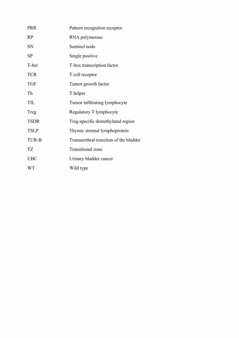
Figure 1. Basic concept of naïve T helper lymphocyte activation. To become activated, a
naïve T lymphocyte requires three distinct sets of signals. 1. TCR recognition of a peptide-
MHC complex. 2. Costimulatory signals. 3. Inflammatory cytokines.
3
successfully rearrange their TCRα chain to form a functional α/β TCR complex undergo
positive selection based on their TCR affinity for MHC molecules on cortical thymic epithelial
cells (cTEC). Failure to recognize self-MHC results in elimination by apoptosis, while
positively selected cells down-regulate CD4 or CD8 expression based on their recognition of
major histocompatibility complex class (MHC) I or MHC II respectively and migrate to the
thymic medulla (Starr et al., 2003). Interestingly, the positive selection process goes on for
several days during which it appears that thymocytes must receive continuous TCR signals
with associated activation of the mitogen activated protein kinase (MAPK) pathway in order
to pass this check point in development (McNeil et al., 2005; Wilkinson et al., 1995).
Negative selection encompasses the process where T lymphocytes with TCRs that strongly
recognize self peptide-MHC complexes are deleted. This is a crucial step in central T cell
tolerance, preventing autoreactive T lymphocytes to exit the thymus to the periphery. Medullar
thymic epithelial cells (mTECs) as well as thymic dendritic cells (tDCs) are key players at this
check point, where presentation of a diverse repertoire of self-antigens on MHC-molecules in
combination with co-stimulation drive the negative selection process (Palmer, 2003).
1.1.2 T helper cell subsets
Once outside the thymus T lymphocytes circulate the secondary lymphoid organs in search of
APCs presenting their cognate antigen. If encountered in the context of co-stimulation and the
appropriate cytokines, the naïve CD4+ T lymphocyte is activated as illustrated in Figure 1. The
local cytokine milieu caused by the triggering factors, and inflammatory cell composition at
the site of activation influences the type of T lymphocyte response that is elicited. The major
T helper lineages that have been described include the classical T helper type 1 (Th1) and T
helper type 2 (Th2) cells, primarily described as stable lineages involved in promoting cellular
and humoral immune responses respectively. With time, new T helper lineages such as the
Th17, Treg and T follicular helper (Tfh) have been characterized adding to the complexity of
T helper differentiation. Intense studies of the factors that drive the evolution of each lineage
have defined transcription factors essential for each subtype as; T-box transcription factor (T-
bet) for Th1, GATA3 for Th2, FOXP3 for Treg and Bcl-6 for Tfh (Weinmann, 2014).
Interestingly, with the increasing number of T helper differentiation fates, reports describing
the co-expression of hallmark cytokines such as IL-17 (Th17) and IFN-γ (Th1) in CD4+ T
lymphocytes have emerged (Wilson et al., 2007). There are now also numerous reports of
combined expression of the lineage defining transcription factors in various contexts (Oestreich
and Weinmann, 2012). With regard to Treg cells, it is interesting to note that the co-expression
of other lineage defining transcription factors such as T-bet and Bcl-6, have been implicated in
the suppression of their respective type of immune response (here; Th1 and Tfh type responses
respectively) (Koch et al., 2009; Linterman et al., 2011). These reports have challenged the
established view of T helper lineages as stable final differentiation states, and suggested a more
dynamic model of T helper differentiation (Weinmann, 2014). In this setting, the ever changing
stimuli that a T cell encounters in combination with its epigenetic and protein phenotype (that
is a consequence of its previous history) will finally determine the type of response elicited.
4
1.2 REGULATORY T CELLS
Regulatory T cells play an integral part in the control of immune responses. The fact that the T
lymphocyte compartment contains cells capable of controlling immune responses has been
known to the scientific community since the early 1970’s (Sakaguchi et al., 2007). At the time
generally known as suppressor T cells, this population attracted strong attention during the
following years, but was finally abandoned much due to the lack of good phenotypic markers
(Sakaguchi et al., 2007). In the mid 1990’s however, the field was revived with Sakaguchi’s
classical report describing the regulatory properties of a subpopulation of CD4+ T lymphocytes
that constitutively expressed the α-chain of the interleukin-2 receptor (IL-2Rα, CD25)
(Sakaguchi et al., 1995). Since then, this population has been the focus of intense research, and
although the knowledge about Treg properties and subtypes has greatly improved, much
remains to be elucidated. Here follows a brief summary of main markers, subsets, and
mechanisms of action of CD4+FOXP3+ Tregs dealt with in this thesis.
1.2.1 Regulatory T cell lineage markers
Numerous molecular markers have been suggested for the delineation of the Treg subsets, some
of which have already been mentioned. The following subsections will discuss some of the
most commonly used markers, with a main focus on the markers used in this thesis.
1.2.1.1 CD25
As mentioned previously, the Treg population was first defined as a subpopulation of CD4+ T
lymphocytes that expressed the IL-2 receptor α-chain (CD25) constitutively (Sakaguchi et al.,
1995). Together with the β-chain (CD122) and the common cytokine receptor γ-chain
(CD132), CD25 forms the high-affinity IL-2R. Interestingly, IL-2R signaling has been shown
to be important for the development and maintenance of Treg (Thornton, 2006). In humans,
Baecher-Allan et al. showed that only the CD25high population (corresponding to between 1
and 2% of CD4+ T lymphocytes in peripheral blood) correlated with suppressive capacity
whereas the CD25low/intermediate cells did not suppress T lymphocyte proliferation (Baecher-Allan
et al., 2001). However, CD25 is upregulated in T cells upon activation, and although CD25
expression is subsequently lost again when the stimulus is removed, this fact decreases its
specificity and usefulness as a marker in clinical settings.
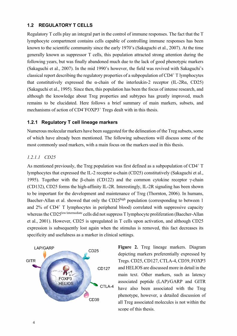
Figure 2. Treg lineage markers. Diagram
depicting markers preferentially expressed by
Tregs. CD25, CD127, CTLA-4, CD39, FOXP3
and HELIOS are discussed more in detail in the
main text. Other markers, such as latency
associated peptide (LAP)/GARP and GITR
have also been associated with the Treg
phenotype, however, a detailed discussion of
all Treg associated molecules is not within the
scope of this thesis.
5
1.2.1.2 FOXP3
The transcription factor FOXP3 has been linked to the suppressive phenotype of both human
(FOXP3) and murine (Foxp3) Treg populations (Sakaguchi et al., 2010). Its close relationship
to the Treg population was first proposed in the Scurfy mouse model, and soon thereafter
confirmed by the fact that the human disease Immune dysregulation, Polyendocrinopathy,
Enteropathy, X-linked syndrome (IPEX) was associated with mutations in the FOXP3 gene
(Bennett et al., 2001; Brunkow et al., 2001). In mice, Foxp3 is generally considered to be a
specific, required and sufficient factor for Treg development (Fontenot et al., 2003). In humans
however, FOXP3 is not as specific since it is transiently upregulated in human CD4+CD25low
T lymphocytes upon activation (Walker et al., 2003). Although this expression is associated
with hypo-responsiveness and decreased cytokine production (Wang et al., 2007), results have
differed regarding the in vitro suppressive capacity of the T lymphocytes with induced FOXP3
expression (Walker et al., 2003; Wang et al., 2007), and it seems that a stable FOXP3
expression is required for suppressive function. Indeed, in vitro induced FOXP3 expression in
conventional human T lymphocytes does confer a regulatory phenotype (Allan et al., 2005;
Walker et al., 2005), and the level of suppressive capacity is both time and dose dependent as
illustrated by a conditional expression model (Allan et al., 2008).
In further contrast to the mouse setting, humans express different splice variants of FOXP3
mRNA that lead to the expression of; full length FOXP3 (FOXP3fl), FOXP3 lacking exon 2
(FOXP3Δ2), and FOXP3 lacking exon 2 and 7 (FOXP3Δ2Δ7) (Allan et al., 2005; Smith et al.,
2006). Although the FOXP3fl and FOXP3Δ2 isoforms have been reported to confer a Treg
phenotype and regulatory activity in vitro (Aarts-Riemens et al., 2008; Smith et al., 2006) this
does not seem to be the case for the FOXP3Δ2Δ7 isoform that instead appears to exert a
dominant negative effect on FOXP3fl upon co-transduction (Mailer et al., 2009). In addition,
it has been shown that the exon 2 domain in the full-length FOXP3 protein interacts with the
transcription factor retinoic acid receptor-related orphan receptor (ROR) α and ROR γt,
suppressing genes related to Th17 development in T lymphocytes, an effect not achieved by
FOXP3Δ2 (Du et al., 2008; Ichiyama et al., 2008). Still, the physiological and clinical impact
of these three isoforms remains to a large extent unknown.
FOXP3 undeniably plays an essential role for the regulatory function of Tregs in both mice and
men. However, the Treg transcriptional profile (in mice) has been demonstrated to be only
partly dependent on Foxp3 expression (Hill et al., 2007). This was emphasized in other studies
where T lymphocytes lacking suppressive capacity but with similar phenotypes to Treg
developed in spite of missing functional Foxp3 (Lin et al., 2007). Together these findings
indicate upstream events of Foxp3 expression as important factors in commitment to the Treg
lineage.
The transient expression of FOXP3 in activated human T lymphocytes combined with the
human heterogeneity with regard to FOXP3 isoforms calls for caution in interpreting human
Treg data based on FOXP3 expression alone, especially in clinical inflammatory settings where
T cell activation is to be expected.

6
1.2.1.3 CD127
Low expression of the IL-7 receptor α-chain (CD127) has been suggested as a complementary
surface marker for Tregs (Liu et al., 2006; Seddiki et al., 2006). CD127 is downregulated in T
lymphocytes upon activation, but whereas effector and memory T lymphocytes resume their
expression, FOXP3+ Tregs remain CD127low/-. Although not perfect, this makes CD127
especially interesting for in vivo studies of immune responses where activated T cells are
expected. The study by Liu et al. related CD127 expression to FOXP3 and found that less than
half of the CD4+CD127low/- population was FOXP3+ (Liu et al., 2006). Categorizing the Treg
population based on CD25high cells did produce much more pure FOXP3+ populations but left
out a considerable number of FOXP3+ Tregs in the CD25int→low population (Liu et al., 2006).
The authors proposed instead that the combination of the two markers be used for surface based
distinction of FOXP3+ cells as the CD4+CD25+CD127lo/- population, which leads to both
improved purity and yield.
1.2.1.4 CTLA-4
The cytotoxic T lymphocyte antigen 4 (CTLA-4 or CD152) is another marker that has been
used to identify the Treg population. It binds the same ligands as the co-stimulatory molecule
CD28 (CD80/CD86 or B7.1/B7.2), but with higher affinity and provides a co-inhibitory signal
(reviewed in (Murakami and Riella, 2014)). In contrast to CD28 which is constitutively
expressed on most resting human T lymphocytes, CTLA-4 expression peaks within 48h of
activation and is only constitutively expressed by the Treg subset (Murakami and Riella, 2014;
Walunas et al., 1994). Its surface expression is tightly regulated and in unstimulated cells
CTLA-4 is mainly localized to intracellular vesicles that upon T cell activation fuse with the
cell membrane to be exposed on the cell surface preferably at the sites of TCR engagement
(Linsley et al., 1996). CTLA-4 can interfere with immune activation through various
mechanisms including the competitive binding of B7 molecules on APCs, interfering with the
intracellular activation signals upon TCR stimulation and induction of regulatory mechanisms
such as indoleamine 2, 3-dioxygenase (IDO) production by the APCs (Murakami and Riella,
2014).
1.2.1.5 Helios
In addition to FOXP3, the ikaros family transcription factor Helios has been proposed as a
complimentary marker for tTreg (Thornton et al., 2010). This study illustrated that Foxp3+
thymocytes are virtually all double positive for Helios, whereas the peripheral Treg
compartment contained only approximately 70% Helios expressing cells. Moreover, they
illustrated that in vitro induced murine and human iTregs do not express Helios (Thornton et
al., 2010). Subsequent studies showed that Helios+FOXP3+ Treg displayed a more
demethylated pattern in their FOXP3 gene compared to Helios- counterparts, suggesting that
these indeed represent a committed Treg lineage (see below) (Kim et al., 2012). However, there
have also been reports challenging Helios as a specific marker of tTreg (Akimova et al., 2011;
Himmel et al., 2013). In one study, Akimova et al. found that Helios expression contrary to
being Treg specific is predominantly associated to activated and proliferating cells (Akimova
7
et al., 2011). Furthermore, Helios’ specificity has been challenged based on the finding of
Helios- Treg expressing markers of naïve T lymphocytes and recent thymic immigrants
(Himmel et al., 2013). In contrast to the earlier studies described, none of the latter were able
to find a correlation between FOXP3 methylation status and Helios expression. In conclusion,
the fact remains that the peripheral CD4+FOXP3+ Treg compartment can be divided into two
subsets based on the expression of the transcription factor Helios, however the functional
significance of which at this point remains unresolved.
1.2.2 Regulatory T cell subsets
As a result of the great scientific interest in the Treg field over the past decades, the knowledge
has vastly expanded together with the number of described Treg subtypes. In addition to the
CD4+FOXP3+ Treg subsets that are the main focus of this thesis, other T lymphocyte
subpopulations with regulatory properties have also been described including the peripherally
IL-10 induced and producing Tr1 and transforming growth factor β (TGF-β) producing Th3
cells as well as CD8+ Tregs (Buckner and Ziegler, 2004; Niederkorn, 2008). Although of great
interest, a detailed discussion of these regulatory populations is beyond the scope of this thesis.
Instead, the following sections will focus on Treg subsets defined by their expression of the
hallmark transcription factor forkhead box P3 (FOXP3). Due to the immense attention that
Tregs have received, the body of litterature on the subject has practically exploded during
recent years together with the proposed number of Treg subsets. In an attempt to simplify the
nomenclature, a group of leading scientists from different parts of the field suggested the
concept of three main subgroups; namely thymus derived Treg (tTreg), peripherally derived
Treg (pTreg) and in vitro induced Treg (iTreg) (Abbas et al., 2013).
1.2.2.1 Regulatory T cell development
The absolute requirement of the thymus for the development of Treg cells is perhaps best
illustrated by animal models such as thymectomy of mice on postnatal day 2-4, which results
in abrogated thymic Treg development and autoimmunity (Sakaguchi et al., 2007). Coherent
with the timing of this model, under normal conditions in mice, CD4+CD25+Foxp3+ Treg are
detectable in the periphery starting from around three days after birth. TCR interactions with
MHC-peptide complexes on APCs together with co-stimulatory signals and cytokines are key
players in Treg selection (Hsieh et al., 2012). The specificity of the Treg TCR repertoire has
been the subject of debate during recent years. However, in general Treg appear to be selected
much based on self-reactivity of their TCR, at an intermediate level between that of thymocytes
that will mature to conventional T lymphocytes and those that will undergo negative selection
due to too high TCR self-affinity (Hsieh et al., 2012).
In the thymus, Foxp3+ thymocytes are found mainly in the medulla, driving the hypothesis that
the Treg lineage is induced mainly in the thymic medulla (Fontenot et al., 2005a). In particular,
mTECs expressing the autoimmune regulator (Aire) transcription factor have been implicated
in the generation of Tregs, since absence of MHC II on these cells resulted in reduction in the
number of Foxp3+ thymocytes in the medulla. Furthermore, the induced expression of a model
8
antigen in mTECs lead to the generation of Tregs specific for the antigen independently of
medullar dendritic cells (Aschenbrenner et al., 2007). However, subsequent studies have shown
that also bone marrow derived tDCs are capable of inducing Foxp3 independently of mTECs
(Hsieh et al., 2012; Proietto et al., 2008). In addition, although the absolute majority of Foxp3+
thymocytes are found as CD4SP cells in the medulla, a few Foxp3+ cells are also observed at
more immature thymocyte stages in the thymic cortex, and Treg development has been
observed in mice with MHC II expression restricted to cTECs (Bensinger et al., 2001). Foxp3+
cells are in fact enriched in the late double positive development stage in the cortex, and the
blocked migration of thymocytes resulted in accumulation of cortical Foxp3+ thymocytes
suggesting that also cTEC may contribute to the induction of Tregs (Liston et al., 2008). In
summary, thymic APCs play a crucial part during tTreg development, however, the
contributions of each APC subtype is still a matter of investigation.
Although the thymic development of Tregs has been studied intensely in mice, relatively little
is known about the mechanisms that drive the development of tTreg in humans (Sakaguchi et
al., 2010). Of note, in humans functional Tregs are present already from week 14 of gestation
in contrast to the murine setting where Treg development is first detected after birth (Darrasse-
Jeze et al., 2005). Moreover, even if many parts of thymocyte development in humans and mice
are comparable, the Hassal’s corpuscles are unique to the histology of the human thymus.
Interestingly, studies have shown that the Hassal’s corpuscles produce thymic stromal
lymphoprotein (TSLP) that is able to activate thymic DCs. These TSLP activated DCs were
subsequently shown to induce FOXP3 expression in CD4SP CD25- thymocytes (Hanabuchi et
al., 2010; Watanabe et al., 2005). Furthermore, in the thymic medulla thymocytes expressing
Treg markers were found to co-localize with Hassal’s corpuscles and activated DCs, supporting
a role for this structure in human Treg development (Watanabe et al., 2005).
1.2.2.2 Role of cytokines in regulatory T cell development
Both in the thymus and the periphery it is clear that the cytokine environment plays a crucial
role for Treg development, as demonstrated by the complete abrogation of Treg development
in mice missing the common γ-chain, involved in the signaling of several cytokines such as IL-
2, IL-7 and IL-15 (Fontenot et al., 2005b). IL-2 signaling in particular has been shown to induce
Foxp3 expression, and IL-2 deficient mice or mice missing IL-2 downstream signaling
molecules display reduced Foxp3 Treg populations in the thymus and the periphery (Fontenot
et al., 2005b; Turka and Walsh, 2008). However, Foxp3+ Tregs do develop in the thymus
despite the lack of IL-2 or CD25, and thus it appears that other cytokines signaling through the
common γ-chain may play complimentary roles to IL-2 during thymic development.
TGF-β is another cytokine that has been implicated in Treg development, and shown to be
important for the peripheral induction and maintenance of Foxp3+ Treg in mice (Marie et al.,
2005). However, its contribution to the thymic development of Treg has been debated, and it
appears that TGF-β signaling contributes to, but is not absolutely essential for thymic Treg
induction (Liu et al., 2008; Marie et al., 2005). Interestingly, the downstream transcription
factor of TGF-β, mothers against decapentaplegic homolog 3 (SMAD3) has been demonstrated
9
to bind a conserved enhancer region within the Foxp3 gene together with nuclear factor of
activated T cells (NFAT) thus promoting Foxp3 transcription (Tone et al., 2008).
1.2.2.3 Functional Treg subsets
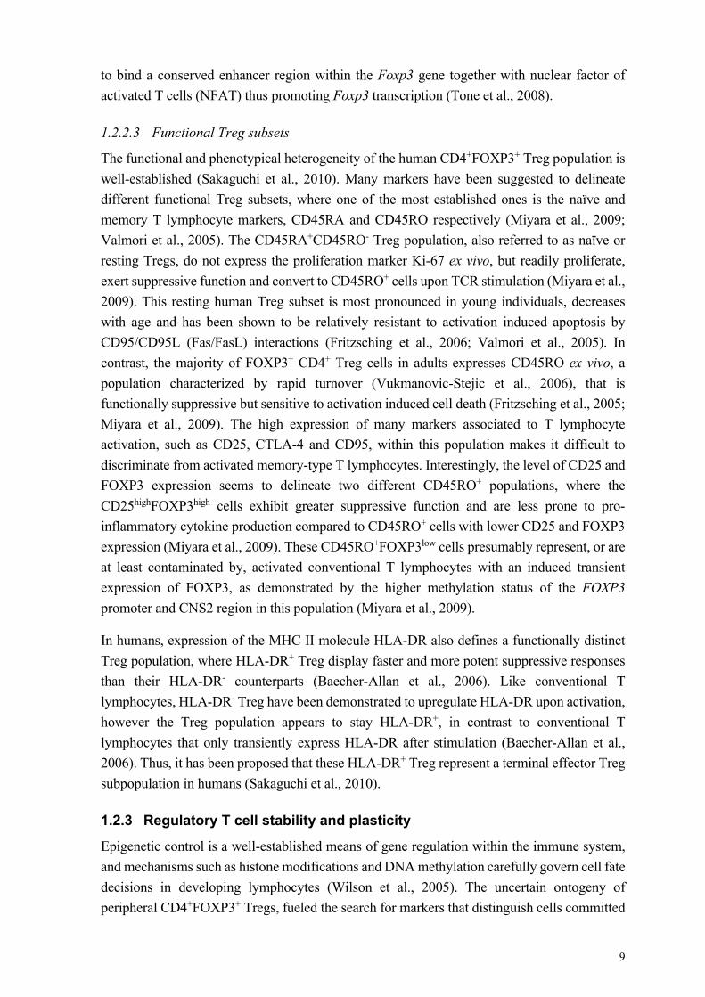
The functional and phenotypical heterogeneity of the human CD4+FOXP3+ Treg population is
well-established (Sakaguchi et al., 2010). Many markers have been suggested to delineate
different functional Treg subsets, where one of the most established ones is the naïve and
memory T lymphocyte markers, CD45RA and CD45RO respectively (Miyara et al., 2009;
Valmori et al., 2005). The CD45RA+CD45RO- Treg population, also referred to as naïve or
resting Tregs, do not express the proliferation marker Ki-67 ex vivo, but readily proliferate,
exert suppressive function and convert to CD45RO+ cells upon TCR stimulation (Miyara et al.,
2009). This resting human Treg subset is most pronounced in young individuals, decreases
with age and has been shown to be relatively resistant to activation induced apoptosis by
CD95/CD95L (Fas/FasL) interactions (Fritzsching et al., 2006; Valmori et al., 2005). In
contrast, the majority of FOXP3+ CD4+ Treg cells in adults expresses CD45RO ex vivo, a
population characterized by rapid turnover (Vukmanovic-Stejic et al., 2006), that is
functionally suppressive but sensitive to activation induced cell death (Fritzsching et al., 2005;
Miyara et al., 2009). The high expression of many markers associated to T lymphocyte
activation, such as CD25, CTLA-4 and CD95, within this population makes it difficult to
discriminate from activated memory-type T lymphocytes. Interestingly, the level of CD25 and
FOXP3 expression seems to delineate two different CD45RO+ populations, where the
CD25highFOXP3high cells exhibit greater suppressive function and are less prone to pro-
inflammatory cytokine production compared to CD45RO+ cells with lower CD25 and FOXP3
expression (Miyara et al., 2009). These CD45RO+FOXP3low cells presumably represent, or are
at least contaminated by, activated conventional T lymphocytes with an induced transient
expression of FOXP3, as demonstrated by the higher methylation status of the FOXP3
promoter and CNS2 region in this population (Miyara et al., 2009).
In humans, expression of the MHC II molecule HLA-DR also defines a functionally distinct
Treg population, where HLA-DR+ Treg display faster and more potent suppressive responses
than their HLA-DR- counterparts (Baecher-Allan et al., 2006). Like conventional T
lymphocytes, HLA-DR- Treg have been demonstrated to upregulate HLA-DR upon activation,
however the Treg population appears to stay HLA-DR+, in contrast to conventional T
lymphocytes that only transiently express HLA-DR after stimulation (Baecher-Allan et al.,
2006). Thus, it has been proposed that these HLA-DR+ Treg represent a terminal effector Treg
subpopulation in humans (Sakaguchi et al., 2010).
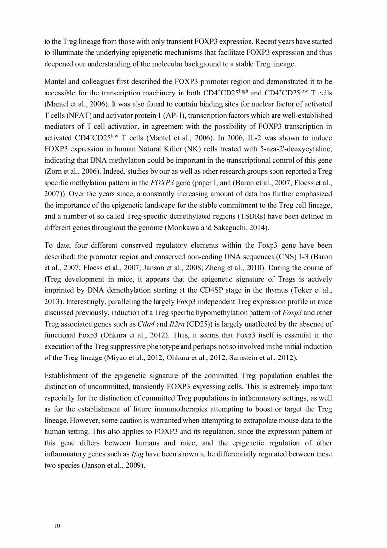
1.2.3 Regulatory T cell stability and plasticity
Epigenetic control is a well-established means of gene regulation within the immune system,
and mechanisms such as histone modifications and DNA methylation carefully govern cell fate
decisions in developing lymphocytes (Wilson et al., 2005). The uncertain ontogeny of
peripheral CD4+FOXP3+ Tregs, fueled the search for markers that distinguish cells committed
10
to the Treg lineage from those with only transient FOXP3 expression. Recent years have started
to illuminate the underlying epigenetic mechanisms that facilitate FOXP3 expression and thus
deepened our understanding of the molecular background to a stable Treg lineage.
Mantel and colleagues first described the FOXP3 promoter region and demonstrated it to be
accessible for the transcription machinery in both CD4+CD25high and CD4+CD25low T cells
(Mantel et al., 2006). It was also found to contain binding sites for nuclear factor of activated
T cells (NFAT) and activator protein 1 (AP-1), transcription factors which are well-established
mediators of T cell activation, in agreement with the possibility of FOXP3 transcription in
activated CD4+CD25low T cells (Mantel et al., 2006). In 2006, IL-2 was shown to induce
FOXP3 expression in human Natural Killer (NK) cells treated with 5-aza-2'-deoxycytidine,
indicating that DNA methylation could be important in the transcriptional control of this gene
(Zorn et al., 2006). Indeed, studies by our as well as other research groups soon reported a Treg
specific methylation pattern in the FOXP3 gene (paper I, and (Baron et al., 2007; Floess et al.,
2007)). Over the years since, a constantly increasing amount of data has further emphasized
the importance of the epigenetic landscape for the stable commitment to the Treg cell lineage,
and a number of so called Treg-specific demethylated regions (TSDRs) have been defined in
different genes throughout the genome (Morikawa and Sakaguchi, 2014).
To date, four different conserved regulatory elements within the Foxp3 gene have been
described; the promoter region and conserved non-coding DNA sequences (CNS) 1-3 (Baron
et al., 2007; Floess et al., 2007; Janson et al., 2008; Zheng et al., 2010). During the course of
tTreg development in mice, it appears that the epigenetic signature of Tregs is actively
imprinted by DNA demethylation starting at the CD4SP stage in the thymus (Toker et al.,
2013). Interestingly, paralleling the largely Foxp3 independent Treg expression profile in mice
discussed previously, induction of a Treg specific hypomethylation pattern (of Foxp3 and other
Treg associated genes such as Ctla4 and Il2ra (CD25)) is largely unaffected by the absence of
functional Foxp3 (Ohkura et al., 2012). Thus, it seems that Foxp3 itself is essential in the
execution of the Treg suppressive phenotype and perhaps not so involved in the initial induction
of the Treg lineage (Miyao et al., 2012; Ohkura et al., 2012; Samstein et al., 2012).
Establishment of the epigenetic signature of the committed Treg population enables the
distinction of uncommitted, transiently FOXP3 expressing cells. This is extremely important
especially for the distinction of committed Treg populations in inflammatory settings, as well
as for the establishment of future immunotherapies attempting to boost or target the Treg
lineage. However, some caution is warranted when attempting to extrapolate mouse data to the
human setting. This also applies to FOXP3 and its regulation, since the expression pattern of
this gene differs between humans and mice, and the epigenetic regulation of other
inflammatory genes such as Ifng have been shown to be differentially regulated between these
two species (Janson et al., 2009).
11
1.2.4 Regulatory T cell effector mechanisms
Multiple mechanisms have been proposed for how Tregs exert their suppression on other
immune cells including both cell contact dependent and independent modes of action. In vitro
studies have shown that Tregs require both antigen specific TCR stimulation and co-
stimulation to exert suppression, however, once activated the Treg population is non-specific
in its suppression (Sakaguchi, 2004).
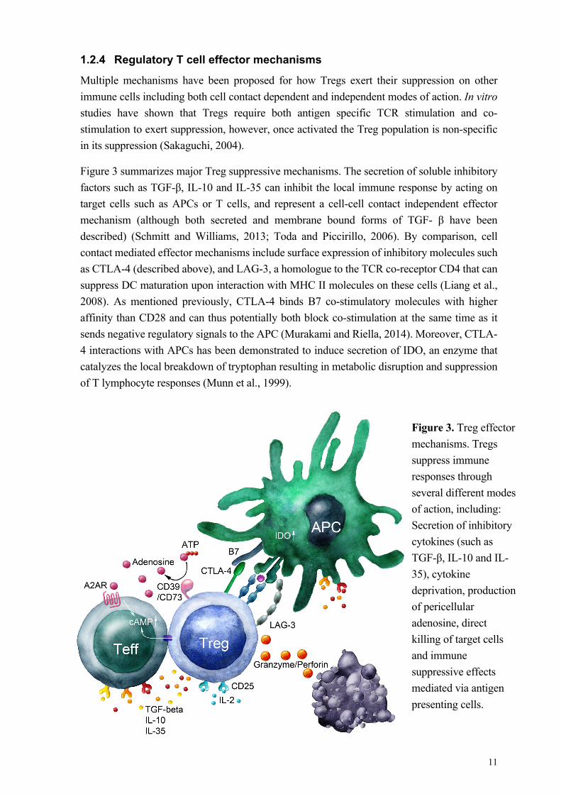
Figure 3 summarizes major Treg suppressive mechanisms. The secretion of soluble inhibitory
factors such as TGF-β, IL-10 and IL-35 can inhibit the local immune response by acting on
target cells such as APCs or T cells, and represent a cell-cell contact independent effector
mechanism (although both secreted and membrane bound forms of TGF- β have been
described) (Schmitt and Williams, 2013; Toda and Piccirillo, 2006). By comparison, cell
contact mediated effector mechanisms include surface expression of inhibitory molecules such
as CTLA-4 (described above), and LAG-3, a homologue to the TCR co-receptor CD4 that can
suppress DC maturation upon interaction with MHC II molecules on these cells (Liang et al.,
2008). As mentioned previously, CTLA-4 binds B7 co-stimulatory molecules with higher
affinity than CD28 and can thus potentially both block co-stimulation at the same time as it
sends negative regulatory signals to the APC (Murakami and Riella, 2014). Moreover, CTLA-
4 interactions with APCs has been demonstrated to induce secretion of IDO, an enzyme that
catalyzes the local breakdown of tryptophan resulting in metabolic disruption and suppression
of T lymphocyte responses (Munn et al., 1999).
Figure 3. Treg effector
mechanisms. Tregs
suppress immune
responses through
several different modes
of action, including:
Secretion of inhibitory
cytokines (such as
TGF-β, IL-10 and IL-
35), cytokine
deprivation, production
of pericellular
adenosine, direct
killing of target cells
and immune
suppressive effects
mediated via antigen
presenting cells.
12
In addition, several distinct mechanisms have been described whereby Tregs can inhibit T
lymphocytes through the elevation of intracellular cAMP levels in the responder cells. Firstly,
Tregs can induce a local anti-inflammatory environment by the production of pericellular
adenosine generated from ATP by CD39 (nucleoside triphosphate diphosphohydralase-1
(NTPDase1)) on the Treg cell surface and CD73 (ecto-5’-nucleotidase) on Tregs or other cells
in the local environment (Antonioli et al., 2013b). The adenosine signaling system is complex
and has several implications in immune regulation (see also section 1.3). Local adenosine
production limits T lymphocyte immune responses primarily through engagement of the
adenosine A2A receptors that leads to increased cAMP levels in the target cells (Ohta and
Sitkovsky, 2001). Moreover, human iTreg have been shown to express cyclooxygenase-2
(COX-2) that result in prostaglandin E2 production dependent elevation of intracellular cAMP
and suppression of immune responses (Mahic et al., 2006). Strikingly, Treg have also been
described to suppress responder cells by direct transfer of cAMP through gap junctions (Bopp
et al., 2007).
FOXP3 in itself has been shown to repress IL-2 expression, coherent with the fact that FOXP3+
Treg do not express this cytokine (Schubert et al., 2001). They are however, inherently
dependent on IL-2 signaling for survival and maintenance (Maloy and Powrie, 2005) and given
their constitutively high expression of CD25, the local consumption of IL-2 depriving other T
lymphocyte subsets of this growth factor, has been suggested as a contributing effector
mechanism (Pandiyan et al., 2007).
Finally, Tregs have also been demonstrated to directly kill their target cells by a variety of
mechanisms involving either apoptosis inducing ligands such as FasL (Janssens et al., 2003)
or granzyme/perforin secretion (Grossman et al., 2004a; Grossman et al., 2004b). Taken
together, the above mechanisms most probably represent complimentary pathways utilized by
Treg populations under different conditions to control immune responses, and mirror the
complexity of the Treg phenotypes described in various contexts.
1.3 ADENOSINE RECEPTOR SIGNALING
The purine adenosine is ubiquitously present throughout the body as an important player in cell
metabolism. Its extracellular concentration is highly dependent on the metabolic state of the
tissues and influenced by various stimuli such as stress, hypoxia and inflammation (Fredholm,
2007). The importance of adenosine receptor signaling has been demonstrated in multiple both
physiological and pathological contexts ranging from cardiovascular effects and temperature
regulation to cell differentiation, migration and immune regulation (Fredholm, 2007).
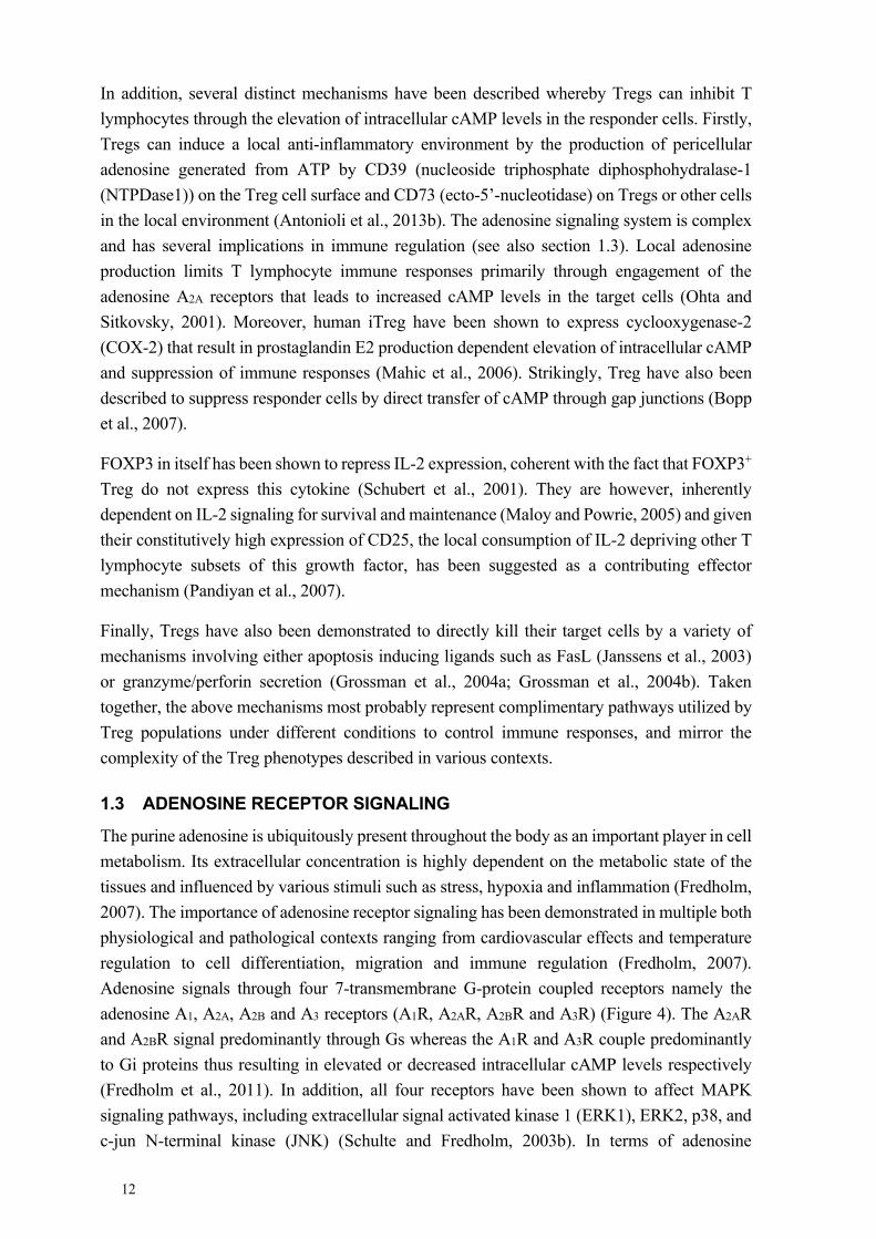
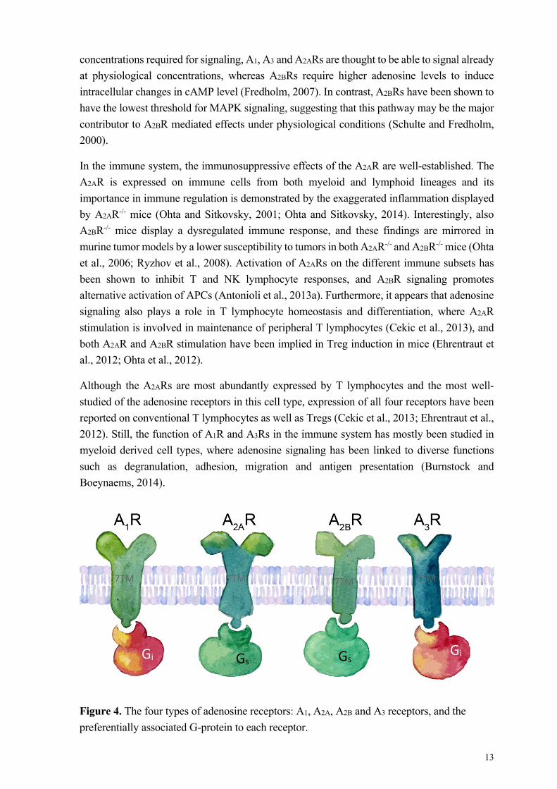
Adenosine signals through four 7-transmembrane G-protein coupled receptors namely the
adenosine A1, A2A, A2B and A3 receptors (A1R, A2AR, A2BR and A3R) (Figure 4). The A2AR
and A2BR signal predominantly through Gs whereas the A1R and A3R couple predominantly
to Gi proteins thus resulting in elevated or decreased intracellular cAMP levels respectively
(Fredholm et al., 2011). In addition, all four receptors have been shown to affect MAPK
signaling pathways, including extracellular signal activated kinase 1 (ERK1), ERK2, p38, and
c-jun N-terminal kinase (JNK) (Schulte and Fredholm, 2003b). In terms of adenosine
13
concentrations required for signaling, A1, A3 and A2ARs are thought to be able to signal already
at physiological concentrations, whereas A2BRs require higher adenosine levels to induce
intracellular changes in cAMP level (Fredholm, 2007). In contrast, A2BRs have been shown to
have the lowest threshold for MAPK signaling, suggesting that this pathway may be the major
contributor to A2BR mediated effects under physiological conditions (Schulte and Fredholm,
2000).
In the immune system, the immunosuppressive effects of the A2AR are well-established. The
A2AR is expressed on immune cells from both myeloid and lymphoid lineages and its
importance in immune regulation is demonstrated by the exaggerated inflammation displayed
by A2AR-/- mice (Ohta and Sitkovsky, 2001; Ohta and Sitkovsky, 2014). Interestingly, also
A2BR-/- mice display a dysregulated immune response, and these findings are mirrored in
murine tumor models by a lower susceptibility to tumors in both A2AR-/- and A2BR-/- mice (Ohta
et al., 2006; Ryzhov et al., 2008). Activation of A2ARs on the different immune subsets has
been shown to inhibit T and NK lymphocyte responses, and A2BR signaling promotes
alternative activation of APCs (Antonioli et al., 2013a). Furthermore, it appears that adenosine
signaling also plays a role in T lymphocyte homeostasis and differentiation, where A2AR
stimulation is involved in maintenance of peripheral T lymphocytes (Cekic et al., 2013), and
both A2AR and A2BR stimulation have been implied in Treg induction in mice (Ehrentraut et
al., 2012; Ohta et al., 2012).
Although the A2ARs are most abundantly expressed by T lymphocytes and the most well-
studied of the adenosine receptors in this cell type, expression of all four receptors have been
reported on conventional T lymphocytes as well as Tregs (Cekic et al., 2013; Ehrentraut et al.,
2012). Still, the function of A1R and A3Rs in the immune system has mostly been studied in
myeloid derived cell types, where adenosine signaling has been linked to diverse functions
such as degranulation, adhesion, migration and antigen presentation (Burnstock and
Boeynaems, 2014).
Figure 4. The four types of adenosine receptors: A1, A2A, A2B and A3 receptors, and the
preferentially associated G-protein to each receptor.
14
1.4 THE CONCEPT OF CANCER
Cancer as a disease has captivated the scientific community for generations. Intense research
has resulted in a deepened understanding of the traits that characterize human malignancies,
first summarized by Hanahan and Weinberg in 2000 (Hanahan and Weinberg, 2000). Their
theory stipulates six basic hallmarks of cancer that are acquired during the multistep
progression of human tumors, namely: (1) Independence from exogenous growth signals, (2)
resistance to antigrowth signals as well as (3) apoptosis, (4) limitless proliferative ability, (5)
angiogenesis induction and (6) invasion/metastasis capability (Hanahan and Weinberg, 2000).
The multistep cancer evolution theory is supported by pathological data from several tumors
where an evolution from premalignant lesions to invasive tumors is observed (Foulds, 1954).
In addition, the theory is further strengthened by epidemiological evidence from solid tumors,
such as e.g. bladder cancer, with an age related incidence where between seven and eight
separate evolutionary events have been inferred (Renan, 1993). These tumor specific changes
are acquired through a variety of different molecular changes, enabled by genetic instability
and DNA mutations in the tumor cells. Consequently, throughout the process of tumor
development, tumor cells acquire a protein signature that separates them from their normal
tissue progenitors. Such foreign proteins can potentially elicit an anti-tumor immune response
by which the body is able to recognize and eliminate altered cells. The field of tumor
immunology has vastly expanded over the past decades, and in a recent update of the Hallmarks
of Cancer theorem, tumor immune evasion is now included as an emerging hallmark (Hanahan
and Weinberg, 2011).
In the context of tumor development however, it is interesting to note the ambiguous role that
inflammation plays where the inflammatory process as such has been shown to promote tumor
progression in many tumors (de Visser et al., 2006). Indeed, inflammation facilitates most if
not all of the basic characteristics of cancers, and epidemiological data link chronic
inflammatory conditions to the development of different kinds of tumors in humans (Elinav et
al., 2013; Hanahan and Weinberg, 2011). Myeloid cells of the innate immune system have been
widely implied in promotion of tumor progression through mechanisms such as production of
reactive oxygen species, metalloproteinases, a diverse palette of chemokines and cytokines as
well as suppression of adaptive immune responses (de Visser et al., 2006; Elinav et al., 2013;
Gabrilovich et al., 2001). Of note, even though lymphocyte infiltration is a positive prognostic
indicator in many tumors (Galon et al., 2006; Lipponen et al., 1992; Zhang et al., 2003) and
adoptive immunotherapy of expanded T cells has proven promise in the clinical treatment of
patients (Dudley et al., 2005; Karlsson et al., 2010), the adaptive immune system has also been
implied to promote tumor progression under certain circumstances (Alizadeh et al., 2013;
DeNardo et al., 2009). In summary, it appears the range of immune effects on tumors can span
from tumor cell elimination to promotion of tumor growth and invasion. Understanding the
cellular and molecular pathways that shift the response in either direction will help us exploit
these mechanisms in the design of future immunotherapies of cancer.
15
1.5 TUMOR IMMUNITY
Cancer immune surveillance – the idea that cells of the immune system continuously circulates,
monitors and protects the body from developing cancers – as a means to eliminate tumors is
now widely accepted (Dunn et al., 2004). Studies on IFN-γ and perforin in mice revealed the
importance of these key immunological substances to prevent the development of both
spontaneous and chemically induced tumors, providing a basic proof for the hypothesis of
tumor immune surveillance (Shankaran et al., 2001; Street et al., 2001). In humans, tumors are
often infiltrated by T lymphocytes, the presence of which correlates positively to disease
outcome (Galon et al., 2006; Lipponen et al., 1992; Zhang et al., 2003), and tumor antigen-
specific functional T lymphocytes have been observed in human cancer patients (Guckel et al.,
2006). The concept of immunoediting and its “three E’s” comprises: 1) Elimination, which
basically encompasses the concept of immune surveillance, 2) equilibrium, a process where
the immune system interacts with the tumor, contributes to the evolutionary selection of tumor
cell clones and may serve to “edit” the tumor phenotype, and finally, 3) escape, where tumor
cells manage to evade the immune system and grow in otherwise apparently immunocompetent
hosts (Dunn et al., 2004).
1.5.1 Immune escape
Tumor immune escape encompasses multiple mechanisms by which tumors manage to elude
the immune system and involves diverse immune cell types. T lymphocyte responses are
evaded mainly by avoiding recognition or by disabling the effector T cells (Teff), each of which
is executed through a number of different mechanisms, including induction of tolerance, altered
antigen presentation, immunosuppressive microenvironment, co-inhibition, and/or
involvement of regulatory cell populations (Rabinovich et al., 2007).
1.5.1.1 Induction of tolerance
A major obstacle in mounting an immune response against tumors is that not all tumor antigens
are tumor specific, i.e. some are also expressed by normal tissues. Thus the immune system
may fail to recognize the cancer as foreign and be rendered unresponsive, which has indeed
been shown in both CD4+ and CD8+ T lymphocyte populations (Lee et al., 1999; Rabinovich
et al., 2007; Staveley-O'Carroll et al., 1998).
A major mechanism of tolerance induction is mediated through APCs, where especially
dendritic cells, play a central role in the development of immune responses against cancer, but
also contribute to the control of immune responses (Rabinovich et al., 2007; Steinman et al.,
2003). DC antigen capture and presentation of antigen in the absence of an inflammatory
environment fails to trigger DC maturation and expression of co-stimulatory molecules,
rendering the DCs incapable of eliciting a robust antigen-specific response. Studies of cancer
patients revealed decreased numbers of differentiated DCs, but an accumulation of immature
DCs with reduced capacity to activate T cells (Pinzon-Charry et al., 2005). Many different
pathways have been implicated by which DCs may suppress anti-tumor immune responses (Ma
et al., 2012). Such mechanisms include expression of IDO (Munn et al., 2004) and arginase
16
(Liu et al., 2009), secretion of immunoregulatory cytokines (Shurin et al., 2013) and expression
of inhibitory molecules such as programmed death ligand 1 (PD-L1) (Mu et al., 2011). Thus
DCs are very much implied in tumor escape mechanisms, however, a more in-depth description
of DC subtypes and their involvement in tumor escape are beyond the scope of this text.
1.5.1.2 Altered antigen presentation
Genetic instability is a hallmark trait of cancer cells (Hanahan and Weinberg, 2011), and forms
the basis for the production of cancer-specific antigens that the body can recognize as foreign.
How then, is it that tumor cells manage to hide these changes from the immune system? One
classical way of tumor immune evasion is the defective presentation of antigens through the
MHC I pathway. Downregulation of MHC I expression has been observed in many forms of
human cancers, where the most common mechanism for total loss of expression is mutations
in or deletion of the ß2-microglobulin genes (Hicklin et al., 1999; Rabinovich et al., 2007).
Alternatively, a variety of different mechanisms can work to downregulate the transcription of
MHC I genes, or affect the antigen processing through the transporter associated with antigen
processing (TAP) and to some extent proteosomal subunits low molecular mass polypeptide
(LMP) 2 and 7 (Hicklin et al., 1999).
1.5.1.3 Importance of the tumor microenvironment
The tumor microenvironment and its molecular interactions between the tumor and immune as
well as other cells in the tumor stroma are central in both development and progression of
tumors. Of note, chronic inflammation in tumor tissue sometimes not only fails to elicit an
adequate response but can also promote cancer development (de Visser et al., 2006). Tumors
release inhibitory factors that affect both the innate and the adaptive immune system, recruit
stromal cells and regulatory cell populations such as Tregs and myeloid derived suppressor
cells that during recent years have been attributed increasing importance in the development
and progression of solid tumors (Hanahan and Weinberg, 2011).
The enzyme IDO (discussed briefly in previous sections) is one mediator of T cell suppression
that has been linked to cancer, where it can be expressed not only by immune cell populations
but also by the tumor itself (Uyttenhove et al., 2003). IDO catalyzes the rate-limiting step in
the degradation of the amino acid tryptophan, and is expressed by a variety of cell types in
response to inflammatory signals, within the immune system normally by APCs in response to
external stimuli (Munn et al., 1999). The depletion of tryptophan and the generation of
immunosuppressive metabolites, result in direct T cell suppression as well as enhancement of
local Treg function (Munn and Mellor, 2007). IDO expression has been observed in human
tumors as well as tumor-draining lymph nodes, and is associated with poor prognosis
(Brandacher et al., 2006; Ino et al., 2006; Munn et al., 2004; Okamoto et al., 2005).
Many cytokines have been implicated in tumor immune suppression, of which TGF-ß is one
of the most prominent. This cytokine has a dual role in cancer development as it inhibits
proliferation of normal cells, but its expression is associated with immunosuppression in
cancers; TGF-ß promotes the generation and maintenance of Tregs, but can also directly
17
suppress T and NK cell anti-tumor immune responses (Wan and Flavell, 2007). Besides TGF-
ß, several other suspected microenvironmental molecular culprits have been implicated in
tumor immune escape, including IL-10 and prostaglandin E2 (Rabinovich et al., 2007).
Moreover, the relatively hypoxic environment within the tumor can in itself suppress immune
responses, e.g. through the accumulation of local adenosine. The recruitment of activated Tregs
to the tumor tissue can further increase adenosine levels through the action of the membrane
bound enzymes CD39 and CD73 as discussed previously, and contribute to immune
suppression by adenosine receptor signaling (Antonioli et al., 2013b). Interestingly, many
tumors have also been shown to express these enzymes (Antonioli et al., 2013b; Bastid et al.,
2013), which have been linked to higher stage tumors and poor prognosis (Kunzli et al., 2011;
Stella et al., 2010). Furthermore, the metabolism of ATP to adenosine may also contribute to
tumor immune escape by the decreased engagement of ATP receptors such as P2Y2 and P2RX7
on APCs and thereby reducing the activation of these cells (Bastid et al., 2013).
Yet another way to achieve immunosuppression is through the engagement of negative co-
stimulatory pathways. CTLA-4, discussed previously, is one such molecule that can be
expressed by tumor cells and has been shown to induce apoptosis in target cells (Contardi et
al., 2005). Furthermore, the interactions of programmed death receptor 1 (PD-1) and
programmed death receptor ligand 1 (PD-L1) have also been implied in this context, where
PD-L1 expression on tumor cells show a strong inverse correlation to patient survival (Blank
and Mackensen, 2007). Of note, the importance of these pathways has been emphasized during
recent years with the successful development of immunotherapies such as ipilimumab and
Nivolumab, targeting CTLA-4 and PD-1/PD-L1 pathways respectively (Hodi et al., 2010;
Philips and Atkins, 2015).
1.5.1.4 Regulatory T cells and FOXP3 in human cancers
The induction and/or recruitment of CD4+ Tregs by tumors represent a possible means of tumor
immune escape. In concordance with this hypothesis CD4+ Tregs have been observed to
increase in the peripheral blood of patients with several types of cancers, and accumulate in
tumor tissue and draining lymph nodes (Nishikawa and Sakaguchi, 2010). It is at present
unclear whether these cells represent a non-specific increase in the Treg pool, expansion of
tumor-specific tTreg or pTreg cells, although antigen-specific Treg clones have been described
(Wang et al., 2004). Interestingly, cancer resection has been reported to normalize the elevated
Treg levels, indicating that the increase of the Treg population is indeed caused by the cancer
(Kono et al., 2006). In tumor draining lymph nodes, one study demonstrated increasing
numbers of Tregs the closer the proximity of the tumor (Kawaida et al., 2005). Intratumoral
localization of FOXP3+ Tregs has also been shown in several tumor types, but whether these
cells are activated T helper cells with transiently induced FOXP3 expression or committed Treg
remains to be determined (Adeegbe and Nishikawa, 2013). Indeed the known heterogeneity of
human Treg populations is mirrored by the clinical prognostic implications of tumor infiltrating
FOXP3+ Tregs, which range from poor to good in a wide collection of studies in different
human tumors (reviewed by (deLeeuw et al., 2012)). For example, Curiel and colleagues
18
demonstrated that ovarian tumor infiltrating Tregs were linked directly to reduced survival, and
also illustrated that the chemokine CCL22 secreted by ovarian tumors was responsible for Treg
recruitment through the interaction with CCR4 on these cells (Curiel et al., 2004). In contrast,
the presence of tumor infiltrating FOXP3+ cells is associated to improved survival in other
types of cancer such as colorectal cancer (Salama et al., 2009) and as we have shown urinary
bladder cancer (Winerdal et al., 2011). Studies have even reached opposite conclusions
regarding the same cancer location; as different papers have reported good, neutral or poor
prognostic claim for FOXP3+ TILs in e.g. oral and gastric cancer, and this general diversity
appeared independent of antibody clone used or quantification method (deLeeuw et al., 2012).
The molecular subtype of tumor could possibly contribute to this diversity, as exemplified by
breast cancer where the prognostic significance of FOXP3+ TIL differs between estrogen
receptor positive and negative tumors (Mahmoud et al., 2011). It would thus seem that both
location and type of tumor influences the clinical impact of FOXP3+ TIL.
Interestingly, recent studies have shown that tumors can also express FOXP3 (Hinz et al., 2007;
Karanikas et al., 2008). This expression correlated to IL-10 and TGF-β (Karanikas et al., 2008),
and co-culture of naïve T cells with FOXP3-expressing tumor cells from pancreatic carcinoma
inhibited T cell proliferation (Hinz et al., 2007). Interestingly, although FOXP3 has been shown
to function as a tumor suppressor in vitro e.g. by the repression of c-Myc expression (Wang et
al., 2009), it is generally associated to bad clinical prognosis and higher risk of metastasis
(Triulzi et al., 2013). Different models have been proposed to explain this apparent
discrepancy: Tumor related mutations within the FOXP3 protein could abrogate the tumor
suppressor effect, FOXP3 could play a dual role by limiting proliferation but supporting tumor
progression, or FOXP3 expression could putatively bestow the tumor with immunoregulatory
properties, fostering immune escape (Triulzi et al., 2013). Of note, at least the first and last of
these alternatives are not mutually exclusive. In conclusion, it appears that FOXP3 is involved
on more than one side in tumor-immune interactions, and still much remains to be elucidated
regarding its biological implications.
19
2 AIMS OF THE THESIS
The overall aim of this thesis was to study regulatory T cells in both the physiological context
and the pathological cancer setting, in order to better understand their natural characteristics,
illuminate their impact disease progression and find potential ways to target them in
immunotherapy. The specific aims of each paper were:
Paper I. To map the relationship of FOXP3 promoter methylation and FOXP3 expression in
Treg and conventional CD4+ T helper lymphocytes in order to assess whether this may be used
as a marker of committed human Tregs.
Paper II. To investigate the impact of CD3+ and FOXP3+ tumor infiltrating lymphocytes as
well as tumor FOXP3 expression on survival in urinary bladder cancer.
Paper III. To examine how the T lymphocyte responses vary with tissue location and with
therapy in patients with urinary bladder cancer.
Paper IV. To study the impact of adenosine receptor signaling on T lymphocyte development
in general and Treg development in particular in adenosine receptor knockout mice.

21
3 MATERIALS AND METHODS
Here follows a summary of the materials and methods used. For more detailed descriptions,
please refer to the respective papers (I-IV).
3.1 PATIENTS
3.1.1 Patient characteristics (Paper I-III)
In paper I, only buffy coats from male healthy blood donors were used in the methylation
analyses to avoid the risk of artefacts due to random X-chromosome inactivation in women.
Paper II and III both encompass urinary bladder cancer (UBC) patients. Paper II is a
retrospective study of 37 UBC patients with muscle invasive disease that underwent radical
cystectomy at Karolinska University Hospital between the years 1999-2002. The mean age of
the included patients was 67 (median 69, range 46-81) at the time of the diagnostic TUR-B and
11 out of 37 patients in this study were female. At the time, neoadjuvant chemotherapy was not
routinely given to this group of patients, however, 10 of the patients received postoperative
chemotherapy, a fact which was accounted for in the study analyses.
In comparison, paper III is a prospective study of 32 UBC patients with suspected muscle
invasive disease at the time of inclusion at one of the collaborating study centers (Umeå
University Hospital, Sundsvall-Härnösand County hospital, Gävle Hospital, Falun Central
Hospital, Enköping Hospital and Uppsala University Hospital) 2013-2014. The mean age of
the included patients in this study was 69.2 years (median 70, range 55-87) and 10 out of 32
patients were female. 7 out of the 15 patients that underwent cystectomy received neoadjuvant
chemotherapy prior to cystectomy according to the MVAC (Methotrexate, Vinblastine,
Adriamycin and Cisplatinum) routine.
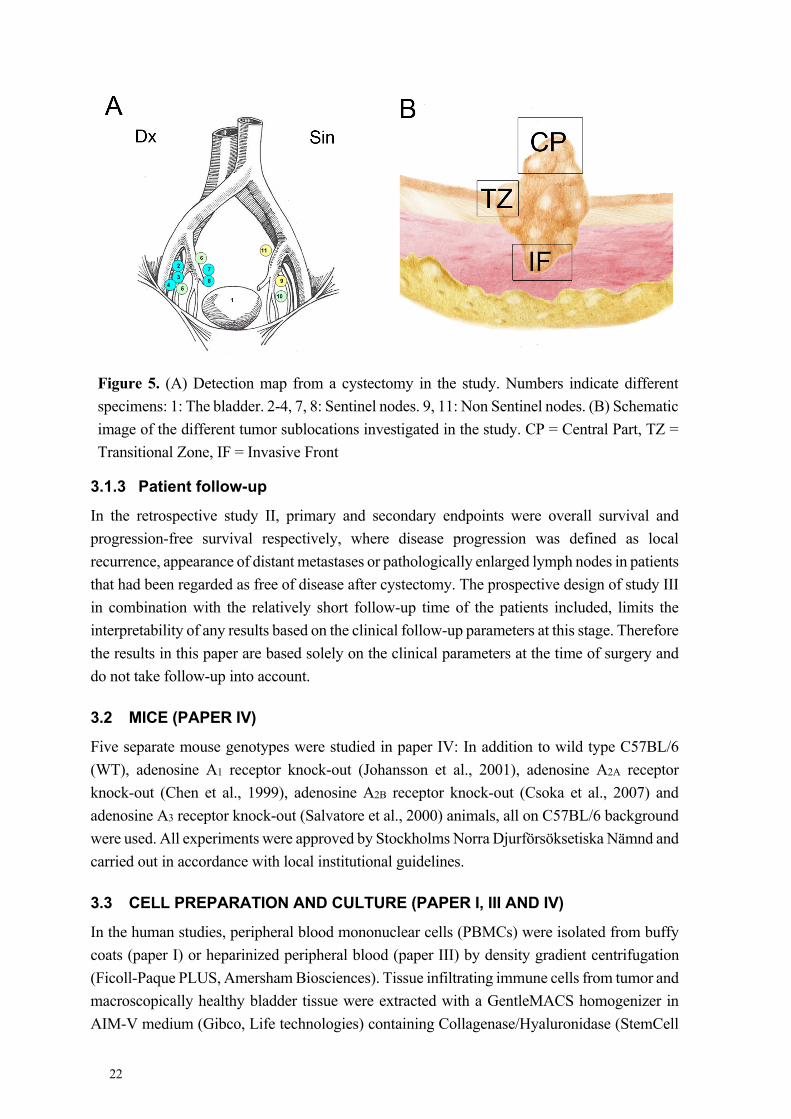
3.1.2 Sentinel node detection and surgical methods
In study III, patient tissue samples were received both at the initial transurethral resection of
the bladder (TUR-B) and at cystectomy. At the TUR-B, peripheral blood and cold-cup biopsies
from macroscopically healthy bladder tissue were received in addition to tumor samples.
Whenever possible, separate tumor fractions were taken from the central part (CP), transitional
zone (TZ) and invasive front (IF) of the tumor. At cystectomy, sentinel node detection was
carried out preoperatively as previously described (Sherif et al., 2001) by transurethral injection
of the radioactive tracer Nanocoll® around the edge of the tumor area, and sentinel nodes (SNs)
were identified after excision of the main specimen both in vivo and ex vivo. An example of a
lymph node map from a cystectomized patient in the study is shown in Figure 5. In addition to
the tumor-draining sentinel lymph nodes (SNs), non-draining lymph nodes (nSNs), peripheral
blood, tumor and macroscopically healthy bladder tissue samples were collected at the time of
cystectomy whenever possible.
22
3.1.3 Patient follow-up
In the retrospective study II, primary and secondary endpoints were overall survival and
progression-free survival respectively, where disease progression was defined as local
recurrence, appearance of distant metastases or pathologically enlarged lymph nodes in patients
that had been regarded as free of disease after cystectomy. The prospective design of study III
in combination with the relatively short follow-up time of the patients included, limits the
interpretability of any results based on the clinical follow-up parameters at this stage. Therefore
the results in this paper are based solely on the clinical parameters at the time of surgery and
do not take follow-up into account.
3.2 MICE (PAPER IV)
Five separate mouse genotypes were studied in paper IV: In addition to wild type C57BL/6
(WT), adenosine A1 receptor knock-out (Johansson et al., 2001), adenosine A2A receptor
knock-out (Chen et al., 1999), adenosine A2B receptor knock-out (Csoka et al., 2007) and
adenosine A3 receptor knock-out (Salvatore et al., 2000) animals, all on C57BL/6 background
were used. All experiments were approved by Stockholms Norra Djurförsöksetiska Nämnd and
carried out in accordance with local institutional guidelines.
3.3 CELL PREPARATION AND CULTURE (PAPER I, III AND IV)
In the human studies, peripheral blood mononuclear cells (PBMCs) were isolated from buffy
coats (paper I) or heparinized peripheral blood (paper III) by density gradient centrifugation
(Ficoll-Paque PLUS, Amersham Biosciences). Tissue infiltrating immune cells from tumor and
macroscopically healthy bladder tissue were extracted with a GentleMACS homogenizer in
AIM-V medium (Gibco, Life technologies) containing Collagenase/Hyaluronidase (StemCell
Figure 5. (A) Detection map from a cystectomy in the study. Numbers indicate different
specimens: 1: The bladder. 2-4, 7, 8: Sentinel nodes. 9, 11: Non Sentinel nodes. (B) Schematic
image of the different tumor sublocations investigated in the study. CP = Central Part, TZ =
Transitional Zone, IF = Invasive Front
23
Technologies). Lymph node leukocytes were extracted by gentle homogenization through a
40µm cell strainer in AIM-V medium. Murine cells from thymi, lymph nodes and spleens were
extracted by gentle homogenization and filtration through a 70µm cell strainer.
Magnetic cell sorting (MACS) in combination sorting of immune cell subpopulations on a
FACS Aria Cell sorter was used to isolate lymphocyte populations of interest (paper I and III).
All sorted cell populations were confirmed >95% pure by FACS.
In paper I, cells were cultured in AIM-V or RPMI 1640 supplemented with 10% human serum,
180U rIL-2, 100µg/mL streptomycin, 100U/mL penicillin and 2mM L-glutamine (all additives
from Sigma) and anti-CD3/CD28 Dynabeads (Invitrogen) or anti-CD3 and anti-CD28
antibodies were used to stimulate the cells. In paper III, AIM-V alone was used as culture
medium, only L-glutamine was added, and cells were stimulated with tumor extract as
described previously (Marits et al., 2006).
3.4 IMMUNOLOGICAL EVALUATION
3.4.1 Flow cytometry (Paper I, III and IV)
For surface staining, leukocyte cell populations of interest were labelled with antibodies as
indicated in the respective materials and methods section of paper I, III and IV. Intracellular
staining for FOXP3 (paper I and III) or Foxp3 and Helios (paper IV) was performed using the
Foxp3 staining buffer set from eBioscience according to the manufacturer’s protocol.
Acquisition of flow cytometry data was performed on a FACSCalibur or FACSAria (paper I)
alternatively an LSRForstessa II (paper III and IV) (all from BD Biosciences). Data analysis
was performed with Cell Quest Pro or FACSDiva (paper I) or FlowJo X 10.0.7r2 (paper III
and IV) software.
3.4.2 Functional T lymphocyte assays (Paper I and III)
3.4.2.1 Proliferation assays (Paper I)
The suppressive capacity of isolated Treg populations was assessed by co-culture with
CD4+CD25- responder cells at Treg:Tresponder ratio 1:1-1:4. The cells were stimulated with
plate bound anti-CD3 and soluble anti-CD28 antibodies and proliferation evaluated by
[3H]Thymidine incorporation after 4-5 days of culture. Alternatively, proliferation was
evaluated by dilution of carboxyfluorescein succinimidyl ester (CFSE) staining in the
responder population.
3.4.2.2 Flow cytometric assay of cell mediated immune response in activated lymphocytes (Paper III)
In order to assess the responsiveness of cells isolated from the different locations to tumor
antigens, 5x10⁵ cells from peripheral blood or lymph nodes were cultured in U-bottomed 96-
well plates with tumor homogenate prepared as described in paper III. As control for tumor
specific reactivity macroscopically healthy bladder tissue homogenate was used, and in
24
addition 5µg/mL of pokeweed mitogen or medium only were included as positive and negative
controls respectively.
3.4.3 Immunohistochemistry (Paper II)
Serial, 4µm sections of paraffin embedded tumor samples were mounted on glass slides and
incubated at 60ºC for 1h before the sections were deparaffinized in xylene and subsequently
rehydrated in an ethanol dilution series. Heat induced epitope retrieval was performed in a
microwave oven in Citrate buffer pH 6.0 (for CD3 staining) or Tris-EDTA buffer pH 9.0 (for
FOXP3) for 20 min. Blocking was performed with diluted serum from the host species of the
secondary antibody (goat). Mouse anti human CD3 antibody (clone PS1; Vector Laboratories,
dilution 1:200) and mouse anti human FOXP3 (clone 259D; BioLegend, dilution 1:100) were
used as primary antibodies, diluted in PBS with 1% bovine serum albumin and 0.05% Tween-
20. Subsequently, the VECTASTAIN Elite ABC kit (Vector Laboratories) was used according
to the manufacturer’s instructions. Biotinylated goat anti-mouse IgG (Vector Laboratories) was
used as a secondary antibody (dilution 1:200). In parallel, stainings without the primary
antibody were performed as negative control and sections of human spleen were used as
positive control. All washing steps were done in PBS with 0.05% Tween-20. Slides were
developed in 3, 3’ diaminobenzidine solution (Vector Laboratories) and counterstained with
Mayer’s hematoxylin before mounting in Pertex solution (HistoLab).
For analysis, three separate photographs of high power fields (X40) of randomly selected tumor
areas were taken in corresponding areas of consecutive slides for CD3 and FOXP3 stainings
respectively. The number of CD3+ and FOXP3+ tumor infiltrating lymphocytes (TILs) was
quantified, and in addition, FOXP3 expression in tumor cells was defined as positive if >25%
of the tumor cells were positively stained. Immunohistochemical evaluation was performed by
two independent researchers blinded to clinicopathological data.
3.4.4 Polymerase chain reaction (PCR) (Paper I)
For quantitative real time PCR, total RNA was extracted using the RNA Mini kit (Bio-Rad)
alternatively TRIZOL reagent (Invitrogen) according to the manufacturers’ protocols.
Conversion to cDNA was performed with the iScript cDNA Synthesis Kit (Bio-Rad).
Quantitative PCR was performed with iQSYBR Green Supermix (Bio-Rad) on an iCyclerIQ
Real Time Detection System (Bio-Rad), and data analyzed with iCycler IQ™ Optical System
Software Version 3.1 (Bio-Rad). The reliability of different commonly used housekeeping
genes has been questioned, especially with regard to their stability in activated cell populations
(Bas et al., 2004; Radonic et al., 2004). With this in mind, we chose RNA polymerase II (RPII)
as a housekeeping gene since it has a documented stability in many cell types including
lymphocytes (Radonic et al., 2004). Expression levels were normalized to the housekeeping
gene using the 2-ΔΔCt method (Livak and Schmittgen, 2001). For regular PCR, ThermoPol
reaction buffer and Taq DNA polymerase (New England Biolabs) were used on a MyCycler
Thermal Cycler (Bio-Rad). Primers, with sequences specified in paper I, were obtained from
Cybergene.
25
3.4.5 DNA methylation analysis (Paper I and III)
3.4.5.1 Bisulphite sequencing (Paper I)
DNA from sorted cell populations was isolated with the DNeasy Blood & Tisse Kit (QIAGEN),
and bisulphite conversion of all non-methylated cytosine bases in the DNA to uracil was carried
out with the EZ DNA Methylation Kit (ZYMO Research) according the manufacturers’
protocols. Subsequently, the FOXP3 promoter region was amplified by PCR and the PCR
product cloned into a pCR®4-TOPO® vector using a TOPO TA Cloning® Kit (Invitrogen).
DNA from individual bacterial clones was sequenced using the BigDye® Terminator v1.1
Cycle Sequencing Kit (Applied Biosystems) with T3 and T7 primers according to the
manufacturer’s instructions, and run on a 310 Genetic Analyzer (Applied Biosystems).
Sequence data was analyzed with ABI Prism Sequencing Analysis Software 3.7 (Applied
Biosystems).
3.4.5.2 Combined bisulphite restriction enzyme analysis (COBRA) (Paper I)
Bisulphite converted DNA, prepared as described above, was used as template to amplify the
FOXP3 promoter region with an unconjugated forward primer and a 5’ 6-FAM conjugated
reverse primer. The PCR product was treated with the restriction enzyme Aci1 in excess, which
would cleave the PCR product only if the -77 cytosine in the FOXP3 promoter in original
template was methylated and thus conserved through the bisulphite conversion. The digested
DNA product was isolated with a phenol-chloroform extraction and run on a 310 Genetic
Analyzer (Applied Biosystems) where fragment length was analyzed. Gene Scan v3.7
application software (Applied Biosystems) was used to analyze the data.
3.4.5.3 Pyrosequencing (Paper III)
DNA isolation and bisulphite conversion was carried out with the EZ DNA methylation Direct
kit (Zymo Research). The FOXP3 target sequence was amplified with biotinylated primers
(Thermo Scientific and Biomers.net) and pyrosequenced on a PSQ96 Pyrosequencer (Qiagen)
with Pyromark Gold 96Q reagents (Qiagen) and sequence primers. PyroMark CpG Software
1.0.11 (Qiagen) was used to analyze the resulting data sequences.
3.5 BONE MARROW TRANSFER EXPERIMENT (PAPER IV)
Bone marrow cell suspensions in sterile PBS were prepared from donor knockout mice and
5x10⁶ cells injected in the tail vein of irradiated (900 rad) WT C57BL/6 recipients. Recipient
mice received prophylactic antibiotics (neomycin sulphate) for two weeks after the transplant.
Resulting immunophenotypes were evaluated after 8 weeks.
3.6 STATISTICAL ANALYSIS (PAPER I-IV)
Categorical data (paper I) was analyzed with Fisher’s exact test. Survival data was related to
categorical predictor variables with the log-rank test and Cox’s proportional hazards model
(paper II). Shapiro-Wilk’s test of normality was used to evaluate if a particular data set was
parametric or not (paper III and IV). For parametric data, two-sided T-test or one-way ANOVA

26
was used, whereas the Mann-Whitney U test or Kruskall-Wallis ANOVA with Dunn’s test was
used for non-parametric datasets (paper III and IV). Statistical calculations were done in
Statistica (StatSoft, Inc. (2013). STATISTICA (data analysis software system), version 12.
www.statsoft.com) or GraphPad Prism (Version 5.04). P-values <0.05 were considered
significant.
27
4 RESULTS AND DISCUSSION
4.1 FOXP3 PROMOTER DEMETHYLATION REVEALS THE COMMITTED TREG POPULATION IN HUMANS (PAPER I)
In the years preceding this study, many Treg markers had been suggested, several of them
useful in an unchallenged setting but less specific in inflammatory environments as they were
also upregulated in conventional CD4+ T lymphocytes upon activation as discussed in section
1.2. When FOXP3 was first discovered as the master transcription factor of the Treg lineage it
was initially considered a specific Treg marker in both mice and humans (Sakaguchi et al.,
2007). However, before long, reports came of transient FOXP3 induction in human activated
conventional CD4+ T lymphocytes (Walker et al., 2003; Wang et al., 2007) making them
impossible to distinguish from Treg based on their protein expression alone. This prompted us
to ask whether Treg may be distinguished from conventional CD4+ T lymphocytes by the
epigenetic signature of the FOXP3 gene. We identified a CpG rich region within the conserved
FOXP3 promoter region and hypothesized that differential methylation of these sites may
account for the differential stability of FOXP3 expression in CD4+ T lymphocytes.
Human CD4+CD25hi Tregs displayed a demethylated FOXP3 promoter (1.4%±0.95% SEM
methylated) in contrast to CD4+CD25lo T lymphocytes, which were partially methylated
(27.9%±7.1%), and to B cells which were fully methylated (93.8%±1.7%). This was in
concordance with parallel study by Kim et al. describing the methylation status of the murine
Foxp3 promoter (Kim and Leonard, 2007). In addition, the promoter appears to have a more
accessible chromatin configuration in Tregs compared to conventional T lymphocytes, as
indicated by increased histone acetylation in this region (Mantel et al., 2006), further
strengthening the role of epigenetic control over FOXP3 transcription.
Furthermore, we examined the methylation status of the FOXP3 promoter in sorted CD25hi and
CD25lo cell populations during the course of activation of CD4+CD25lo T lymphocytes (Paper
I, Figure 6A). Interestingly, there was an initial difference in methylation status in the sorted
populations during early activation, but over time this difference leveled off at approximately
60% demethylation. One could speculate that the initial difference in methylation between the
CD25hi and CD25lo populations during activation may reflect a small contaminating resting
CD45RA+ Treg subset in the starting population. This population has a demethylated FOXP3
promoter region, lower CD25 and FOXP3 expression, but higher proliferative capacity than
their CD45RA- Treg counterparts (Miyara et al., 2009; Valmori et al., 2005), and could
potentially respond faster to activation than conventional CD4+ T lymphocytes completely
lacking CD25 expression at the start of the culture. At later time points, both the CD25hi and
the CD25lo population are more likely to consist of cells of mixed origin, possibly explaining
why the initial difference gradually disappears. Still, the FOXP3 promoter in CD4+CD25hi T
lymphocytes sorted ex vivo remained demethylated and these cells stayed FOXP3+ throughout
the culture. In contrast, although a limited decrease in FOXP3 promoter methylation was
observed in the stimulated CD4+CD25lo T lymphocytes, it remained partially methylated
corresponding to the transient FOXP3 expression observed in these cells. Of note, murine
28
CD4+CD25- T lymphocytes reportedly increase their promoter methylation upon TCR
stimulation in the absence of exogenous TGF-β, possibly preventing the induction of Foxp3
expression in these cells (Kim and Leonard, 2007), and potentially contributing to the
differences in FOXP3 expression pattern observed in human and murine conventional CD4+ T
lymphocytes.
In addition, we investigated the effects of various culture conditions such as addition of TGF-
β and/or IL-10, on FOXP3 promoter methylation. Of note, no additional effects of these
substances on promoter demethylation were observed. In contrast, addition of TGF-β during
activation of murine naïve CD4+ T lymphocytes has been demonstrated to induce
demethylation of the murine Foxp3 promoter as well as partial demethylation of the CNS3
intronic region (Floess et al., 2007; Kim and Leonard, 2007). These results could indicate that
TGF-β is more proficient at inducing a Tregs in mice consistent with in vitro findings (Chen et
al., 2003; Tran et al., 2007). However, they may also reflect different responsiveness to low
levels of TGF-β in humans versus mice, as the neutralization of this cytokine in the culture
medium reportedly blocks the induction of transient FOXP3 expression in naïve human
CD4+CD25- T lymphocytes (Tran et al., 2007).
The unique FOXP3 promoter methylation profile in Tregs compared to conventional CD4+ T
lymphocytes suggested that a demethylated pattern is a prerequisite for stable FOXP3
expression and suppressive phenotype. This was one of the first reports regarding the epigenetic
regulation of FOXP3 transcription. Since then, with expanding knowledge and technological
advances within the field of epigenetics, extensive mapping of the chromatin landscape and
methylation pattern in Treg versus conventional CD4+ T lymphocytes has deepened our
understanding of the epigenetic control of the Treg lineage (Morikawa and Sakaguchi, 2014).
Although most studies have been performed on mice, it is now clear that epigenetic
modifications are crucial in Treg development, and currently represent the most specific marker
of commitment to the Treg lineage available (Sakaguchi et al., 2013).
4.2 FOXP3 AND SURVIVAL IN URINARY BLADDER CANCER (PAPER II)
Urinary bladder cancer (UBC) is a malignancy highly associated to inflammation during its
development and also harbors great potential for immunotherapeutic strategies as illustrated by
the long-standing tradition of successful BCG treatment of superficial UBC (Herr and Morales,
2008). In study II, the prognostic significance of tumor infiltrating CD3+ and FOXP3+
lymphocytes as well as tumor expression of FOXP3 was evaluated in a retrospective material
of 37 patients with muscle invasive UBC.
In accordance with previous studies in UBC and other malignancies, the presence of CD3+
tumor infiltrating lymphocytes (TIL) in the tumor was a positive prognostic factor for survival
in our study (Galon et al., 2006; Lipponen et al., 1992; Zhang et al., 2003). This supports the
notion that T lymphocytes are important players in the anti-tumor immune response to UBC
and validates further studies to better understand how to potentiate this response in patients.
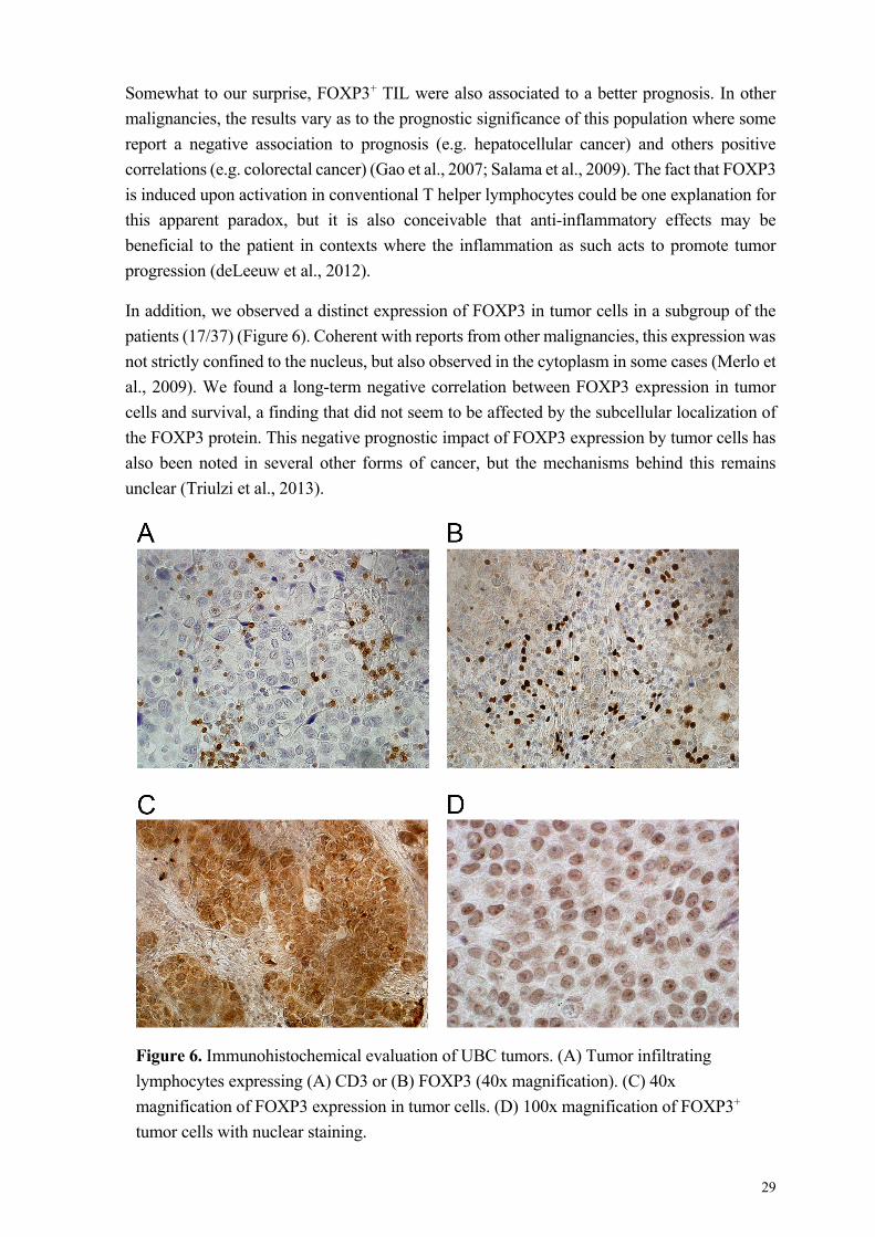
29
Somewhat to our surprise, FOXP3+ TIL were also associated to a better prognosis. In other
malignancies, the results vary as to the prognostic significance of this population where some
report a negative association to prognosis (e.g. hepatocellular cancer) and others positive
correlations (e.g. colorectal cancer) (Gao et al., 2007; Salama et al., 2009). The fact that FOXP3
is induced upon activation in conventional T helper lymphocytes could be one explanation for
this apparent paradox, but it is also conceivable that anti-inflammatory effects may be
beneficial to the patient in contexts where the inflammation as such acts to promote tumor
progression (deLeeuw et al., 2012).
In addition, we observed a distinct expression of FOXP3 in tumor cells in a subgroup of the
patients (17/37) (Figure 6). Coherent with reports from other malignancies, this expression was
not strictly confined to the nucleus, but also observed in the cytoplasm in some cases (Merlo et
al., 2009). We found a long-term negative correlation between FOXP3 expression in tumor
cells and survival, a finding that did not seem to be affected by the subcellular localization of
the FOXP3 protein. This negative prognostic impact of FOXP3 expression by tumor cells has
also been noted in several other forms of cancer, but the mechanisms behind this remains
unclear (Triulzi et al., 2013).
Figure 6. Immunohistochemical evaluation of UBC tumors. (A) Tumor infiltrating
lymphocytes expressing (A) CD3 or (B) FOXP3 (40x magnification). (C) 40x
magnification of FOXP3 expression in tumor cells. (D) 100x magnification of FOXP3+
tumor cells with nuclear staining.
30
In conclusion, tumor infiltrating T lymphocytes are indeed important for patient outcome in
urinary bladder cancer, as both CD3+ and FOXP3+ TIL were positively correlated to survival.
The negative impact of tumor FOXP3 expression in urinary bladder cancer warrants further
investigation to elucidate the biological impact of this expression on the tumor cells themselves
as well as the consequent interactions with the immune system.
4.3 ANTI-TUMOR IMMUNE REPONSES ARE SHAPED BY LOCATION AND THERAPY (PAPER III)
As study II indicated a clear role for T lymphocytes in determining the prognosis of the disease
in UBC, we set out to investigate the T lymphocyte immune response in more detail in a
prospective study. In order to illuminate the effects of tumor-immune interactions on the T cell
compartment at multiple levels, study III examines the immunophenotype of T lymphocytes in
peripheral blood, tumor draining lymph nodes (sentinel node, SN), non-draining lymph nodes
(non sentinel node, nSN) as well as tumor and macroscopically healthy bladder tissue.
We demonstrate that the FOXP3+ fraction of CD4+ T lymphocytes was significantly increased
in TIL populations as compared to all other locations. In addition, these CD4+FOXP3+ TIL T
lymphocytes displayed high levels of the Treg effector markers surface CTLA-4 and CD39 as
well as the activation and memory markers CD69, HLA-DR and CD45RO. Interestingly, this
activated phenotype was to some extent mirrored by both CD4+FOXP3- and CD8+ TIL T
lymphocytes. Furthermore, methylation analysis of tumor CD4+ TIL revealed a highly
methylated FOXP3 promoter region despite the pronounced FOXP3 expression in these cells,
in contrast to the CD4+ T lymphocyte population in peripheral blood where FOXP3 expression
seemed to correlate better with the methylation level observed. Just as the previous study, these
results indicate that the FOXP3+ TIL populations may represent an activated T lymphocyte
subpopulation rather than committed Tregs. It is possible that the differing prognostic impact
of FOXP3+ TILs in human cancers could relate to this finding (deLeeuw et al., 2012). Active
recruitment of Treg to the tumor has been implied in other forms of cancer such as ovarian
cancer, and were then associated to negative prognosis (Curiel et al., 2004). Notably, a recent
study of tumor infiltrating Tregs in head and neck cancer, where this population also has been
linked to worse prognosis (Sun et al., 2012), described elevated levels of immunosuppressive
markers which correlated to more potent suppressor activity of these cells (Jie et al., 2013).
Future studies comparing the epigenetic commitment and suppressive capacity of this subset
in cancers with differing prognostic impact may help decipher the role of FOXP3+ TIL in
humans.
Since the immune infiltrates in tumors have been noted to differ also within the tumor itself
(Fridman et al., 2012), we examined different tumor sublocalizations at the initial TUR-B.
Interestingly, the invasive front (IF) of the tumor stood out with higher CD8+ T lymphocyte
fraction compared to the central part (CP) or transitional zone (TZ) of the tumor. Furthermore,
the IF also had a lower FOXP3+ fraction of CD4+ T lymphocyte in patients with muscle
invasive disease. Thus again, higher FOXP3 appears to associate to better prognosis (lower
31
stage). Whether this association is linked to FOXP3 as an activation marker or directly to some
immunomodulatory effect of these FOXP3+ cells remains to be investigated.
In addition to direct anti-tumor effects, chemotherapy is known to potentiate immune responses
by the induction of immunogenic cell death in the tumor (Krysko et al., 2012), and furthermore,
different chemotherapeutic agents have been demonstrated to exert direct immunostimulatory
effects (Maccubbin et al., 1992; Markasz et al., 2008). Interestingly, patients that had received
chemotherapy in this study had higher lymph node T lymphocyte fractions compared to
patients that had not received such therapy. Moreover, the neoadjuvant treated patients
displayed higher T lymphocyte anti-tumor reactivity in their lymph nodes, as assessed with a
flow cytometry based assay evaluating the proportion of blasting T lymphocytes to certain
stimuli. Of note, a corresponding difference was not observed in the peripheral blood from
these patients, in agreement with the hypothesis that the draining lymph nodes are the most
probable sites for an anti-tumor response to be elicited.
The prospective study design limits the conclusions that can be drawn from clinical parameters
at this time due to at present relatively short follow-up time (3-18 months). In the future, it will
be interesting to follow the disease in these patients and correlate clinical outcomes to the
immunological parameters investigated.
We conclude from this study that anti-tumor T lymphocyte responses are shaped by the
environment in the different T lymphocyte compartments investigated, where T lymphocytes
in lymph nodes and tumor display higher levels of both activation and effector suppressor
markers. The greater anti-tumor reactivity in regional lymph nodes following chemotherapy
suggests a future for therapeutic combinations of chemotherapy with adoptive T cell based
immunotherapy of cancer.
4.4 ADENOSINE RECEPTOR SIGNALING AFFECTS THE DEVELOPMENT OF TREG AND CONVENTIONAL T LYMPHOCYTES (PAPER IV)
Given the recognized pronounced effects of adenosine receptor signaling in both tumor
pathology and the immune system at large, we were interested in examining the impact of these
receptors on the T lymphocyte compartment in mice, in order to set the base line for future
studies in murine tumor models. Thus, in study IV, the influence of each individual adenosine
receptor on the central and peripheral T lymphocyte compartments was evaluated in adenosine
receptor knockout mice (A1R-/-, A2AR-/-, A2BR-/- and A3R-/-).
Immunophenotyping was performed on thymus, spleen and lymph nodes, and compared to WT
controls. In the thymus, there was an overall effect of knocking out any one of the four
adenosine receptors, resulting in decreased fractions of CD4 and CD8 single positive
thymocytes. The most pronounced effects were observed in A2BR-/- mice, which displayed a
prominent reduction in cell yield from the thymus as well as alterations in relative and absolute
numbers of thymic subpopulations. Previous studies in mice have implicated a role for A2AR
signaling in the induction of apoptosis in DP thymocytes, but interestingly also depicted an
A2AR independent role for adenosine signaling in inhibition of TCR induced activation of
32
thymocytes (Apasov et al., 2000). Furthermore, although the A2BR has the highest adenosine
concentration threshold for induction intracellular of cAMP signaling, it has been shown to
affect MAPK-signaling already at physiological adenosine concentrations (Schulte and
Fredholm, 2000; Schulte and Fredholm, 2003a). In relation to the pronounced thymic
phenotype of A2BR-/- mice, it is thus interesting to note that MAPK-signaling is essential during
thymic selection and development of thymocytes (McNeil et al., 2005; Palmer, 2003). Since
all four adenosine receptors can affect signaling via the MAPK pathway (Schulte and
Fredholm, 2003b), it is possible that alterations in the balance of these receptors may explain
their apparent non-redundant functions in thymic selection in our study. Interestingly, BM
transfer to lethally irradiated WT hosts did not restore normal SP thymocyte frequencies,
indicating that the changes observed were not attributable to the radioresistant stromal cells.
The thymic CD4+Foxp3+Helios+ Treg population was also addressed and not surprisingly
found to be decreased in number, given the reduced frequency of CD4SP thymocytes in all of
the knockouts. Only the A1R-/- animals stood out with a reduced fraction of Foxp3+Helios+
CD4SP thymocytes, which normalized after bone marrow transfer to WT hosts, suggesting that
adenosine A1R signaling in the thymic stromal cells may account for this part of the A1R-/-
phenotype. This is in line with the fact that most studies describe very low expression level of
the A1R in T lymphocytes (Bours et al., 2006).
In the periphery, again the A2BR-/- animals stood out with reduced splenetic cell yield and T
cell fraction as well as an altered CD4/CD8 ratio. The peripheral Treg compartment was also
affected in these animals with the Foxp3+Helios+ fraction of CD4+ T lymphocytes found to
approximately double of that found in WT mice. Of note, we observed a distinct Foxp3-Helios+
CD4+ T lymphocyte population, particularly in the spleen of A2BR-/- mice. In light of, the
marked reduction in thymic cell count and reports linking Helios expression to activation and
proliferation (Akimova et al., 2011), one could speculate that this population represents a
peripheral expansion of T lymphocytes to compensate for the reduced thymic output.
Interestingly, after bone marrow transfer to WT hosts, A2BR-/- T lymphocytes displayed normal
CD4/CD8 ratio and peripheral T lymphocyte fractions, as well as an almost normalized
Foxp3+Helios+ Treg fraction. This would suggest that adenosine A2BR signaling in the WT
environment is largely able to rescue the peripheral T lymphocyte phenotype of A2BR-/- T
lymphocytes, although the reduction in CD4SP thymocytes remained.
It is clear that adenosine signaling contributes to the control of peripheral T lymphocyte
responses via Treg dependent and independent mechanisms (Antonioli et al., 2013a). This
study illustrates that also the thymic development of T lymphocytes is affected by abrogation
of either one of the four adenosine receptors. In light of these findings, a more detailed
examination of the functional consequences of adenosine receptor deletion in both Treg and
conventional T lymphocyte subsets would be of great interest.
33
5 CONCLUDING REMARKS AND FUTURE PERSPECTIVES
Tregs are vital in the maintenance of peripheral self-tolerance and play a pivotal role in cancer
immunology as demonstrated by the prognostic significance of these cells in several forms of
cancer. The intriguing, and at first glance contradictory findings regarding the prognostic
impact of FOXP3+ tumor infiltrating T lymphocytes, underscore the heterogeneity of this
population and the importance of considering the whole immune contexture of the tumor
(Fridman et al., 2012). Transient expression of FOXP3 in human activated T lymphocytes as
well as the existence of FOXP3 splice variants in humans that are not present in mice deepen
the complexity further and highlights the necessity to validate findings from murine models in
humans.
Our studies suggest that in the case of urinary bladder cancer, tumor infiltrating FOXP3+ CD4+
T lymphocytes do not represent committed tTreg, but rather a CD4+ conventional T
lymphocyte population with induced expression of FOXP3. Still, FOXP3 expression in these
cells is associated to high surface expression of effector Treg markers such as CD39 and
CTLA-4, which indicates that these cells do harbor suppressive function. Since urinary bladder
cancer development has been linked to chronic inflammation (Thompson et al., 2014), it may
be that the positive prognostic impact associated with FOXP3+ TILs relates to suppression of
this inflammatory response. Alternatively, given the methylated state of the FOXP3 promoter
in these cells and the transient expression of FOXP3 upon activation in conventional CD4+ T
lymphocytes, one could speculate that the observed FOXP3 expression mirrors a higher level
of activation and thus anti-tumor responsiveness. Ongoing studies in our laboratory are
currently addressing the functional suppressor capacity and expression profile of these tumor
infiltrating Tregs. In light of the expanding view on T helper lineage plasticity (Weinmann,
2014), it will also be of great interest to assess the cytokine profile and possible expression of
transcription factors other than FOXP3 within this cell population.
Increased understanding of the mechanisms that govern stable FOXP3 expression and a
suppressive Treg phenotype, is instrumental for the development of future therapies targeting
the Treg population. Multiple murine studies have illustrated the value of Treg depletion to
augment anti-tumor immune responses in different cancer models, and in humans there are
currently clinical trials underway that target this population by various mechanisms (Nishikawa
and Sakaguchi, 2014), which stresses the importance to advance knowledge concerning the
clinical impact and implications of Tregs in cancer. Furthermore, the same issues become of
importance at the opposite end of the clinical spectrum, when attempting to harvest and expand
Tregs for immunotherapy of autoimmunity. The evaluation of stable Treg commitment as
opposed to the expression of FOXP3 in activated effector T lymphocytes, assessed by e.g.
methylation analysis, is likely to be essential for the development of optimal expansion
protocols.
34
As demonstrated by us and others (Jie et al., 2013), both Treg and conventional T lymphocyte
populations appear to upregulate immunosuppressive markers in the context of the tumor. With
this in mind, it is interesting to note the successful introduction of therapeutics designed to
target co-inhibitory pathways such as ipilimumab (Hodi et al., 2010). However, the limited
response rate combined with the autoimmune adverse effects of these treatments leave room
for improvement (Weber et al., 2012). Since many pathways converge to elevate adenosine
levels within the tumor, which in turn has been associated with immune suppression,
modulation of adenosine signaling pathways represent a promising area of therapeutic
development. Such approaches include CD39/CD73 inhibitors and A2AR antagonists that
potentially could tip the balance between Treg and T effector cells in favor of an anti-tumor
immune response (Bastid et al., 2013; Ohta and Sitkovsky, 2014).
The immune effects of different conventional therapeutic regimens such as chemotherapy
should also be considered in the evaluation of anti-tumor immune responses, as illustrated by
study III in this thesis. Other studies have noted direct immunostimulatory effects of certain
chemotherapeutic drugs in addition to their direct anti-tumor effects and promotion of
immunogenic cell death (Krysko et al., 2012; Maccubbin et al., 1992; Markasz et al., 2008). In
the clinical setting, further characterization of each of these different routes is essential to allow
optimization of therapeutic protocols designed to promote the anti-tumor effects combined with
the pro-immune effects as much as possible.
In addition to immunotherapies aiming to enhance the anti-tumor immune response in vivo,
multiple studies describing adoptive cell therapy of cancer have been reported (Dudley et al.,
2005; Karlsson et al., 2010; Qian et al., 2014; Sherif et al., 2010). Current adoptive
immunotherapeutic strategies with in vitro expanded anti-tumor T lymphocytes have shown
much promise but are cumbersome and still have a limited response rate in patients, possibly
in part due to immune escape mechanisms employed by the tumor. It is tempting to speculate
that the combination of adoptive T cell therapy with immunomodulatory strategies such as
those discussed above may represent ways to overcome this local immune suppression.
The immune system hosts great potential in the fight against cancer. Recent years have seen
the emergence of many successful immunotherapies and undoubtedly more are still to come.
All the same, a lot remains to be elucidated in the intricate interplay between tumor and immune
cells. Further clarification of the immune contexture of human tumors will pave the road for
new immunotherapeutic strategies and optimization of current clinical treatments to promote
anti-tumor immunity.
35
6 POPULÄRVETENSKAPLIG SAMMANFATTNING
I människokroppen pågår en ständig kamp för överlevnad där immunförsvaret kämpar för att
försvara kroppens egna vävnader mot olika typer av faror såsom bakterier och virus. Samtidigt
är förmågan att särskilja kroppens egna celler av yttersta vikt då oförmåga att upprätthålla
tolerans mot dessa kan leda till immunattack mot kroppsegna organ, så kallad autoimmunitet.
Cancerceller har förvärvat förändringar i sin arvsmassa, DNA, som dels ligger till grund för
sjukdomsutvecklingen, men som också betyder att dessa celler uttrycker proteiner som till viss
del är kroppsfrämmande. Dessa förändringar kan kännas igen av immunförsvaret som i det
bästa scenariot identifierar förändrade celler redan på ett tidigt stadium och eliminerar dessa
innan någon klinisk sjukdom hunnit bryta ut. Framgångrika tumörer utvecklar därför olika
mekanismer för att undvika igenkänning.
T celler är centrala spelare i både det specifika adaptiva immunförsvaret mot cancer och i
upprätthållandet av tolerans mot kroppens egna vävnader. De så kallade regulatoriska T
cellerna är essentiella för balansen i kroppens immunförsvar och kontrollen av det
inflammatoriska svaret. Dessa celler karaktäriseras till stor del baserat på deras uttryck av
proteinet FOXP3. I människor kompliceras dock studierna av dessa celler av att vanliga
aktiverade T celler också visat sig kunna uttrycka detta protein. Den första studien i denna
avhandling handlar om regleringen av FOXP3 uttryck i T celler. Här studerar vi så kallad
epigenetik, vilket handlar om förändringar av kroppens arvsmassa som kan påverka
proteinuttrycket i cellen, men som inte påverkar själva DNAsekvensen. En sådan förändring är
tillägg av metylgrupper på DNAt (metylering). Generellt kan man säga att gener med en hög
grad av metylering är mindre aktiva än ometylerade motsvarigheter. I vår första studie visar vi
att regulatoriska T celler som kontinuerligt uttrycker FOXP3, har en ometylerad konfiguration
i början av genen som kodar för detta protein, till skillnad från de vanliga T cellerna som är
halvmetylerade i detta område. Som kontrast är B celler, som inte alls uttrycker FOXP3 helt
metylerade. Vår slutsats från denna studie var att metyleringsgraden av FOXP3 genen kan
förutspå ett kontinuerligt eller övergående uttryck av proteinet och således kan användas som
en markör för stabila regulatoriska T celler.
I nästa steg undersökte vi vilken effekt tumörinfiltrerande regulatoriska T celler har på den
kliniska prognosen vid urinblåsecancer. Intressant nog fann vi att både T cellsinfiltration
generellt och T celler som uttryckte proteinet FOXP3 förutspådde bättre prognos för
patienterna. Detta kan tyckas motsägelsefullt då regulatoriska T celler generellt anses trycka
ned det tumörspecifika immunförsvaret och således borde vara associerade till sämre prognos.
I nuläget har dock många rapporter publicerats där betydelsen av tumörinfiltrerande
regulatoriska T celler undersöks i olika former av cancer och det är tydligt att den prognostiska
betydelsen kan variera alltifrån god till dålig. Orsakerna till detta är i nuläget inte helt klarlagda,
men det tycks som om både tumörtyp och lokalisation kan spela en roll. Det har spekulerats att
den goda prognoskopplingen i vissa cancerformer har att göra med kontroll av den kroniska
inflammation som annars kan agera som en drivande faktor i tumörutvecklingen, alternativt
skulle detta FOXP3 uttryck kunna vara en reflektion av allmän T cellsaktivering i
36
konventionella T celler. Intressant nog noterade vi även ett uttryck av FOXP3 i tumörceller hos
en andel av patienterna. Detta uttryck var kopplat till sämre klinisk prognos, men de
bakomliggande mekanismerna till detta är fortfarande oklara.
För att vidare undersöka hur vanliga och regulatoriska T celler blir påverkade av tumören i
urinblåsecancer, undersökte vi dessa cellpopulationer från blod, lymfknutor, och tumör med
flödescytometri. De tumörinfiltrerande T cellerna generellt, men särskilt de regulatoriska T
cellerna uppvisade en mycket hög aktiveringsgrad och uttryckte höga nivåer av proteiner som
är aktiva vid nedreglering och kontroll av immunsvar. Intressant var också att vi fann skillnader
i sammansättningen av de tumörinfiltrerande cellerna i olika delar av tumören, där den invasiva
kanten stod ut i jämförelse med centrala delar av tumören. Vi fann att patienter med
muskelinvasiva tumörer hade lägre andel FOXP3 uttryckande T celler vid den invasiva kanten
jämfört med ytligt växande tumörer. Intressant nog visade sig de tumörinfiltrerande T cellerna
vara till hög grad metylerade i sin FOXP3 gen, vilket tyder på att dessa celler inte representerar
en dedikerade regulatoriska T celler utan snarare T celler med ett aktiveringsrelaterat uttryck
av FOXP3. Dessutom noterade vi en tydlig effekt av genomgången cellgiftsbehandling på
immunsvaret mot tumören, där denna behandling tycktes potentiera förmågan av T celler från
tumördränerande lymfknutor att reagera på tumörproteiner.
En metod som regulatoriska T celler utnyttjar för att kontrollera immunreaktioner är produktion
av adenosin som i sig är immunhämmande i närmiljön. Det har visat sig att vissa tumörer också
aktivt producerar adenosin, som kan hjälpa dessa att kontrollera ett eventuellt immunsvar mot
tumören. För att studera vilka effekter adenosin har på T celler använde vi oss av så kallade
knockoutmöss som saknar endera av de fyra definierade receptorerna för adenosin. I dessa
möss karaktäriserade vi sedan T cellernas utveckling från deras utbildning i thymus (brässen)
till deras sammansättning i mjälte och lymfknutor. Vi fann att adenosinsignalering tycks bidra
till T cellsutveckling med både T cellsberoende och oberoende mekanismer. Vidare studier
behövs för att utvärdera den funktionella betydelsen av dessa fynd.
Sammanfattningsvis har dessa studier påvisat en epigenetisk signatur hos mänskliga
regulatoriska T celler som kan användas för identifiering av denna population i patienter.
Genom studier av denna cellpopulation hos patienter med urinblåsecancer har vi börjat
kartlägga en del av de mekanismer som påverkar T cellssvaret och fortsatt prognos hos dessa
patienter. Studier av T cellsutvecklingen i möss som saknar specifika adenosinreceptorer har
belyst betydelsen av adenosinsignallering i thymus. Bättre förståelse för hur tumören
interagerar med kroppens immunförsvar i patienter kan i kombination med grundläggande
studier av hur regulatoriska system i kroppen fungerar, leda till nya behandlingsstrategier och
sätt att väcka det immunförsvar som sövts av tumören.
37
7 ACKNOWLEDGEMENTS
This work came together as a result of numerous fruitful collaborations and discussions over
the past years, and many have contributed to the work presented herein. I would like to
specifically recognize and extend my appreciation to the following people:
Ola Winqvist, my supervisor, for your everlasting enthusiasm and contagious positivity with
the attitude that we can do anything that we set our minds to – the impossible just takes a bit
longer (as some may say is illustrated by this very book). Thank you for taking me in and
allowing me to become a part of your research family, for sharing your vast knowledge about
immunology and the rules that makes the world of science tick.
My co-supervisor Per Marits, who has stood by me ever since I stepped into the lab. Always
available with good advice and always with an article or two up your sleeve. Thank you for all
your practical guidance as I took my first baby steps into the world of immunology as well as
your fantastic support and engaging discussions on everything from science to politics.
Our fantastic collaborator Amir Sherif, for your extraordinary commitment and passion for
science. Thank you for your dedication, valuable discussions and for helping us keep the ever
so important clinical focus in our research.
Ann-Charlotte Wikström, for being such an inspiring person and my mentor during these past
years.
To all present and former members of the Winqvist group I am eternally grateful for the crazy,
fun and stimulating years that we have spent together.
My present co-soldiers on the urinary bladder cancer project: David Krantz, for your
enthusiasm, support in the lab, ability to keep your head cool during late work nights, and good
company on trips within Sweden and to the US. Ali Zirakzadeh, for all your efforts during our
work in the lab and enjoyable scientific discussions. Emma Ahlén Bergman, for fun
conversations and for keeping the epigenetics part of my thesis in place. My co-author Robert
Rosenblatt, for keeping track of the clinical data, and for putting up with my never-ending
stream of e-mails.
All other present members and student in the group: Ludvig Linton, for your good spirits,
enjoyable company at conferences, and for racing the last months of our PhD studies together.
Jin Hu, for your insights into Chinese culture and technological inventions as well as intriguing
discussions about the real estate market. Petra Jones and Martina Jones at ITH for fun
conversations and company across the lab bench during the past years. Robert Wallin, for
interesting scientific input and entrepreneurial spirit. Eduardo Villablanca, for your
overwhelming and contagious dedication to science. Martina Parigi, for your scientific
enthusiasm and pleasant conversations. Johan Kinn, Michael Mints, Love von Euler, Sara
Andréasson and Christian Lundgren for good collaborations, assistance and discussions. Lu
Zhang, Kurt Arkestål, Katarina Glise Sandblad, and Ciputra Adijaya “Adi” Hartana for good

38
company and enjoyable conversations in the office – and especially Adi, for letting us call you
Adi. All students, past and present that have contributed to the group atmosphere with their
enthusiasm and hard work.
Research group members who regrettably are no longer in the group but always considered
family: Evelina Lindmark, for your great social attitude and for your crucial help in study IV.
Peter Jansson, my co-author and friend-in-arms in my first project at the lab – thank you for
introducing me to epigenetics and for your always easygoing attitude. Mona Karlsson, for your
amazing sarcasm, and for being my idol in terms of how to keep the “messier” members of the
lab in check. Emma Lindh, always with a smile and a great sense of humor to save the day.
All of my fantastic colleagues at L2:04, thank you for creating such a great working
atmosphere!
Professor Annika Scheynius who was the head of the unit during the main part of my PhD
studies, for your support and encouragement.
Catharina Johansson, for your indispensable help to get through all the intricate administrative
turns of my clinical, parental and scientific careers.
A special thank you also to Stephanie Hiltbrunner, for our nice collaboration on the urinary
bladder cancer project, fun conversations and great company during late nights at the lab and
trips to Umeå, and to Anne-Laure Joly for your fantastic personality, your help and all the
interesting discussions.
To our collaborators on the adenosine projects: Professor Bertil Fredholm, for your dedication
to science and exquisite scientific input. Ulrika Ådén, for taking care of my husband during his
PhD and for great collaborations. Mattias Carlström and Ting Yang, for vital input and
assistance with the knockout mice as well as stimulating discussions.
Our former neighbors at the lab, Mikael Karlsson and his whole group, for great company, and
fun discussions! A special thank you to Emilie Grasset for invaluable support and help with
the confocal microscope.
Our clinical collaborators all over Sweden: Börje Ljungberg, Johan Hansson, Benny
Holmström and Markus Johansson. Thank you for your commitment and for making our
research possible. Furthermore, a special thank you to the research nurses in Umeå, Britt-Inger
Dahlin and Kerstin Almroth for all their hard work and assistance on paper III.
Anna Tolf, thank you for your dedication and scientific input on paper II.
To my friends of old: Linn, Jenny, Saga, Isabelle, Lotta, and Lina – thank you for putting up
with me during all our years in school, and for staying in contact and staying great!
Hanna, for standing by me through upper secondary school and for being one of the most
passionate people I know.

39
Erik and Angelica, for wonderful long discussions during late evenings with good wine, for
being the most loveable people in the world, and for all the smoked ham. Karin and Anders,
for your great company and creativity.
Pearl, Alex, Ullot, Kay and Julian – thanks for all of the good times, and for making our time
in Med school so much fun!
To all my family, with new members and those that I have known my entire life, for your never-
ending support and love! My mother, Lisen, for all your love and support. For being a fantastic
mother to me as you are a fantastic grandmother to my children, for being my friend and for
always listening. My father, Mats, for your support and encouragement over all the past years.
For believing in me and for the love that you show as a grandfather. Min mormor, Gunvor, du
är en så fantastisk och underbart varm människa som rymmer hela världen i ditt hjärta! Tack
för allt stöd och hjälp genom åren! Min farmor, Märta, för ditt hjärta av guld och alla varma
kramar! My brothers, Mattias and Victor, for all the good times and the rest as well, I am
forever grateful. My new sister, Ana, for your energy and never-ending enthusiasm to life, and
for all your help with “the little ladies”. My godmother, Ulla, for all your care, love and support
over the years. My parents-in-law, Elisabet and Lars, for becoming my second family, taking
care of Otis and for being such wonderful grandparents to my children.
My daughters, Mira and Matilda, for being the miracles you are.
Finally, Max, my husband, co-author and best friend. For you – a “thank you” would just seem
petty. You inspire me to do things I would have never thought possible. You are my life.

41
8 REFERENCES
Aarts-Riemens, T., M.E. Emmelot, L.F. Verdonck, and T. Mutis. 2008. Forced overexpression of either of the two common human Foxp3 isoforms can induce regulatory T cells from CD4(+)CD25(-) cells. European journal of immunology 38:1381-1390.
Abbas, A.K., C. Benoist, J.A. Bluestone, D.J. Campbell, S. Ghosh, S. Hori, S. Jiang, V.K. Kuchroo, D. Mathis, M.G. Roncarolo, A. Rudensky, S. Sakaguchi, E.M. Shevach, D.A. Vignali, and S.F. Ziegler. 2013. Regulatory T cells: recommendations to simplify the nomenclature. Nature immunology 14:307-308.
Abbas, A.K., J. Lohr, B. Knoechel, and V. Nagabhushanam. 2004. T cell tolerance and autoimmunity. Autoimmunity reviews 3:471-475.
Adeegbe, D.O., and H. Nishikawa. 2013. Natural and induced T regulatory cells in cancer. Frontiers in immunology 4:190.
Akimova, T., U.H. Beier, L. Wang, M.H. Levine, and W.W. Hancock. 2011. Helios expression is a marker of T cell activation and proliferation. PloS one 6:e24226.
Alizadeh, D., E. Katsanis, and N. Larmonier. 2013. The multifaceted role of Th17 lymphocytes and their associated cytokines in cancer. Clinical & developmental immunology 2013:957878.
Allan, S.E., L. Passerini, R. Bacchetta, N. Crellin, M. Dai, P.C. Orban, S.F. Ziegler, M.G. Roncarolo, and M.K. Levings. 2005. The role of 2 FOXP3 isoforms in the generation of human CD4+ Tregs. The Journal of clinical investigation 115:3276-3284.
Allan, S.E., G.X. Song-Zhao, T. Abraham, A.N. McMurchy, and M.K. Levings. 2008. Inducible reprogramming of human T cells into Treg cells by a conditionally active form of FOXP3. European journal of immunology 38:3282-3289.
Antonioli, L., C. Blandizzi, P. Pacher, and G. Hasko. 2013a. Immunity, inflammation and cancer: a leading role for adenosine. Nature reviews. Cancer 13:842-857.
Antonioli, L., P. Pacher, E.S. Vizi, and G. Hasko. 2013b. CD39 and CD73 in immunity and inflammation. Trends in molecular medicine 19:355-367.
Apasov, S., J.F. Chen, P. Smith, and M. Sitkovsky. 2000. A(2A) receptor dependent and A(2A) receptor independent effects of extracellular adenosine on murine thymocytes in conditions of adenosine deaminase deficiency. Blood 95:3859-3867.
Aschenbrenner, K., L.M. D'Cruz, E.H. Vollmann, M. Hinterberger, J. Emmerich, L.K. Swee, A. Rolink, and L. Klein. 2007. Selection of Foxp3+ regulatory T cells specific for self antigen expressed and presented by Aire+ medullary thymic epithelial cells. Nature immunology 8:351-358.
Baecher-Allan, C., J.A. Brown, G.J. Freeman, and D.A. Hafler. 2001. CD4+CD25high regulatory cells in human peripheral blood. Journal of immunology 167:1245-1253.
Baecher-Allan, C., E. Wolf, and D.A. Hafler. 2006. MHC class II expression identifies functionally distinct human regulatory T cells. Journal of immunology 176:4622-4631.
Baron, U., S. Floess, G. Wieczorek, K. Baumann, A. Grutzkau, J. Dong, A. Thiel, T.J. Boeld, P. Hoffmann, M. Edinger, I. Turbachova, A. Hamann, S. Olek, and J. Huehn. 2007. DNA demethylation in the human FOXP3 locus discriminates regulatory T cells from
42
activated FOXP3(+) conventional T cells. European journal of immunology 37:2378-2389.
Bas, A., G. Forsberg, S. Hammarstrom, and M.L. Hammarstrom. 2004. Utility of the housekeeping genes 18S rRNA, beta-actin and glyceraldehyde-3-phosphate-dehydrogenase for normalization in real-time quantitative reverse transcriptase-polymerase chain reaction analysis of gene expression in human T lymphocytes. Scandinavian journal of immunology 59:566-573.
Bastid, J., A. Cottalorda-Regairaz, G. Alberici, N. Bonnefoy, J.F. Eliaou, and A. Bensussan. 2013. ENTPD1/CD39 is a promising therapeutic target in oncology. Oncogene 32:1743-1751.
Bennett, C.L., J. Christie, F. Ramsdell, M.E. Brunkow, P.J. Ferguson, L. Whitesell, T.E. Kelly, F.T. Saulsbury, P.F. Chance, and H.D. Ochs. 2001. The immune dysregulation, polyendocrinopathy, enteropathy, X-linked syndrome (IPEX) is caused by mutations of FOXP3. Nature genetics 27:20-21.
Bensinger, S.J., A. Bandeira, M.S. Jordan, A.J. Caton, and T.M. Laufer. 2001. Major histocompatibility complex class II-positive cortical epithelium mediates the selection of CD4(+)25(+) immunoregulatory T cells. The Journal of experimental medicine 194:427-438.
Blank, C., and A. Mackensen. 2007. Contribution of the PD-L1/PD-1 pathway to T-cell exhaustion: an update on implications for chronic infections and tumor evasion. Cancer immunology, immunotherapy : CII 56:739-745.
Bopp, T., C. Becker, M. Klein, S. Klein-Hessling, A. Palmetshofer, E. Serfling, V. Heib, M. Becker, J. Kubach, S. Schmitt, S. Stoll, H. Schild, M.S. Staege, M. Stassen, H. Jonuleit, and E. Schmitt. 2007. Cyclic adenosine monophosphate is a key component of regulatory T cell-mediated suppression. The Journal of experimental medicine 204:1303-1310.
Bours, M.J., E.L. Swennen, F. Di Virgilio, B.N. Cronstein, and P.C. Dagnelie. 2006. Adenosine 5'-triphosphate and adenosine as endogenous signaling molecules in immunity and inflammation. Pharmacology & therapeutics 112:358-404.
Brandacher, G., A. Perathoner, R. Ladurner, S. Schneeberger, P. Obrist, C. Winkler, E.R. Werner, G. Werner-Felmayer, H.G. Weiss, G. Gobel, R. Margreiter, A. Konigsrainer, D. Fuchs, and A. Amberger. 2006. Prognostic value of indoleamine 2,3-dioxygenase expression in colorectal cancer: effect on tumor-infiltrating T cells. Clinical cancer research : an official journal of the American Association for Cancer Research 12:1144-1151.
Brunkow, M.E., E.W. Jeffery, K.A. Hjerrild, B. Paeper, L.B. Clark, S.A. Yasayko, J.E. Wilkinson, D. Galas, S.F. Ziegler, and F. Ramsdell. 2001. Disruption of a new forkhead/winged-helix protein, scurfin, results in the fatal lymphoproliferative disorder of the scurfy mouse. Nature genetics 27:68-73.
Buckner, J.H., and S.F. Ziegler. 2004. Regulating the immune system: the induction of regulatory T cells in the periphery. Arthritis research & therapy 6:215-222.
Burnstock, G., and J.M. Boeynaems. 2014. Purinergic signalling and immune cells. Purinergic signalling 10:529-564.
43
Cekic, C., D. Sag, Y.J. Day, and J. Linden. 2013. Extracellular adenosine regulates naive T cell development and peripheral maintenance. The Journal of experimental medicine 210:2693-2706.
Chen, J.F., Z. Huang, J. Ma, J. Zhu, R. Moratalla, D. Standaert, M.A. Moskowitz, J.S. Fink, and M.A. Schwarzschild. 1999. A(2A) adenosine receptor deficiency attenuates brain injury induced by transient focal ischemia in mice. The Journal of neuroscience : the official journal of the Society for Neuroscience 19:9192-9200.
Chen, W., W. Jin, N. Hardegen, K.J. Lei, L. Li, N. Marinos, G. McGrady, and S.M. Wahl. 2003. Conversion of peripheral CD4+CD25- naive T cells to CD4+CD25+ regulatory T cells by TGF-beta induction of transcription factor Foxp3. The Journal of experimental medicine 198:1875-1886.
Contardi, E., G.L. Palmisano, P.L. Tazzari, A.M. Martelli, F. Fala, M. Fabbi, T. Kato, E. Lucarelli, D. Donati, L. Polito, A. Bolognesi, F. Ricci, S. Salvi, V. Gargaglione, S. Mantero, M. Alberghini, G.B. Ferrara, and M.P. Pistillo. 2005. CTLA-4 is constitutively expressed on tumor cells and can trigger apoptosis upon ligand interaction. International journal of cancer. Journal international du cancer 117:538-550.
Csoka, B., Z.H. Nemeth, L. Virag, P. Gergely, S.J. Leibovich, P. Pacher, C.X. Sun, M.R. Blackburn, E.S. Vizi, E.A. Deitch, and G. Hasko. 2007. A2A adenosine receptors and C/EBPbeta are crucially required for IL-10 production by macrophages exposed to Escherichia coli. Blood 110:2685-2695.
Curiel, T.J., G. Coukos, L. Zou, X. Alvarez, P. Cheng, P. Mottram, M. Evdemon-Hogan, J.R. Conejo-Garcia, L. Zhang, M. Burow, Y. Zhu, S. Wei, I. Kryczek, B. Daniel, A. Gordon, L. Myers, A. Lackner, M.L. Disis, K.L. Knutson, L. Chen, and W. Zou. 2004. Specific recruitment of regulatory T cells in ovarian carcinoma fosters immune privilege and predicts reduced survival. Nature medicine 10:942-949.
Darrasse-Jeze, G., G. Marodon, B.L. Salomon, M. Catala, and D. Klatzmann. 2005. Ontogeny of CD4+CD25+ regulatory/suppressor T cells in human fetuses. Blood 105:4715-4721.
de Visser, K.E., A. Eichten, and L.M. Coussens. 2006. Paradoxical roles of the immune system during cancer development. Nature reviews. Cancer 6:24-37.
deLeeuw, R.J., S.E. Kost, J.A. Kakal, and B.H. Nelson. 2012. The prognostic value of FoxP3+ tumor-infiltrating lymphocytes in cancer: a critical review of the literature. Clinical cancer research : an official journal of the American Association for Cancer Research 18:3022-3029.
DeNardo, D.G., J.B. Barreto, P. Andreu, L. Vasquez, D. Tawfik, N. Kolhatkar, and L.M. Coussens. 2009. CD4(+) T cells regulate pulmonary metastasis of mammary carcinomas by enhancing protumor properties of macrophages. Cancer cell 16:91-102.
Du, J., C. Huang, B. Zhou, and S.F. Ziegler. 2008. Isoform-specific inhibition of ROR alpha-mediated transcriptional activation by human FOXP3. Journal of immunology 180:4785-4792.
Dudley, M.E., J.R. Wunderlich, J.C. Yang, R.M. Sherry, S.L. Topalian, N.P. Restifo, R.E. Royal, U. Kammula, D.E. White, S.A. Mavroukakis, L.J. Rogers, G.J. Gracia, S.A. Jones, D.P. Mangiameli, M.M. Pelletier, J. Gea-Banacloche, M.R. Robinson, D.M. Berman, A.C. Filie, A. Abati, and S.A. Rosenberg. 2005. Adoptive cell transfer
44
therapy following non-myeloablative but lymphodepleting chemotherapy for the treatment of patients with refractory metastatic melanoma. Journal of clinical oncology : official journal of the American Society of Clinical Oncology 23:2346-2357.
Dunn, G.P., L.J. Old, and R.D. Schreiber. 2004. The three Es of cancer immunoediting. Annual review of immunology 22:329-360.
Ehrentraut, H., J.A. Westrich, H.K. Eltzschig, and E.T. Clambey. 2012. Adora2b adenosine receptor engagement enhances regulatory T cell abundance during endotoxin-induced pulmonary inflammation. PloS one 7:e32416.
Elinav, E., R. Nowarski, C.A. Thaiss, B. Hu, C. Jin, and R.A. Flavell. 2013. Inflammation-induced cancer: crosstalk between tumours, immune cells and microorganisms. Nature reviews. Cancer 13:759-771.
Floess, S., J. Freyer, C. Siewert, U. Baron, S. Olek, J. Polansky, K. Schlawe, H.D. Chang, T. Bopp, E. Schmitt, S. Klein-Hessling, E. Serfling, A. Hamann, and J. Huehn. 2007. Epigenetic control of the foxp3 locus in regulatory T cells. PLoS biology 5:e38.
Fontenot, J.D., J.L. Dooley, A.G. Farr, and A.Y. Rudensky. 2005a. Developmental regulation of Foxp3 expression during ontogeny. The Journal of experimental medicine 202:901-906.
Fontenot, J.D., M.A. Gavin, and A.Y. Rudensky. 2003. Foxp3 programs the development and function of CD4+CD25+ regulatory T cells. Nature immunology 4:330-336.
Fontenot, J.D., J.P. Rasmussen, M.A. Gavin, and A.Y. Rudensky. 2005b. A function for interleukin 2 in Foxp3-expressing regulatory T cells. Nature immunology 6:1142-1151.
Foulds, L. 1954. The experimental study of tumor progression: a review. Cancer research 14:327-339.
Fredholm, B.B. 2007. Adenosine, an endogenous distress signal, modulates tissue damage and repair. Cell death and differentiation 14:1315-1323.
Fredholm, B.B., I.J. AP, K.A. Jacobson, J. Linden, and C.E. Muller. 2011. International Union of Basic and Clinical Pharmacology. LXXXI. Nomenclature and classification of adenosine receptors--an update. Pharmacological reviews 63:1-34.
Fridman, W.H., F. Pages, C. Sautes-Fridman, and J. Galon. 2012. The immune contexture in human tumours: impact on clinical outcome. Nature reviews. Cancer 12:298-306.
Fritzsching, B., N. Oberle, N. Eberhardt, S. Quick, J. Haas, B. Wildemann, P.H. Krammer, and E. Suri-Payer. 2005. In contrast to effector T cells, CD4+CD25+FoxP3+ regulatory T cells are highly susceptible to CD95 ligand- but not to TCR-mediated cell death. Journal of immunology 175:32-36.
Fritzsching, B., N. Oberle, E. Pauly, R. Geffers, J. Buer, J. Poschl, P. Krammer, O. Linderkamp, and E. Suri-Payer. 2006. Naive regulatory T cells: a novel subpopulation defined by resistance toward CD95L-mediated cell death. Blood 108:3371-3378.
Gabrilovich, D.I., M.P. Velders, E.M. Sotomayor, and W.M. Kast. 2001. Mechanism of immune dysfunction in cancer mediated by immature Gr-1+ myeloid cells. Journal of immunology 166:5398-5406.
Galon, J., A. Costes, F. Sanchez-Cabo, A. Kirilovsky, B. Mlecnik, C. Lagorce-Pages, M. Tosolini, M. Camus, A. Berger, P. Wind, F. Zinzindohoue, P. Bruneval, P.H.
45
Cugnenc, Z. Trajanoski, W.H. Fridman, and F. Pages. 2006. Type, density, and location of immune cells within human colorectal tumors predict clinical outcome. Science 313:1960-1964.
Gao, Q., S.J. Qiu, J. Fan, J. Zhou, X.Y. Wang, Y.S. Xiao, Y. Xu, Y.W. Li, and Z.Y. Tang. 2007. Intratumoral balance of regulatory and cytotoxic T cells is associated with prognosis of hepatocellular carcinoma after resection. Journal of clinical oncology : official journal of the American Society of Clinical Oncology 25:2586-2593.
Godfrey, D.I., J. Kennedy, T. Suda, and A. Zlotnik. 1993. A developmental pathway involving four phenotypically and functionally distinct subsets of CD3-CD4-CD8- triple-negative adult mouse thymocytes defined by CD44 and CD25 expression. Journal of immunology 150:4244-4252.
Grossman, W.J., J.W. Verbsky, W. Barchet, M. Colonna, J.P. Atkinson, and T.J. Ley. 2004a. Human T regulatory cells can use the perforin pathway to cause autologous target cell death. Immunity 21:589-601.
Grossman, W.J., J.W. Verbsky, B.L. Tollefsen, C. Kemper, J.P. Atkinson, and T.J. Ley. 2004b. Differential expression of granzymes A and B in human cytotoxic lymphocyte subsets and T regulatory cells. Blood 104:2840-2848.
Guckel, B., C. Rentzsch, M.D. Nastke, A. Marme, I. Gruber, S. Stevanovic, S. Kayser, and D. Wallwiener. 2006. Pre-existing T-cell immunity against mucin-1 in breast cancer patients and healthy volunteers. Journal of cancer research and clinical oncology 132:265-274.
Hanabuchi, S., T. Ito, W.R. Park, N. Watanabe, J.L. Shaw, E. Roman, K. Arima, Y.H. Wang, K.S. Voo, W. Cao, and Y.J. Liu. 2010. Thymic stromal lymphopoietin-activated plasmacytoid dendritic cells induce the generation of FOXP3+ regulatory T cells in human thymus. Journal of immunology 184:2999-3007.
Hanahan, D., and R.A. Weinberg. 2000. The hallmarks of cancer. Cell 100:57-70.
Hanahan, D., and R.A. Weinberg. 2011. Hallmarks of cancer: the next generation. Cell 144:646-674.
Herr, H.W., and A. Morales. 2008. History of bacillus Calmette-Guerin and bladder cancer: an immunotherapy success story. The Journal of urology 179:53-56.
Hicklin, D.J., F.M. Marincola, and S. Ferrone. 1999. HLA class I antigen downregulation in human cancers: T-cell immunotherapy revives an old story. Molecular medicine today 5:178-186.
Hill, J.A., M. Feuerer, K. Tash, S. Haxhinasto, J. Perez, R. Melamed, D. Mathis, and C. Benoist. 2007. Foxp3 transcription-factor-dependent and -independent regulation of the regulatory T cell transcriptional signature. Immunity 27:786-800.
Himmel, M.E., K.G. MacDonald, R.V. Garcia, T.S. Steiner, and M.K. Levings. 2013. Helios+ and Helios- cells coexist within the natural FOXP3+ T regulatory cell subset in humans. Journal of immunology 190:2001-2008.
Hinz, S., L. Pagerols-Raluy, H.H. Oberg, O. Ammerpohl, S. Grussel, B. Sipos, R. Grutzmann, C. Pilarsky, H. Ungefroren, H.D. Saeger, G. Kloppel, D. Kabelitz, and H. Kalthoff. 2007. Foxp3 expression in pancreatic carcinoma cells as a novel mechanism of immune evasion in cancer. Cancer research 67:8344-8350.
46
Hodi, F.S., S.J. O'Day, D.F. McDermott, R.W. Weber, J.A. Sosman, J.B. Haanen, R. Gonzalez, C. Robert, D. Schadendorf, J.C. Hassel, W. Akerley, A.J. van den Eertwegh, J. Lutzky, P. Lorigan, J.M. Vaubel, G.P. Linette, D. Hogg, C.H. Ottensmeier, C. Lebbe, C. Peschel, I. Quirt, J.I. Clark, J.D. Wolchok, J.S. Weber, J. Tian, M.J. Yellin, G.M. Nichol, A. Hoos, and W.J. Urba. 2010. Improved survival with ipilimumab in patients with metastatic melanoma. The New England journal of medicine 363:711-723.
Hoffman, E.S., L. Passoni, T. Crompton, T.M. Leu, D.G. Schatz, A. Koff, M.J. Owen, and A.C. Hayday. 1996. Productive T-cell receptor beta-chain gene rearrangement: coincident regulation of cell cycle and clonality during development in vivo. Genes & development 10:948-962.
Hsieh, C.S., H.M. Lee, and C.W. Lio. 2012. Selection of regulatory T cells in the thymus. Nature reviews. Immunology 12:157-167.
Ichiyama, K., H. Yoshida, Y. Wakabayashi, T. Chinen, K. Saeki, M. Nakaya, G. Takaesu, S. Hori, A. Yoshimura, and T. Kobayashi. 2008. Foxp3 inhibits RORgammat-mediated IL-17A mRNA transcription through direct interaction with RORgammat. The Journal of biological chemistry 283:17003-17008.
Ino, K., N. Yoshida, H. Kajiyama, K. Shibata, E. Yamamoto, K. Kidokoro, N. Takahashi, M. Terauchi, A. Nawa, S. Nomura, T. Nagasaka, O. Takikawa, and F. Kikkawa. 2006. Indoleamine 2,3-dioxygenase is a novel prognostic indicator for endometrial cancer. British journal of cancer 95:1555-1561.
Janson, P.C., M.E. Winerdal, P. Marits, M. Thorn, R. Ohlsson, and O. Winqvist. 2008. FOXP3 promoter demethylation reveals the committed Treg population in humans. PloS one 3:e1612.
Janson, P.C., M.E. Winerdal, and O. Winqvist. 2009. At the crossroads of T helper lineage commitment-Epigenetics points the way. Biochimica et biophysica acta 1790:906-919.
Janssens, W., V. Carlier, B. Wu, L. VanderElst, M.G. Jacquemin, and J.M. Saint-Remy. 2003. CD4+CD25+ T cells lyse antigen-presenting B cells by Fas-Fas ligand interaction in an epitope-specific manner. Journal of immunology 171:4604-4612.
Jie, H.B., N. Gildener-Leapman, J. Li, R.M. Srivastava, S.P. Gibson, T.L. Whiteside, and R.L. Ferris. 2013. Intratumoral regulatory T cells upregulate immunosuppressive molecules in head and neck cancer patients. British journal of cancer 109:2629-2635.
Johansson, B., L. Halldner, T.V. Dunwiddie, S.A. Masino, W. Poelchen, L. Gimenez-Llort, R.M. Escorihuela, A. Fernandez-Teruel, Z. Wiesenfeld-Hallin, X.J. Xu, A. Hardemark, C. Betsholtz, E. Herlenius, and B.B. Fredholm. 2001. Hyperalgesia, anxiety, and decreased hypoxic neuroprotection in mice lacking the adenosine A1 receptor. Proceedings of the National Academy of Sciences of the United States of America 98:9407-9412.
Karanikas, V., M. Speletas, M. Zamanakou, F. Kalala, G. Loules, T. Kerenidi, A.K. Barda, K.I. Gourgoulianis, and A.E. Germenis. 2008. Foxp3 expression in human cancer cells. Journal of translational medicine 6:19.
Karlsson, M., P. Marits, K. Dahl, T. Dagoo, S. Enerback, M. Thorn, and O. Winqvist. 2010. Pilot study of sentinel-node-based adoptive immunotherapy in advanced colorectal cancer. Annals of surgical oncology 17:1747-1757.
47
Kawaida, H., K. Kono, A. Takahashi, H. Sugai, K. Mimura, N. Miyagawa, H. Omata, A. Ooi, and H. Fujii. 2005. Distribution of CD4+CD25high regulatory T-cells in tumor-draining lymph nodes in patients with gastric cancer. The Journal of surgical research 124:151-157.
Kim, H.P., and W.J. Leonard. 2007. CREB/ATF-dependent T cell receptor-induced FoxP3 gene expression: a role for DNA methylation. The Journal of experimental medicine 204:1543-1551.
Kim, Y.C., R. Bhairavabhotla, J. Yoon, A. Golding, A.M. Thornton, D.Q. Tran, and E.M. Shevach. 2012. Oligodeoxynucleotides stabilize Helios-expressing Foxp3+ human T regulatory cells during in vitro expansion. Blood 119:2810-2818.
Koch, M.A., G. Tucker-Heard, N.R. Perdue, J.R. Killebrew, K.B. Urdahl, and D.J. Campbell. 2009. The transcription factor T-bet controls regulatory T cell homeostasis and function during type 1 inflammation. Nature immunology 10:595-602.
Kono, H., and K.L. Rock. 2008. How dying cells alert the immune system to danger. Nature reviews. Immunology 8:279-289.
Kono, K., H. Kawaida, A. Takahashi, H. Sugai, K. Mimura, N. Miyagawa, H. Omata, and H. Fujii. 2006. CD4(+)CD25high regulatory T cells increase with tumor stage in patients with gastric and esophageal cancers. Cancer immunology, immunotherapy : CII 55:1064-1071.
Krysko, D.V., A.D. Garg, A. Kaczmarek, O. Krysko, P. Agostinis, and P. Vandenabeele. 2012. Immunogenic cell death and DAMPs in cancer therapy. Nature reviews. Cancer 12:860-875.
Kunzli, B.M., M.I. Bernlochner, S. Rath, S. Kaser, E. Csizmadia, K. Enjyoji, P. Cowan, A. d'Apice, K. Dwyer, R. Rosenberg, A. Perren, H. Friess, C.A. Maurer, and S.C. Robson. 2011. Impact of CD39 and purinergic signalling on the growth and metastasis of colorectal cancer. Purinergic signalling 7:231-241.
Lee, P.P., C. Yee, P.A. Savage, L. Fong, D. Brockstedt, J.S. Weber, D. Johnson, S. Swetter, J. Thompson, P.D. Greenberg, M. Roederer, and M.M. Davis. 1999. Characterization of circulating T cells specific for tumor-associated antigens in melanoma patients. Nature medicine 5:677-685.
Liang, B., C. Workman, J. Lee, C. Chew, B.M. Dale, L. Colonna, M. Flores, N. Li, E. Schweighoffer, S. Greenberg, V. Tybulewicz, D. Vignali, and R. Clynes. 2008. Regulatory T cells inhibit dendritic cells by lymphocyte activation gene-3 engagement of MHC class II. Journal of immunology 180:5916-5926.
Lin, W., D. Haribhai, L.M. Relland, N. Truong, M.R. Carlson, C.B. Williams, and T.A. Chatila. 2007. Regulatory T cell development in the absence of functional Foxp3. Nature immunology 8:359-368.
Lind, E.F., S.E. Prockop, H.E. Porritt, and H.T. Petrie. 2001. Mapping precursor movement through the postnatal thymus reveals specific microenvironments supporting defined stages of early lymphoid development. The Journal of experimental medicine 194:127-134.
Linsley, P.S., J. Bradshaw, J. Greene, R. Peach, K.L. Bennett, and R.S. Mittler. 1996. Intracellular trafficking of CTLA-4 and focal localization towards sites of TCR engagement. Immunity 4:535-543.
48
Linterman, M.A., W. Pierson, S.K. Lee, A. Kallies, S. Kawamoto, T.F. Rayner, M. Srivastava, D.P. Divekar, L. Beaton, J.J. Hogan, S. Fagarasan, A. Liston, K.G. Smith, and C.G. Vinuesa. 2011. Foxp3+ follicular regulatory T cells control the germinal center response. Nature medicine 17:975-982.
Lipponen, P.K., M.J. Eskelinen, K. Jauhiainen, E. Harju, and R. Terho. 1992. Tumour infiltrating lymphocytes as an independent prognostic factor in transitional cell bladder cancer. European journal of cancer 29A:69-75.
Liston, A., K.M. Nutsch, A.G. Farr, J.M. Lund, J.P. Rasmussen, P.A. Koni, and A.Y. Rudensky. 2008. Differentiation of regulatory Foxp3+ T cells in the thymic cortex. Proceedings of the National Academy of Sciences of the United States of America 105:11903-11908.
Liu, Q., C. Zhang, A. Sun, Y. Zheng, L. Wang, and X. Cao. 2009. Tumor-educated CD11bhighIalow regulatory dendritic cells suppress T cell response through arginase I. Journal of immunology 182:6207-6216.
Liu, W., A.L. Putnam, Z. Xu-Yu, G.L. Szot, M.R. Lee, S. Zhu, P.A. Gottlieb, P. Kapranov, T.R. Gingeras, B. Fazekas de St Groth, C. Clayberger, D.M. Soper, S.F. Ziegler, and J.A. Bluestone. 2006. CD127 expression inversely correlates with FoxP3 and suppressive function of human CD4+ T reg cells. The Journal of experimental medicine 203:1701-1711.
Liu, Y., P. Zhang, J. Li, A.B. Kulkarni, S. Perruche, and W. Chen. 2008. A critical function for TGF-beta signaling in the development of natural CD4+CD25+Foxp3+ regulatory T cells. Nature immunology 9:632-640.
Livak, K.J., and T.D. Schmittgen. 2001. Analysis of relative gene expression data using real-time quantitative PCR and the 2(-Delta Delta C(T)) Method. Methods 25:402-408.
Ma, Y., G.V. Shurin, D.W. Gutkin, and M.R. Shurin. 2012. Tumor associated regulatory dendritic cells. Seminars in cancer biology 22:298-306.
Maccubbin, D.L., K.R. Wing, K.F. Mace, R.L. Ho, M.J. Ehrke, and E. Mihich. 1992. Adriamycin-induced modulation of host defenses in tumor-bearing mice. Cancer research 52:3572-3576.
Mahic, M., S. Yaqub, C.C. Johansson, K. Tasken, and E.M. Aandahl. 2006. FOXP3+CD4+CD25+ adaptive regulatory T cells express cyclooxygenase-2 and suppress effector T cells by a prostaglandin E2-dependent mechanism. Journal of immunology 177:246-254.
Mahmoud, S.M., E.C. Paish, D.G. Powe, R.D. Macmillan, A.H. Lee, I.O. Ellis, and A.R. Green. 2011. An evaluation of the clinical significance of FOXP3+ infiltrating cells in human breast cancer. Breast cancer research and treatment 127:99-108.
Mailer, R.K., K. Falk, and O. Rotzschke. 2009. Absence of leucine zipper in the natural FOXP3Delta2Delta7 isoform does not affect dimerization but abrogates suppressive capacity. PloS one 4:e6104.
Maloy, K.J., and F. Powrie. 2005. Fueling regulation: IL-2 keeps CD4+ Treg cells fit. Nature immunology 6:1071-1072.
Mantel, P.Y., N. Ouaked, B. Ruckert, C. Karagiannidis, R. Welz, K. Blaser, and C.B. Schmidt-Weber. 2006. Molecular mechanisms underlying FOXP3 induction in human T cells. Journal of immunology 176:3593-3602.
49
Marie, J.C., J.J. Letterio, M. Gavin, and A.Y. Rudensky. 2005. TGF-beta1 maintains suppressor function and Foxp3 expression in CD4+CD25+ regulatory T cells. The Journal of experimental medicine 201:1061-1067.
Marits, P., M. Karlsson, A. Sherif, U. Garske, M. Thorn, and O. Winqvist. 2006. Detection of immune responses against urinary bladder cancer in sentinel lymph nodes. European urology 49:59-70.
Markasz, L., H. Skribek, M. Uhlin, R. Otvos, E. Flaberg, S. Eksborg, E. Olah, G. Stuber, and L. Szekely. 2008. Effect of frequently used chemotherapeutic drugs on cytotoxic activity of human cytotoxic T-lymphocytes. Journal of immunotherapy 31:283-293.
McNeil, L.K., T.K. Starr, and K.A. Hogquist. 2005. A requirement for sustained ERK signaling during thymocyte positive selection in vivo. Proceedings of the National Academy of Sciences of the United States of America 102:13574-13579.
Merlo, A., P. Casalini, M.L. Carcangiu, C. Malventano, T. Triulzi, S. Menard, E. Tagliabue, and A. Balsari. 2009. FOXP3 expression and overall survival in breast cancer. Journal of clinical oncology : official journal of the American Society of Clinical Oncology 27:1746-1752.
Miyao, T., S. Floess, R. Setoguchi, H. Luche, H.J. Fehling, H. Waldmann, J. Huehn, and S. Hori. 2012. Plasticity of Foxp3(+) T cells reflects promiscuous Foxp3 expression in conventional T cells but not reprogramming of regulatory T cells. Immunity 36:262-275.
Miyara, M., Y. Yoshioka, A. Kitoh, T. Shima, K. Wing, A. Niwa, C. Parizot, C. Taflin, T. Heike, D. Valeyre, A. Mathian, T. Nakahata, T. Yamaguchi, T. Nomura, M. Ono, Z. Amoura, G. Gorochov, and S. Sakaguchi. 2009. Functional delineation and differentiation dynamics of human CD4+ T cells expressing the FoxP3 transcription factor. Immunity 30:899-911.
Morikawa, H., and S. Sakaguchi. 2014. Genetic and epigenetic basis of Treg cell development and function: from a FoxP3-centered view to an epigenome-defined view of natural Treg cells. Immunological reviews 259:192-205.
Mu, C.Y., J.A. Huang, Y. Chen, C. Chen, and X.G. Zhang. 2011. High expression of PD-L1 in lung cancer may contribute to poor prognosis and tumor cells immune escape through suppressing tumor infiltrating dendritic cells maturation. Medical oncology 28:682-688.
Munn, D.H., and A.L. Mellor. 2007. Indoleamine 2,3-dioxygenase and tumor-induced tolerance. The Journal of clinical investigation 117:1147-1154.
Munn, D.H., E. Shafizadeh, J.T. Attwood, I. Bondarev, A. Pashine, and A.L. Mellor. 1999. Inhibition of T cell proliferation by macrophage tryptophan catabolism. The Journal of experimental medicine 189:1363-1372.
Munn, D.H., M.D. Sharma, D. Hou, B. Baban, J.R. Lee, S.J. Antonia, J.L. Messina, P. Chandler, P.A. Koni, and A.L. Mellor. 2004. Expression of indoleamine 2,3-dioxygenase by plasmacytoid dendritic cells in tumor-draining lymph nodes. The Journal of clinical investigation 114:280-290.
Murakami, N., and L.V. Riella. 2014. Co-inhibitory pathways and their importance in immune regulation. Transplantation 98:3-14.
Niederkorn, J.Y. 2008. Emerging concepts in CD8(+) T regulatory cells. Current opinion in immunology 20:327-331.
50
Nishikawa, H., and S. Sakaguchi. 2010. Regulatory T cells in tumor immunity. International journal of cancer. Journal international du cancer 127:759-767.
Nishikawa, H., and S. Sakaguchi. 2014. Regulatory T cells in cancer immunotherapy. Current opinion in immunology 27:1-7.
Oestreich, K.J., and A.S. Weinmann. 2012. Master regulators or lineage-specifying? Changing views on CD4+ T cell transcription factors. Nature reviews. Immunology 12:799-804.
Ohkura, N., M. Hamaguchi, H. Morikawa, K. Sugimura, A. Tanaka, Y. Ito, M. Osaki, Y. Tanaka, R. Yamashita, N. Nakano, J. Huehn, H.J. Fehling, T. Sparwasser, K. Nakai, and S. Sakaguchi. 2012. T cell receptor stimulation-induced epigenetic changes and Foxp3 expression are independent and complementary events required for Treg cell development. Immunity 37:785-799.
Ohta, A., E. Gorelik, S.J. Prasad, F. Ronchese, D. Lukashev, M.K. Wong, X. Huang, S. Caldwell, K. Liu, P. Smith, J.F. Chen, E.K. Jackson, S. Apasov, S. Abrams, and M. Sitkovsky. 2006. A2A adenosine receptor protects tumors from antitumor T cells. Proceedings of the National Academy of Sciences of the United States of America 103:13132-13137.
Ohta, A., R. Kini, A. Ohta, M. Subramanian, M. Madasu, and M. Sitkovsky. 2012. The development and immunosuppressive functions of CD4(+) CD25(+) FoxP3(+) regulatory T cells are under influence of the adenosine-A2A adenosine receptor pathway. Frontiers in immunology 3:190.
Ohta, A., and M. Sitkovsky. 2001. Role of G-protein-coupled adenosine receptors in downregulation of inflammation and protection from tissue damage. Nature 414:916-920.
Ohta, A., and M. Sitkovsky. 2014. Extracellular adenosine-mediated modulation of regulatory T cells. Frontiers in immunology 5:304.
Okamoto, A., T. Nikaido, K. Ochiai, S. Takakura, M. Saito, Y. Aoki, N. Ishii, N. Yanaihara, K. Yamada, O. Takikawa, R. Kawaguchi, S. Isonishi, T. Tanaka, and M. Urashima. 2005. Indoleamine 2,3-dioxygenase serves as a marker of poor prognosis in gene expression profiles of serous ovarian cancer cells. Clinical cancer research : an official journal of the American Association for Cancer Research 11:6030-6039.
Palmer, E. 2003. Negative selection--clearing out the bad apples from the T-cell repertoire. Nature reviews. Immunology 3:383-391.
Pandiyan, P., L. Zheng, S. Ishihara, J. Reed, and M.J. Lenardo. 2007. CD4+CD25+Foxp3+ regulatory T cells induce cytokine deprivation-mediated apoptosis of effector CD4+ T cells. Nature immunology 8:1353-1362.
Perez, V.L., L. Van Parijs, A. Biuckians, X.X. Zheng, T.B. Strom, and A.K. Abbas. 1997. Induction of peripheral T cell tolerance in vivo requires CTLA-4 engagement. Immunity 6:411-417.
Philips, G.K., and M. Atkins. 2015. Therapeutic uses of anti-PD-1 and anti-PD-L1 antibodies. International immunology 27:39-46.
Pinzon-Charry, A., C.S. Ho, R. Laherty, T. Maxwell, D. Walker, R.A. Gardiner, L. O'Connor, C. Pyke, C. Schmidt, C. Furnival, and J.A. Lopez. 2005. A population of HLA-DR+ immature cells accumulates in the blood dendritic cell compartment of patients with different types of cancer. Neoplasia 7:1112-1122.
51
Proietto, A.I., S. van Dommelen, P. Zhou, A. Rizzitelli, A. D'Amico, R.J. Steptoe, S.H. Naik, M.H. Lahoud, Y. Liu, P. Zheng, K. Shortman, and L. Wu. 2008. Dendritic cells in the thymus contribute to T-regulatory cell induction. Proceedings of the National Academy of Sciences of the United States of America 105:19869-19874.
Qian, X., X. Wang, and H. Jin. 2014. Cell transfer therapy for cancer: past, present, and future. Journal of immunology research 2014:525913.
Rabinovich, G.A., D. Gabrilovich, and E.M. Sotomayor. 2007. Immunosuppressive strategies that are mediated by tumor cells. Annual review of immunology 25:267-296.
Radonic, A., S. Thulke, I.M. Mackay, O. Landt, W. Siegert, and A. Nitsche. 2004. Guideline to reference gene selection for quantitative real-time PCR. Biochemical and biophysical research communications 313:856-862.
Redmond, W.L., and L.A. Sherman. 2005. Peripheral tolerance of CD8 T lymphocytes. Immunity 22:275-284.
Renan, M.J. 1993. How many mutations are required for tumorigenesis? Implications from human cancer data. Molecular carcinogenesis 7:139-146.
Ryzhov, S., S.V. Novitskiy, R. Zaynagetdinov, A.E. Goldstein, D.P. Carbone, I. Biaggioni, M.M. Dikov, and I. Feoktistov. 2008. Host A(2B) adenosine receptors promote carcinoma growth. Neoplasia 10:987-995.
Sakaguchi, S. 2004. Naturally arising CD4+ regulatory t cells for immunologic self-tolerance and negative control of immune responses. Annu Rev Immunol 22:531-562.
Sakaguchi, S., M. Miyara, C.M. Costantino, and D.A. Hafler. 2010. FOXP3+ regulatory T cells in the human immune system. Nature reviews. Immunology 10:490-500.
Sakaguchi, S., N. Sakaguchi, M. Asano, M. Itoh, and M. Toda. 1995. Immunologic self-tolerance maintained by activated T cells expressing IL-2 receptor alpha-chains (CD25). Breakdown of a single mechanism of self-tolerance causes various autoimmune diseases. Journal of immunology 155:1151-1164.
Sakaguchi, S., D.A. Vignali, A.Y. Rudensky, R.E. Niec, and H. Waldmann. 2013. The plasticity and stability of regulatory T cells. Nature reviews. Immunology 13:461-467.
Sakaguchi, S., K. Wing, and M. Miyara. 2007. Regulatory T cells - a brief history and perspective. European journal of immunology 37 Suppl 1:S116-123.
Salama, P., M. Phillips, F. Grieu, M. Morris, N. Zeps, D. Joseph, C. Platell, and B. Iacopetta. 2009. Tumor-infiltrating FOXP3+ T regulatory cells show strong prognostic significance in colorectal cancer. Journal of clinical oncology : official journal of the American Society of Clinical Oncology 27:186-192.
Salvatore, C.A., S.L. Tilley, A.M. Latour, D.S. Fletcher, B.H. Koller, and M.A. Jacobson. 2000. Disruption of the A(3) adenosine receptor gene in mice and its effect on stimulated inflammatory cells. The Journal of biological chemistry 275:4429-4434.
Samstein, R.M., A. Arvey, S.Z. Josefowicz, X. Peng, A. Reynolds, R. Sandstrom, S. Neph, P. Sabo, J.M. Kim, W. Liao, M.O. Li, C. Leslie, J.A. Stamatoyannopoulos, and A.Y. Rudensky. 2012. Foxp3 exploits a pre-existent enhancer landscape for regulatory T cell lineage specification. Cell 151:153-166.
Schmitt, E.G., and C.B. Williams. 2013. Generation and function of induced regulatory T cells. Frontiers in immunology 4:152.
52
Schubert, L.A., E. Jeffery, Y. Zhang, F. Ramsdell, and S.F. Ziegler. 2001. Scurfin (FOXP3) acts as a repressor of transcription and regulates T cell activation. The Journal of biological chemistry 276:37672-37679.
Schulte, G., and B.B. Fredholm. 2000. Human adenosine A(1), A(2A), A(2B), and A(3) receptors expressed in Chinese hamster ovary cells all mediate the phosphorylation of extracellular-regulated kinase 1/2. Molecular pharmacology 58:477-482.
Schulte, G., and B.B. Fredholm. 2003a. The G(s)-coupled adenosine A(2B) receptor recruits divergent pathways to regulate ERK1/2 and p38. Experimental cell research 290:168-176.
Schulte, G., and B.B. Fredholm. 2003b. Signalling from adenosine receptors to mitogen-activated protein kinases. Cellular signalling 15:813-827.
Seddiki, N., B. Santner-Nanan, J. Martinson, J. Zaunders, S. Sasson, A. Landay, M. Solomon, W. Selby, S.I. Alexander, R. Nanan, A. Kelleher, and B. Fazekas de St Groth. 2006. Expression of interleukin (IL)-2 and IL-7 receptors discriminates between human regulatory and activated T cells. The Journal of experimental medicine 203:1693-1700.
Shankaran, V., H. Ikeda, A.T. Bruce, J.M. White, P.E. Swanson, L.J. Old, and R.D. Schreiber. 2001. IFNgamma and lymphocytes prevent primary tumour development and shape tumour immunogenicity. Nature 410:1107-1111.
Sherif, A., M. De La Torre, P.U. Malmstrom, and M. Thorn. 2001. Lymphatic mapping and detection of sentinel nodes in patients with bladder cancer. The Journal of urology 166:812-815.
Sherif, A., M.N. Hasan, P. Marits, M. Karlsson, O. Winqvist, and M. Thorn. 2010. Feasibility of T-cell-based adoptive immunotherapy in the first 12 patients with advanced urothelial urinary bladder cancer. Preliminary data on a new immunologic treatment based on the sentinel node concept. European urology 58:105-111.
Shurin, G.V., Y. Ma, and M.R. Shurin. 2013. Immunosuppressive mechanisms of regulatory dendritic cells in cancer. Cancer microenvironment : official journal of the International Cancer Microenvironment Society 6:159-167.
Smith, E.L., H.M. Finney, A.M. Nesbitt, F. Ramsdell, and M.K. Robinson. 2006. Splice variants of human FOXP3 are functional inhibitors of human CD4+ T-cell activation. Immunology 119:203-211.
Starr, T.K., S.C. Jameson, and K.A. Hogquist. 2003. Positive and negative selection of T cells. Annual review of immunology 21:139-176.
Staveley-O'Carroll, K., E. Sotomayor, J. Montgomery, I. Borrello, L. Hwang, S. Fein, D. Pardoll, and H. Levitsky. 1998. Induction of antigen-specific T cell anergy: An early event in the course of tumor progression. Proceedings of the National Academy of Sciences of the United States of America 95:1178-1183.
Steinman, R.M., D. Hawiger, and M.C. Nussenzweig. 2003. Tolerogenic dendritic cells. Annual review of immunology 21:685-711.
Stella, J., L. Bavaresco, E. Braganhol, L. Rockenbach, P.F. Farias, M.R. Wink, A.A. Azambuja, C.H. Barrios, F.B. Morrone, and A.M. Oliveira Battastini. 2010. Differential ectonucleotidase expression in human bladder cancer cell lines. Urologic oncology 28:260-267.
53
Street, S.E., E. Cretney, and M.J. Smyth. 2001. Perforin and interferon-gamma activities independently control tumor initiation, growth, and metastasis. Blood 97:192-197.
Sun, D.S., M.Q. Zhao, M. Xia, L. Li, and Y.H. Jiang. 2012. The correlation between tumor-infiltrating Foxp3+ regulatory T cells and cyclooxygenase-2 expression and their association with recurrence in resected head and neck cancers. Medical oncology 29:707-713.
Thompson, D.B., L.E. Siref, M.P. Feloney, R.J. Hauke, and D.K. Agrawal. 2014. Immunological basis in the pathogenesis and treatment of bladder cancer. Expert review of clinical immunology 1-15.
Thornton, A.M. 2006. Signal transduction in CD4+CD25+ regulatory T cells: CD25 and IL-2. Frontiers in bioscience : a journal and virtual library 11:921-927.
Thornton, A.M., P.E. Korty, D.Q. Tran, E.A. Wohlfert, P.E. Murray, Y. Belkaid, and E.M. Shevach. 2010. Expression of Helios, an Ikaros transcription factor family member, differentiates thymic-derived from peripherally induced Foxp3+ T regulatory cells. Journal of immunology 184:3433-3441.
Toda, A., and C.A. Piccirillo. 2006. Development and function of naturally occurring CD4+CD25+ regulatory T cells. Journal of leukocyte biology 80:458-470.
Toker, A., D. Engelbert, G. Garg, J.K. Polansky, S. Floess, T. Miyao, U. Baron, S. Duber, R. Geffers, P. Giehr, S. Schallenberg, K. Kretschmer, S. Olek, J. Walter, S. Weiss, S. Hori, A. Hamann, and J. Huehn. 2013. Active demethylation of the Foxp3 locus leads to the generation of stable regulatory T cells within the thymus. Journal of immunology 190:3180-3188.
Tone, Y., K. Furuuchi, Y. Kojima, M.L. Tykocinski, M.I. Greene, and M. Tone. 2008. Smad3 and NFAT cooperate to induce Foxp3 expression through its enhancer. Nature immunology 9:194-202.
Tran, D.Q., H. Ramsey, and E.M. Shevach. 2007. Induction of FOXP3 expression in naive human CD4+FOXP3 T cells by T-cell receptor stimulation is transforming growth factor-beta dependent but does not confer a regulatory phenotype. Blood 110:2983-2990.
Triulzi, T., E. Tagliabue, A. Balsari, and P. Casalini. 2013. FOXP3 expression in tumor cells and implications for cancer progression. Journal of cellular physiology 228:30-35.
Turka, L.A., and P.T. Walsh. 2008. IL-2 signaling and CD4+ CD25+ Foxp3+ regulatory T cells. Frontiers in bioscience : a journal and virtual library 13:1440-1446.
Uyttenhove, C., L. Pilotte, I. Theate, V. Stroobant, D. Colau, N. Parmentier, T. Boon, and B.J. Van den Eynde. 2003. Evidence for a tumoral immune resistance mechanism based on tryptophan degradation by indoleamine 2,3-dioxygenase. Nature medicine 9:1269-1274.
Walker, M.R., B.D. Carson, G.T. Nepom, S.F. Ziegler, and J.H. Buckner. 2005. De novo generation of antigen-specific CD4+CD25+ regulatory T cells from human CD4+CD25- cells. Proceedings of the National Academy of Sciences of the United States of America 102:4103-4108.
Walker, M.R., D.J. Kasprowicz, V.H. Gersuk, A. Benard, M. Van Landeghen, J.H. Buckner, and S.F. Ziegler. 2003. Induction of FoxP3 and acquisition of T regulatory activity by stimulated human CD4+CD25- T cells. The Journal of clinical investigation 112:1437-1443.
54
Valmori, D., A. Merlo, N.E. Souleimanian, C.S. Hesdorffer, and M. Ayyoub. 2005. A peripheral circulating compartment of natural naive CD4 Tregs. The Journal of clinical investigation 115:1953-1962.
Walunas, T.L., D.J. Lenschow, C.Y. Bakker, P.S. Linsley, G.J. Freeman, J.M. Green, C.B. Thompson, and J.A. Bluestone. 1994. CTLA-4 can function as a negative regulator of T cell activation. Immunity 1:405-413.
Wan, Y.Y., and R.A. Flavell. 2007. 'Yin-Yang' functions of transforming growth factor-beta and T regulatory cells in immune regulation. Immunological reviews 220:199-213.
Wang, H.Y., D.A. Lee, G. Peng, Z. Guo, Y. Li, Y. Kiniwa, E.M. Shevach, and R.F. Wang. 2004. Tumor-specific human CD4+ regulatory T cells and their ligands: implications for immunotherapy. Immunity 20:107-118.
Wang, J., A. Ioan-Facsinay, E.I. van der Voort, T.W. Huizinga, and R.E. Toes. 2007. Transient expression of FOXP3 in human activated nonregulatory CD4+ T cells. European journal of immunology 37:129-138.
Wang, L., R. Liu, W. Li, C. Chen, H. Katoh, G.Y. Chen, B. McNally, L. Lin, P. Zhou, T. Zuo, K.A. Cooney, Y. Liu, and P. Zheng. 2009. Somatic single hits inactivate the X-linked tumor suppressor FOXP3 in the prostate. Cancer cell 16:336-346.
Watanabe, N., Y.H. Wang, H.K. Lee, T. Ito, Y.H. Wang, W. Cao, and Y.J. Liu. 2005. Hassall's corpuscles instruct dendritic cells to induce CD4+CD25+ regulatory T cells in human thymus. Nature 436:1181-1185.
Weber, J.S., K.C. Kahler, and A. Hauschild. 2012. Management of immune-related adverse events and kinetics of response with ipilimumab. Journal of clinical oncology : official journal of the American Society of Clinical Oncology 30:2691-2697.
Weinmann, A.S. 2014. Roles for helper T cell lineage-specifying transcription factors in cellular specialization. Advances in immunology 124:171-206.
Wilkinson, R.W., G. Anderson, J.J. Owen, and E.J. Jenkinson. 1995. Positive selection of thymocytes involves sustained interactions with the thymic microenvironment. Journal of immunology 155:5234-5240.
Wilson, C.B., K.W. Makar, M. Shnyreva, and D.R. Fitzpatrick. 2005. DNA methylation and the expanding epigenetics of T cell lineage commitment. Seminars in immunology 17:105-119.
Wilson, N.J., K. Boniface, J.R. Chan, B.S. McKenzie, W.M. Blumenschein, J.D. Mattson, B. Basham, K. Smith, T. Chen, F. Morel, J.C. Lecron, R.A. Kastelein, D.J. Cua, T.K. McClanahan, E.P. Bowman, and R. de Waal Malefyt. 2007. Development, cytokine profile and function of human interleukin 17-producing helper T cells. Nature immunology 8:950-957.
Winerdal, M.E., P. Marits, M. Winerdal, M. Hasan, R. Rosenblatt, A. Tolf, K. Selling, A. Sherif, and O. Winqvist. 2011. FOXP3 and survival in urinary bladder cancer. BJU international 108:1672-1678.
Vukmanovic-Stejic, M., Y. Zhang, J.E. Cook, J.M. Fletcher, A. McQuaid, J.E. Masters, M.H. Rustin, L.S. Taams, P.C. Beverley, D.C. Macallan, and A.N. Akbar. 2006. Human CD4+ CD25hi Foxp3+ regulatory T cells are derived by rapid turnover of memory populations in vivo. The Journal of clinical investigation 116:2423-2433.

55
Zhang, L., J.R. Conejo-Garcia, D. Katsaros, P.A. Gimotty, M. Massobrio, G. Regnani, A. Makrigiannakis, H. Gray, K. Schlienger, M.N. Liebman, S.C. Rubin, and G. Coukos. 2003. Intratumoral T cells, recurrence, and survival in epithelial ovarian cancer. The New England journal of medicine 348:203-213.
Zheng, Y., S. Josefowicz, A. Chaudhry, X.P. Peng, K. Forbush, and A.Y. Rudensky. 2010. Role of conserved non-coding DNA elements in the Foxp3 gene in regulatory T-cell fate. Nature 463:808-812.
Zorn, E., E.A. Nelson, M. Mohseni, F. Porcheray, H. Kim, D. Litsa, R. Bellucci, E. Raderschall, C. Canning, R.J. Soiffer, D.A. Frank, and J. Ritz. 2006. IL-2 regulates FOXP3 expression in human CD4+CD25+ regulatory T cells through a STAT-dependent mechanism and induces the expansion of these cells in vivo. Blood 108:1571-1579.
Recommended