3/2/2011
1
DENTAL RADIOLOGY FOR
THE PEDIATRIC AND
SPECIAL NEEDS PATIENT
Dr. Tannen
St. Barnabas Hospital
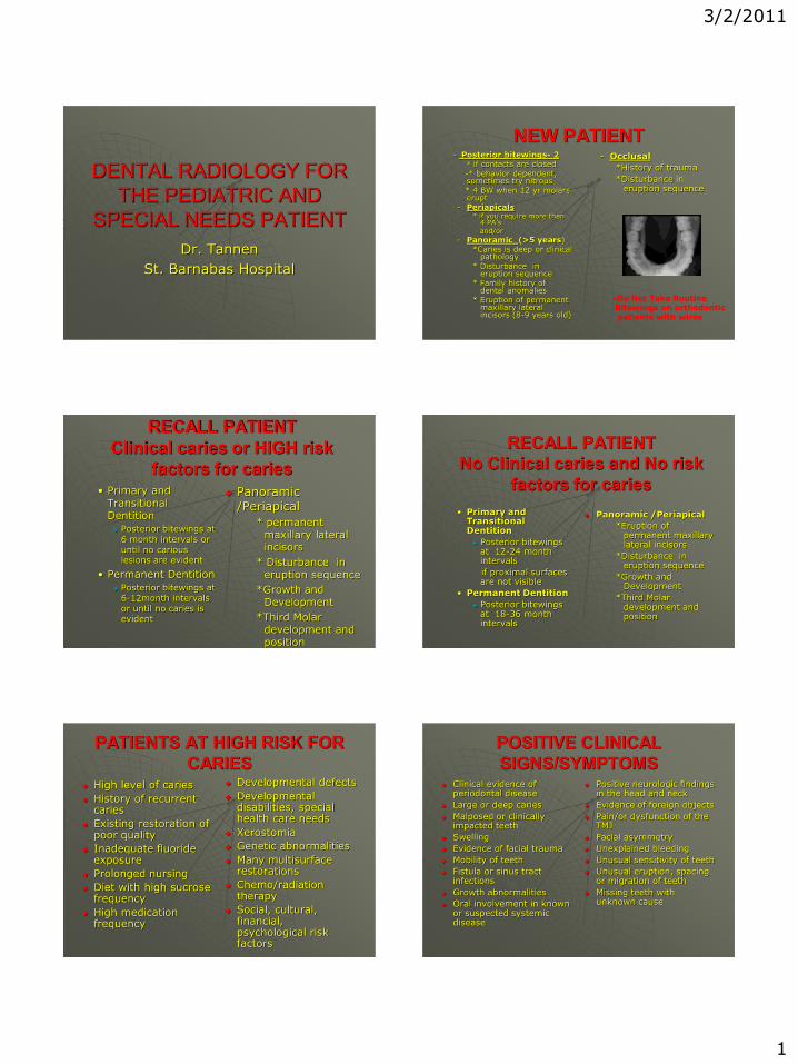
NEW PATIENT- Posterior bitewings- 2
* if contacts are closed-* behavior dependent, sometimes try nitrous* 4 BW when 12 yr molars erupt
- Periapicals * if you require more than
4 PA’sand/or
- Panoramic (>5 years)*Caries is deep or clinical
pathology* Disturbance in
eruption sequence* Family history of
dental anomalies* Eruption of permanent
maxillary lateral incisors (8-9 years old)
- Occlusal
*History of trauma
*Disturbance in eruption sequence
•Do Not Take Routine Bitewings on orthodontic patients with wires
RECALL PATIENT
Clinical caries or HIGH risk
factors for caries• Primary and
Transitional Dentition
Posterior bitewings at 6 month intervals or until no carious lesions are evident
• Permanent Dentition
Posterior bitewings at 6-12month intervals or until no caries is evident
Panoramic /Periapical
* permanent maxillary lateral incisors
* Disturbance in eruption sequence
*Growth and Development
*Third Molar development and position
RECALL PATIENT
No Clinical caries and No risk
factors for caries
• Primary and Transitional Dentition
Posterior bitewings at 12-24 month intervals
if proximal surfaces are not visible
• Permanent Dentition
Posterior bitewings at 18-36 month intervals
Panoramic /Periapical
*Eruption of permanent maxillary lateral incisors
*Disturbance in eruption sequence
*Growth and Development
*Third Molar development and position
PATIENTS AT HIGH RISK FOR
CARIES High level of caries
History of recurrent caries
Existing restoration of poor quality
Inadequate fluoride exposure
Prolonged nursing
Diet with high sucrose frequency
High medication frequency
Developmental defects
Developmental disabilities, special health care needs
Xerostomia
Genetic abnormalities
Many multisurface restorations
Chemo/radiation therapy
Social, cultural, financial, psychological risk factors
POSITIVE CLINICAL
SIGNS/SYMPTOMS Clinical evidence of
periodontal disease
Large or deep caries
Malposed or clinically impacted teeth
Swelling
Evidence of facial trauma
Mobility of teeth
Fistula or sinus tract infections
Growth abnormalities
Oral involvement in known or suspected systemic disease
Positive neurologic findings in the head and neck
Evidence of foreign objects
Pain/or dysfunction of the TMJ
Facial asymmetry
Unexplained bleeding
Unusual sensitivity of teeth
Unusual eruption, spacing or migration of teeth
Missing teeth with unknown cause

3/2/2011
2
PANORAMIC RADIOGRAPH PERIODONTAL EVALUATION CLINICAL
Oral Hygiene
Width of attached gingiva and recession
Attachment Levels• Permanent incisors and
molars
Calculus• 10% children
• 33% adolescents
Gingival tissues• Redness
• Enlargement
• Bleeding
• Edema
RADIOGRAPHIC
Periapical• Evaluate peripapically
Bitewings• Normal crestal height
should be within 1-2 mm of the cemento-enamel junction
• Interdental horizontal or vertical bone loss
• Furcation involvement
RADIOGRAPHIC TECHNIQUES
FOR THE
* INFANT
* UNCOOPERATIVE CHILD
* SPECIAL NEEDS PATIENT
Parental, family, or friend assistance Informed consent Pharmacologic assistance (nitrous oxide,
sedative, oral/IM/IV sedation, general anesthesia)
Protective stabilization Digital Radiography is Visual!
POSITIVE INDICATIONS FOR
ALTERNATIVE TECHNIQUES
Developmentally disabled patient
Patients with exaggerated gag reflex
Pediatric patients
Dental phobic patients
Trauma/trismus patients
ALTERNATIVE TECHNIQUES
Horizontal versus vertical bitewings
Snap-a-ray/ rings / finger
Extra-oral
Digital radiography (visual)
Others (salt, distraction, accupuncture)
RADIATION PHYSICS
The image is a "photographic negative" of the object - the "shadows" are white regions (where the X-rays were blocked by the object)
90% of the xray photons are absorbed by the tissues

3/2/2011
3
RADIATION PHYSICS
A chest x-ray has almost three times the exposure of a periapical film because of the chest projection’s much larger field size
A full mouth series is not equal to the sum of the individual exposures because of movement of the tube (1-30% of total beam exposure, Alcox/Jameson, 1974)
RADIATION PHYSICS
As kVp (tube voltage) is increased there is an increase in the energy each electron has when it strikes the target
Increase the quality of the xray beam by removing the less penetrating photons with an aluminum filter in the path of the beam
Collimation reduces patient exposure and increases film quality by reducing the size of the xray beam and volume of irradiated tissue within the patient by reducing scattered radiation
If the kilovoltage is increased to reduce contrast than the mAs must be decreased or the radiograph will be over exposed
For a given beam, the intensity is inversely proportional to the square of the distance from the source (modify kVp or mA)
TECHNIQUES
The film should ideally be parallel to the object and the central ray should be perpendicular to the object and film
Bisecting the angle technique
Paralleling technique
What is the difference?
Foreshortening results when the central ray is perpendicular to film but object is not parallel to film
Elongation of radiographic image results when central ray is perpendicular to object but not film
TECHNIQUES
SLOB rule- if the tooth moves in the same direction of the central X-ray beam from the first film to the second, the tooth is lingual or palatal to the other teeth ( opposite – buccal)
OFFICE MAINTENANCE AND
QUALITY ASSURANCE
Protection of patients
Protection of personnel
Lead apron and thyroid collar
DIGITAL
RADIOGRAPHY Digital systems are
compared with film and those studies which have evaluated the effects • on diagnostic accuracy
of contrast and edge enhancement
• image size,• variations in radiation
dose and image compression are reviewed together with the use of automated image analysis for caries diagnosis
• as accurate as the currently available dental films for the detection of caries, sensitivities are relatively high (0.6-0.8) for detection of occlusal lesions into dentine with false positive fractions of 5-10%.
• for detection of approximal dentinal lesions, sensitivities, specificities as well as the predictive values are fair, but are very poor for lesions known to be confined to enamel

3/2/2011
4
THE SENSOR
is radiated precisely by your existing system at about 10% the exposure to radiation when compared to conventional film X-rays.
is connected to a computer in the operatory for file management of captured images
sensors are shared between rooms minimizing the cost of equipment duplication. Concise, Simple, Precise.
DIGITAL RADIOGRAPHY
Patient education
Patient visualization / distraction
Less radiation
ADVANTAGES OF
DIGITAL
RADIOGRAPHY
improve the contrast and enhance the density of the image immediately
computer based imaging facilitates automatic analysis of images and image reconstruction from two or more component images
EXTRAORAL RADIOGRAPHIC
TECHNIQUE MAXILLARY
• Open mouth as wide as possible
• Sensor is placed on the external surface of the cheek directly buccal to the tooth with a cotton roll between the sensor and face
• Xray cone is angled -55 degrees from the horizontal and perpendicular to the sensor
MANDIBULAR• Patient’s chin is raised
• Sensor is placed on the external surface of the cheek directly buccal to the tooth with a cotton roll between the sensor and face
• Xray cone is angled -35 degrees from the horizontal and perpendicular to the sensor
DOCUMENTATION
Document radiographic analysis and indication(s) for taking or not takingradiographs
Document number of radiographs, type and who took them
Documentation of succedaneous tooth when restorative or extraction is indicated
Document when you cannot obtain a radiograph and reason. Explain risks/benefits/alternatives
REVIEW AAPD GUIDELINES

3/2/2011
5
THE END
RADIATION EXPOSURE
Recommended