Pathology of Respiratory System
Collage of Veterinary Medicine and Animal Sciences
University of Gondar,
Gondar, Ethiopia
4/22/2020 1Girma B. UoG, CVMAS

Objectives
• At the end of the session students will able to
explain and determine
– General consideration of respiratory system.
– Pathology of nasal cavity, larynx, trachea and
bronchi.
– Deferent the anomalies of the respiratory system
4/22/2020 2Girma B. UoG, CVMAS

Pre test for respiratory system
• Discuss in group the important anatomic features of
the respiratory apparatus as well as their function(s)
in respiration, beginning with the nasal mucosa and
ending with the alveolar walls?
4/22/2020 Girma B. UoG, CVMAS 3

• Animal’s or human’s life is highly dependent onoxygen.
• Oxygen is vital for normal oxidative metabolismof body cells.
• Carbon dioxide is major end product of cellularrespiration, which if not removed, accumulate &poisons cells.
• The respiratory system is well equipped system tomediate exchange of these gases between the body& the atmosphere.
4/22/2020 4Girma B. UoG, CVMAS

• The respiratory tract is arbitrarily divided into three continuous systems
1. Conducting system: The conducting system includes the nasal cavity, sinuses, larynx, trachea and bronchi.
– The mucosa of the conducting system is lined primarily by ciliated epithelium and goblet cells.
2. Transitional system: The transitional system is formed by the bronchioles that are lined by a specialized mucosa containing several types of ciliated and secretory cells such as Clara cells.
– Unlike the conducting system, the normal bronchiolar mucosa contains no goblet cells.
4/22/2020 5Girma B. UoG, CVMAS

3. Exchange system: This system is composed
of the alveoli that are lined externally by
epithelial cells called pneumonocytes.
– The type I (membranous) pneumonocytes are thin
cells and together with the capillary endothelium
and basement membrane constitute the air-blood
barrier.
– Type II pneumonocytes are cuboidal and produce
surfactant.
4/22/2020 6Girma B. UoG, CVMAS

4/22/2020 7Girma B. UoG, CVMAS

Pathology of Nasal Cavities and Sinuses
• Inflammatory diseases are the most common
disorders affecting the nose and paranasal sinuses.
1. Rhinitis
• Rhinitis is inflammation of the nasal cavities.
• The condition may be acute or chronic, and the
exudate may be serous, catarrhal, purulent, pseudome
membranous.
4/22/2020 8Girma B. UoG, CVMAS

• The cause of rhinitis is based on the interplay of
viruses, bacteria, and allergens.
• Acute rhinitis is usually initiated by viruses which
commonly evoke a profuse catarrhal discharge.
Also, allergens may initiate an acute rhinitis, but
they are important only in cattle.
4/22/2020 9Girma B. UoG, CVMAS

• Pathogenesis of viral rhinitis / tracheitis /
bronchitis
• Virus in air or saliva virus replication in epithelial
cells cell degeneration loss cellular attachment
cell exfoliation ulceration exudation (fluid
and cellular) cell mitosis repair.
– Complete repair occurs in approximately 14 days.
4/22/2020 10Girma B. UoG, CVMAS

• Grossly: During the initial acute stage of rhinitis, the
nasal mucosa is swollen, edematous, and pale grey to
red (depending on the degree of hyperemia).
• The mucosal surfaces are covered by a thin watery to
mucoid discharge which is relatively clear.
• When such acute reactions persist for a few days,
bacterial infection modifies the character of the
discharge and produces a mucopurulent or
suppurative exudate.
4/22/2020 11Girma B. UoG, CVMAS
• Microscopically, edema is the most prominent
feature; the lamina propria is sparsely infiltrated by
inflammatory cells.
• Swelling of the mucous membrane may cause mild
respiratory discomfort.
• Chronic rhinitis is characterized by the presence of
fibrous connective tissue scarring; the epithelium
becomes atrophic, foci of squamous metaplasia may
develop and there is progressive atrophy of the
mucous secreting glands.
4/22/2020 12Girma B. UoG, CVMAS

2. Amyloidosis:
• Amyloid is sometimes deposited in the nasal submucosa
of horses.
• The deposition is not part of a generalized amyloidosis
and the cause is unknown.
• The amyloid is deposited in the anterior portion of the
nasal cavity and stenosis can be severe enough to cause
clinical signs of nasal obstruction.
4/22/2020 13Girma B. UoG, CVMAS
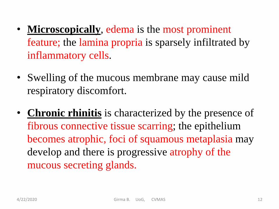
3. Epistaxis:
• The term epistaxis refers to nasal hemorrhage.
• Hemorrhage from the nose may be due to traumatic, Bacterial (Anthrax), Parasites (Eimeriacanis in dogs; Oestrous ovis in sheep).
• Blood-stained foam emitted from the nose of carcasses, especially sheep is indicative of severe pulmonary congestion and edema with seepage of blood from the congested alveolar walls.
4/22/2020 14Girma B. UoG, CVMAS
4. Nasal Polyps:
• Nasal polyps are inflammatory new growths which resemble true neoplasms.
• Polyps are non neoplastic masses that resemble tumours.
• They represent focal accumulations of edematous fluid accompanied by hyperplasia of submucosa connective tissue and inflammatory cells (neutrophils, lymphocytes, plasma cells).
• Older polyps may contain considerable fibrous connective tissue.
4/22/2020 15Girma B. UoG, CVMAS

5. Nasal Parasites:
• The larvae of a number of flies of the family Oestridaeare parasites of the nasal cavities of domestic animals.
• However, Oestrus ovis (nasal bot of sheep) is most commonly encountered.
• The adult flies are relatively harmless from a pathologic point of view; however, they do cause considerable annoyance to their host.
4/22/2020 16Girma B. UoG, CVMAS

6. Nasal Neoplasms:
• Neoplasms of the nasal cavity are uncommon;
however, a wide histogenic variety may be found
(benign and malignant).
• Fibrosarcomas are the most common of the
mesenchymal neoplasms and squamous cell
carcinomas are the predominate neoplasm of
epithelial cell origin.
4/22/2020 17Girma B. UoG, CVMAS

7. Congenital Anomalies:
• The most common congenital anomalies of
the nose and pharynx are cleft lip and palate
4/22/2020 18Girma B. UoG, CVMAS

8. Sinusitis:
• Sinusitis is Inflammation of the paranasal sinuses.
• Sinusitis is common in sheep as a response to the larvae of Oestrus ovis. And it is most significant in the horse, because this species has the largest and most complex sinus structure, coupled with the poorest drainage.
• In addition, in horses, periodontitis often extends into the sinuses.
4/22/2020 19Girma B. UoG, CVMAS

Specific Diseases of the Nasal Cavities and
Related Structures
Strangles of Horses
• Strangles is an acute contagious disease of horses
caused by Streptococcus equi and characterized by
inflammation of the upper respiratory tract and
abscessation in the regional lymph nodes.
• Clinicaly indicated by fever, a slight cough, and
bilateral nasal discharge (which changes from serous
to catarrhal and then purulent).
4/22/2020 20Girma B. UoG, CVMAS

• In the nasal cavities, a purulent rhinitis develops and large amounts of creamy yellow pus collect in the folds of turbinates. The nasal mucosa is edematous and hyperemic and occasionally small ulcers may develop.
Necrotic Rhinitis of Swine
• ("Bull Nose," Rhinohyperplasia) is characterized by irregular proliferation of fibrous connective tissue and bone over the snout region.
• Apparently, a variety of infectious agents can initiate the condition. Such agents usually enter through defects in the gums.
4/22/2020 21Girma B. UoG, CVMAS

Glanders
• Glanders is a disease of horses caused by Burkholderia mallei.
• The bacteria enter through the nasal cavity and cause big caseo-necrotizing tracts through the sinuses, within the subcutis, and in the lung.
• When the lesions occur in the subcutis of the skin, the disease is called “farcy.”
4/22/2020 22Girma B. UoG, CVMAS

Pharynx And Guttural Pouches
• The pharynx is common to both the respiratory and digestive systems and shares the misfortunes of both.
• Guttural pouches are diverticula of the eustachian tubes and they are found only in the horse.
• Occasionally, the guttural pouches become infected by extension of infections from the eustachian tubes. Such infections are entirely comparable with those of the paranasal sinuses.
4/22/2020 23Girma B. UoG, CVMAS

Larynx and Trachea
Laryngitis/Tracheitis:
• Inflammation is the most common and important disorder affecting the larynx and trachea.
• Because of their location, these structures frequently become inflamed as a part of inflammatory diseases of either the upper or lower parts of the respiratory tract.
• Thus, laryngitis and, to a lesser extent, tracheitis is expected to accompany rhinitis; whereas tracheitis and, to a lesser extent, laryngitis, is expected to accompany acute pneumonias.
4/22/2020 24Girma B. UoG, CVMAS

• Inflammations of the air passages usually involve all levels
and they are characterized by coughing, noisy inspiration
and some degree of inspiratory embarrassment.
• Diseases in which laryngitis and tracheitis are a prominent
feature include infectious bovine rhinotracheitis, calf
diphtheria, equine viral rhinopneumonitis, strangles, swine
influenza, feline infectious respiratory diseases, etc.
4/22/2020 25Girma B. UoG, CVMAS

• Grossly: Mucosa of larynx is swollen. The mucosa is haemorrhagic and dry at first, later becomes coated with mucus or mucopurulent discharge.
• Tracheal mucosa is congested and the lumen contains mucus or blood tinged mucus containing red worms.
• Microscopically: Mucosa is covered with exudate -mucus, blood or necrotic material Lamina propria is infiltrated with leucocytes.
• Lesions may vary from catarrhal to chronic granulomatous involvement, depending on the durationand severity of infection.
4/22/2020 26Girma B. UoG, CVMAS

Hemorrhages:
• Laryngeal and tracheal hemorrhages may occur as
a result of infection, trauma, violent coughing, etc.
• Laryngeal hemorrhages occur in many septicemic
diseases (hog cholera, salmonellosis, etc.).
• In the trachea, agonal hemorrhages are often times
associated with severe dyspnea and hypoxia.
4/22/2020 27Girma B. UoG, CVMAS

Edema:
• Edema of the larynx is usually inflammatory and part of the picture of acute respiratory infection.
• laryngeal edema may be associated with allergic reactions, inhalation of irritants, insertion of tracheal tubes, etc.
• In the tracheal lumen, foamy fluid is commonly observed. Such foamy fluid is associated with severe pulmonary edema. The foams are actually formed in the alveoli.
4/22/2020 28Girma B. UoG, CVMAS

Reading assignments
• Specific Diseases Involving The Larynx And Trachea
– Calf Diphtheria
– Infectious Bovine Rhinotracheitis
– Feline Upper Respiratory Disease
– Feline Viral Rhinotracheitis
– Feline Pneumonitis
– Infectious Laryngotracheitis
– Syngamus Trachea Infection
4/22/2020 Girma B. UoG, CVMAS 29

Bronchi and Bronchioles
• The bronchi and bronchioles have a hybrid
nature. Their function is conducting air.
• However, they are embedded within the lung
parenchyma, which is predominantly focused
on gaseous exchange.
4/22/2020 Girma B. UoG, CVMAS 30

1. Bronchitis and Bronchiolitis
• The consequences of inflammation of the large bronchi
may be quite different from those of inflammation of
smaller bronchi and bronchioles.
• The larger bronchi, for example, lie in interstitial tissue
outside of the lung lobules. The epithelium is
pseudostratified and well supplied with mucous-secreting
and ciliated cell and the peribronchial connective tissue is
abundant. The lumen is large enough to remain patent
even in the presence of copious exudate and such exudate
can be expelled by an effective cough reflex.
4/22/2020 Girma B. UoG, CVMAS 31

• By contrast, the small bronchi and bronchioles lie within
the lung parenchyma. The epithelium is simple and the
ciliated and mucous-secreting cells decrease in number
and disappear from the smallest branches.
• The walls are thin and the small lumens are easily
occluded by exudate which may be too far distal for the
cough reflex to be particularly effective.
4/22/2020 Girma B. UoG, CVMAS 32

• Therefore, inflammation of larger bronchi may or may not
have significant consequences for the lung, but
inflammation of small bronchi and bronchioles almost
inevitably leads to occlusion with extension of
inflammation to the lung parenchyma.
• The causes of bronchitis/bronchiolitis are chemical agents,
bacteria, viruses, helminths.
• The condition may be acute or chronic, focal or
generalized and characterized by a catarrhal, purulent,
fibrinous or pseudomembranous exudation
4/22/2020 Girma B. UoG, CVMAS 33

• Grossly, bronchitis/bronchiolitis are usually accompanied
by hyperemia of the mucosa and exudation of materials
into the lumen.
• If fibrin is present, this mass of material may solidify and
form a "cast" within the lumen.
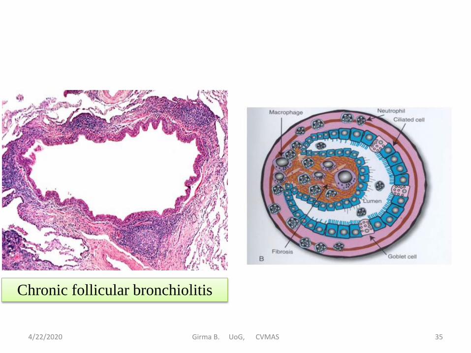
• Microscopically, Inflammation of bronchi bronchioles
should be diagnosed only if inflammatory cells are found
in their wall. If the inflammatory cells are in the lumen but
not in the wall, it is not a bronchitis or bronchiolitis
4/22/2020 Girma B. UoG, CVMAS 34
4/22/2020 Girma B. UoG, CVMAS 35
Chronic follicular bronchiolitis

2. Stenosis of Bronchi/Bronchioles
• Stenosis is narrowing of the lumen. It can be caused
inflammation, parasites, Aspiration of foreign bodies,
peribronchial pressure, Accumulation of exudates and
infiltration etc..
• There may be partial or complete closure of the bronchial
lumen and this results increased resistance in the airways.
If there is partial obstruction, greater effort is required to
force air into the alveoli on inspiration. During this
difficult and slow passage of air, the alveolar walls are
momentarily stretched.
4/22/2020 Girma B. UoG, CVMAS 36

• If this process is repeated over a prolonged period,
this continued over-stretching of the alveolar walls
eventually causes them to break resulting in a
condition referred to as emphysema.
• Then upon expiration, not all of the air is expelled.
This results in incomplete exchange of gases.
– Over-inflated alveoli without rupture is also
referred to as emphysema
4/22/2020 Girma B. UoG, CVMAS 37

If complete stenosis/obstruction of the smaller
bronchioles occurs, at first the air is trapped in the
alveoli because it cannot escape during expiration.
• Eventually, the body will absorb the air. As air is
absorbed from the alveoli, they collapse and this area
of the lung is no longer functional
– This process leads to partial stasis of blood in the
alveolar capillaries, and mild edema
– This is a choice place for infection to start if bacteria
gain entrance to this part of the lung.
4/22/2020 Girma B. UoG, CVMAS 38

3. Bronchiectasis
• Bronchiectasis refers to dilatation of bronchi and
bronchioles.
• Mainy due to chronic inflammation (chronic
bronchitis) there is repeated coughing.
• Repeated coughing results in continuous over-stretching of
the alveolar walls as well as over-dilatation of the
bronchioles as they stretch to accommodate the cough.
4/22/2020 Girma B. UoG, CVMAS 39

• As this over-distention and over-stretching is repeated
over long periods of time, the smooth muscle in the wall
of these bronchioles loses its tone and undergoes
degeneration.
• Eventually, the lumens remain open instead of
contracting as they do when they are relaxed and not
being used.
• The mucosa gradually becomes thinner and the walls are
gradually destroyed and replaced by fibrous connective
tissue. This connective tissue helps to tie the walls of
these bronchioles to the surrounding tissue, which in turn
aids in keeping them over-dilated.
4/22/2020 Girma B. UoG, CVMAS 40

4/22/2020 Girma B. UoG, CVMAS 41

4/22/2020 Girma B. UoG, CVMAS 42
• Grossly: Cylindrical form of bronchiectasis is more
common in cattle and Saccular form is less common
• Microscopically: Destruction and disappearance of the
elastic tissue, lamina propria infiltrated with
mononuclear cells

The Lungs
• There are notable differences in lung morphology
among animal species; i.e., equine poorly defined
pulmonary lobes; bovine and canine well defined lobes.
• Pulmonary lobes are subdivided into lobules by
interlobular septa.
• Lobules are prominent in bovines and pigs, poorly
defined in horses and man, and
absent in dogs and cats.
4/22/2020 Girma B. UoG, CVMAS 43

Inflation Disturbances of the Lung
1. Atelectasis of the Lungs
• Atelectasis is imperfect or incomplete expansion.
• The lungs contain no air and nothing else.
• Atelectasis may be focal or involve entire lobes.
• It may be classified as fetal or acquired.
• Atelectasis occurs in all species except birds where the rigid structure of the parabronchi prevents collapse of the alveoli.
4/22/2020 Girma B. UoG, CVMAS 44

a. Fetal Atelectasis
• The lung is dark-red, liver-like, and will not float in water.
• If the lungs of a newborn are not inflated, it can be assumed that it was born dead.
• However, a newborn animal may take a few breaths and then die. In this case, areas of the lungs will be partially inflated while other areas remain atelectatic.
– Fetal atelectasis is common in newborn foals .why? (reading assignment)
4/22/2020 Girma B. UoG, CVMAS 45

b. Acquired Atelectasis
• May be caused by either compression or obstruction.
• Compression atelectasis is due to pressure on the lungs due to fluid, air, neoplasms, etc., which drives air out of the lungs resulting in collapse of the walls of alveoli.
• Grossly, the lung tissue is deflated, depressed, dark red, and will not float in water.
• If the cause is removed, the lung tissue will reinflateand no damage will follow.
• Respiratory distress is not noticeable unless a considerable portion of the lung tissue is involved.
4/22/2020 Girma B. UoG, CVMAS 46

• Obstructive atelectasis is due to an accumulation of material in the bronchioles.
• If the airway is completely obstructed, air cannot enter the alveolus and it collapses.
• The effect on the body depends on the size of the affected area.
• Histologically, Atelectatic lung showed collapse of the alveolar space and devoid of air with increased thickness of the septa. Alveoli appeared as small or elongated cleft. The blood vessel of the affected part was congested
4/22/2020 Girma B. UoG, CVMAS 47

4/22/2020 Girma B. UoG, CVMAS 48

2. Emphysema of the Lungs
• Emphysema is Over-inflated alveoli without rupture.
• Emphysema is an abnormal and permanent enlargement
of airspaces distal to terminal bronchioles with
destruction of their alveolar walls.
• Primary lung emphysema is rare in animals but
extremely important in human beings.
4/22/2020 Girma B. UoG, CVMAS 49

2.1. Acute Pulmonary Alveolar Emphysema
• Caused by
– Physical: Over exertion during struggling leads to
over ventilation.
– Nutritional: Feeding on lush green pastures.
– Pathological
• Over exertion during coughing (Bronchitis/
Bronchopneumonia).
• Compensatory at areas adjacent to pneumonia.
• Loss of elasticity of wall of alveoli.
• Allergy: Feeding mouldy forage/potato
• Poisons: Parathion poisoning.
4/22/2020 Girma B. UoG, CVMAS 50

• Gross pathology
– Certain areas of lung are unduly distended with air.
– Pale or white areas project above surrounding tissue.
– Cut surface is dry.
• Histopathology
– Alveolar size greatly distended (Giant alveoli).
– Wall of the lung : Sometimes ruptured combined to
form bullae.
4/22/2020 Girma B. UoG, CVMAS 51

2.2. Acute Interstitial Emphysema
• In this condition air collects in the interlobular septa beneath the pleura and wherever there is interstitial tissue in the lungs.
• Often it accompanies with acute alveolar emphysema.
• More often seen in cattle and sheep.
• Caused by :
– Violent respiratory effort (struggling) during death from loss of blood.
– Perforation of lung by mechanical means- foreign body through rumen and reticulum.
– Excessive bellowing in estrum or when separated from calf.
– Forced breathing as in old hunting dogs.
– Pulmonary strongylosis.
4/22/2020 Girma B. UoG, CVMAS 52

• Gross pathology
– Air may escape through thoracic inlet into the subcutis of neck.
– In Severe case,
• Air collects beneath the pleura and other interstitial tissue of lung.
• Air accumulate along the spine – from pole to base of tail.
– Interlobular septa are thickened and seen as criss cross straight lines usually at wider intervals.
– Interlobular septum: shiny, well outlined and filled with large and small air bubbles.
• Histopathology
– Lung interstitial tissue is widely separated.
• Chronic alveolar emphysema (Broken Wind/Heaves) reading assignment.
4/22/2020 Girma B. UoG, CVMAS 53

4/22/2020 Girma B. UoG, CVMAS 54
Bulle

Circulatory disturbance of the lung
1. Congestion of The Lungs
• Pulmonary congestion and edema develop acutely
in a variety of diseases.
• Chronic congestion and edema are usually attributable
to some functional defect in the left heart (the left
ventricle and atrium are prevented from clearing the
blood that comes from the lungs).
4/22/2020 Girma B. UoG, CVMAS 55

• Hypostatic congestion develops due to the effects of
gravity on blood flow through the lungs.
• If an animal lies on one side for a long period of time
blood accumulates in the lower lung. The affected lung
will be nonfunctional, extremely heavy, dark-red, and
firm to the touch.
• In contrast, the upper lung will be pale, inflated, and
light in weight.
4/22/2020 Girma B. UoG, CVMAS 56

2. Edema of the Lungs
• Pulmonary edema is the accumulation of fluid (plasma
protein filtrate) in alveoli. This is due to:
– Altered hemodynamics - (disturbance of normal fluid
exchange in the lungs such as in cardiac failure
backup of blood, and consequently increased
hydrostatic pressure in the alveoli.
– Sudden diffuse and direct damage to capillary
endothelium - (usually the peracute stage of
inflammation).
4/22/2020 Girma B. UoG, CVMAS 57

• Grossly, the lungs are doughy, heavy, firm, and foamy.
Fluid flows from the cut surface.
• Microscopically: Bronchi and alveolar lumen contain
a pink stained homogeneous material proportional to
the amount of protein present.
4/22/2020 Girma B. UoG, CVMAS 58

• Clinically: the head is held low, the nose is flecked with
froth, dyspnea and gasping are prominent, and wheezing,
bubbly sounds of respiration are characteristic.
• Here are two clues when doing the post mortem:
– Look in the trachea – if there is foam, that is a good
indication that there was pulmonary edema.
– Remove the lungs from the thorax and set them in a
dry spot. If, after 5 minutes, the lungs are sitting in a
puddle of fluid, that is evidence of pulmonary edema.
4/22/2020 Girma B. UoG, CVMAS 59

Inflammation of The Lungs
• Pneumonitis is the correct term for inflammation
of the lungs.
• However, pneumonia is the conventional term for lung
inflammation (correctly, the term pneumonia refers to
filling of alveoli with cellular exudate).
• The causes of pneumonia are numerous and may include
bacteria, viruses, aspirated foreign matter, fungi,
parasites, etc.
4/22/2020 Girma B. UoG, CVMAS 60

• There are numerous classifications such as :
– Etiological: Viral pneumonia, Mannheimiosis, Histophilosis
pneumonia, distemper pneumonia, allergic pneumonia, etc.
– Epidemiological: Enzootic pneumonia, contagious bovine
pleuropneumonia, etc.
– Exudate: Suppurative, fibrinous, or granulomatous pneumonias.
– Topographical (distribution): Lobar, lobular, diffuse,
interstitial, focal, etc.
– Miscellaneous: Progressive pneumonia, proliferative
pneumonia, atypical pneumonia, pneumonitis, etc.
4/22/2020 Girma B. UoG, CVMAS 61
Stages of Pneumonia:
– Traditionally, bronchopneumonias caused by bacterial
agents have been divided into four successive stages:
1. Stage of congestion,
2. Stage of red hepatization.
3. Stage of grey hepatization, and
4. Stage of resolution.
4/22/2020 Girma B. UoG, CVMAS 62
Stage of congestion represents the developing bacterial
infection and lasts for approximately 24 hr.
– Microscopically, it is characterized by hyperemia, fluid
within the alveoli (edema), a few neutrophils, and often
by the presence of numerous bacteria.
– Grossly, the involved lung is heavy, boggy, and red (the
affected areas are not consolidated).
4/22/2020 Girma B. UoG, CVMAS 63

Stage of red hepatization is characterized by
increasing numbers of neutrophils and precipitation
of fibrin to fill the alveolar spaces.
• Affected areas of the lungs are consolidated (liver-
like in consistency). Completely consolidated lung
tissue sinks in water.
– Grossly, involved lung is distinctly red, firm, and
airless, with a liver-like consistency.
4/22/2020 Girma B. UoG, CVMAS 64

Stage of gray hepatization is characterized by a
continuing accumulation of fibrin, associated with
progressive disinte-gration of neutrophils and
erythrocytes.
• The hyperemia is decreased and erythrocytes have
disappeared from the alveolar contents. Mononuclear
cells and fibrin predominate, with fewer neutrophils.
• Grossly, the affected lung tissue appears grayish (or
less red). Tissues are still consolidated (liver-like) and
will sink in water.
4/22/2020 Girma B. UoG, CVMAS 65

Stage of resolution is the final stage. this stage may
take in 7 to 10 days after the onset of pneumonia and a
favorable outcome may follow.
• During this stage, the exudate within alveoli (fibrin,
inflammatory cells, etc.) undergoes progressive
enzymatic digestion to produce a semifluid debris that
is either reabsorbed, ingested by macrophages, or
coughed up.
• In favorable cases, the normal lung parenchyma is
restored to its normal state. However, complete
resolution may not occur and complications develop.
4/22/2020 Girma B. UoG, CVMAS 66

4/22/2020 Girma B. UoG, CVMAS 67

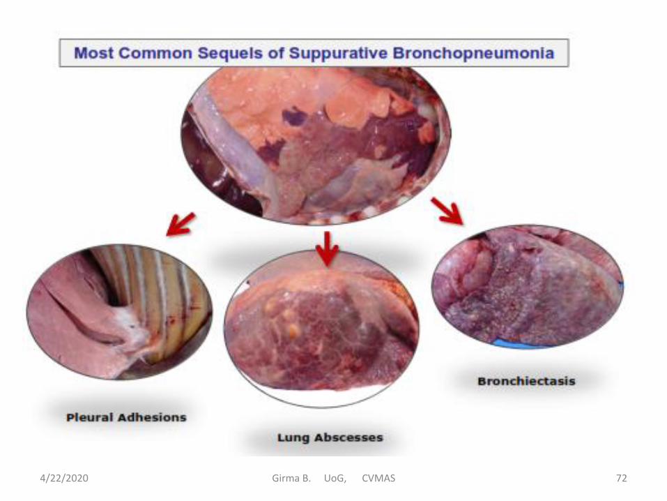
Suppurative Bronchopneumonia
• Bronchopneumonia is the most common form of lung
inflammation.
• It is particularly common in young calves and pigs and in
sheep of any age.
• Bronchopneumonias in animals are primarily bacterial in
origin and characterized by intense exudation of
inflammatory cells (chiefly neutrophils) into alveoli.
• Also, whenever the lesions are lobular and centered
around the bronchiolar tree, that is why the term
bronchopneumonia is used.
4/22/2020 Girma B. UoG, CVMAS 68
• Many species of pathogenic bacteria may cause
bronchopneumonia.
– In calves, pigs, and sheep, Pasteurella spp (P. multocida
and/or P. hemolytica) and Corynebacterium pyogenes are
usually found.
– In horses, Streptococcus spp. Staphylococcus spp.
Corynebacterium equi and E. coli are important.
– In dogs canine distemper virus is considered to be a
primary etiologic agent.
• Bacteria will pathogenic potential only when the respiratory
environment is suitably altered by other agents (viruses,
external environmental factors, etc.). In other words, bacterial
agents usually act as secondary invaders.
4/22/2020 Girma B. UoG, CVMAS 69

• Gross lesions: Affected lung is consolidated and the
lobular pattern is accentuated. Color varies from red
(acute, hyperaemia) to grey (chronic inflammation,
atelectasis, fibrosis).
– Typically, purulent/pus exudate can be expressed from
airways; in chronic bronchopneumonia the exudate takes a
mucoid appearance.
• Microscopically: Large number of polymorphonuclear
leukocytes in bronchoalveolar space in acute cases and a
mixture of PMN,PAM and mucus (goblet cell hyperplasia)
in the more chronic cases.
4/22/2020 Girma B. UoG, CVMAS 70

4/22/2020 Girma B. UoG, CVMAS 71
4/22/2020 Girma B. UoG, CVMAS 72

Fibrinous Bronchopneumonia
• Fibrinous pneumonia is basically a bronchopneumonia characterized by a marked exudation of fibrin.
• Occasionally, fibrinous pneumonia is hematogenous in the course of septicemic salmonellosis.
• Fibrinous bronchopneumonia is an important disease in cattle, sheep and swine and is usually caused by Pasteurellaspp. (P. multocidia and P. hemolytica).
• Examples of disease causing fibrinous bronchopneumonia: Pneumonic Mannheimiosis (shipping fever), Porcine Pleuropneumonia, CBPP.
4/22/2020 Girma B. UoG, CVMAS 73
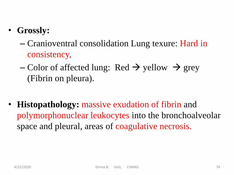
• Grossly:
– Cranioventral consolidation Lung texure: Hard in
consistency,
– Color of affected lung: Red yellow grey
(Fibrin on pleura).
• Histopathology: massive exudation of fibrin and
polymorphonuclear leukocytes into the bronchoalveolar
space and pleural, areas of coagulative necrosis.
4/22/2020 Girma B. UoG, CVMAS 74

4/22/2020 Girma B. UoG, CVMAS 75

4/22/2020 Girma B. UoG, CVMAS 76

4/22/2020 Girma B. UoG, CVMAS 77

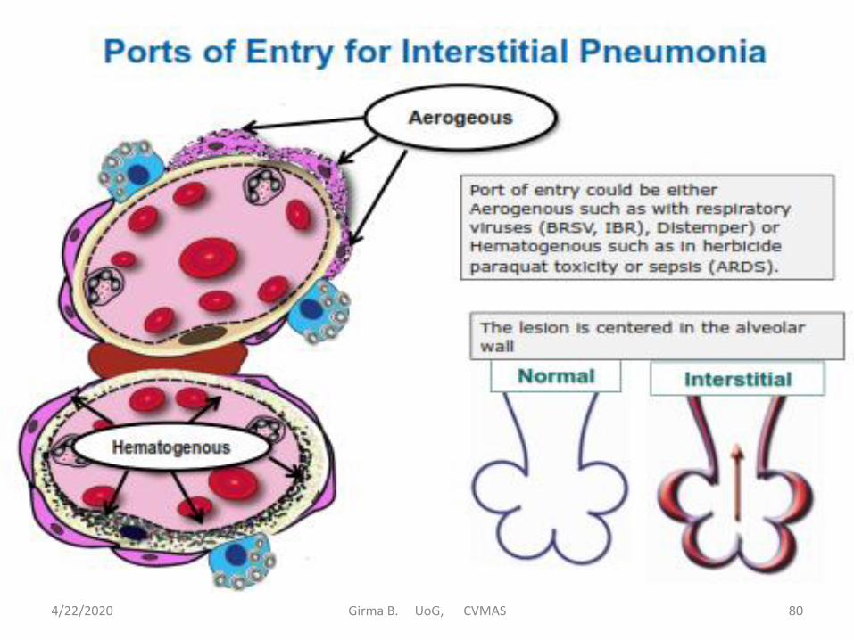
Interstitial Pneumonia (Primary Atypical Pneumonia)
• Interstitial pneumonia (pneumonitis) is characterized by inflammation of the alveolar walls with an absence or minimum exudation of inflammatory cells (neutrophils, etc.) into alveolar lumens.
• The causes of IP are numerous and include viral infections (feline pneumonitis, canine distemper, equine viral rhinopneumonitis, etc.), chemical toxicosis(turpentine, kerosene, etc.), mycoplasma infection (enzootic pig pneumonia, etc.), and some allergic reactions.
4/22/2020 Girma B. UoG, CVMAS 78

• In addition, interstitial pneumonia is common in
septicemic diseases and/or those characterized by
sustained or intermittent bacteremia (colibacillosis,
salmonellosis, erysipelas, leptospirosis, etc.).
• A diagnosis of interstitial pneumonia depends on the
detection of any one of a combination of the following:
– Thickening of the alveolar walls by exudates (fibrin,
etc.), and by an infiltration of inflammatory cells
(macrophages, plasma cells, and lymphocytes).
– Proliferation of alveolar epithelial cells.
– Formation of hyaline membranes in the alveoli and
covering alveolar ducts.
4/22/2020 Girma B. UoG, CVMAS 79
4/22/2020 Girma B. UoG, CVMAS 80

• Gross lesions:
– The lungs fail to collapse when the thorax is opened;
– occasional costal imprints are visible on the pleural surface.
Meaty appearance on the cut surface.
– The color depends on blood: tissue ratio and type of exudate
or fibrous scarring.
• It is difficult to diagnose grossly generally requiring
histopathologic confirmation.
• Histopathology: The primary lesion is centered in the
alveolar wall.
– Thickening of alveolar walls.
– Interstitial exudation or proliferation of type II pneumonocyt
– In chronic interstitial pneumonia there is alveolar fibrosis. 4/22/2020 Girma B. UoG, CVMAS 81

4/22/2020 Girma B. UoG, CVMAS 82

4/22/2020 Girma B. UoG, CVMAS 83

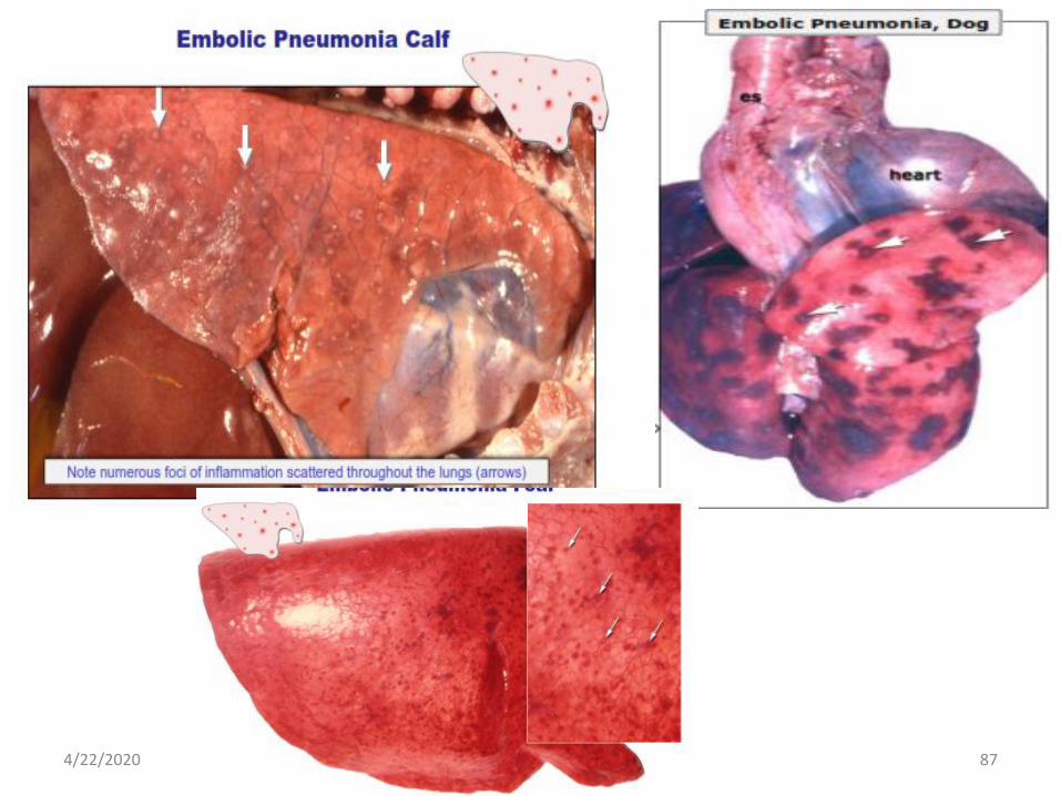
Embolic (Hematogenous) Pneumonia
• Embolic pneumonia refers to foci of lung inflammation
initiated by the lodgement of emboli of pathogenic
organisms or septic fragments of thrombi in the pulmonary
arteries or capillaries.
• Most organisms which cause embolic Pneumonia are
pyogenic and abscessation is to be expected.
• Endocarditis, ruptured hepatic abscess (vena cava
thrombosis in cattle), Omphalophlebitis cause embolic
Pneumonia
4/22/2020 Girma B. UoG, CVMAS 84

4/22/2020 Girma B. UoG, CVMAS 85

• Gross lesions: Variable number of foci, often with a
white center and red hemorrhagic margins. Eventually
embolic lesions may progress to abscesses.
• Histopathology: Septic emboli attached to pulmonary
capillaries, pulmonary edema, microabscesses.
• Common sequels: Abscesses in all pulmonary lobes.
4/22/2020 Girma B. UoG, CVMAS 86
4/22/2020 Girma B. UoG, CVMAS 87

4/22/2020 Girma B. UoG, CVMAS 88

Granulomatous Pneumonia
• Refers to a distinctive pattern of chronic lung inflammation evoked by certain etiologic agents (usually higher bacteria and fungi).
• Etiology: Tuberculosis, systemic mycosis, some parasites (Muellerius capillaris; larva migrans). Usually caused by microorganisms, parasites (ova, larvae) or foreign material (inhaled food particles) difficult to eliminate by phagocytosis.
• The typical granuloma consists of collections of modified macrophages (epithelial cells), usually surrounded by a rim of lymphocytes.
4/22/2020 Girma B. UoG, CVMAS 89

• Gross lesions: Granulomas in the lung and sometimes in other organs too.
– Be aware that granulomatous pneumonia can resemble lung cancer and may require histopathologicalconfirmation.
• Histopathology: Variable size nodules with a necrotic center infiltrated by macrophages and giant cells and surrounded by connective tissue mixed with lymphocytes and plasma cells.
4/22/2020 Girma B. UoG, CVMAS 90

4/22/2020 Girma B. UoG, CVMAS 91

4/22/2020 Girma B. UoG, CVMAS 92

o Special Forms of Pneumonia
1. Gangrenous Pneumonia
• Is not an independent type of pneumonia, but a complication
of other forms in which there is necrosis and invasion of
lung tissue by saprophytic and putrefactive bacteria.
• The usual cause of this condition is the introduction into the
lungs of materials (medicines, etc.) intended for the
gastrointestinal tract (saprophytic and putrefactive bacteria
are usually introduced with the foreign material).
• At necropsy, the lungs may be yellowish-green to
greenish-black, foul smelling, with extensive cavitations.
4/22/2020 Girma B. UoG, CVMAS 93

• NOTE: Gangrenous pneumonia is also referred to as
aspiration pneumonia, inhalation pneumonia, foreign body
pneumonia, medication pneumonia, and lipid pneumonia..
2. Verminous Pneumonia
• Refers to lung inflammation caused by parasites (larvae
and/or adults)
3. Hypostatic Pneumonia
• Refers to lung inflammation that develops subsequent to the
accumulation of blood and the inhalation of upper
respiratory pathogens in the ventral portions of the lungs.
4/22/2020 Girma B. UoG, CVMAS 94

Specific Diseases Characterized by Lung Lesions
I. Paramyxoviruses
a) Parainfluenza-3
• Parainfluenza type 3 virus induces acute respiratory
disease in a wide variety of species including cattle,
sheep and goats, and horses. It attacks cells of the
conducting airways.
• It can cause pneumonia alone, but is more
commonly part of the etiologic complex of enzootic
pneumonia in calves or shipping fever in adults.
4/22/2020 Girma B. UoG, CVMAS 95

b) Bovine respiratory syncytial virus
• Bovine respiratory syncytial virus occurs either alone in
an outbreak form of pneumonia or in concert with other
agents, especially bacteria, in the shipping fever
syndrome.
• The virus attacks conducting airway epithelium, most
severely that at the broncho-alveolar junction.
• Histologically there are frequently syncytial giant cells
formed by fusing bronchiolar epithelial cells.
4/22/2020 Girma B. UoG, CVMAS 96

c) Canine distemper
• Canine distemper virus is pantropic, which means that a
number of tissues and organs are targeted by the virus.
• The virus infects epithelium in multiple organs, so for the
respiratory system, this would include nares, trachea,
bronchi, bronchioles, and alveolar cells.
• Consequently, the pneumonia could look like a
bronchopneumonia or an interstitial pneumonia. And, if
the dog survives the pneumonia, the virus usually goes on
the brain.
4/22/2020 Girma B. UoG, CVMAS 97

d) Peste des petits ruminants (PPR)
• This virus is closely related to rinderpest, and causes
enteritis and pneumonia in sheep and goats.
• The disease is endemic in much of Africa and the
Middle East, but is foreign to this hemisphere.
4/22/2020 Girma B. UoG, CVMAS 98

II. Orthomyxoviruses
• The Orthmyxoviridae include the influenza viruses,
which infect several domestic species.
Equine influenza
– Disease usually occurs in young animals that are
stressed and/or grouped with older horses. It may
occur in outbreak form, with high morbidity but low
mortality.
– The virus infects both ciliated and alveolar cells, so
it can look like a bronchopneumonia or an interstitial
pneumonia.
4/22/2020 Girma B. UoG, CVMAS 99

III. Maedi
• Maedi is a chronic progressive interstitial pneumonia of
sheep which is caused by a viral Maedi occurs principally
in sheep (2-years old or older), but it has been reported in
goats.
• Grossly, the lungs are larger, and 2-4 times as heavy as
normal lungs.
– They do not collapse completely when the chest cavity
is open and appear grey to pink in color.
– Enlargement of the bronchial and mediastinal lymph
nodes is a constant feature.
4/22/2020 Girma B. UoG, CVMAS 100
Maedi cont…
• Microscopically, changes are characteristic of a
chronic interstitial pneumonia.
– There is rather uniform thickening of alveolar walls
by hyperplastic alveolar lining cells, lymphocytes,
and mononuclear cells.
• There is an absence of healing.
• No treatment has been successful.
Jaagsiekte disease is a chronic progressive interstitial
pneumonia of sheep, characterized by rather uniform
thickening of alveolar walls and by hyperplastic
alveolar lining cells. The disease is similar in character
to Maedi.
4/22/2020 Girma B. UoG, CVMAS 101

• Bacterial pneumonias
1. Bovine Enzootic Pneumonia
• Bovine enzootic pneumonia is composed of a number of
etiologies, all of which can be interacting in synergy. It
usually starts with a viral infection, maybe PI-3, or BHV-1,
or a mycoplasmal infection, Mycoplasma bovis.
• After those are set up, then opportunists move in and set up
camp - Pasteurella multocida, Arcanobacterium pyogenes,
or E. coli. In some cases, Mannheimia hemolytica can move
in and cause a full-fledged shipping pneumonia.
4/22/2020 Girma B. UoG, CVMAS 102

2. Pneumonic Pasteurellosis
• The members of the Pasteurella genus (p. multocida
and Mannheimia haemolytica) are widely distributed
among a variety of animal species.
• They are carried subclinically, usually in the upper
respiratory tract, and migrate down the bronchial tree to
cause disease when the opportunity presents itself.
4/22/2020 Girma B. UoG, CVMAS 103
3. Tuberculosis
• Bovine tuberculosis, caused by Mycobacterium bovis, is the principal respiratory tuberculous disease seen in animals.
• The tuberculous pulmonary process starts at the bronchiolo-alveolar junction and develops into a tuberculous granuloma, or tubercle.
• This tubercle is the result of an ongoing battle between the tubercle bacilli and the cell mediated immune response.
4/22/2020 Girma B. UoG, CVMAS 104

• Parasitic diseases of the lungs
• Lesions due to parasites in the lung come in two
categories.
–There are those parasites who live specifically in
lung,
– then there are others who merely do damage as
they migrate through to somewhere else.
4/22/2020 Girma B. UoG, CVMAS 105

Host Parasite
Cattle Dictyocaulus viviparus
Cat Aelurostrongylus abstrusus
Dog Filaroides milski and F. osleri
Horse Dictyocaulus arnfieldi
Sheep Dictyocaulus filaria
Sheep Protostrongylus rufescens
Sheep Muellerius capillaris
Cattle Dictyocaulus viviparus
Swine Metastrongylus apri, M. salmi, and M. pudendotectus
Important Lung Worms
4/22/2020 Girma B. UoG, CVMAS 106

• Calcification Of The Lungs
– Calcification of the lungs occurs rather frequently in
the dog. It usually occurs as a consequence of vitamin
D toxicosis in young dogs, whereas in older dogs, it is
usually associated with chronic renal diseases and
hyperparathyroidism (metastatic calcification).
– Grossly, the lungs are gritty, firm to hard, and they do
not collapse when the thoracic cavity is opened.
– Microscopically, calcium concretions are found in
alveolar walls.
4/22/2020 Girma B. UoG, CVMAS 107

• Melanosis of The Lungs
– Melanosis refers to a deposition of melanin
pigments in various organs and tissues:
– the lung is a common site of involvement.
– Affected animals are in a state of normal health.
4/22/2020 Girma B. UoG, CVMAS 108

• Lung Neoplasia
• Most neoplasms encountered in the lungs are metastatic growths (lungs are especially prone to the lodgement of tumor emboli).
• However, any component of lung tissue can give rise to primary neoplasms.
• Primary lung neoplasms are rather infrequent; but the incidence is much higher in dogs than in any other species.
• The common tumour is lymphocytoma
4/22/2020 Girma B. UoG, CVMAS 109

Pathology of Thoracic Pleura
• Pathologic involvement of the pleura is usually a
secondary complication of some underlying disease.
1. Pleuritis
• Pleuritis (inflammation of the pleura) is the most
common condition encountered and it is usually
secondary to pneumonia.
• When copious exudation into the pleural sacs
accompanies the inflammation, the lesion is
commonly designated as "pleurisy with effusion."
4/22/2020 Girma B. UoG, CVMAS 110

• primary pleuritis is associated with bovine blackleg,
Glasser's disease, sporadic bovine encephalomyelitis,
etc.
• The exudate is voluminous, the diaphragm is depressed,
and the lungs are compressed and displaced to the
caudodorsal portion of the thorax.
• Pasteurella multocida, Escherichia coli, and Sterptococci
are present in most cases.
2. Abnormal contents
• Noninflammatory pleural collections (fluid, blood, chyle,
air) may be observed. Hydrothorax, hemothorax,
chylothorax, and pneumothorax.
4/22/2020 Girma B. UoG, CVMAS 111

4/22/2020 Girma B. UoG, CVMAS 112

Air Sacculitis
• Inflammation of the air sacs is called air sacculitis
• Mainly caused by Mycoplasma gallisepticum, Escherichia coli
• Grossly: Pin point pale foci seen in early change
– Cloudy –Mild inflammation
– Thick – Moderate
– Thick with whitish cheesy exudate – severe
– Thick with yellowish cheesy material indicate complicated infection
4/22/2020 Girma B. UoG, CVMAS 113

4/22/2020 Girma B. UoG, CVMAS 114
QUESTIONS
Recommended