Title
Preparation and Properties of Hydrophilic PolypeptideMembranes as Biodegradable Materials (CommemorationIssue Dedicated to Professor Hiroshi Ibagaki, Professor MichioKurata, Professor Ryozo Kitamura, On the Occasion of TheirRetirments)
Author(s) Hayashi, Toshio; Nakanishi, Eiji; Nakajima, Akio
Citation Bulletin of the Institute for Chemical Research, KyotoUniversity (1989), 66(3): 251-262
Issue Date 1989-02-15
URL http://hdl.handle.net/2433/77236
Right
Type Departmental Bulletin Paper
Textversion publisher
Kyoto University

Bull. Inst. Chem. Res., Kyoto Univ., Vol. 66, No. 3, 1988
Preparation and Properties of Hydrophilic Polypeptide
Membranes as Biodegradable Materials
Toshio HAYASHI*, Eiji NAKANISHI**, and Akio NAKAJIMA***
Received July 29, 1988
Three-component random copolypeptides consisting of N-hydroxyalkyl-L-glutamine, L-glutamic acid, and L-lysine were prepared by carring out aminoalcoholysis reaction with aminoalcohols, such as 2-amino-l-ethanol(E) and 4-amino-l-butanol(B), followed by crosslinking reaction with 1,8-octa-methylenediamine (OMDA) on starting polymer membranes consisting of y-methyl-L-glutamate, L-glutamic acid, and L-lysine. The effective crosslink density was shown to be proportional to the content of the crosslinker (OMDA) in the reaction mixture. The tensile properties of these hydrophilic membranes were highly dependent on the degree of swelling in the pseudo-extracellular fluid (PECF), hydrophobicity of the side chains, and the effective charge density of membranes, and their behavior was typical of an elastomer. A higher rate of water permeability was obtained with charged membranes than non-charged or compensated charged membranes with the same order of the degree of swelling in PECF. Biodegradation of the samples in vitro by actinase and papain indicated that the degradation could be regarded as a bulk rather than a surface phenomenon. The rate of degradation was also highly dependent on the degree of swelling of membranes, as well as on the hydrophobicity and effective charge density of side chains of the samples.
KEY WORDS: Charged Copolypeptide/ Water Permeability/ Enzyme/ Hyd- rophilic Polypeptide/ Membrane/ Biodegradable Materials/
INTRODUCTION
Poly(a-amino acid)s and their copolymers may be useful for biodegradable medical applications such as a; temporary artificial skin substitutes in burn therapy, b; tempor-ary barriers to prevent adhesion between natural tissues planes damaged eigher by accident or surgery, and between the pericardium and heart wall during open-heart surgery, c; polymer carriers for conjugates coupled to proteins for therapeutic use, and d; in drug delivery systems.1l On the other hand, proteins contain both anionic and cationic groups in their molecules. Thus, it is interesting to investigate chain con-formations as well as membrane properties of copolypeptides carrying both negative and positive charges in the side chains of the same molecules.
In this paper, we prepared three-component random copolypeptides (MBK) consist-ing of L-glutamic acid (B), L-lysine (K), and N-hydroxyalkyl-L-glutamine (M), as well as two-component random copolypeptides such as copoly (N-hydroxyalkyl-L-
glutamine/L-glutamic acid)(MB) and copoly (N-hydroxyalkyl-L-glutamine/L-
* : Research Center for Medical Polymers and Biomaterials, Kyoto University, Sakyo-ku, Kyoto 606, Japan.
** criN- : Department of Material Science, Nagoya Institute of Technology, Syowa-ku, Nagoya 466, Japan.
*** A** : Department of Applied Chemistry, Osaka Institute of Technology, Asahi-ku, Osaka 535, Japan.
(251)

T. HAYASHI, E. NAKANISHI and A. NAKAJIMA
lysine)(MK), and homopolymers such as poly (L-glutamic acid), poly (L-lysine), poly
(N-hydroxyethyl-L-glutamine) and poly (N-hydroxybutyl-L-glutamine), and investi-gated the relation between their bulk structures and membrane properties such as the degree of swelling in the pseudo-extracellular fluid (PECF), tensile properties in PECF, water vapor permeability, and enzymatic degradation behavior in vitro of the hydrophi-lic membranes in PECF, from application view points as biomedical materials.
EXPERIMENTAL
Materials
Synthesis of Copolypeptides Three-component random copolypeptide, copoly(y-methyl-L-glutamate/y-benzyl-L-glutamate/e-N-carbobenzyloxy-L-lysine)(MBK), two-component random copolypeptides, copoly(y-methyl-L-glutamate/y-benzyl-L-
glutamate)(MB) and copoly (y-methyl-L-glutamate/e-N-carbobenzyloxy-L-lysine)(MK), and homopolypeptides, poly(y-methyl-L-glutamate)(PMLG), poly(y-benzyl-L-
glutamate)(PBLG), and poly(e-N-carbobenzyloxy-L-lysine)(PCBL) were synthesized by the N-carboxyanhydride(NCA) method. The monomers, M-NCA, B-NCA, and K-NCA, were prepared according to the method reported in the previous paper,2) and
purified by recrystallization from an ethyl acetate solution with the addition of pet-roleum ether. Recrystallization was repeated more than three times. The polymeriza-tion was initiated with triethylamine (TEA) at an NCA-to-TEA molar ratio of 50. All starting polymers were purified and fractionated as described in the previous paper.3) The results of all the polymerizations are summarized in Table I.
Table I. Copolymerization of random copolypeptides
Sample NCA (mol-%)dl/ CodeMB K(DCA,25~C)My,
MBK-11 90 5 51.69 323,000 MBK-21 80 15 51.44 252,000
MBK-22 80 10 101.56 281,000
MBK-23 80 5 151.89 367,000
MBK-32 70 15 151.44 248,000
MB-190 10 02.01 375,000
MB-280 20 01.98 371,000
MK-190 0 101.55 276,000
MK-280 0 201.26 176,000
PMLG-1 100 0 00.98 163,000
PBLG-10 100 01.22 224,000
PCBL-10 0 1001.88 312,000
( 252 )

Hydrophilic Polypeptide Membranes as Biodegradable Materials
Preparation of Hydrophilic Polymer Membranes The debenzylation of y-BLG
as well as the decarbobenzylation of e-CBL residues in copolymers was performed by anhydrous HBr treatment according to the method of Idelson and Blout.4)
After a membrane of ca. 100 in thickness cast from an appropriate mixed solvent was immersed in a mixture of 2-amino-l-ethanol (E) and the crosslinker
1,8-octamethylenediamine (OMDA), as well as in a mixture of 4-amino-1-butanol (B), n-butanol, and OMDA, at 60°C for 48 hr, the membrane was linsed with pure water, ethyl ether, and stored in pure ethanol. Figure 1 shows a schematic diagram for the
preparation of the hydrophilic membranes.
NBK
H-(-NH-CH-CO-)-(-NH-CH-CO-)-(-NH-'H-CO-)-OH CO2 CH2 CH2
'H2 CH 2 002 C=0 C=0CH2 OCH3 O
ICHZPh " CO2 HHCOCH2Ph
HBr/AcOH
H-(-NH-CH-CO-)-(-NH-CH-CO-)-(-NH-CH-CO-)-OH
CH, CHZ H2 iHZ CHZ CH2 t-0 i-0 iHZ
OCH3 OHi112 NH2
I Aminoalcohol
)-(-NH-CH-CO-)-OH
LHZ iH2 CH2 iHZ CH2CH COCOCH2 NHOH CH2 (LH2)mOHNHZ
(-COO) (-NH3*)
Fig. 1. Schematic diagram for preparation of hydrophilic membranes.
Measurements
Molecular Characterization of the Starting Copolymers The molecular weights of the starting polymers were determined in N, N'-diemethylformamide (DMF) or in m-cresol at 25°C by sedimentation equilibrium using a MOM Type-3170-b ultracentri-fuge equipped with a Reyleigh interference optical system and a 12 mm double sector cel1.5) The intrinsic viscosity was measured with dichloroacetic acid (DCA) solution, using an Ubbelohde type viscometer at 25°C. These data are also listed in Table I.
The chain conformation of the hydrophilic polymers in PECF was examined by optical rotatory dispersion (ORD) measurements with water-soluble samples prepared without crosslinker OMDA. The Moffitt-Yang parameter b0 was evaluated from the ORD data obtained with a Yanagimoto OR-100 Type spectrophotometer, using a tungsten lamp at 37°C. Table II summarized the experimental data of b° for MBK (E), MB (E), MK (E), MBK (B), MB (B), MK (B), PHEG, PHBG, poly-L-glutamic acid, and
poly-L-lysine, in PECF at pH 7.4 and 37°C.
( 253 )

T. HAYASHI, E. NAKANISHI and A. NAKAJIMA
Table II. Moffitt parameter b0 for samples in PECF (pH 7.4, 37°C)
Sample Code Starting Polymer bo
MBK (E)-110 MBK-11—30
MBK (E)-220 MBK-22—45
MBK (E)-320 MBK-32—45
MB (E)-20 MB-2—30 MK (E)-20 MK-2—15
PHEG-10PMLG-1—30
MBK (B)-110 MBK-11—270
MBK (B)-220 MBK-22—260
MB (B)-20 MB-2—150
MK (B)-20 MK-2—180
PHBG-1PMLG-1—290
PLG1u-10 PBLG-160
PLLys-10 PCBL-140
Table III. Preparative data of hydrophilic membranes
Sample Starting OMDA Qµ. (%) Code Polymer (mol-%)
MBK (E)-221 MBK-22 1.0 1350
MBK (E)-222 MBK-22 2.0 960
MBK (E)-223 MBK-22 3.0 750
MBK (E)-223 MBK-22 4.0 630
MBK (E)-224 MBK-22 4.0 630
MB (E)-22 MB-2 2.0 1200
MB (E)-23 MB-2 3.0 980
MB (E)-24 MB-2 4.0 800
MK (E)-22 MK-2 2.0 1250 MK (E)-23 MK-2 3.0 1010
PHEG-11 PMLG-1 1.0 1150 PHEG-12 PMLG-1 2.0 790
PHEG-13 PMLG-1 3.0 560
PHEG-14 PMLG-1 4.0 480
MBK (B)-221 MBK-22 1.0 920
MBK (B)-222 MBK-22 2.0 630
MBK (B)-223 MBK-22 3.0 500
MB (B)-21 MB-2 1.0 1210
MB (B)-22 MB-2 2.0 840 MB (B)-23 MB-2 3.0 620
PHBG-11 PMLG-1 1.0 850
PHBG-12 PMLG-1 2.0 550
PHBG-13 PMLG-1 3.0 460
( 254 )

Hydrophilic Polypeptide Membranes as Biodegradable Materials
Physical Properties of Hydrophilic Membranes The degree of swelling Q.,,, in PECF was determined by equilibrating the membrane in PECF at 37°C. The mem-brane taken out of PECF was blotted to remove surface PECF, and weighed until a constant weight was achieved. The membrane was then dried in a vacuum oven. The Q,, was defined as the ratio of the weight of PECF in swollen sample to the weight of the dried crosslinked hydrophilic membrane. Table III summarized experimental data of Qu, for membranes with the different molar ratio of OMDA in aminoalcohol solution. The tensile properties of hydrophilic membranes were measured in PECF at 25°C by a Tensilon UTM-II-20 (Toyo-Boldwin Co.) using the standard techniques. All the samples were tested at an elongation rate of 40% per minute.
Water vapor permeation through the membranes was measured with a cylindrical
glass cell') at 37°C. The exposed membrane area was 12.57 cm2.
Biodegradation of Hydrophilic Membranes in vitro: Enzymatic degradation studies in vitro were carried out by using papain and actinase. These enzymes were
purchased from Nakarai Chem. Co. Enzyme solution were prepared by standard techniques') at 37°C. A series of the crosslinked hydrophilic membranes were exposed to PECF solution at pH 7.4 with appropriate activators at 37°C. Polymer membranes were removed from the enzyme solution at appropriate time intervals, weighed, and then vacuum dried at 60°C to constant weights.
RESULTS AND DISCUSSION
Molecular Conformations of Copolymers in PECF
In Table II, it is shown that PHEG homopolymer exists in random coil conforma-tion at pH 7.4 and 37°C in PECF, while poly-L-glutamic acid and poly-L-lysine exist in charged coil conformation.8) On the other hand, PHBG exists in an interrupted a-helix conformation at the same experimental condition.
The equimolar-charged (molar ratio of B to K is unity) hydrophilic membranes, MBK (B)-110 and MBK (B)-220, exist in the same content of the interrupted a-helix conformation as PHBG, indicating that the interaction between B and K residues leads to helix stabilization.9 While, the helix content of MB (B)-20 and MK (B)-20 was, respectively, less than that of MBK (B)-220 according to the charge effect on the helix destabilization.
Degree of Swelling of Hydrophilic Membranes in PECF
The degree of swelling in a solvent is determined by the free energy of mixing between the solvent and polymer and the elastic free energy consequential to the expansion of the network structure in the solvent.
The precise crosslink density has not been determined because of the uncertainty in the relative reactivity of aminoalcohol to OMDA, and also because of the difficulty to estimate the fraction of reacted diamine molecules which form effective crosslinks. However, here the effect of crosslinker (OMDA) concentration in the reaction mixture
( 255 )

T. HAYASHI, E. NAKANISHI and A. NAKAJIMA
15 — •
10 3 o ^ 4
x _4 2
4. 6 1
• 5 —• 4
^ 5 7
1 2 3 4 5
OMDA (mol '/o) Fig. 2. The degree of swelling Q,,(%) of hydrophilic membranes in PECF against
the molar percent of OMDA in double logarithmic scale: (1) MBK(E) (0), (2) MB(E) (a), (3) MK(E) (s), (4) PHEG(0), (5) MBK(B) (^), (6) MB(B)
(0), and (7) PHBG(0)•
on the degree of swelling Qu, of the crosslinked membranes in PECF is shown in Figure 2. The degree of swelling Qu, in PECF decreases with increasing OMDA molar concentration in the reaction solution.
If the degree of swelling Qu, is quite large, the following equation holds according to rubber elasticity theory"
Qw513— (vMM)(1— 2Mr/ M)— 1(1/ 2— Xi)/ V1(1)
where Me is the molecular weight per crosslinked unit, M the primary molecular weight, v the specific volume of polymer, V1 the molar volume of solvent, and X1 the interaction parameter. The factor (1-2M,/M) expresses the correction for network inperfections resulting from chain ends. For a quite high molecular weight polymer chain, it reduces to unity. As the effective crosslink density f. is proportional to the value of Mo/Me where Ma is the molecular weight of the repeating unit (monomeric unit), equation (1) may be simplified as
Qws"3_ K(vM0)(1/2— Xs)/(2)
where K is a constant. The slope of log-log plots in Figure 2 leads to a value of — 3/5 as predicted by equation (2). This means that the effective crosslink density f. is
proportional to the crosslinker concentration (mol %) in the reaction solution. The Qu, values obtained with the charged membrans MB (E) and MK (E) were higher than those obtained with the compensated charged membranes, MBK (E), as well as the non-charged membranes, PHEG, of the same order of OMDA molar concentration,
( 256 )

Hydrophilic Polypeptide Membranes as Biodegradable Materials
indicating that Qu, value depends on the charge density in membranes.
Tensile Properties of Hydrophilic Membranes in PECF
The tensile properties of hydrophilic membranes are highly dependent on the
degree of swelling in PECF. Further, elastomeric membranes are highly suited to
biomedical applications, such as membranes for artificial organs, reconstractive prosth-
esis, and cosmesis.
Figure 3 illustrated the stress-strain curves of hydrophilic membranes in PECF at
25°C. While MB (E)-24 and PHEG membranes gave lower strength, MBK (E)-222
attained higher strength with moderate modulus. Table IV lists the experimental
findings of Young's modulus E at an elongation of 1%, the tensile strength 8B and
elongation eB at the breaking point together with Qw values for membranes in PECF.
Tensile strength of MBK (B)-221 membrane is higher than that of PHBG-11, even
though the Q, value of MBK (B)-221 is higher than that of PHBG-11, showing that the
charge interactions between cation and anion in the molecular chains affect the tensile
strength of the membrane. Further, it is pointed out that the degree of swelling in
PECF for membranes containing N-hydroxybutyl-L-glutamine (B) becomes much lower
than those for membranes containing N-hydroxyethyl-L-glutamine (E) (see Figure 2) at
the same order of OMDA molar concentration. The hydrophobic nature of glutamine
side chain increases from E to B. Lotan, et al.11) have shown that the helix content in
I------------------------------------------------------------------------------------------------I I II I I I I I I
8 --
5
7 --
0 6 -4
x N
E_ 5
C
4 -3 VI N
in 3 _-
.67"2 i'' 2 --
1
1 _
0 I 1 I I--------- 050100
Strain (%)
Fig. 3. Stress-strain behaviors of membranes in PECF at 25°C: (1) MB(E)-24, (2) PHEG-12, (3) MBK(E)-222, (4) PHBG-12, and (5) MBK(B)-221.
( 257 )

T. HAYASHI, E. NAKANISHI and A. NAKAJIMA
Table IV. Mechanical properties of membranes in PECF at 25°C
SampleQw (%) ESsrB (%) Code(dyne/cm2) (dyne/cm2)
MBK (E)-222960 8.4 X 10⁷ 4.5 X 10780 MBK (E)-224630 1.2 X 10⁸ 5.9 X 10780 MB (E)-221200 1.8 x 10⁷ 1.0 x 10740 MB (E)-24800 2.5 x 1071.7x 10755 MK (E)-231010 1.9 x 10⁷ 1.1 x 10736 PHEG-111150 3.0 x 10⁷ 1.8 x 10775
PHEG-12800 4.3 X 10⁷ 2.5 X 10780
MBK (B)-221920 1.5 x 10⁸ 7.8 x 107120 MBK (B)-222630 2.1 x 10⁸ 9.8 x 10795
MB (B)-211210 2.6 x 10⁷ 1.6 x 10770 MB (B)-22840 3.7 x 10⁷ 2.6 x 10785 PHBG-11850 8.0 x 10⁷ 4.1 x 10790
PHBG-12550 1.4 x 10⁸ 6.5 x 10780
aqueous solution increases with increasing length of hydrocarbon of side chain, and PHBG exists in the interrupted a-helix conformation, while PHEG exists in random coil conformations. Thus, the Q„, value may be affected by molecular conformation.
Furthermore, it is pointed out from Figure 3 as well as from Table IV that the hydrophobic nature of the glutamine side chain affected the mechanical properties of membranes.
Water Vapor Permeability of Membranes
A large variety of synthetic polymer membranes has been investigated in the treatment of burns.12-14) Among them, for example, the formulation of a crosslinked
polymer in the form of hydrogel appears to have added capability for encouraging cellular migration into the graft and vascularization.15) In designining an effective wound closure or an artificial skin inner-layer substrate, at least two functions of skin are urgently essential for survival. The first is the ability of skin to keep most bacteria out. The second is its ability to control water passage moderately from tissue and organ. Thus, it is important to know the value of the rate of water vapor permeability Vf through the membrane. If the Vf value is excessively low, water accumulates at the interface between the wound-bed and impermeable graft, and edma results. The
graft/wound-bed interfacial contact is thereby undermined. Accordingly, to maintain the ability to wet the wound-bed and thereby maintain an air-free interface, an inner skin substitute membrane should have a higher Vf value than that of the human
physiological level of about 500 g/m2 day. Figure 4 illustrates the relation between the rate of water vapor permeability Vf of
PECF and the degree of swelling Qu, for membranes at 37°C. It may be also shown that the Vf value is highly dependent on the Qu, value in PECF. Further, it may be
judged in Figure 4 that the Vf value of the charged membranes, MB (E) and MK (B), is
( 258 )

Hydrophilic Polypeptide Membranes as Biodegradable Materials
_------------------------------------------------------------------------------------------------ I I I I II I 1 1 I I-
7 -3
12 rq 5 — 01
• 1 t7
• •
rn 3
• •
^•
2 I I I I I I I I 4571015
Q (%) x 10-2 Fig. 4. Rate of water vapor permeability Vf (g/m2 day) against the degree of
swelling Qu, (%) in double logarithmic scale: (1) PHEG(o) and PHBG(p), (2) MBK(E) (•) and MBK(B) (•), and (3) MB(E) (0), MB(B) (0), and
MK(E) (®).
higher than those of the compensated charged or non-charged membranes, MBK (E), MBK (B), PHEG, or PHBG, at the same value of Qw, indicating that the charge density in membranes affects the state of water in membranes.
Biodegradation of Membranes in vitro
Numerous proteases may be present at a wound site.16) These proteases are divided into some classes depending on the structure of active sites. Enzymes of inflammatory response that are likely to degrade poly(a-amino acid)s include the endopeptidase Cathepsin B and the exopeptidases Carboxypeptidase and Leucine amino peptidase.17) In the present investigation, a plant thiol endopeptidase papain, as a commercially available analog of Cathepsin B, and actinase, as a commercially available analog of an exopeptidase released during the acute and chronic stages of the inflammatory response, were used. Although papain is a general plant thiol endopepti-dase, the mechanism of the degradation of peptide bonds with papain was reported to be similar with that with Cathepsin B.18) Actinase obtained from Streptomyces Griseus has been separated into eight proteases19) of which four are serine endopeptidases.20) These endopeptidases are not expected to hydrolyze N-hydroxyalkyl-L-glutamine series. The remaining four proteases are exopeptidases; two are aminopeptidases21) and two are carboxypeptidases.22) The actinase activity observed in the crosslinked membranes in this study almost certainly comes from these exopeptidases.
Pre-weighed MBK (E)-224 membranes were exposed to papain or actinase and the results are illustrated in Figure 5. The dry weights of these membranes began to decrease immediately and the weight loss continued steadily thereafter for actinase
( 259

T. HAYASHI, E. NAKANISHI and A. NAKAJIMA
_I I I
10- 1' 0
a,a- 3 Ln
6 -I I
I 1.0-
-••
1
2• N 0.5—
• 0 — • — I I I
0 1 2 Time (hr)
Fig. 5. Dry weight ratio (Wr/Wo), and swelling ratio in PECF for MBK (E)-224 membrane against enzymatic digestion time (hr): (1) papain (0) (0.15 mg/
ml) and (2) actinase (•) (0.4 mg/ml) at 37°C and pH 7.4.
system. Simultaneously, the swelling ratio increased steadily. On the other hand, the MBK (E)-224 membrane that was exposed to papain showed a lag time with respect to weight loss so that when the experiment was half over, i.e., at half the time needed to dissolve the specimen, about 75% of the dry weight remained. The increase in swelling ratio for papain system was always higher order than that for actinase system.
Judging from the immediate increase in the swelling ratio, the degradation of the MBK
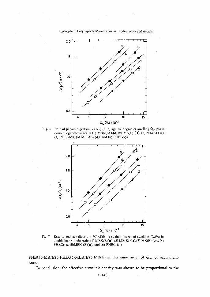
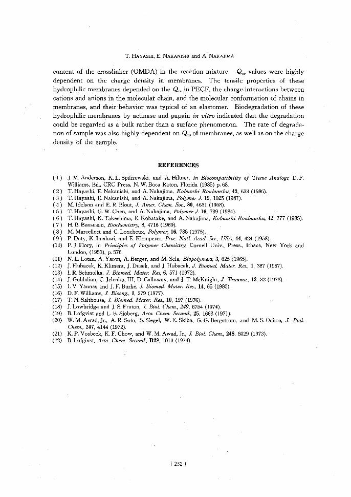
(E)-224 membrane should be a bulk rather than a surface phenomenon. Qualitative differences in the action of actinase and papain are clearly shown in Figure 5. The immediate and steady weight loss observed with actinase system is consistent with the release of monomers from the chain ends by exopeptidases. On the other hand, an endopeptidase, such as papain, must make two incisions in a chain segment if a soluble fragment is produced, but a single cleavage will decrease the effective crosslink density, resulting in increased swelling ratio of membranes. Thus, the initial effect of papain is therefore to decrease the effective crosslink density without producing soluble mate-rials. Figures 6 and 7 summarize the rate of papain and actinase digestion, V(1/2)(h-1), respectively, as a function of the degree of swelling Q.u, in PECF for hydrophilic
polymer membranes. V(1/2) is defined as the reciprocal of the time required for the sample weight to be reduced to one-half its initial value. It is clearly shown that the
order of degradation rates among the hydrophilic membranes is as follows: MBK(B)>
( 260 )
Hydrophilic Polypeptide Membranes as Biodegradable Materials
2.0 ' II---------------------------------------------------------------------------------------------------------------------------------------------------------I I I I I 1I 5 3
6 1 1.5 —
^4
• 2
1.0- •
• •
•
•
0.5 ——
4 5 710 15
Ow (%) x10-2
Fig. 6. Rate of papain digestion V(1/2) (h—') against degree of swelling Qw (%) in double logarithmic scale: (1) MBK(E) (•), (2) MB(E) (0), (3) MK(E) (0),
(4) PHEG(o), (5) MBK(B) (^), and (6) PHBG(0).
5 3 2.0 -1 —
6
4
1.5 —• • 2
L
•
•
0 I
4 5 7 10 15
Ow (MO x102
Fig. 7. Rate of actinase digestion V(1/2)(h—') against degree of swelling Q,„(%) in double logarithmic scale: (1) MBK(E)(•), (2) MB(E) (4),(3) MK(E) (0), (4)
PHEG(0), (5)MBK (B)(^), and (6) PHBG (0).
PHBG>MK(E)>PHEG>MBK(E)>MB(E) at the same order of Q. for each mem-
brane.
In conclusion, the effective crosslink density was shown to be proportional to the
( 261 )
T. HAYASHI, E. NAKANISHI and A. NAKAJIMA
content of the crosslinker (OMDA) in the reaction mixture. Qu, values were highly dependent on the charge density in membranes. The tensile properties of these hydrophilic membranes depended on the Qu, in PECF, the charge interactions between cations and anions in the molecular chain, and the molecular conformation of chains in membranes, and their behavior was typical of an elastomer. Biodegradation of these hydrophilic membranes by actinase and papain in vitro indicated that the degradation could be regarded as a bulk rather than a surface phenomenon. The rate of degrada-tion of sample was also highly dependent on Qu, of membranes, as well as on the charge density of the sample.
REFERENCES
(1) J. M. Anderson, K. L. Spilizewski, and A. Hiltner, in Biocompatibility of Tissue Analogs, D. F. Williams, Ed., CRC Press, N. W. Boca Raton, Florida (1985) p. 68.
(2) T. Hayashi, E. Nakanishi, and A. Nakajima, Kobunshi Ronbunshu, 43, 633 (1986). (3) T. Hayashi, E. Nakanishi, and A. Nakajima, Polymer J. 19, 1025 (1987). (4) M. Idelson and E. R. Blout, J. Amer. Chem. Soc., 80, 4631 (1958). (5) T. Hayashi, G. W. Chen, and A. Nakajima, Polymer J. 16, 739 (1984). (6) T. Hayashi, K. Takeshima, E. Kobatake, and A. Nakajima, Kobunshi Ronbunshu, 42, 777 (1985). (7) H. B. Bensusan, Biochemistry, 8, 4716 (1969). (8) M. Marcellect and C. Loucheux, Polymer, 16, 785 (1975). (9) P. Doty, K. Imahori, and E. Klemperer, Proc. NatL Acad. Sci., USA, 44, 424 (1958). (10) P. J. Flory, in Principles of Polymer Chemistry, Cornell Univ., Press., Ithaca, New York and
London, (1953), p. 576. (11) N. L. Lotan, A. Yaron, A. Berger, and M. Sela, Biopolymers, 3, 625 (1965). (12) J. Hubacek, K. Kliment, J. Dusek, and J. Hubacek, J. Biomed Mater. Res., 1, 387 (1967). (13) I. R. Schmolka, J. Biomed Mater. Res, 6, 571 (1972). (14) J. Guldalian, C. Jelenko, III, D. Calloway, and J. T. McKnight, J. Trauma., 13, 32 (1973). (15) I. V. Yannas and J. F. Burke, J. Biomed. Mater. Res., 14, 65 (1980). (16) D. F. Williams, J. Bioeng., 1, 279 (1977). (17) T. N. Salthouse, J. Biomed Mater. Res., 10, 197 (1976). (18) J. Lowbridge and J. S. Fruton, J. Biol Chem., 249, 6754 (1974). (19) B. Lofgvist and L. B. Sjoberg, Acta. Chem. Secand, 25, 1663 (1971). (20) W. M. Awad, Jr., A. R. Soto, S. Siegel, W. E. Skiba, G. G. Bergstrom, and M. S. Ochoa, J. BioL
Chem., 247, 4144 (1972). (21) K. P. Vosbeck, K. F. Chow, and W. M. Awad, Jr., J. Biol. Chem., 248, 6029 (1973). (22) B. Lofgivst, Acta. Chem. Secand, B28, 1013 (1974).
( 262 )
Recommended