review article
T h e n e w e ngl a nd j o u r na l o f m e dic i n e
n engl j med 369;9 nejm.org august 29, 2013840
Critical Care MedicineSimon R. Finfer, M.D., and Jean-Louis Vincent, M.D., Ph.D., Editors
Severe Sepsis and Septic ShockDerek C. Angus, M.D., M.P.H., and Tom van der Poll, M.D., Ph.D.
From the CRISMA (Clinical Research, Inves-tigation, and Systems Modeling of Acute Illness) Center, Department of Critical Care Medicine, University of Pittsburgh School of Medicine, Pittsburgh (D.C.A.); and the Center for Experimental and Mo-lecular Medicine, Division of Infectious Diseases, and Center for Infection and Immunity Amsterdam, Academic Medical Center, University of Amsterdam, Am-sterdam (T.P.). Address reprint requests to Dr. Angus at the Department of Criti-cal Care Medicine, University of Pitts-burgh, 614 Scaife Hall, 3550 Terrace St., Pittsburgh, PA 15261, or at [email protected]; or to Dr. van der Poll at the Division of Infectious Diseases, Academ-ic Medical Center, Meibergdreef 9, Rm. G2-130, 1105 AZ Amsterdam, the Nether-lands, or at [email protected].
N Engl J Med 2013;369:840-51.DOI: 10.1056/NEJMra1208623Copyright © 2013 Massachusetts Medical Society.
Sepsis is one of the oldest and most elusive syndromes in medicine. Hippocrates claimed that sepsis (σηψις) was the process by which flesh rots, swamps generate foul airs, and wounds fester.1 Galen later considered sepsis
a laudable event, necessary for wound healing.2 With the confirmation of germ theory by Semmelweis, Pasteur, and others, sepsis was recast as a systemic infec-tion, often described as “blood poisoning,” and assumed to be the result of the host’s invasion by pathogenic organisms that then spread in the bloodstream. However, with the advent of modern antibiotics, germ theory did not fully explain the pathogenesis of sepsis: many patients with sepsis died despite successful erad-ication of the inciting pathogen. Thus, researchers suggested that it was the host, not the germ, that drove the pathogenesis of sepsis.3
In 1992, an international consensus panel defined sepsis as a systemic inflam-matory response to infection, noting that sepsis could arise in response to mul-tiple infectious causes and that septicemia was neither a necessary condition nor a helpful term.4 Instead, the panel proposed the term “severe sepsis” to describe instances in which sepsis is complicated by acute organ dysfunction, and they codified “septic shock” as sepsis complicated by either hypotension that is refrac-tory to fluid resuscitation or by hyperlactatemia. In 2003, a second consensus panel endorsed most of these concepts, with the caveat that signs of a systemic inflammatory response, such as tachycardia or an elevated white-cell count, occur in many infectious and noninfectious conditions and therefore are not helpful in distinguishing sepsis from other conditions.5 Thus, “severe sepsis” and “sepsis” are sometimes used interchangeably to describe the syndrome of infection com-plicated by acute organ dysfunction.
Incidence a nd C auses
The incidence of severe sepsis depends on how acute organ dysfunction is defined and on whether that dysfunction is attributed to an underlying infection. Organ dysfunction is often defined by the provision of supportive therapy (e.g., mechani-cal ventilation), and epidemiologic studies thus count the “treated incidence” rath-er than the actual incidence. In the United States, severe sepsis is recorded in 2% of patients admitted to the hospital. Of these patients, half are treated in the intensive care unit (ICU), representing 10% of all ICU admissions.6,7 The number of cases in the United States exceeds 750,000 per year7 and was recently reported to be rising.8 However, several factors — new International Classification of Diseases, 9th Revision (ICD-9) coding rules, confusion over the distinction between septicemia and severe sepsis, the increasing capacity to provide intensive care, and increased awareness and surveillance — confound the interpretation of temporal trends.
Studies from other high-income countries show similar rates of sepsis in the ICU.9 The incidence of severe sepsis outside modern ICUs, especially in parts of
The New England Journal of Medicine Downloaded from nejm.org by LISA HUZEL on September 4, 2013. For personal use only. No other uses without permission.
Copyright © 2013 Massachusetts Medical Society. All rights reserved.
critical care medicine
n engl j med 369;9 nejm.org august 29, 2013 841
the world in which ICU care is scarce, is largely unknown. Extrapolating from treated incidence rates in the United States, Adhikari et al. estimated up to 19 million cases worldwide per year.10 The true incidence is presumably far higher.
Severe sepsis occurs as a result of both com-munity-acquired and health care–associated in-fections. Pneumonia is the most common cause, accounting for about half of all cases, followed by intraabdominal and urinary tract infections.7,8,11,12 Blood cultures are typically positive in only one third of cases, and in up to a third of cases, cultures from all sites are negative.7,11,13,14 Staphy-lococcus aureus and Streptococcus pneumoniae are the most common gram-positive isolates, whereas Escherichia coli, klebsiella species, and Pseudomonas aeruginosa predominate among gram-negative iso-lates.11,14 An epidemiologic study of sepsis showed that during the period from 1979 to 2000, gram-positive infections overtook gram-negative infections.15 However, in a more recent study involving 14,000 ICU patients in 75 coun-tries, gram-negative bacteria were isolated in 62% of patients with severe sepsis who had positive cultures, gram-positive bacteria in 47%, and fungi in 19%.12
Risk factors for severe sepsis are related both to a patient’s predisposition for infection and to the likelihood of acute organ dysfunction if in-fection develops. There are many well-known risk factors for the infections that most commonly precipitate severe sepsis and septic shock, includ-ing chronic diseases (e.g., the acquired immuno-deficiency syndrome, chronic obstructive pul-monary disease, and many cancers) and the use of immunosuppressive agents.7 Among patients with such infections, however, the risk factors for organ dysfunction are less well studied but probably include the causative organism and the patient’s genetic composition, underlying health status, and preexisting organ function, along with the timeliness of therapeutic intervention.16 Age, sex, and race or ethnic group all influence the incidence of severe sepsis, which is higher in infants and elderly persons than in other age groups, higher in males than in females, and higher in blacks than in whites.7,17
There is considerable interest in the contribu-tion of host genetic characteristics to the inci-dence and outcome of sepsis, in part because of strong evidence of inherited risk factors.18 Many studies have focused on polymorphisms in genes
encoding proteins implicated in the pathogene-sis of sepsis, including cytokines and other me-diators involved in innate immunity, coagula-tion, and fibrinolysis. However, findings are often inconsistent, owing at least in part to the heterogeneity of the patient populations stud-ied.19,20 Although a recent genomewide associa-tion study21 explored drug responsiveness in sepsis, no such large-scale studies of susceptibil-ity to or outcome of sepsis have been performed.
Clinic a l Fe at ur es
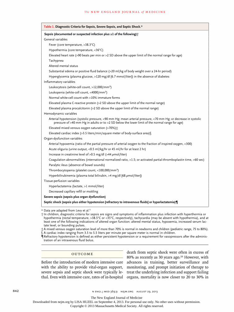
The clinical manifestations of sepsis are highly variable, depending on the initial site of infec-tion, the causative organism, the pattern of acute organ dysfunction, the underlying health status of the patient, and the interval before initiation of treatment. The signs of both infection and or-gan dysfunction may be subtle, and thus the most recent international consensus guidelines provide a long list of warning signs of incipient sepsis (Table 1).5 Acute organ dysfunction most commonly affects the respiratory and cardiovas-cular systems. Respiratory compromise is classi-cally manifested as the acute respiratory distress syndrome (ARDS), which is defined as hypox-emia with bilateral infiltrates of noncardiac ori-gin.22 Cardiovascular compromise is manifested primarily as hypotension or an elevated serum lactate level. After adequate volume expansion, hypotension frequently persists, requiring the use of vasopressors, and myocardial dysfunction may occur.23
The brain and kidneys are also often affected. Central nervous system dysfunction is typically manifested as obtundation or delirium. Imaging studies generally show no focal lesions, and findings on electroencephalography are usually consistent with nonfocal encephalopathy. Criti-cal illness polyneuropathy and myopathy are also common, especially in patients with a pro-longed ICU stay.24 Acute kidney injury is mani-fested as decreasing urine output and an in-creasing serum creatinine level and frequently requires treatment with renal-replacement ther-apy. Paralytic ileus, elevated aminotransferase levels, altered glycemic control, thrombocytope-nia and disseminated intravascular coagulation, adrenal dysfunction, and the euthyroid sick syn-drome are all common in patients with severe sepsis.5
The New England Journal of Medicine Downloaded from nejm.org by LISA HUZEL on September 4, 2013. For personal use only. No other uses without permission.
Copyright © 2013 Massachusetts Medical Society. All rights reserved.
T h e n e w e ngl a nd j o u r na l o f m e dic i n e
n engl j med 369;9 nejm.org august 29, 2013842
Ou t come
Before the introduction of modern intensive care with the ability to provide vital-organ support, severe sepsis and septic shock were typically le-thal. Even with intensive care, rates of in-hospital
death from septic shock were often in excess of 80% as recently as 30 years ago.25 However, with advances in training, better surveillance and monitoring, and prompt initiation of therapy to treat the underlying infection and support failing organs, mortality is now closer to 20 to 30% in
Table 1. Diagnostic Criteria for Sepsis, Severe Sepsis, and Septic Shock.*
Sepsis (documented or suspected infection plus ≥1 of the following)†
General variables
Fever (core temperature, >38.3°C)
Hypothermia (core temperature, <36°C)
Elevated heart rate (>90 beats per min or >2 SD above the upper limit of the normal range for age)
Tachypnea
Altered mental status
Substantial edema or positive fluid balance (>20 ml/kg of body weight over a 24-hr period)
Hyperglycemia (plasma glucose, >120 mg/dl [6.7 mmol/liter]) in the absence of diabetes
Inflammatory variables
Leukocytosis (white-cell count, >12,000/mm3)
Leukopenia (white-cell count, <4000/mm3)
Normal white-cell count with >10% immature forms
Elevated plasma C-reactive protein (>2 SD above the upper limit of the normal range)
Elevated plasma procalcitonin (>2 SD above the upper limit of the normal range)
Hemodynamic variables
Arterial hypotension (systolic pressure, <90 mm Hg; mean arterial pressure, <70 mm Hg; or decrease in systolic pressure of >40 mm Hg in adults or to >2 SD below the lower limit of the normal range for age)
Elevated mixed venous oxygen saturation (>70%)‡
Elevated cardiac index (>3.5 liters/min/square meter of body-surface area)§
Organ-dysfunction variables
Arterial hypoxemia (ratio of the partial pressure of arterial oxygen to the fraction of inspired oxygen, <300)
Acute oliguria (urine output, <0.5 ml/kg/hr or 45 ml/hr for at least 2 hr)
Increase in creatinine level of >0.5 mg/dl (>44 μmol/liter)
Coagulation abnormalities (international normalized ratio, >1.5; or activated partial-thromboplastin time, >60 sec)
Paralytic ileus (absence of bowel sounds)
Thrombocytopenia (platelet count, <100,000/mm3)
Hyperbilirubinemia (plasma total bilirubin, >4 mg/dl [68 μmol/liter])
Tissue-perfusion variables
Hyperlactatemia (lactate, >1 mmol/liter)
Decreased capillary refill or mottling
Severe sepsis (sepsis plus organ dysfunction)
Septic shock (sepsis plus either hypotension [refractory to intravenous fluids] or hyperlactatemia)¶
* Data are adapted from Levy et al.5
† In children, diagnostic criteria for sepsis are signs and symptoms of inflammation plus infection with hyperthermia or hypothermia (rectal temperature, >38.5°C or <35°C, respectively), tachycardia (may be absent with hypothermia), and at least one of the following indications of altered organ function: altered mental status, hypoxemia, increased serum lac-tate level, or bounding pulses.
‡ A mixed venous oxygen saturation level of more than 70% is normal in newborns and children (pediatric range, 75 to 80%).§ A cardiac index ranging from 3.5 to 5.5 liters per minute per square meter is normal in children.¶ Refractory hypotension is defined as either persistent hypotension or a requirement for vasopressors after the adminis-
tration of an intravenous fluid bolus.
The New England Journal of Medicine Downloaded from nejm.org by LISA HUZEL on September 4, 2013. For personal use only. No other uses without permission.
Copyright © 2013 Massachusetts Medical Society. All rights reserved.
critical care medicine
n engl j med 369;9 nejm.org august 29, 2013 843
many series.7,26 With decreasing death rates, at-tention has focused on the trajectory of recovery among survivors. Numerous studies have sug-gested that patients who survive to hospital dis-charge after sepsis remain at increased risk for death in the following months and years. Those who survive often have impaired physical or neu-rocognitive functioning, mood disorders, and a low quality of life.27 In most studies, determining the causal role of sepsis in such subsequent disor-ders has been difficult. However, a recent analy-sis of the Health and Retirement Study, involving a large, longitudinal cohort of aging Americans, suggested that severe sepsis significantly acceler-ated physical and neurocognitive decline.28
Pathoph ysiol o gy
Host Response
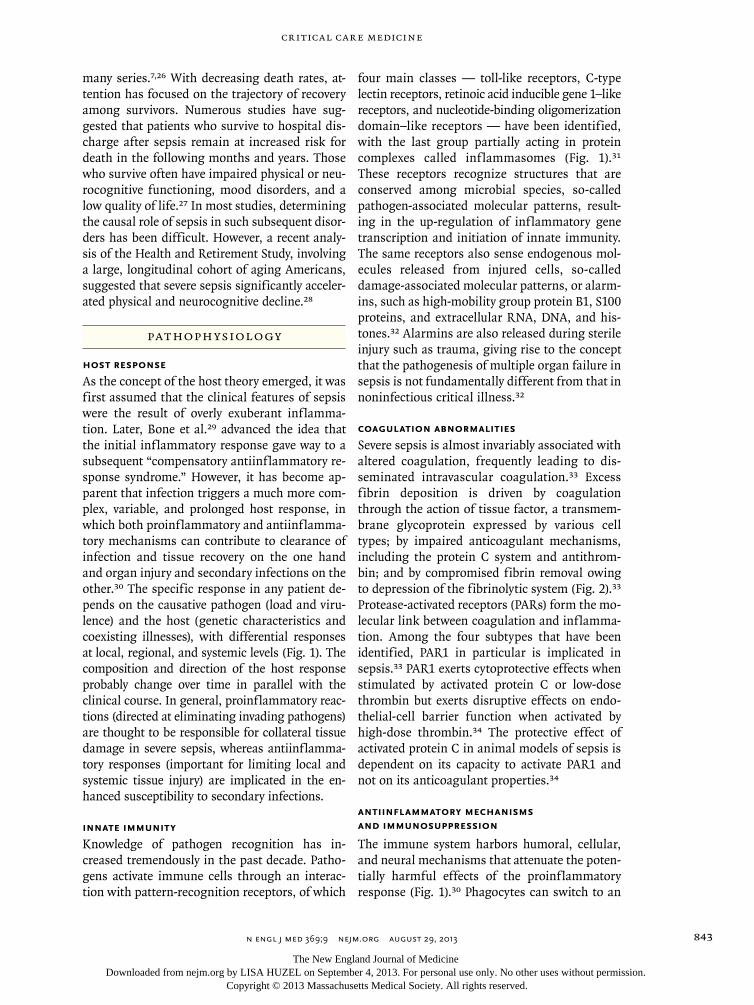
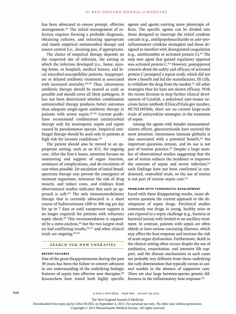
As the concept of the host theory emerged, it was first assumed that the clinical features of sepsis were the result of overly exuberant inflamma-tion. Later, Bone et al.29 advanced the idea that the initial inflammatory response gave way to a subsequent “compensatory antiinflammatory re-sponse syndrome.” However, it has become ap-parent that infection triggers a much more com-plex, variable, and prolonged host response, in which both proinflammatory and antiinflamma-tory mechanisms can contribute to clearance of infection and tissue recovery on the one hand and organ injury and secondary infections on the other.30 The specific response in any patient de-pends on the causative pathogen (load and viru-lence) and the host (genetic characteristics and coexisting illnesses), with differential responses at local, regional, and systemic levels (Fig. 1). The composition and direction of the host response probably change over time in parallel with the clinical course. In general, proinflammatory reac-tions (directed at eliminating invading pathogens) are thought to be responsible for collateral tissue damage in severe sepsis, whereas antiinflamma-tory responses (important for limiting local and systemic tissue injury) are implicated in the en-hanced susceptibility to secondary infections.
Innate Immunity
Knowledge of pathogen recognition has in-creased tremendously in the past decade. Patho-gens activate immune cells through an interac-tion with pattern-recognition receptors, of which
four main classes — toll-like receptors, C-type lectin receptors, retinoic acid inducible gene 1–like receptors, and nucleotide-binding oligomerization domain–like receptors — have been identified, with the last group partially acting in protein complexes called inflammasomes (Fig. 1).31 These receptors recognize structures that are conserved among microbial species, so-called pathogen-associated molecular patterns, result-ing in the up-regulation of inflammatory gene transcription and initiation of innate immunity. The same receptors also sense endogenous mol-ecules released from injured cells, so-called damage-associated molecular patterns, or alarm-ins, such as high-mobility group protein B1, S100 proteins, and extracellular RNA, DNA, and his-tones.32 Alarmins are also released during sterile injury such as trauma, giving rise to the concept that the pathogenesis of multiple organ failure in sepsis is not fundamentally different from that in noninfectious critical illness.32
Coagulation Abnormalities
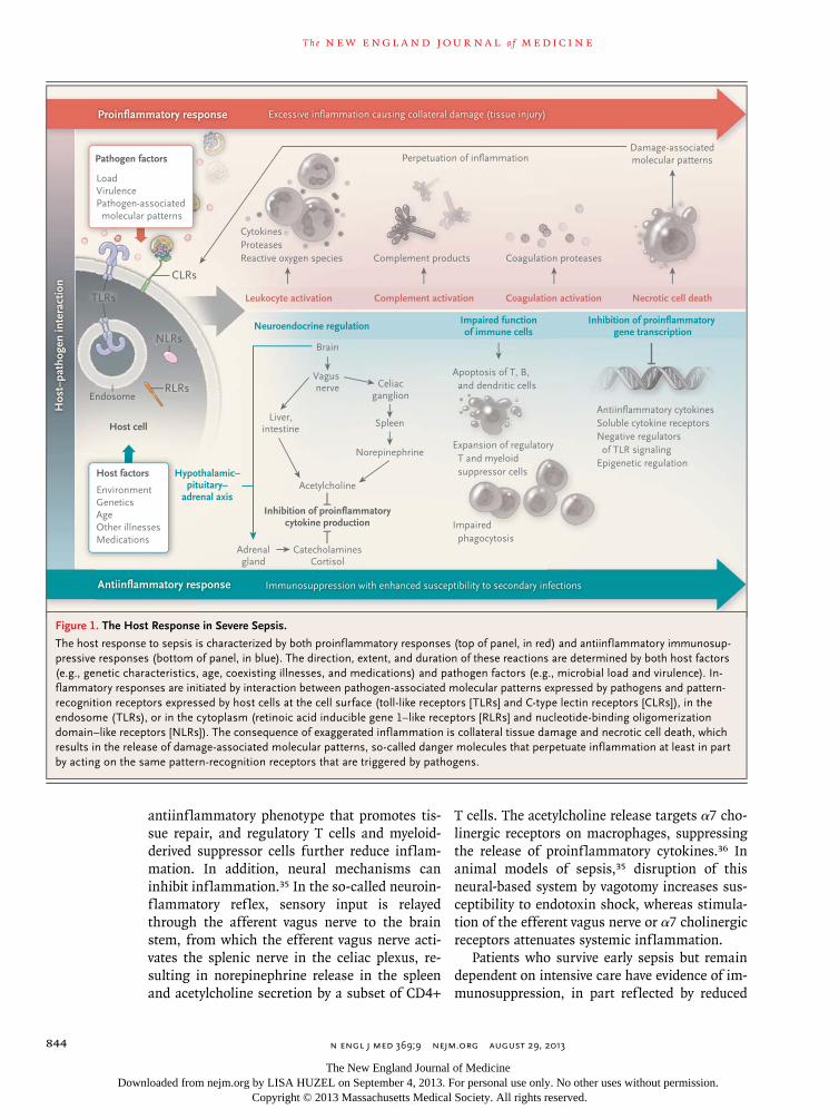
Severe sepsis is almost invariably associated with altered coagulation, frequently leading to dis-seminated intravascular coagulation.33 Excess fibrin deposition is driven by coagulation through the action of tissue factor, a transmem-brane glycoprotein expressed by various cell types; by impaired anticoagulant mechanisms, including the protein C system and antithrom-bin; and by compromised fibrin removal owing to depression of the fibrinolytic system (Fig. 2).33 Protease-activated receptors (PARs) form the mo-lecular link between coagulation and inflamma-tion. Among the four subtypes that have been identified, PAR1 in particular is implicated in sepsis.33 PAR1 exerts cytoprotective effects when stimulated by activated protein C or low-dose thrombin but exerts disruptive effects on endo-thelial-cell barrier function when activated by high-dose thrombin.34 The protective effect of activated protein C in animal models of sepsis is dependent on its capacity to activate PAR1 and not on its anticoagulant properties.34
Antiinflammatory Mechanisms and Immunosuppression
The immune system harbors humoral, cellular, and neural mechanisms that attenuate the poten-tially harmful effects of the proinflammatory response (Fig. 1).30 Phagocytes can switch to an
The New England Journal of Medicine Downloaded from nejm.org by LISA HUZEL on September 4, 2013. For personal use only. No other uses without permission.
Copyright © 2013 Massachusetts Medical Society. All rights reserved.
T h e n e w e ngl a nd j o u r na l o f m e dic i n e
n engl j med 369;9 nejm.org august 29, 2013844
antiinflammatory phenotype that promotes tis-sue repair, and regulatory T cells and myeloid-derived suppressor cells further reduce inflam-mation. In addition, neural mechanisms can inhibit inflammation.35 In the so-called neuroin-flammatory reflex, sensory input is relayed through the afferent vagus nerve to the brain stem, from which the efferent vagus nerve acti-vates the splenic nerve in the celiac plexus, re-sulting in norepinephrine release in the spleen and acetylcholine secretion by a subset of CD4+
T cells. The acetylcholine release targets α7 cho-linergic receptors on macrophages, suppressing the release of proinflammatory cytokines.36 In animal models of sepsis,35 disruption of this neural-based system by vagotomy increases sus-ceptibility to endotoxin shock, whereas stimula-tion of the efferent vagus nerve or α7 cholinergic receptors attenuates systemic inflammation.
Patients who survive early sepsis but remain dependent on intensive care have evidence of im-munosuppression, in part reflected by reduced
1
Drazen
8/29/13
8/09/13
AUTHOR PLEASE NOTE:Figure has been redrawn and type has been reset
Please check carefully
Author
Fig #
Title
ME
DEArtist
Issue date
COLOR FIGURE
Draft 6Angus
Knoper
Hos
t–pa
thog
en in
tera
ctio
n
Proinflammatory response Excessive inflammation causing collateral damage (tissue injury)
Antiinflammatory response
Pathogen factors
Host factors
EnvironmentGeneticsAgeOther illnessesMedications
Load VirulencePathogen-associated molecular patterns
Immunosuppression with enhanced susceptibility to secondary infections
CytokinesProteasesReactive oxygen species Complement products
Perpetuation of inflammation
Coagulation proteases
Damage-associatedmolecular patterns
Leukocyte activation
Neuroendocrine regulationImpaired functionof immune cells
Inhibition of proinflammatory gene transcription
Complement activation Coagulation activation Necrotic cell death
NLRs
RLRs
TLRs
CLRs
Vagus nerve
Apoptosis of T, B, and dendritic cells
Antiinflammatory cytokinesSoluble cytokine receptorsNegative regulators of TLR signalingEpigenetic regulation
Brain
Celiacganglion
Liver,intestine
Norepinephrine
Acetylcholine
Spleen
Adrenalgland
Inhibition of proinflammatorycytokine production
CatecholaminesCortisol
Hypothalamic–pituitary–
adrenal axis
Expansion of regulatory T and myeloid suppressor cells
Impaired phagocytosis
Endosome
Host cell
Figure 1. The Host Response in Severe Sepsis.
The host response to sepsis is characterized by both proinflammatory responses (top of panel, in red) and antiinflammatory immunosup-pressive responses (bottom of panel, in blue). The direction, extent, and duration of these reactions are determined by both host factors (e.g., genetic characteristics, age, coexisting illnesses, and medications) and pathogen factors (e.g., microbial load and virulence). In-flammatory responses are initiated by interaction between pathogen-associated molecular patterns expressed by pathogens and pattern-recognition receptors expressed by host cells at the cell surface (toll-like receptors [TLRs] and C-type lectin receptors [CLRs]), in the endosome (TLRs), or in the cytoplasm (retinoic acid inducible gene 1–like receptors [RLRs] and nucleotide-binding oligomerization domain–like receptors [NLRs]). The consequence of exaggerated inflammation is collateral tissue damage and necrotic cell death, which results in the release of damage-associated molecular patterns, so-called danger molecules that perpetuate inflammation at least in part by acting on the same pattern-recognition receptors that are triggered by pathogens.
The New England Journal of Medicine Downloaded from nejm.org by LISA HUZEL on September 4, 2013. For personal use only. No other uses without permission.
Copyright © 2013 Massachusetts Medical Society. All rights reserved.
critical care medicine
n engl j med 369;9 nejm.org august 29, 2013 845
expression of HLA-DR on myeloid cells.37 These patients frequently have ongoing infectious foci, despite antimicrobial therapy, or reactivation of latent viral infection.38,39 Multiple studies have documented reduced responsiveness of blood leukocytes to pathogens in patients with sep-sis,30 findings that were recently corroborated by postmortem studies revealing strong functional impairments of splenocytes obtained from pa-
tients who had died of sepsis in the ICU.37 Be-sides the spleen, the lungs also showed evidence of immunosuppression; both organs had en-hanced expression of ligands for T-cell inhibi-tory receptors on parenchymal cells.37 Enhanced apoptosis, especially of B cells, CD4+ T cells, and follicular dendritic cells, has been implicat-ed in sepsis-associated immunosuppression and death.40,41 Epigenetic regulation of gene expres-
2
Drazen
8/29/13
7/24/13
AUTHOR PLEASE NOTE:Figure has been redrawn and type has been reset
Please check carefully
Author
Fig #
Title
ME
DEArtist
Issue date
COLOR FIGURE
Draft 6Angus
Knoper
Mic
roci
rcul
atio
nTi
ssue
Release of mitochondrial
contents
Mitochondrialdysfunction
Increased coagulation Decreased anticoagulation
Monocyte
Neutrophil
NETs with trapped
platelets
Tissuefactor
↓ Antithrombin
Endothelial cell↓Tissue
factor pathway inhibitor ↓ TM ↓ Endothelial
protein C receptor
↓ Protein C
↓ Activatedprotein C
↓ Activated protein Cand ↑ thrombin
↓Fibrinolysis↑ PAI-1
Thrombosis
Tissue hypoperfusionLoss of
barrier function
↓Tissue oxygenation
Organ failure
↑PAR1
S1P3 S1P1
↑ S1P3 and ↓ S1P1
↑ Angiopoietin 2
↓ VE cadherin and↓Tight junctions
Cell shrinkageand cell death
Capillary leak and interstitial
edema
Vasodilatation
↓ Blood pressure
↓ Red-cell deformability
Thrombus
Tissue hypoperfusion Loss of barrier function
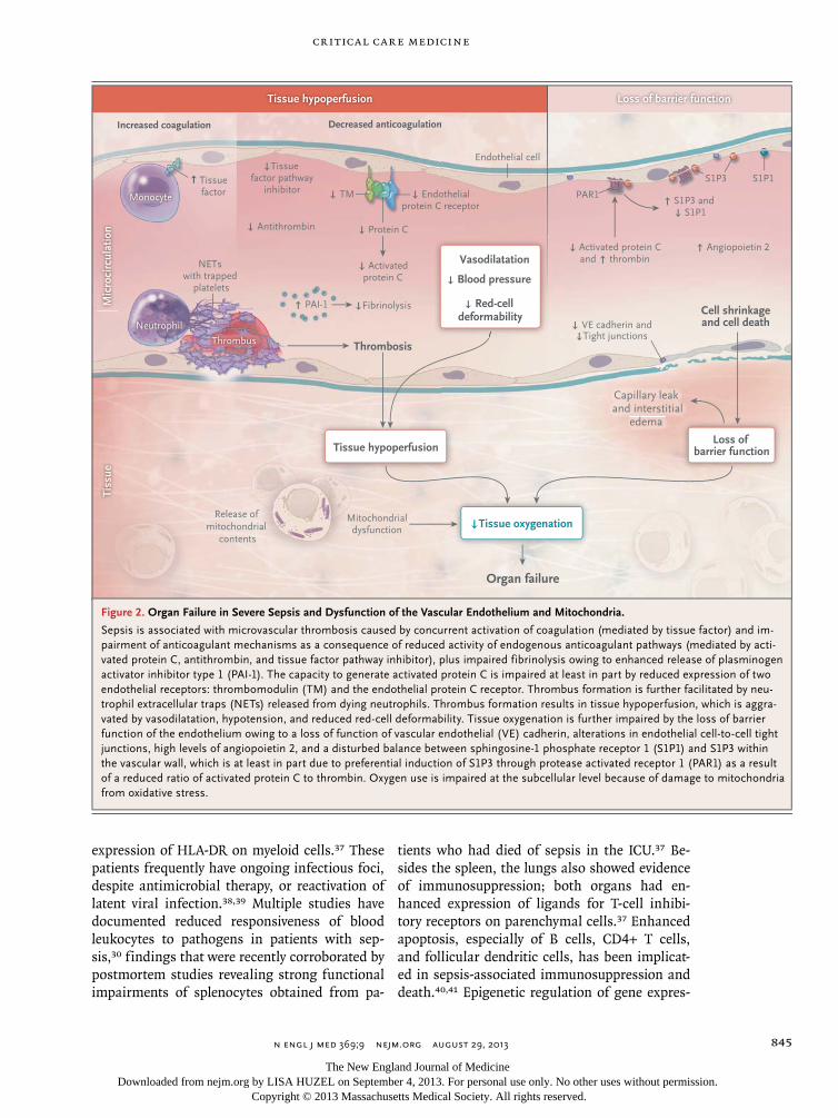
Figure 2. Organ Failure in Severe Sepsis and Dysfunction of the Vascular Endothelium and Mitochondria.
Sepsis is associated with microvascular thrombosis caused by concurrent activation of coagulation (mediated by tissue factor) and im-pairment of anticoagulant mechanisms as a consequence of reduced activity of endogenous anticoagulant pathways (mediated by acti-vated protein C, antithrombin, and tissue factor pathway inhibitor), plus impaired fibrinolysis owing to enhanced release of plasminogen activator inhibitor type 1 (PAI-1). The capacity to generate activated protein C is impaired at least in part by reduced expression of two endothelial receptors: thrombomodulin (TM) and the endothelial protein C receptor. Thrombus formation is further facilitated by neu-trophil extracellular traps (NETs) released from dying neutrophils. Thrombus formation results in tissue hypoperfusion, which is aggra-vated by vasodilatation, hypotension, and reduced red-cell deformability. Tissue oxygenation is further impaired by the loss of barrier function of the endothelium owing to a loss of function of vascular endothelial (VE) cadherin, alterations in endothelial cell-to-cell tight junctions, high levels of angiopoietin 2, and a disturbed balance between sphingosine-1 phosphate receptor 1 (S1P1) and S1P3 within the vascular wall, which is at least in part due to preferential induction of S1P3 through protease activated receptor 1 (PAR1) as a result of a reduced ratio of activated protein C to thrombin. Oxygen use is impaired at the subcellular level because of damage to mitochondria from oxidative stress.
The New England Journal of Medicine Downloaded from nejm.org by LISA HUZEL on September 4, 2013. For personal use only. No other uses without permission.
Copyright © 2013 Massachusetts Medical Society. All rights reserved.
T h e n e w e ngl a nd j o u r na l o f m e dic i n e
n engl j med 369;9 nejm.org august 29, 2013846
Tabl
e 2.
Gui
delin
es fo
r th
e Tr
eatm
ent o
f Sev
ere
Seps
is a
nd S
eptic
Sho
ck fr
om th
e Su
rviv
ing
Seps
is C
ampa
ign.
*
Elem
ent
of C
are
Gra
de†
Res
usci
tatio
n
Beg
in g
oal-d
irec
ted
resu
scita
tion
duri
ng fi
rst 6
hr
afte
r re
cogn
ition
1C
Beg
in in
itial
flui
d re
susc
itatio
n w
ith c
ryst
allo
id a
nd c
onsi
der
the
addi
tion
of a
lbum
in1B
Con
side
r th
e ad
ditio
n of
alb
umin
whe
n su
bsta
ntia
l am
ount
s of
cry
stal
loid
are
req
uire
d to
mai
ntai
n ad
equa
te a
rter
ial p
ress
ure
2C
Avo
id h
etas
tarc
h fo
rmul
atio
ns1C
Beg
in in
itial
flui
d ch
alle
nge
in p
atie
nts
with
tiss
ue h
ypop
erfu
sion
and
sus
pect
ed h
ypov
olem
ia, t
o ac
hiev
e ≥3
0 m
l of c
ryst
allo
ids
per
kilo
gram
of b
ody
wei
ght‡
1C
Con
tinue
flui
d-ch
alle
nge
tech
niqu
e as
long
as
ther
e is
hem
odyn
amic
impr
ovem
ent
1C
Use
nor
epin
ephr
ine
as th
e fir
st-c
hoic
e va
sopr
esso
r to
mai
ntai
n a
mea
n ar
teri
al p
ress
ure
of ≥
65 m
m H
g1B
Use
epi
neph
rine
whe
n an
add
ition
al a
gent
is n
eede
d to
mai
ntai
n ad
equa
te b
lood
pre
ssur
e2B
Add
vas
opre
ssin
(at
a d
ose
of 0
.03
units
/min
) w
ith w
eani
ng o
f nor
epin
ephr
ine,
if to
lera
ted
UG
Avo
id th
e us
e of
dop
amin
e ex
cept
in c
aref
ully
sel
ecte
d pa
tient
s (e
.g.,
patie
nts
with
a lo
w r
isk
of a
rrhy
thm
ias
and
eith
er k
now
n m
arke
d le
ft v
entr
icul
ar s
ysto
lic d
ys-
func
tion
or lo
w h
eart
rat
e)2C
Infu
se d
obut
amin
e or
add
it to
vas
opre
ssor
ther
apy
in th
e pr
esen
ce o
f myo
card
ial d
ysfu
nctio
n (e
.g.,
elev
ated
car
diac
filli
ng p
ress
ures
or
low
car
diac
out
put)
or
on-
goin
g hy
pope
rfus
ion
desp
ite a
dequ
ate
intr
avas
cula
r vo
lum
e an
d m
ean
arte
rial
pre
ssur
e1C
Avo
id th
e us
e of
intr
aven
ous
hydr
ocor
tison
e if
adeq
uate
flui
d re
susc
itatio
n an
d va
sopr
esso
r th
erap
y re
stor
e he
mod
ynam
ic s
tabi
lity;
if h
ydro
cort
ison
e is
use
d, a
d-m
inis
ter
at a
dos
e of
200
mg/
day
2C
Targ
et a
hem
oglo
bin
leve
l of 7
to 9
g/d
l in
patie
nts
with
out h
ypop
erfu
sion
, cri
tical
cor
onar
y ar
tery
dis
ease
or
myo
card
ial i
sche
mia
, or
acut
e he
mor
rhag
e1B
Infe
ctio
n co
ntro
l
Obt
ain
bloo
d cu
lture
s be
fore
ant
ibio
tic th
erap
y is
adm
inis
tere
d1C
Perf
orm
imag
ing
stud
ies
prom
ptly
to c
onfir
m s
ourc
e of
infe
ctio
nU
G
Adm
inis
ter
broa
d-sp
ectr
um a
ntib
iotic
ther
apy
with
in 1
hr
afte
r di
agno
sis
of e
ither
sev
ere
seps
is o
r se
ptic
sho
ck1B
/1C
Rea
sses
s an
tibio
tic th
erap
y da
ily fo
r de
-esc
alat
ion
whe
n ap
prop
riat
e1B
Perf
orm
sou
rce
cont
rol w
ith a
tten
tion
to r
isks
and
ben
efits
of t
he c
hose
n m
etho
d w
ithin
12
hr a
fter
dia
gnos
is1C
Res
pira
tory
sup
port
Use
a lo
w ti
dal v
olum
e an
d lim
itatio
n of
insp
irat
ory-
plat
eau-
pres
sure
str
ateg
y fo
r A
RD
S1A
/1B
App
ly a
min
imal
am
ount
of p
ositi
ve e
nd-e
xpir
ator
y pr
essu
re in
AR
DS
1B
Adm
inis
ter
high
er r
athe
r th
an lo
wer
pos
itive
end
-exp
irat
ory
pres
sure
for
patie
nts
with
sep
sis-
indu
ced
AR
DS
2C
Use
rec
ruitm
ent m
aneu
vers
in p
atie
nts
with
sev
ere
refr
acto
ry h
ypox
emia
due
to A
RD
S2C
Use
pro
ne p
ositi
onin
g in
pat
ient
s w
ith s
epsi
s-in
duce
d A
RD
S an
d a
ratio
of t
he p
artia
l pre
ssur
e of
art
eria
l oxy
gen
(mm
Hg)
to th
e fr
actio
n of
insp
ired
oxy
gen
of
<100
, in
faci
litie
s th
at h
ave
expe
rien
ce w
ith s
uch
prac
tice
2C
Elev
ate
the
head
of t
he b
ed in
pat
ient
s un
derg
oing
mec
hani
cal v
entil
atio
n, u
nles
s co
ntra
indi
cate
d1B
Use
a c
onse
rvat
ive
fluid
str
ateg
y fo
r es
tabl
ishe
d ac
ute
lung
inju
ry o
r A
RD
S w
ith n
o ev
iden
ce o
f tis
sue
hypo
perf
usio
n1C
Use
wea
ning
pro
toco
ls1A
The New England Journal of Medicine Downloaded from nejm.org by LISA HUZEL on September 4, 2013. For personal use only. No other uses without permission.
Copyright © 2013 Massachusetts Medical Society. All rights reserved.
critical care medicine
n engl j med 369;9 nejm.org august 29, 2013 847
sion may also contribute to sepsis-associated immunosuppression.42
Organ Dysfunction
Although the mechanisms that underlie organ failure in sepsis have been only partially eluci-dated, impaired tissue oxygenation plays a key role (Fig. 2). Several factors — including hypo-tension, reduced red-cell deformability, and microvascular thrombosis — contribute to dimin-ished oxygen delivery in septic shock. Inflamma-tion can cause dysfunction of the vascular endo-thelium, accompanied by cell death and loss of barrier integrity, giving rise to subcutaneous and body-cavity edema.43 In addition, mitochondrial damage caused by oxidative stress and other mech-anisms impairs cellular oxygen use.44 Moreover, injured mitochondria release alarmins into the extracellular environment, including mitochon-drial DNA and formyl peptides, which can acti-vate neutrophils and cause further tissue injury.45
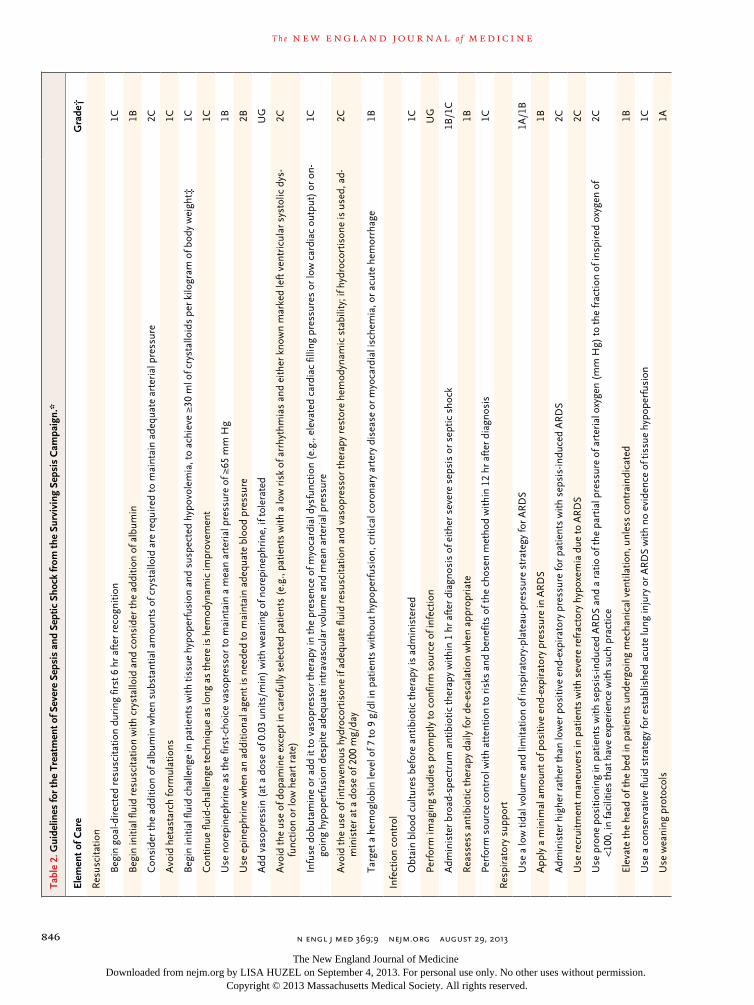
Tr e atmen t
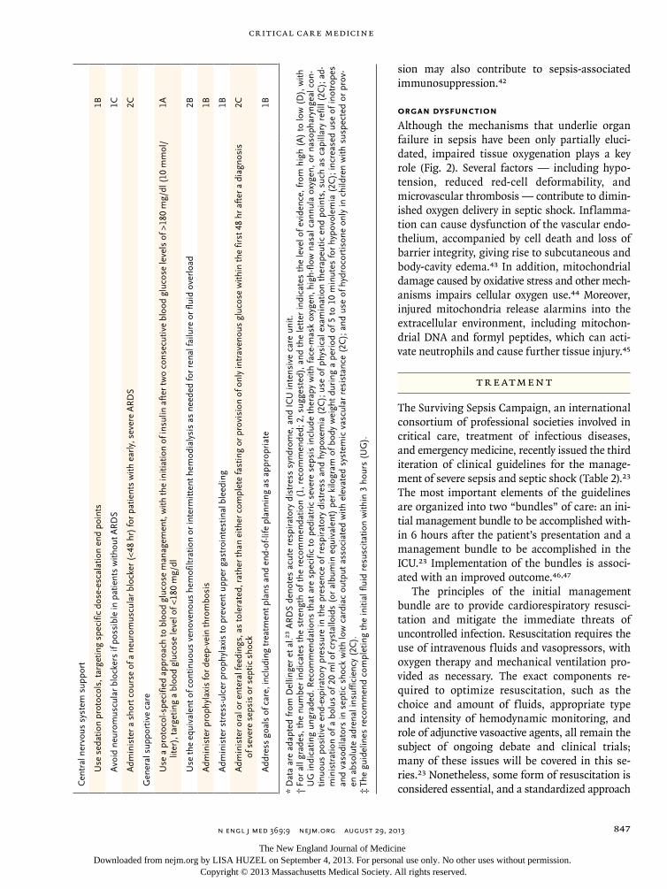
The Surviving Sepsis Campaign, an international consortium of professional societies involved in critical care, treatment of infectious diseases, and emergency medicine, recently issued the third iteration of clinical guidelines for the manage-ment of severe sepsis and septic shock (Table 2).23 The most important elements of the guidelines are organized into two “bundles” of care: an ini-tial management bundle to be accomplished with-in 6 hours after the patient’s presentation and a management bundle to be accomplished in the ICU.23 Implementation of the bundles is associ-ated with an improved outcome.46,47
The principles of the initial management bundle are to provide cardiorespiratory resusci-tation and mitigate the immediate threats of uncontrolled infection. Resuscitation requires the use of intravenous fluids and vasopressors, with oxygen therapy and mechanical ventilation pro-vided as necessary. The exact components re-quired to optimize resuscitation, such as the choice and amount of fluids, appropriate type and intensity of hemodynamic monitoring, and role of adjunctive vasoactive agents, all remain the subject of ongoing debate and clinical trials; many of these issues will be covered in this se-ries.23 Nonetheless, some form of resuscitation is considered essential, and a standardized approach C
entr
al n
ervo
us s
yste
m s
uppo
rt
Use
sed
atio
n pr
otoc
ols,
targ
etin
g sp
ecifi
c do
se-e
scal
atio
n en
d po
ints
1B
Avo
id n
euro
mus
cula
r bl
ocke
rs if
pos
sibl
e in
pat
ient
s w
ithou
t AR
DS
1C
Adm
inis
ter
a sh
ort c
ours
e of
a n
euro
mus
cula
r bl
ocke
r (<
48 h
r) fo
r pa
tient
s w
ith e
arly
, sev
ere
AR
DS
2C
Gen
eral
sup
port
ive
care
Use
a p
roto
col-s
peci
fied
appr
oach
to b
lood
glu
cose
man
agem
ent,
with
the
initi
atio
n of
insu
lin a
fter
two
cons
ecut
ive
bloo
d gl
ucos
e le
vels
of >
180
mg/
dl (
10 m
mol
/lit
er),
targ
etin
g a
bloo
d gl
ucos
e le
vel o
f <18
0 m
g/dl
1A
Use
the
equi
vale
nt o
f con
tinuo
us v
enov
enou
s he
mof
iltra
tion
or in
term
itten
t hem
odia
lysi
s as
nee
ded
for
rena
l fai
lure
or
fluid
ove
rloa
d2B
Adm
inis
ter
prop
hyla
xis
for
deep
-vei
n th
rom
bosi
s1B
Adm
inis
ter
stre
ss-u
lcer
pro
phyl
axis
to p
reve
nt u
pper
gas
troi
ntes
tinal
ble
edin
g1B
Adm
inis
ter
oral
or
ente
ral f
eedi
ngs,
as
tole
rate
d, r
athe
r th
an e
ither
com
plet
e fa
stin
g or
pro
visi
on o
f onl
y in
trav
enou
s gl
ucos
e w
ithin
the
first
48
hr a
fter
a d
iagn
osis
of
sev
ere
seps
is o
r se
ptic
sho
ck2C
Add
ress
goa
ls o
f car
e, in
clud
ing
trea
tmen
t pla
ns a
nd e
nd-o
f-life
pla
nnin
g as
app
ropr
iate
1B
* D
ata
are
adap
ted
from
Del
linge
r et
al.23
AR
DS
deno
tes
acut
e re
spir
ator
y di
stre
ss s
yndr
ome,
and
IC
U in
tens
ive
care
uni
t.†
For
all
grad
es, t
he n
umbe
r in
dica
tes
the
stre
ngth
of t
he r
ecom
men
datio
n (1
, rec
omm
ende
d; 2
, sug
gest
ed),
and
the
lett
er in
dica
tes
the
leve
l of e
vide
nce,
from
hig
h (A
) to
low
(D
), w
ith
UG
indi
catin
g un
grad
ed. R
ecom
men
datio
ns t
hat
are
spec
ific
to p
edia
tric
sev
ere
seps
is in
clud
e th
erap
y w
ith fa
ce-m
ask
oxyg
en, h
igh-
flow
nas
al c
annu
la o
xyge
n, o
r na
soph
aryn
geal
con
-tin
uous
pos
itive
end
-exp
irat
ory
pres
sure
in t
he p
rese
nce
of r
espi
rato
ry d
istr
ess
and
hypo
xem
ia (
2C);
use
of p
hysi
cal e
xam
inat
ion
ther
apeu
tic e
nd p
oint
s, s
uch
as c
apill
ary
refil
l (2C
); a
d-m
inis
trat
ion
of a
bol
us o
f 20
ml o
f cry
stal
loid
s (o
r al
bum
in e
quiv
alen
t) p
er k
ilogr
am o
f bod
y w
eigh
t du
ring
a p
erio
d of
5 t
o 10
min
utes
for
hypo
vole
mia
(2C
); in
crea
sed
use
of in
otro
pes
and
vaso
dila
tors
in s
eptic
sho
ck w
ith lo
w c
ardi
ac o
utpu
t as
soci
ated
with
ele
vate
d sy
stem
ic v
ascu
lar
resi
stan
ce (
2C);
and
use
of h
ydro
cort
ison
e on
ly in
chi
ldre
n w
ith s
uspe
cted
or
prov
-en
abs
olut
e ad
rena
l ins
uffic
ienc
y (2
C).
‡ T
he g
uide
lines
rec
omm
end
com
plet
ing
the
initi
al fl
uid
resu
scita
tion
with
in 3
hou
rs (
UG
).
The New England Journal of Medicine Downloaded from nejm.org by LISA HUZEL on September 4, 2013. For personal use only. No other uses without permission.
Copyright © 2013 Massachusetts Medical Society. All rights reserved.
T h e n e w e ngl a nd j o u r na l o f m e dic i n e
n engl j med 369;9 nejm.org august 29, 2013848
has been advocated to ensure prompt, effective management.23 The initial management of in-fection requires forming a probable diagnosis, obtaining cultures, and initiating appropriate and timely empirical antimicrobial therapy and source control (i.e., draining pus, if appropriate).
The choice of empirical therapy depends on the suspected site of infection, the setting in which the infection developed (i.e., home, nurs-ing home, or hospital), medical history, and lo-cal microbial-susceptibility patterns. Inappropri-ate or delayed antibiotic treatment is associated with increased mortality.48,49 Thus, intravenous antibiotic therapy should be started as early as possible and should cover all likely pathogens. It has not been determined whether combination antimicrobial therapy produces better outcomes than adequate single-agent antibiotic therapy in patients with severe sepsis.50-53 Current guide-lines recommend combination antimicrobial therapy only for neutropenic sepsis and sepsis caused by pseudomonas species. Empirical anti-fungal therapy should be used only in patients at high risk for invasive candidiasis.50
The patient should also be moved to an ap-propriate setting, such as an ICU, for ongoing care. After the first 6 hours, attention focuses on monitoring and support of organ function, avoidance of complications, and de-escalation of care when possible. De-escalation of initial broad-spectrum therapy may prevent the emergence of resistant organisms, minimize the risk of drug toxicity, and reduce costs, and evidence from observational studies indicates that such an ap-proach is safe.54 The only immunomodulatory therapy that is currently advocated is a short course of hydrocortisone (200 to 300 mg per day for up to 7 days or until vasopressor support is no longer required) for patients with refractory septic shock.23 This recommendation is support-ed by a meta-analysis,55 but the two largest stud-ies had conflicting results,56,57 and other clinical trials are ongoing.58,59
se a rch for ne w ther a pies
Recent Failures
One of the great disappointments during the past 30 years has been the failure to convert advances in our understanding of the underlying biologic features of sepsis into effective new therapies.60 Researchers have tested both highly specific
agents and agents exerting more pleiotropic ef-fects. The specific agents can be divided into those designed to interrupt the initial cytokine cascade (e.g., antilipopolysaccharide or anti–pro-inflammatory cytokine strategies) and those de-signed to interfere with dysregulated coagulation (e.g., antithrombin or activated protein C).61 The only new agent that gained regulatory approval was activated protein C.62 However, postapproval concern about the safety and efficacy of activated protein C prompted a repeat study, which did not show a benefit and led the manufacturer, Eli Lilly, to withdraw the drug from the market.11 All other strategies thus far have not shown efficacy. With the recent decision to stop further clinical devel-opment of CytoFab, a polyclonal anti–tumor ne-crosis factor antibody (ClinicalTrials.gov number, NCT01145560), there are no current large-scale trials of anticytokine strategies in the treatment of sepsis.
Among the agents with broader immunomod-ulatory effects, glucocorticoids have received the most attention. Intravenous immune globulin is also associated with a potential benefit,63 but important questions remain, and its use is not part of routine practice.23 Despite a large num-ber of observational studies suggesting that the use of statins reduces the incidence or improves the outcome of sepsis and severe infection,64 such findings have not been confirmed in ran-domized, controlled trials, so the use of statins is not part of routine sepsis care.23
PROBLEMS WITH therapeutic development
Faced with these disappointing results, many ob-servers question the current approach to the de-velopment of sepsis drugs. Preclinical studies commonly test drugs in young, healthy mice or rats exposed to a septic challenge (e.g., bacteria or bacterial toxins) with limited or no ancillary treat-ment. In contrast, patients with sepsis are often elderly or have serious coexisting illnesses, which may affect the host response and increase the risk of acute organ dysfunction. Furthermore, death in the clinical setting often occurs despite the use of antibiotics, resuscitation, and intensive life sup-port, and the disease mechanisms in such cases are probably very different from those underlying the early deterioration that typically occurs in ani-mal models in the absence of supportive care. There are also large between-species genetic dif-ferences in the inflammatory host response.65
The New England Journal of Medicine Downloaded from nejm.org by LISA HUZEL on September 4, 2013. For personal use only. No other uses without permission.
Copyright © 2013 Massachusetts Medical Society. All rights reserved.

critical care medicine
n engl j med 369;9 nejm.org august 29, 2013 849
In clinical studies, the enrollment criteria are typically very broad, the agent is administered on the basis of a standard formula for only a short period, there is little information on how the agent changes the host response and host–pathogen interactions, and the primary end point is death from any cause. Such a research strategy is prob-ably overly simplistic in that it does not select pa-tients who are most likely to benefit, cannot adjust therapy on the basis of the evolving host response and clinical course, and does not capture poten-tially important effects on nonfatal outcomes.
NEW STRATEGIES
Consequently, hope is pinned on newer so-called precision-medicine strategies with better preclin-ical models, more targeted drug development, and clinical trials that incorporate better patient selection, drug delivery, and outcome measure-ment. For example, options to enrich the pre-clinical portfolio include the study of animals that are more genetically diverse, are older, or have preexisting disease. Longer experiments with more advanced supportive care would allow better mimicry of the later stages of sepsis and multiorgan failure, permitting the testing of drugs in a more realistic setting and perhaps fa-cilitating the measurement of outcomes such as cognitive and physical functioning. In addition, preclinical studies could be used to screen for potential biomarkers of a therapeutic response for which there are human homologues.
Activated protein C mutants that lack antico-agulant properties are examples of more target-ed drug development and were shown to provide protection from sepsis-induced death in animals, without an increased risk of bleeding.66 Bio-markers such as whole-genome expression pat-terns in peripheral-blood leukocytes may aid in stratifying patients into more homogeneous sub-groups or in developing more targeted therapeu-tic interventions.67 The insight that severe sepsis can cause immunosuppression raises the possi-bility of using immune-stimulatory therapy (e.g., interleukin-7, granulocyte–macrophage colony-stimulating factor,68 or interferon-γ 69), but ide-ally, such therapy would be used only in patients in whom immunosuppression is identified or predicted. Thus, such therapies could be deployed on the basis of laboratory measures, such as monocyte HLA-DR expression. In addition, con-cern about accelerated neurocognitive decline in
survivors of sepsis opens up avenues to explore agents currently being tested in patients with dementia and related conditions.
The designs of trials could be modified to more easily incorporate these ideas. For exam-ple, the considerable uncertainty at the begin-ning of a trial with regard to the appropriate selection of patients and drug-administration strategy and the possibility of treatment inter-actions may be better handled with the use of a Bayesian design. A trial could commence with multiple study groups that reflect the various un-certainties to be tested but then automatically nar-row assignments to the best-performing groups on the basis of predefined-response adaptive randomization rules. Such designs could be par-ticularly helpful when testing combination ther-apy or incorporating potential biomarkers of drug responsiveness.
Conclusions
Severe sepsis and septic shock represent one of the oldest and most pressing problems in medi-cine. With advances in intensive care, increased awareness, and dissemination of evidence-based guidelines, clinicians have taken large strides in reducing the risk of imminent death associated with sepsis. However, as more patients survive sepsis, concern mounts over the lingering se-quelae of what was previously a lethal event. Strategies are also needed to reach the many mil-lions of patients with sepsis who are far from modern intensive care. At the same time, advanc-es in molecular biology have provided keen in-sight into the complexity of pathogen and alarm recognition by the human host and important clues to a host response that has gone awry. However, harnessing that information to provide effective new therapies has proved to be difficult. To further improve the outcome of patients with sepsis through the development of new therapeu-tic agents, newer, smarter approaches to clinical-trial design and execution are essential.
Dr. Angus reports receiving grant support through his insti-tution from Eisai, consulting fees from Idaho Technology, Pfizer, Eisai, MedImmune, BioAegis, and Ferring, and fees from Eli Lilly for serving as a member of a clinical-trial data and safety monitoring board. Dr. van der Poll reports receiving grant sup-port through his institution from Sirtris Pharmaceuticals and consulting fees from Eisai. No other potential conflict of inter-est relevant to this article was reported.
Disclosure forms provided by the authors are available with the full text of this article at NEJM.org.
The New England Journal of Medicine Downloaded from nejm.org by LISA HUZEL on September 4, 2013. For personal use only. No other uses without permission.
Copyright © 2013 Massachusetts Medical Society. All rights reserved.
T h e n e w e ngl a nd j o u r na l o f m e dic i n e
n engl j med 369;9 nejm.org august 29, 2013850
References
1. Majno G. The ancient riddle of sigma eta psi iota sigma (sepsis). J Infect Dis 1991;163:937-45.2. Funk DJ, Parrillo JE, Kumar A. Sepsis and septic shock: a history. Crit Care Clin 2009;25:83-101.3. Cerra FB. The systemic septic response: multiple systems organ failure. Crit Care Clin 1985;1:591-607.4. Bone RC, Sibbald WJ, Sprung CL. The ACCP-SCCM Consensus Conference on sepsis and organ failure. Chest 1992;101: 1481-3.5. Levy MM, Fink MP, Marshall JC, et al. 2001 SCCM/ESICM/ACCP/ATS/SIS Inter-national Sepsis Definitions Conference. Crit Care Med 2003;31:1250-6.6. Rangel-Frausto MS, Pittet D, Costigan M, Hwang T, Davis CS, Wenzel RP. The natural history of the systemic inflamma-tory response syndrome (SIRS): a prospec-tive study. JAMA 1995;273:117-23.7. Angus DC, Linde-Zwirble WT, Lidick-er J, Clermont G, Carcillo J, Pinsky MR. Epidemiology of severe sepsis in the Unit-ed States: analysis of incidence, outcome, and associated costs of care. Crit Care Med 2001;29:1303-10.8. Lagu T, Rothberg MB, Shieh MS, Pe-kow PS, Steingrub JS, Lindenauer PK. Hospitalizations, costs, and outcomes of severe sepsis in the United States 2003 to 2007. Crit Care Med 2012;40:754-6. [Erra-tum, Crit Care Med 2012;40:2932.]9. Linde-Zwirble WT, Angus DC. Severe sepsis epidemiology: sampling, selection, and society. Crit Care 2004;8:222-6.10. Adhikari NK, Fowler RA, Bhagwanjee S, Rubenfeld GD. Critical care and the global burden of critical illness in adults. Lancet 2010;376:1339-46.11. Ranieri VM, Thompson BT, Barie PS, et al. Drotrecogin alfa (activated) in adults with septic shock. N Engl J Med 2012; 366:2055-64.12. Vincent JL, Rello J, Marshall J, et al. International study of the prevalence and outcomes of infection in intensive care units. JAMA 2009;302:2323-9.13. Abraham E, Reinhart K, Opal S, et al. Efficacy and safety of tifacogin (recombi-nant tissue factor pathway inhibitor) in severe sepsis: a randomized controlled trial. JAMA 2003;290:238-47.14. Opal SM, Garber GE, LaRosa SP, et al. Systemic host responses in severe sepsis analyzed by causative microorganism and treatment effects of drotrecogin alfa (ac-tivated). Clin Infect Dis 2003;37:50-8.15. Martin GS, Mannino DM, Eaton S, Moss M. The epidemiology of sepsis in the United States from 1979 through 2000. N Engl J Med 2003;348:1546-54.16. Angus DC, Wax RS. Epidemiology of sepsis: an update. Crit Care Med 2001;29: Suppl:S109-S116.17. Mayr FB, Yende S, Linde-Zwirble WT,
et al. Infection rate and acute organ dys-function risk as explanations for racial differences in severe sepsis. JAMA 2010; 303:2495-503.18. Sørensen TI, Nielsen GG, Andersen PK, Teasdale TW. Genetic and environ-mental influences on premature death in adult adoptees. N Engl J Med 1988;318:727-32.19. Chung LP, Waterer GW. Genetic pre-disposition to respiratory infection and sepsis. Crit Rev Clin Lab Sci 2011;48:250-68.20. Namath A, Patterson AJ. Genetic polymorphisms in sepsis. Crit Care Nurs Clin North Am 2011;23:181-202.21. Man M, Close SL, Shaw AD, et al. Be-yond single-marker analyses: mining whole genome scans for insights into treatment responses in severe sepsis. Pharmacoge-nomics J 2012 February 7 (Epub ahead of print).22. Ranieri VM, Rubenfeld GD, Thomp-son BT, et al. Acute respiratory distress syndrome: the Berlin Definition. JAMA 2012;307:2526-33.23. Dellinger RP, Levy MM, Rhodes A, et al. Surviving Sepsis Campaign: interna-tional guidelines for management of se-vere sepsis and septic shock: 2012. Crit Care Med 2013;41:580-637.24. De Jonghe B, Sharshar T, Lefaucheur J, et al. Paresis acquired in the intensive care unit: a prospective multicenter study. JAMA 2002;288:2859-67.25. Friedman G, Silva E, Vincent JL. Has the mortality of septic shock changed with time? Crit Care Med 1998;26:2078-86.26. Kumar G, Kumar N, Taneja A, et al. Nationwide trends of severe sepsis in the 21st century (2000-2007). Chest 2011;140: 1223-31.27. Angus DC, Carlet J. Surviving inten-sive care: a report from the 2002 Brussels Roundtable. Intensive Care Med 2003;29: 368-77.28. Iwashyna TJ, Ely EW, Smith DM, Lan-ga KM. Long-term cognitive impairment and functional disability among survivors of severe sepsis. JAMA 2010;304:1787-94.29. Bone RC, Grodzin CJ, Balk RA. Sep-sis: a new hypothesis for pathogenesis of the disease process. Chest 1997;112:235-43.30. van der Poll T, Opal SM. Host-patho-gen interactions in sepsis. Lancet Infect Dis 2008;8:32-43.31. Takeuchi O, Akira S. Pattern recogni-tion receptors and inf lammation. Cell 2010;140:805-20.32. Chan JK, Roth J, Oppenheim JJ, et al. Alarmins: awaiting a clinical response. J Clin Invest 2012;122:2711-9.33. Levi M, van der Poll T. Inflammation and coagulation. Crit Care Med 2010;38: Suppl:S26-S34.
34. Ruf W. New players in the sepsis-pro-tective activated protein C pathway. J Clin Invest 2010;120:3084-7.35. Andersson U, Tracey KJ. Reflex prin-ciples of immunological homeostasis. Annu Rev Immunol 2012;30:313-35.36. Rosas-Ballina M, Olofsson PS, Ochani M, et al. Acetylcholine-synthesizing T cells relay neural signals in a vagus nerve cir-cuit. Science 2011;334:98-101.37. Boomer JS, To K, Chang KC, et al. Im-munosuppression in patients who die of sepsis and multiple organ failure. JAMA 2011;306:2594-605.38. Limaye AP, Kirby KA, Rubenfeld GD, et al. Cytomegalovirus reactivation in critically ill immunocompetent patients. JAMA 2008;300:413-22.39. Torgersen C, Moser P, Luckner G, et al. Macroscopic postmortem findings in 235 surgical intensive care patients with sepsis. Anesth Analg 2009;108:1841-7.40. Hotchkiss RS, Tinsley KW, Swanson PE, et al. Depletion of dendritic cells, but not macrophages, in patients with sepsis. J Immunol 2002;168:2493-500.41. Hotchkiss RS, Tinsley KW, Swanson PE, et al. Sepsis-induced apoptosis causes progressive profound depletion of B and CD4+ T lymphocytes in humans. J Immu-nol 2001;166:6952-63.42. Carson WF, Cavassani KA, Dou Y, Kunkel SL. Epigenetic regulation of im-mune cell functions during post-septic immunosuppression. Epigenetics 2011;6: 273-83.43. Goldenberg NM, Steinberg BE, Slutsky AS, Lee WL. Broken barriers: a new take on sepsis pathogenesis. Sci Transl Med 2011;3:88ps25.44. Galley HF. Oxidative stress and mito-chondrial dysfunction in sepsis. Br J An-aesth 2011;107:57-64.45. Zhang Q, Raoof M, Chen Y, et al. Cir-culating mitochondrial DAMPs cause in-flammatory responses to injury. Nature 2010;464:104-7.46. Ferrer R, Artigas A, Levy MM, et al. Improvement in process of care and out-come after a multicenter severe sepsis edu-cational program in Spain. JAMA 2008; 299:2294-303.47. Levy MM, Dellinger RP, Townsend SR, et al. The Surviving Sepsis Campaign: results of an international guideline-based performance improvement program targeting severe sepsis. Crit Care Med 2010;38:367-74.48. Paul M, Shani V, Muchtar E, Kariv G, Robenshtok E, Leibovici L. Systematic re-view and meta-analysis of the efficacy of appropriate empiric antibiotic therapy for sepsis. Antimicrob Agents Chemother 2010;54:4851-63.49. Kumar A, Roberts D, Wood KE, et al. Duration of hypotension before initiation of effective antimicrobial therapy is the
The New England Journal of Medicine Downloaded from nejm.org by LISA HUZEL on September 4, 2013. For personal use only. No other uses without permission.
Copyright © 2013 Massachusetts Medical Society. All rights reserved.
critical care medicine
n engl j med 369;9 nejm.org august 29, 2013 851
critical determinant of survival in human septic shock. Crit Care Med 2006;34:1589-96.50. Bochud PY, Bonten M, Marchetti O, Calandra T. Antimicrobial therapy for pa-tients with severe sepsis and septic shock: an evidence-based review. Crit Care Med 2004;32:S495-S512.51. Safdar N, Handelsman J, Maki DG. Does combination antimicrobial therapy reduce mortality in Gram-negative bacte-raemia? A meta-analysis. Lancet Infect Dis 2004;4:519-27.52. Brunkhorst FM, Oppert M, Marx G, et al. Effect of empirical treatment with moxifloxacin and meropenem vs merope-nem on sepsis-related organ dysfunction in patients with severe sepsis: a random-ized trial. JAMA 2012;307:2390-9.53. Paul M, Benuri-Silbiger I, Soares-Weiser K, Leibovici L. Beta lactam mono-therapy versus beta lactam-aminoglyco-side combination therapy for sepsis in immunocompetent patients: systematic review and meta-analysis of randomised trials. BMJ 2004;328:668. [Erratum, BMJ 2004;328:884.]54. Heenen S, Jacobs F, Vincent JL. Anti-biotic strategies in severe nosocomial sepsis: why do we not de-escalate more often? Crit Care Med 2012;40:1404-9.55. Annane D, Bellissant E, Bollaert PE, et al. Corticosteroids in the treatment of
severe sepsis and septic shock in adults: a systematic review. JAMA 2009;301:2362-75.56. Annane D, Sebille V, Charpentier C, et al. Effect of treatment with low doses of hydrocortisone and fludrocortisone on mortality in patients with septic shock. JAMA 2002;288:862-71.57. Sprung CL, Annane D, Keh D, et al. Hydrocortisone therapy for patients with septic shock. N Engl J Med 2008;358:111-24.58. ADjunctive coRticosteroid trEatment iN criticAlly ilL Patients With Septic Shock (ADRENAL). ClinicalTrials.gov, 2013 (http://clinicaltrials.gov/ct2/show/NCT01448109).59. Hydrocortisone for Prevention of Sep-tic Shock (HYPRESS). ClinicalTrials.gov, 2013 (http://www.clinicaltrials.gov/ct2/show/NCT00670254).60. Angus DC. The search for effective therapy for sepsis: back to the drawing board? JAMA 2011;306:2614-5.61. Webster NR, Galley HF. Immuno-modulation in the critically ill. Br J An-aesth 2009;103:70-81.62. Bernard GR, Vincent JL, Laterre PF, et al. Efficacy and safety of recombinant hu-man activated protein C for severe sepsis. N Engl J Med 2001;344:699-709.63. Laupland KB, Kirkpatrick AW, Delaney A. Polyclonal intravenous immunoglobu-lin for the treatment of severe sepsis and
septic shock in critically ill adults: a sys-tematic review and meta-analysis. Crit Care Med 2007;35:2686-92.64. Yende S, Milbrandt EB, Kellum JA, et al. Understanding the potential role of statins in pneumonia and sepsis. Crit Care Med 2011;39:1871-8.65. Seok J, Warren HS, Cuenca AG, et al. Genomic responses in mouse models poorly mimic human inflammatory dis-eases. Proc Natl Acad Sci U S A 2013;110: 3507-12.66. Kerschen EJ, Fernandez JA, Cooley BC, et al. Endotoxemia and sepsis mortal-ity reduction by non-anticoagulant acti-vated protein C. J Exp Med 2007;204:2439-48.67. Wong HR. Clinical review: sepsis and septic shock — the potential of gene ar-rays. Crit Care 2012;16:204.68. Meisel C, Schefold JC, Pschowski R, et al. Granulocyte-macrophage colony-stim-ulating factor to reverse sepsis-associated immunosuppression: a double-blind, ran-domized, placebo-controlled multicenter trial. Am J Respir Crit Care Med 2009; 180:640-8.69. Döcke WD, Randow F, Syrbe U, et al. Monocyte deactivation in septic patients: restoration by IFN-gamma treatment. Nat Med 1997;3:678-81.Copyright © 2013 Massachusetts Medical Society.
images in clinical medicine
The Journal welcomes consideration of new submissions for Images in Clinical Medicine. Instructions for authors and procedures for submissions can be found on the Journal’s website at NEJM.org. At the discretion of the editor, images that
are accepted for publication may appear in the print version of the Journal, the electronic version, or both.
The New England Journal of Medicine Downloaded from nejm.org by LISA HUZEL on September 4, 2013. For personal use only. No other uses without permission.
Copyright © 2013 Massachusetts Medical Society. All rights reserved.
Recommended