Study Guide• How do hormones regulate adenylyl cyclase
activity? PLC activity?• Describe the mechanism of regulation of PKA by
cAMP• Contrast diabetes mellitus type I and type II• Describe the architecture of insulin and the insulin
receptor• How does insulin activate the Raf-MEK-ERK
pathway?• How does glucagon produce hyperglycemia?• How does one treat diabetic hypoglycemia?
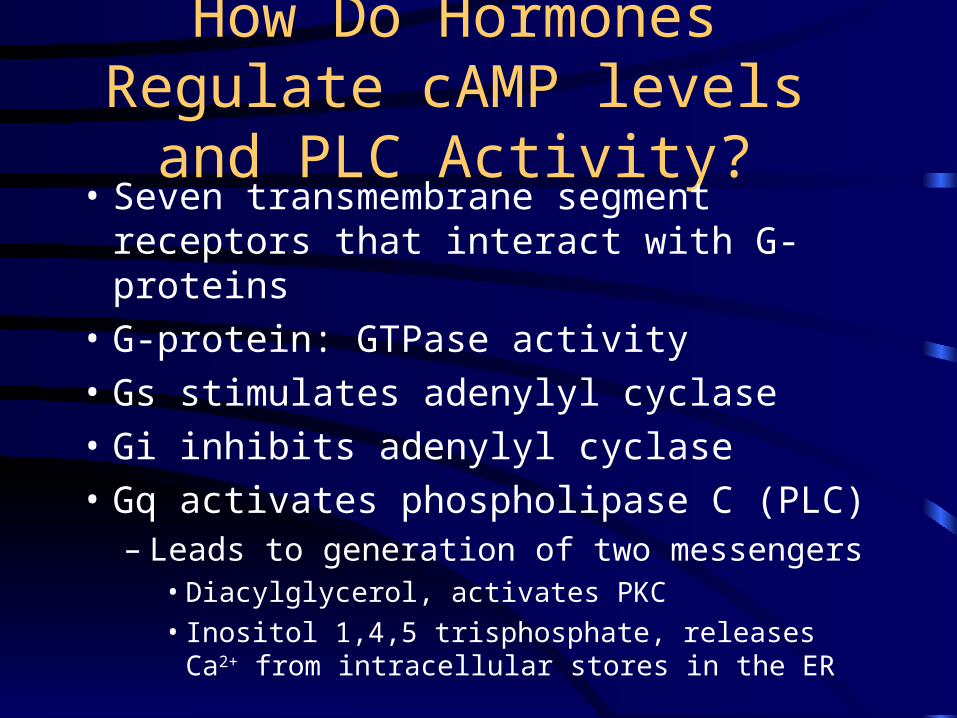
How Do Hormones Regulate cAMP levels and PLC Activity?• Seven transmembrane segment receptors
that interact with G-proteins
• G-protein: GTPase activity
• Gs stimulates adenylyl cyclase
• Gi inhibits adenylyl cyclase
• Gq activates phospholipase C (PLC)– Leads to generation of two messengers
• Diacylglycerol, activates PKC
• Inositol 1,4,5 trisphosphate, releases Ca2+ from intracellular stores in the ER
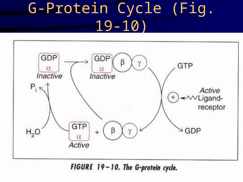
G-Protein Cycle (Fig. 19-10)
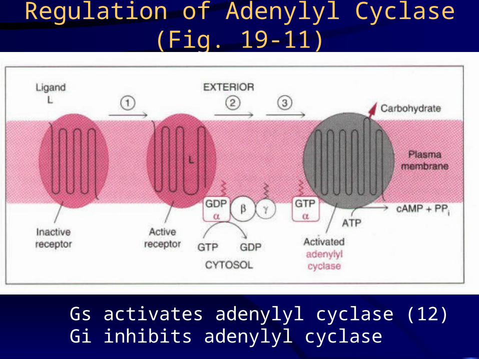
Regulation of Adenylyl Cyclase (Fig. 19-11)
Gs activates adenylyl cyclase (12)Gi inhibits adenylyl cyclase
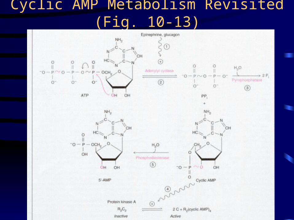
Cyclic AMP Metabolism Revisited (Fig. 10-13)
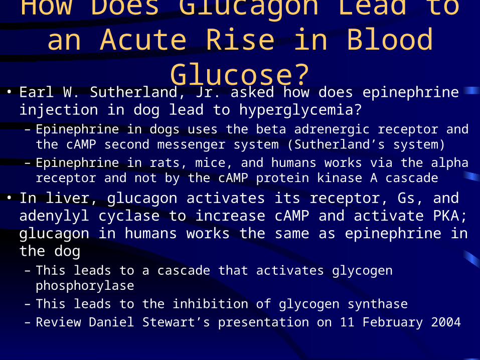
How Does Glucagon Lead to an Acute Rise in Blood Glucose?
• Earl W. Sutherland, Jr. asked how does epinephrine injection in dog lead to hyperglycemia?– Epinephrine in dogs uses the beta adrenergic receptor and the
cAMP second messenger system (Sutherland’s system)
– Epinephrine in rats, mice, and humans works via the alpha receptor and not by the cAMP protein kinase A cascade
• In liver, glucagon activates its receptor, Gs, and adenylyl cyclase to increase cAMP and activate PKA; glucagon in humans works the same as epinephrine in the dog– This leads to a cascade that activates glycogen phosphorylase
– This leads to the inhibition of glycogen synthase
– Review Daniel Stewart’s presentation on 11 February 2004
The Protein Kinase Reaction
• ATP + protein phosphoprotein + ADP
• PKA is a serine/threonine kinase
• It is a broad specificity enzyme with many substrates
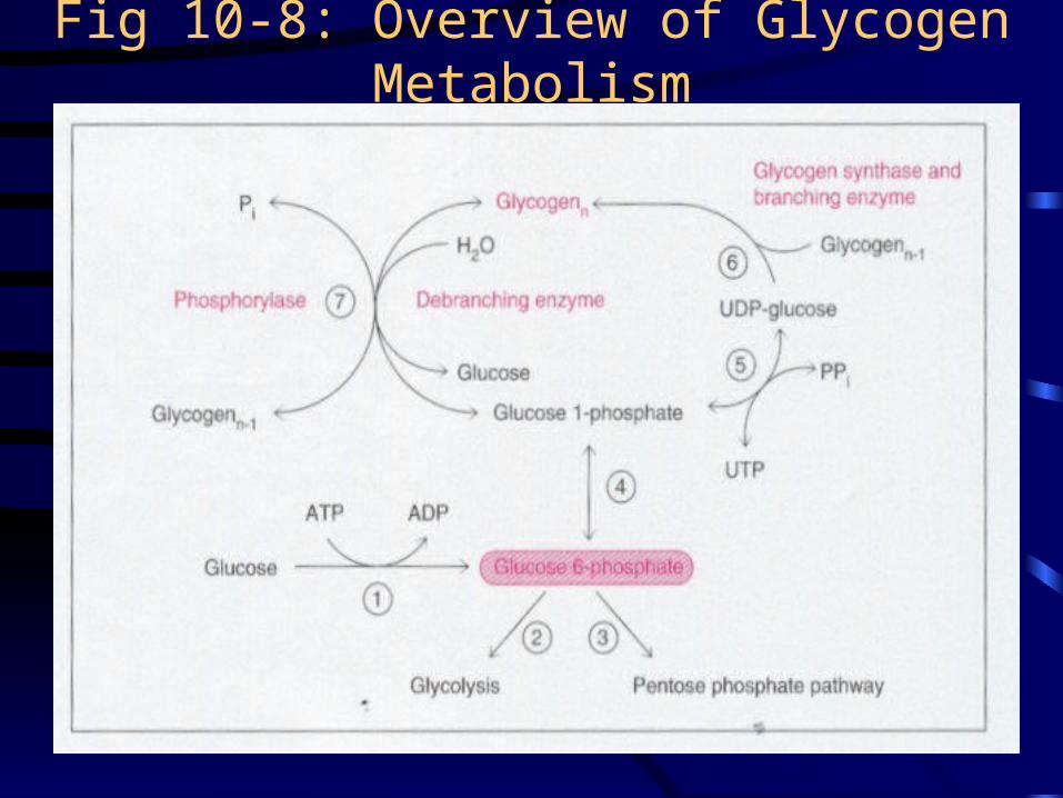
Fig 10-8: Overview of Glycogen Metabolism
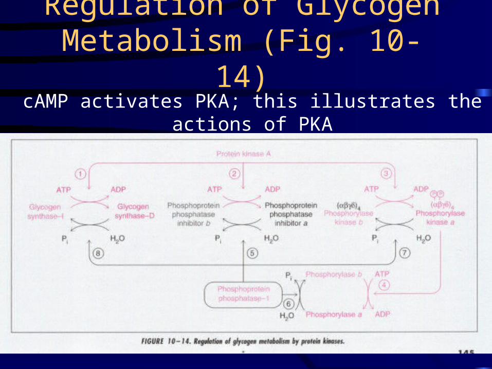
Regulation of Glycogen Metabolism (Fig. 10-14)
cAMP activates PKA; this illustrates the actions of PKA
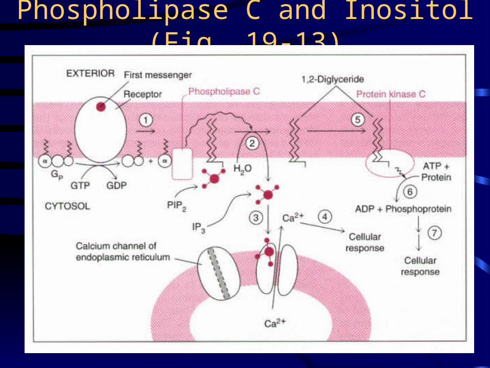
Phospholipase C and Inositol (Fig. 19-13)
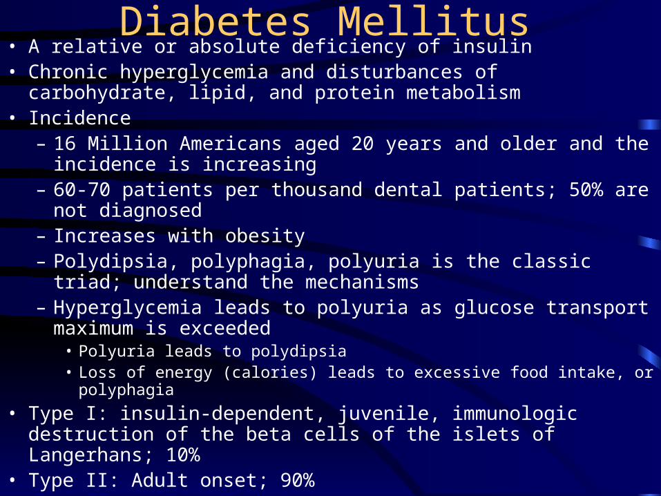
Diabetes Mellitus• A relative or absolute deficiency of insulin• Chronic hyperglycemia and disturbances of carbohydrate, lipid, and
protein metabolism• Incidence
– 16 Million Americans aged 20 years and older and the incidence is increasing
– 60-70 patients per thousand dental patients; 50% are not diagnosed– Increases with obesity– Polydipsia, polyphagia, polyuria is the classic triad; understand the
mechanisms– Hyperglycemia leads to polyuria as glucose transport maximum is
exceeded• Polyuria leads to polydipsia• Loss of energy (calories) leads to excessive food intake, or polyphagia
• Type I: insulin-dependent, juvenile, immunologic destruction of the beta cells of the islets of Langerhans; 10%
• Type II: Adult onset; 90%
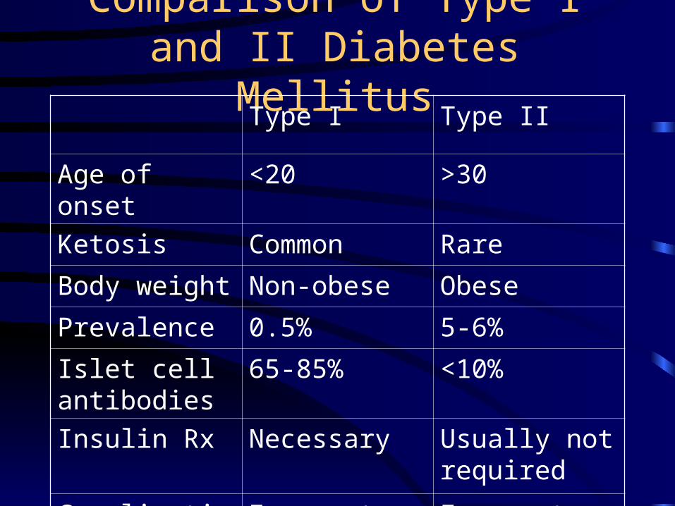
Comparison of Type I and II Diabetes Mellitus
Type I Type II
Age of onset <20 >30
Ketosis Common Rare
Body weight Non-obese Obese
Prevalence 0.5% 5-6%
Islet cell antibodies
65-85% <10%
Insulin Rx Necessary Usually not required
Complications Frequent Frequent
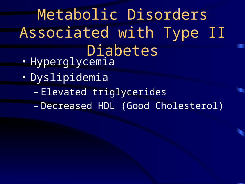
Metabolic Disorders Associated with Type II Diabetes
• Hyperglycemia
• Dyslipidemia– Elevated triglycerides– Decreased HDL (Good Cholesterol)
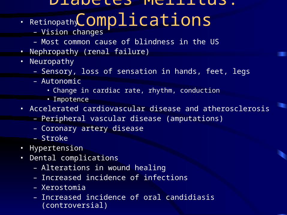
Diabetes Mellitus: Complications• Retinopathy
– Vision changes– Most common cause of blindness in the US
• Nephropathy (renal failure)• Neuropathy
– Sensory, loss of sensation in hands, feet, legs– Autonomic
• Change in cardiac rate, rhythm, conduction• Impotence
• Accelerated cardiovascular disease and atherosclerosis– Peripheral vascular disease (amputations)– Coronary artery disease– Stroke
• Hypertension• Dental complications
– Alterations in wound healing– Increased incidence of infections– Xerostomia– Increased incidence of oral candidiasis (controversial)
Diabetes and Periodontal Health • Risk factor for prevalence and severity of gingivitis and
periodontitis• Altered host defense secondary to diabetes may contribute• Increased collagen breakdown owing to increased collagenase
production• Not only does diabetes promote periodontal disease, but
periodontal disease can make the diabetes more difficult to control (any inflammatory flare up can increase insulin requirement)
• Possible findings in an undiagnosed diabetic– Severe, progressive periodontitis– Enlarged gingiva that bleed easily when manipulated– Multiple periodontal abscesses
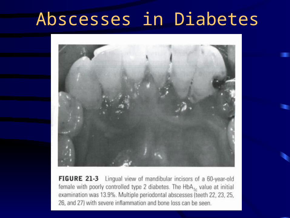
Abscesses in Diabetes
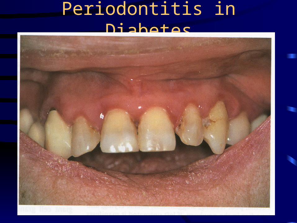
Periodontitis in Diabetes
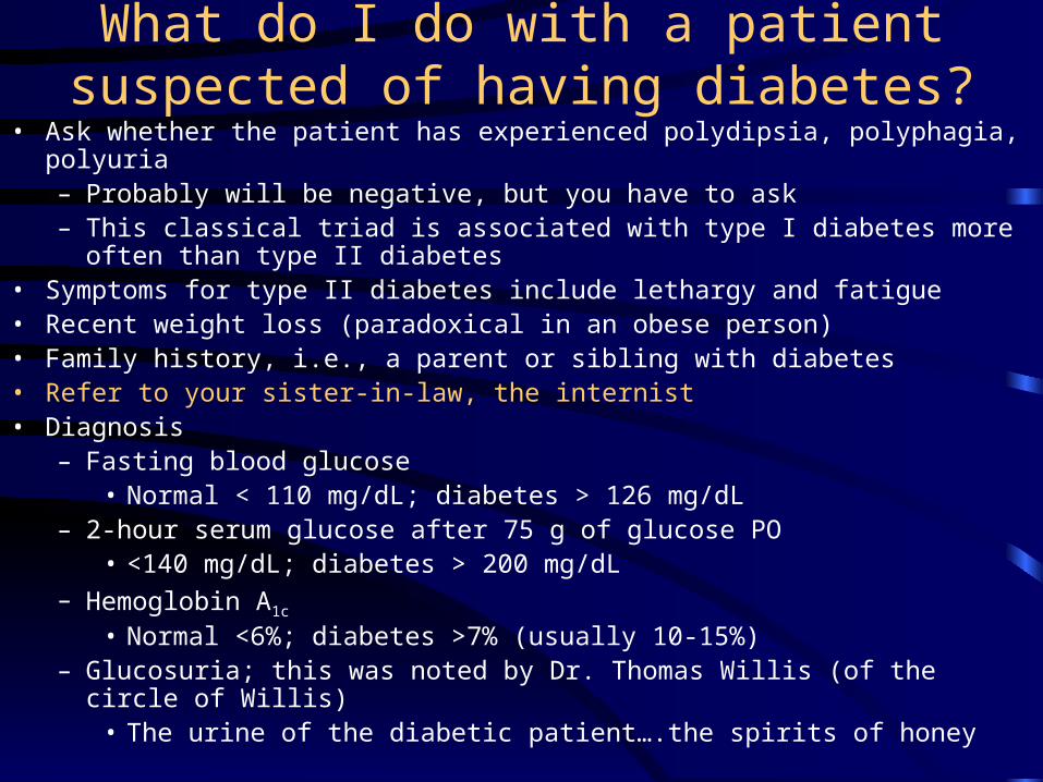
What do I do with a patient suspected of having diabetes?
• Ask whether the patient has experienced polydipsia, polyphagia, polyuria– Probably will be negative, but you have to ask – This classical triad is associated with type I diabetes more often than type II
diabetes • Symptoms for type II diabetes include lethargy and fatigue• Recent weight loss (paradoxical in an obese person)• Family history, i.e., a parent or sibling with diabetes• Refer to your sister-in-law, the internist• Diagnosis
– Fasting blood glucose• Normal < 110 mg/dL; diabetes > 126 mg/dL
– 2-hour serum glucose after 75 g of glucose PO• <140 mg/dL; diabetes > 200 mg/dL
– Hemoglobin A1c
• Normal <6%; diabetes >7% (usually 10-15%)– Glucosuria; this was noted by Dr. Thomas Willis (of the circle of Willis)
• The urine of the diabetic patient….the spirits of honey
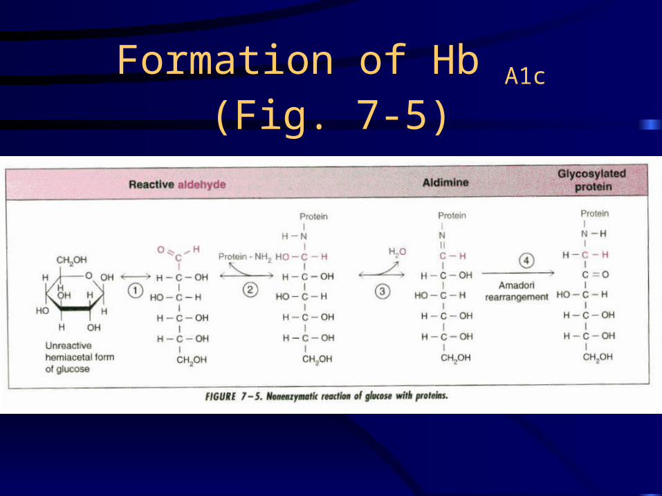
Formation of Hb A1c (Fig. 7-5)
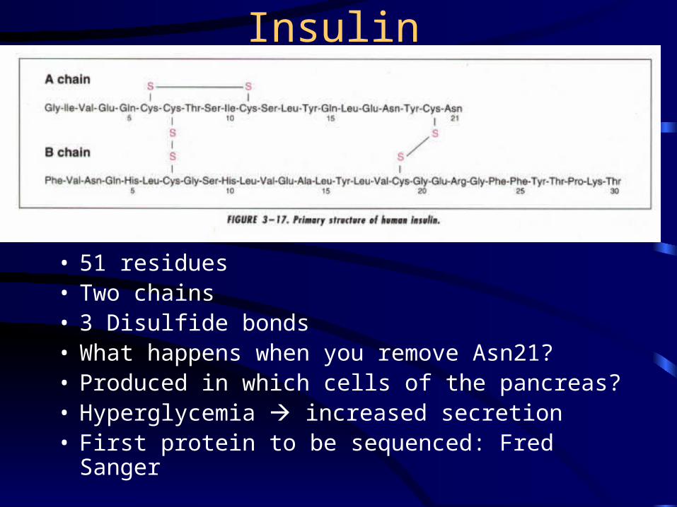
Insulin
• 51 residues• Two chains• 3 Disulfide bonds• What happens when you remove Asn21?• Produced in which cells of the pancreas?• Hyperglycemia increased secretion• First protein to be sequenced: Fred Sanger
Insulin Receptor Protein-Tyrosine Kinase• Insulin stimulates glucose uptake in muscle and fat,
glycogen synthesis, lipogenesis, and protein synthesis, and insulin inhibits lipolysis, proteolysis, and glycogenolysis
• Insulin receptor undergoes autophosphorylation and phosphorylates IRS1-4 (Insulin receptor substrates 1-4), PI3 kinase binding protein, and Shc
• Expressed in almost all cells, but at much higher levels in liver, fat, and muscle
• Insulin does not increase glucose transport into the liver
Protein-Tyrosine Kinase (PTK) Cascades
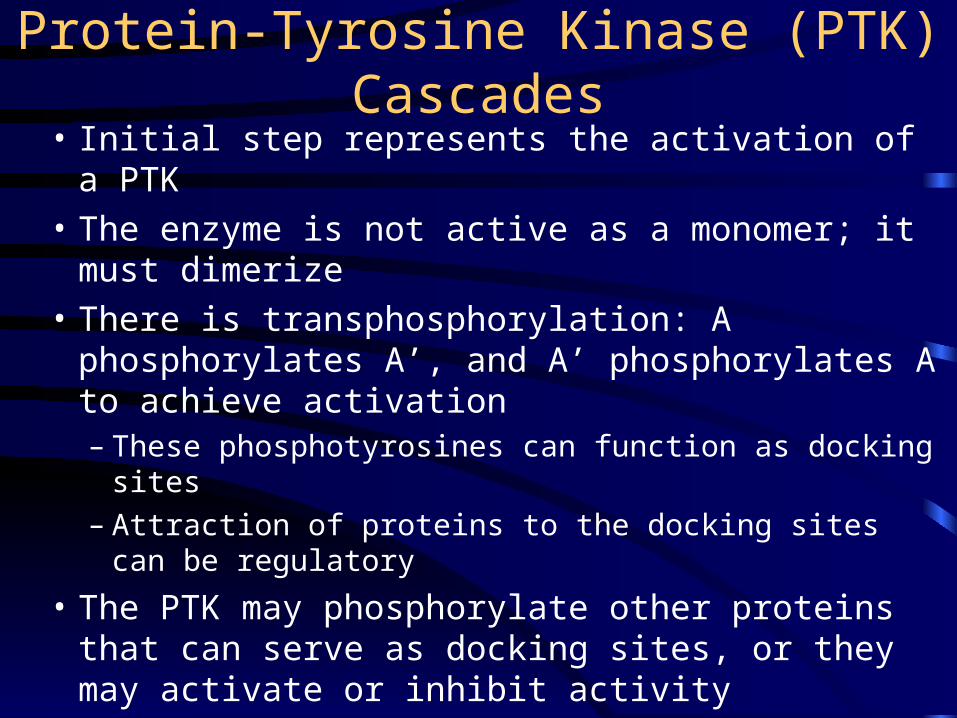
• Initial step represents the activation of a PTK
• The enzyme is not active as a monomer; it must dimerize
• There is transphosphorylation: A phosphorylates A’, and A’ phosphorylates A to achieve activation– These phosphotyrosines can function as docking sites– Attraction of proteins to the docking sites can be
regulatory
• The PTK may phosphorylate other proteins that can serve as docking sites, or they may activate or inhibit activity
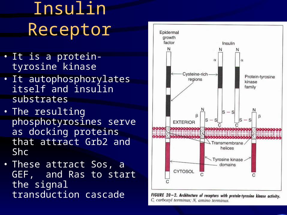
Insulin Receptor
• It is a protein-tyrosine kinase
• It autophosphorylates itself and insulin substrates
• The resulting phosphotyrosines serve as docking proteins that attract Grb2 and Shc
• These attract Sos, a GEF, and Ras to start the signal transduction cascade
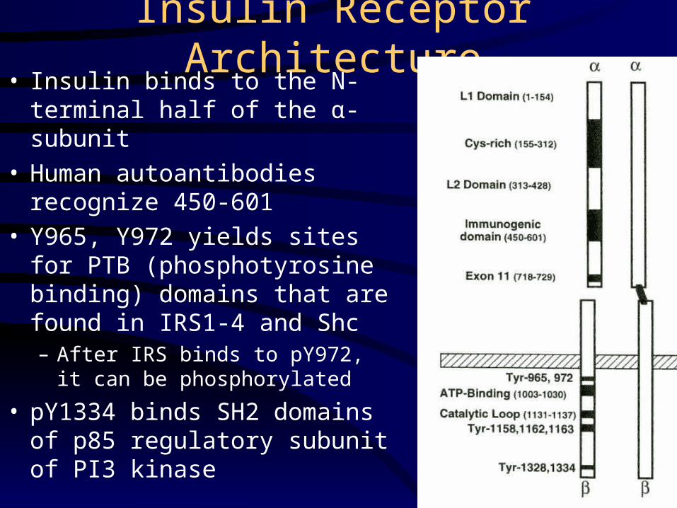
Insulin Receptor Architecture• Insulin binds to the N-terminal
half of the α-subunit• Human autoantibodies recognize
450-601• Y965, Y972 yields sites for PTB
(phosphotyrosine binding) domains that are found in IRS1-4 and Shc– After IRS binds to pY972, it can be
phosphorylated
• pY1334 binds SH2 domains of p85 regulatory subunit of PI3 kinase
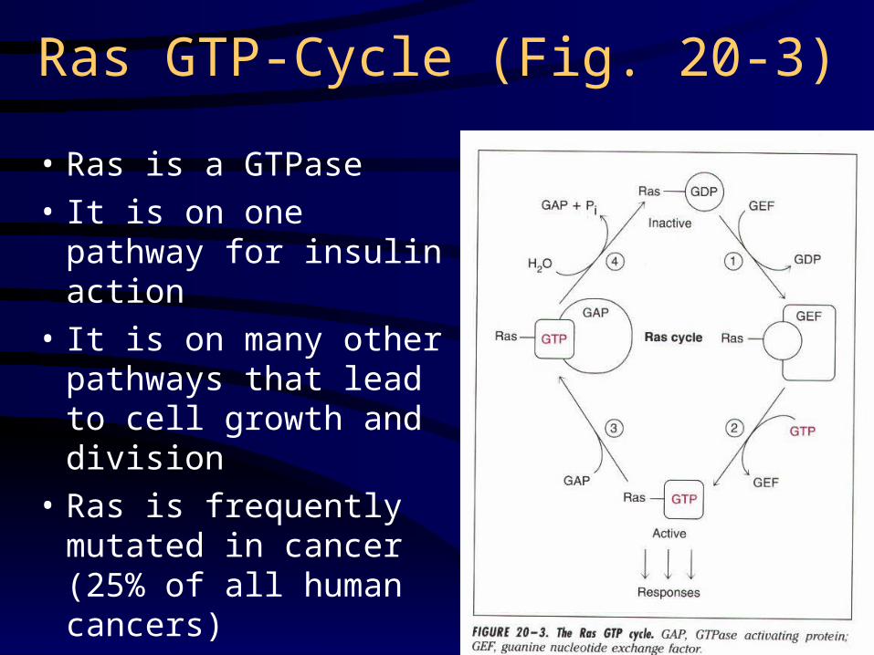
Ras GTP-Cycle (Fig. 20-3)
• Ras is a GTPase
• It is on one pathway for insulin action
• It is on many other pathways that lead to cell growth and division
• Ras is frequently mutated in cancer (25% of all human cancers)
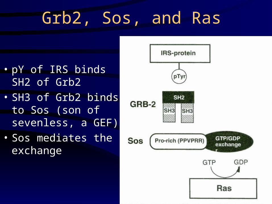
Grb2, Sos, and Ras
• pY of IRS binds SH2 of Grb2
• SH3 of Grb2 binds to Sos (son of sevenless, a GEF)
• Sos mediates the exchange
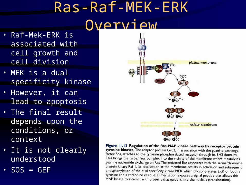
Ras-Raf-MEK-ERK Overview• Raf-Mek-ERK is
associated with cell growth and cell division
• MEK is a dual specificity kinase
• However, it can lead to apoptosis
• The final result depends upon the conditions, or context
• It is not clearly understood
• SOS = GEF
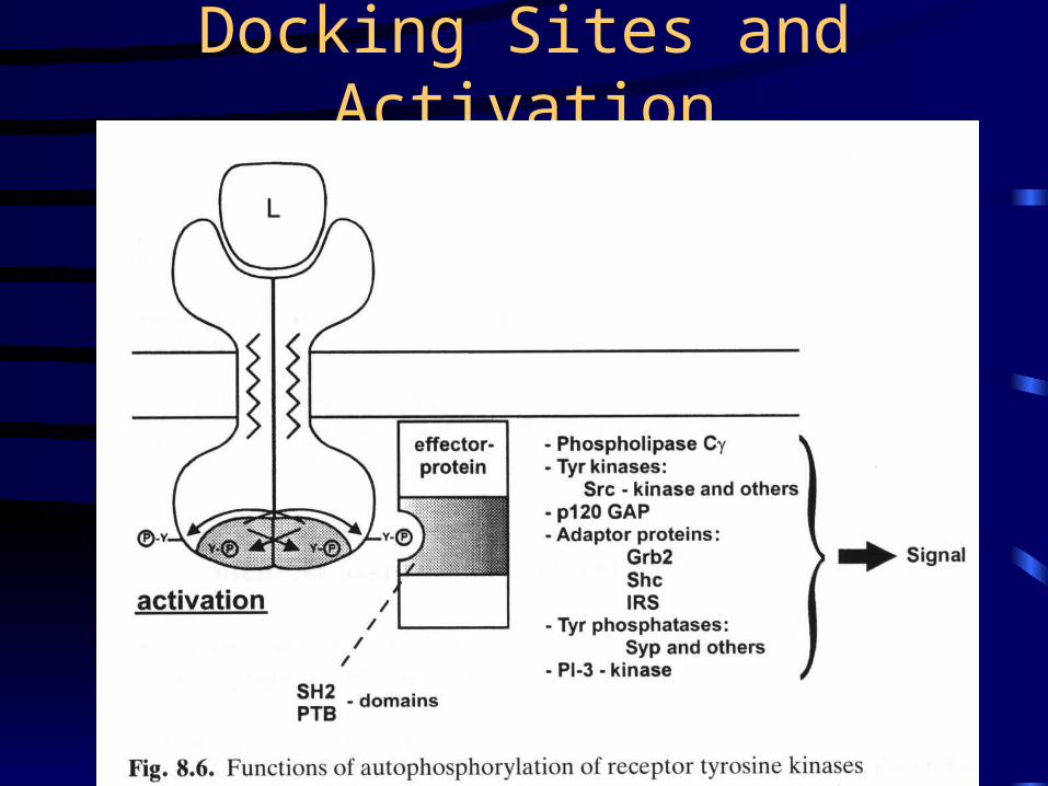
Docking Sites and Activation
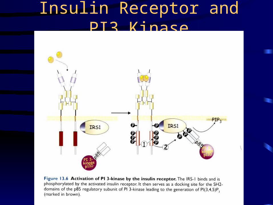
Insulin Receptor and PI3 Kinase
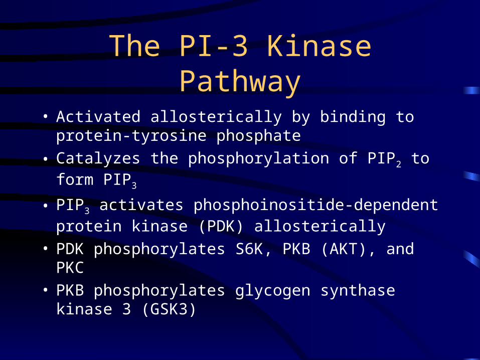
The PI-3 Kinase Pathway
• Activated allosterically by binding to protein-tyrosine phosphate
• Catalyzes the phosphorylation of PIP2 to form PIP3
• PIP3 activates phosphoinositide-dependent protein kinase (PDK) allosterically
• PDK phosphorylates S6K, PKB (AKT), and PKC• PKB phosphorylates glycogen synthase kinase 3
(GSK3)
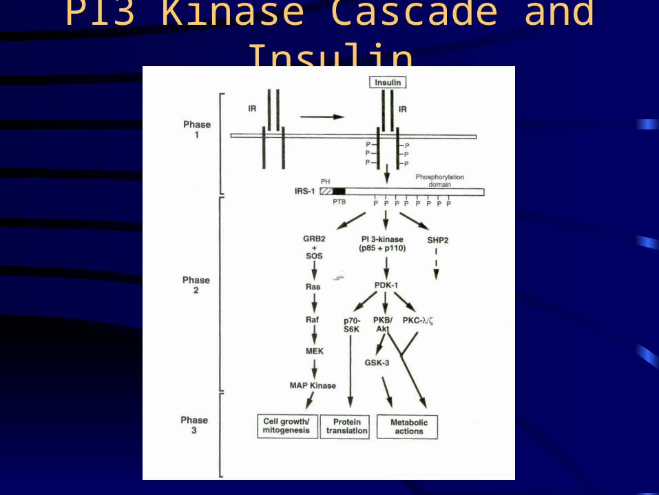
PI3 Kinase Cascade and Insulin
Phosphoprotein Phosphatase-1• Insulin stimulates glycogenesis in muscle, but epinephrine
stimulates glycogenolysis– Glycogenolyis (breakdown) is associated with
phosphorylation (the cascade)– Glycogenesis (build up) is associated with
dephosphorylation• Insulin promotes the dephosphorylation of glycogen
synthase and phosphorylase– These reactions are catalyzed by the catalytic subunit of
PPase-1– Insulin leads to the phosphorylation and activation of
PPase-1– Epinephrine leads to the phosphorylation and
inactivation of PPase-1
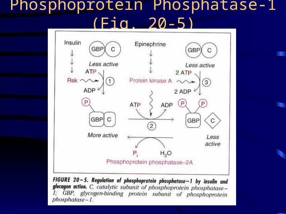
Phosphoprotein Phosphatase-1 (Fig. 20-5)
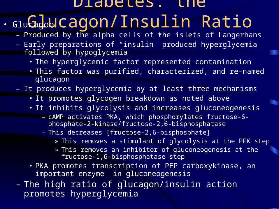
Diabetes: the Glucagon/Insulin Ratio• Glucagon
– Produced by the alpha cells of the islets of Langerhans– Early preparations of “insulin” produced hyperglycemia followed by
hypoglycemia• The hyperglycemic factor represented contamination• This factor was purified, characterized, and re-named glucagon
– It produces hyperglycemia by at least three mechanisms• It promotes glycogen breakdown as noted above• It inhibits glycolysis and increases gluconeogenesis
– cAMP activates PKA, which phosphorylates fructose-6-phosphate-2-kinase/fructose-2,6-bisphosphatase
– This decreases [fructose-2,6-bisphosphate]» This removes a stimulant of glycolysis at the PFK step» This removes an inhibitor of gluconeogenesis at the fructose-1,6-
bisphosphatase step
• PKA promotes transcription of PEP carboxykinase, an important enzyme in gluconeogenesis
– The high ratio of glucagon/insulin action promotes hyperglycemia
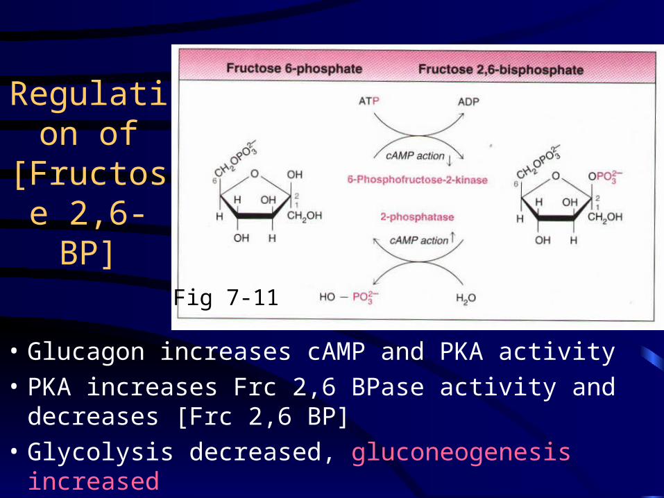
Regulation of [Fructose
2,6-BP]
• Glucagon increases cAMP and PKA activity
• PKA increases Frc 2,6 BPase activity and decreases [Frc 2,6 BP]
• Glycolysis decreased, gluconeogenesis increased
Fig 7-11
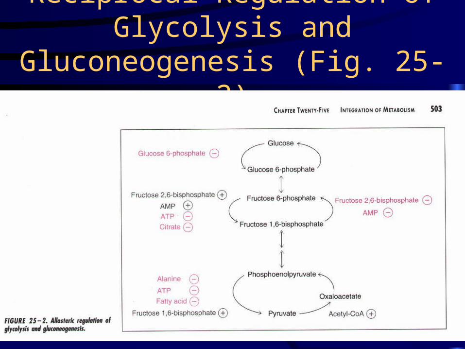
Reciprocal Regulation of Glycolysis and Gluconeogenesis (Fig. 25-2)
Insulin Action • Stimulates glucose transport into muscle, adipose
tissue, and many other cells EXCEPT liver– This results from the recruitment of GLUT4 (of
GLUT1-GLUT7)– Glucose transporters contains 12 transmembrane
segments– Mechanism of recruitment is unclear
• It does not rely on new transporter synthesis
• GLUT4 associated with internal membranes fuses with the plasma membrane
• Insulin promotes glycogen synthesis by inducing the production of glycogen synthase
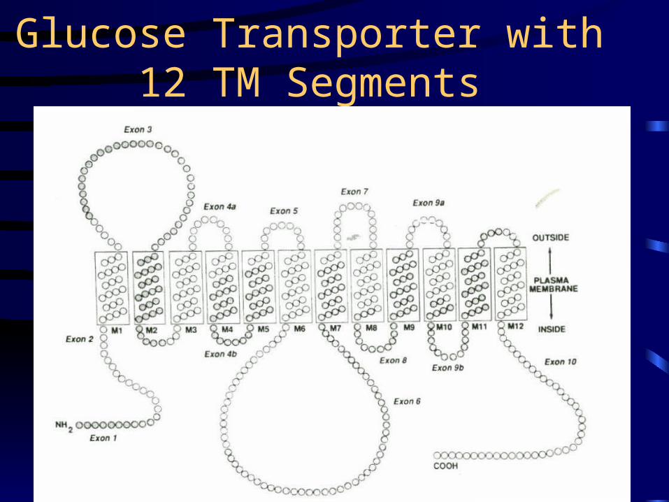
Glucose Transporter with 12 TM Segments
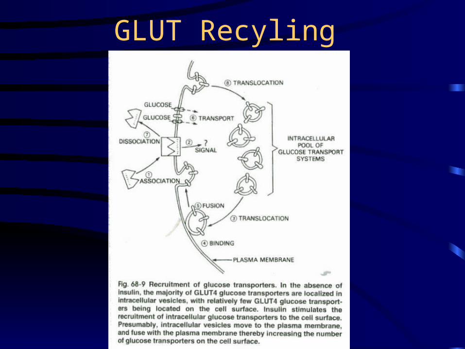
GLUT Recyling
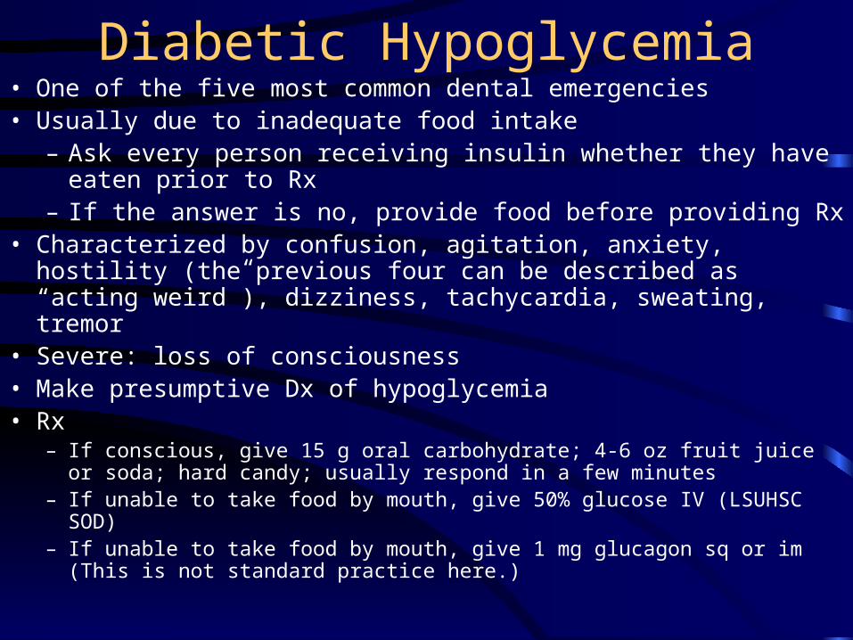
Diabetic Hypoglycemia• One of the five most common dental emergencies• Usually due to inadequate food intake
– Ask every person receiving insulin whether they have eaten prior to Rx
– If the answer is no, provide food before providing Rx• Characterized by confusion, agitation, anxiety, hostility (the previous
four can be described as “acting weird”), dizziness, tachycardia, sweating, tremor
• Severe: loss of consciousness• Make presumptive Dx of hypoglycemia• Rx
– If conscious, give 15 g oral carbohydrate; 4-6 oz fruit juice or soda; hard candy; usually respond in a few minutes
– If unable to take food by mouth, give 50% glucose IV (LSUHSC SOD)– If unable to take food by mouth, give 1 mg glucagon sq or im (This is not
standard practice here.)
Angiotensin System
• Renin, a proteolytic enzyme, is released from the juxtaglomerular (JG) cells of the kidney and converts angiotensinogen to angiotensin I
• Angiotensin converting enzyme (ACE) catalyses the conversion of angiotensin I to angiotensin II– Angiotensin II is a potent vasoconstrictor and promotes
the formation of aldosterone (increases Na+ reabsorption)
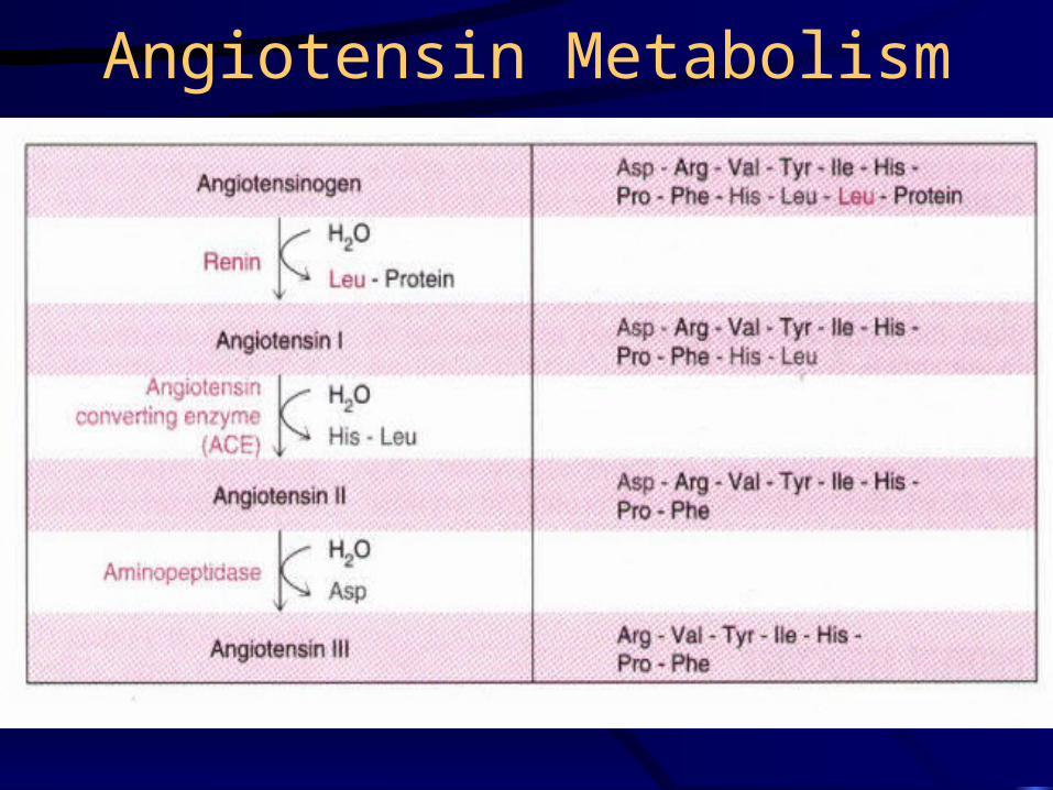
Angiotensin Metabolism
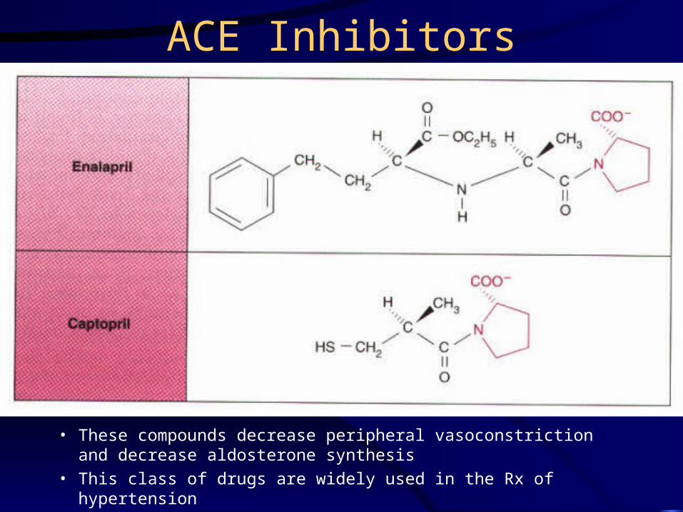
ACE Inhibitors
• These compounds decrease peripheral vasoconstriction and decrease aldosterone synthesis
• This class of drugs are widely used in the Rx of hypertension
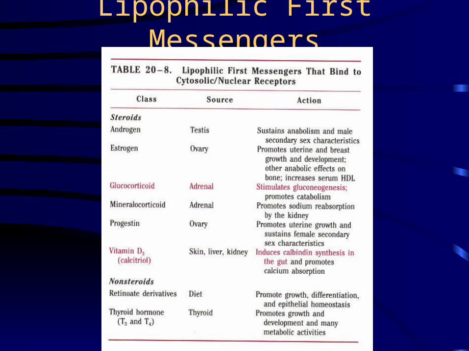
Lipophilic First Messengers
Lipophilic Hormones• These hormones can diffuse through plasma and nuclear
membranes• The intracellular receptors , which constitute the nuclear-
receptor superfamily, function as transcription activators when bound to ligand
• Receptor architecture– C-terminal variable segment
– Middle DNA binding region with a C4 zinc finger segment
– N-terminal hormone (ligand) binding domain
• In some receptors, this domain functions as a repression domain in the absence of ligand
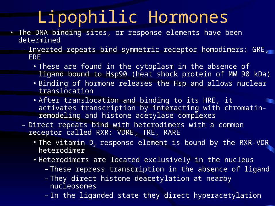
Lipophilic Hormones• The DNA binding sites, or response elements have been determined
– Inverted repeats bind symmetric receptor homodimers: GRE, ERE• These are found in the cytoplasm in the absence of ligand bound to
Hsp90 (heat shock protein of MW 90 kDa)• Binding of hormone releases the Hsp and allows nuclear
translocation• After translocation and binding to its HRE, it activates transcription
by interacting with chromatin-remodeling and histone acetylase complexes
– Direct repeats bind with heterodimers with a common receptor called RXR: VDRE, TRE, RARE
• The vitamin D3 response element is bound by the RXR-VDR heterodimer
• Heterodimers are located exclusively in the nucleus– These repress transcription in the absence of ligand– They direct histone deacetylation at nearby nucleosomes– In the liganded state they direct hyperacetylation
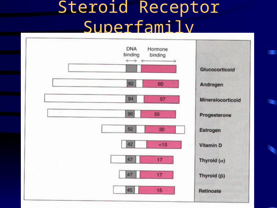
Steroid Receptor Superfamily
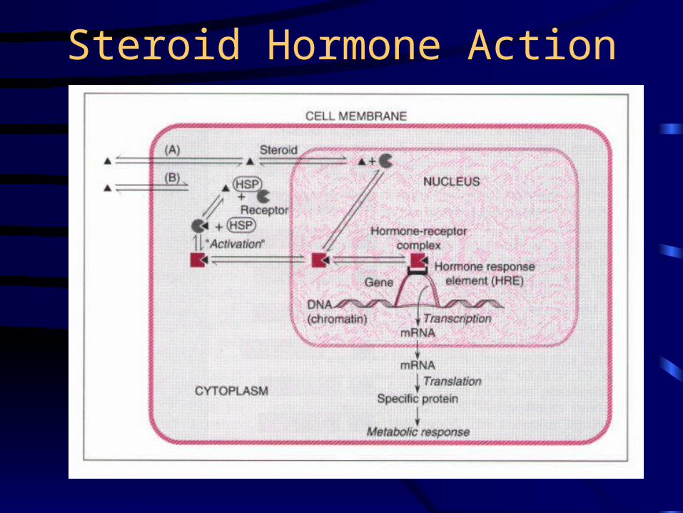
Steroid Hormone Action
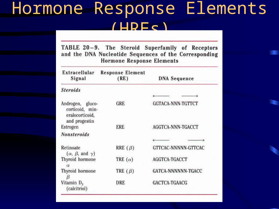
Hormone Response Elements (HREs)
The End
Biochemistry is fun!!!
Recommended