Journal of the American College of Cardiology Vol. 59, No. 18, 2012© 2012 by the American College of Cardiology Foundation ISSN 0735-1097/$36.00
Cardiac Imaging
Usefulness of Fluorine-18 Positron Emission Tomography/Computed Tomography for Identification ofCardiovascular Implantable Electronic Device Infections
Jean-François Sarrazin, MD,* François Philippon, MD,* Michel Tessier, MD,† Jean Guimond, MD,†Franck Molin, MD,* Jean Champagne, MD,* Isabelle Nault, MD,* Louis Blier, MD,* Maxime Nadeau,*Lyne Charbonneau, RN,* Mikaël Trottier, MD,† Gilles O’Hara, MD*
Quebec City, Quebec, Canada
Objectives This study evaluated the usefulness of fluorodesoxyglucose marked by fluorine-18 (18F-FDG) positron emissiontomography (PET) and computed tomography (CT) in patients with suspected cardiovascular implantable elec-tronic device (CIED) infection.
Background CIED infection is sometimes challenging to diagnose. Because extraction is associated with significant morbidi-ty/mortality, new imaging modalities to confirm the infection and its dissemination would be of clinical value.
Methods Three groups were compared. In Group A, 42 patients with suspected CIED infection underwent 18F-FDG PET/CT.Positive PET/CT was defined as abnormal uptake along cardiac devices. Group B included 12 patients withoutinfection who underwent PET/CT 4 to 8 weeks post-implant. Group C included 12 patients implanted for �6months without infection who underwent PET/CT for another indication. Semi-quantitative ratio (SQR) was ob-tained from the ratio between maximal uptake and lung parenchyma uptake.
Results In Group A, 32 of 42 patients with suspected CIED infection had positive PET/CT. Twenty-four patients with posi-tive PET/CT underwent extraction with excellent correlation. In 7 patients with positive PET/CT, 6 were treatedas superficial infection with clinical resolution. One patient with positive PET/CT but negative leukocyte scan wasconsidered false positive due to Dacron pouch. Ten patients with negative-PET/CT were treated with antibioticsand none has relapsed at 12.9 � 1.9 months. In Group B, patients had mild uptake seen at the level of the con-nector. There was no abnormal uptake in Group C patients. Median SQR was significantly higher in Group A(A � 2.02 vs. B � 1.08 vs. C � 0.57; p � 0.001).
Conclusions PET/CT is useful in differentiating between CIED infection and recent post-implant changes. It may guide appro-priate therapy. (J Am Coll Cardiol 2012;59:1616–25) © 2012 by the American College of CardiologyFoundation
Published by Elsevier Inc. doi:10.1016/j.jacc.2011.11.059
Cardiovascular implantable electronic device (CIED) infec-tion is one of the most feared complications of deviceimplantation. The incidence of CIED infections is 1.9 casesby 1,000 implants/year (1,2). The total number of CIEDinfections is increasing, mainly with new clinical indicationsand the growing number of implants worldwide (3). Defin-itive CIED infection diagnosis is often challenging. Inaddition, CIED infection treatment can be invasive, requir-
From the *Department of Medicine, Division of Cardiology, Institut universitaire decardiologie et pneumologie de Québec, Québec City, Quebec, Canada; and the†Department of Medical Imaging, Division of Nuclear Medicine, Institut universi-taire de cardiologie et pneumologie de Québec, Québec City, Quebec, Canada. Allauthors have reported that they have no relationships relevant to the contents to thispaper to disclose.
Manuscript received July 7, 2011; revised manuscript received November 9, 2011,accepted November 22, 2011.
ing complete extraction of the generator and all leads. Leadextraction is associated with significant morbidity (majorcomplications � 1.5% to 2%) and mortality (0.8%) (4,5).New imaging modalities to confirm the infectious processand its dissemination would be of clinical value.
See page 1626
Combined fluorodesoxyglucose marked by fluorine-18(18F-FDG) positron emission tomography (PET) and com-puted tomography (CT) is a well-established imaging mo-dality that allows 3-D measurement of metabolic activitywithin an organ obtained from the emission of positronsfollowing disintegration of an injected radioactive product.The value of 18F-FDG PET/CT is already recognized in
oncology for cancer diagnosis and staging, and in cardiology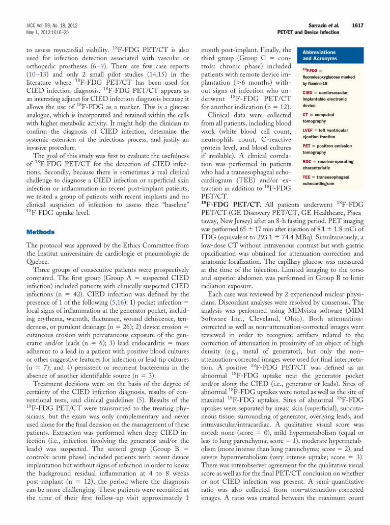
uo(lC
awcsi
otciwc
od
fwnpitwctP
Floaaar
caScrcdat
uninlo
1617JACC Vol. 59, No. 18, 2012 Sarrazin et al.May 1, 2012:1616–25 PET/CT and Device Infection
to assess myocardial viability. 18F-FDG PET/CT is alsosed for infection detection associated with vascular orrthopedic prostheses (6–9). There are few case reports10–13) and only 2 small pilot studies (14,15) in theiterature where 18F-FDG PET/CT has been used forIED infection diagnosis. 18F-FDG PET/CT appears as
an interesting adjunct for CIED infection diagnosis because itallows the use of 18F-FDG as a marker. This is a glucosenalogue, which is incorporated and retained within the cellsith higher metabolic activity. It might help the clinician to
onfirm the diagnosis of CIED infection, determine theystemic extension of the infectious process, and justify annvasive procedure.
The goal of this study was first to evaluate the usefulnessf 18F-FDG PET/CT for the detection of CIED infec-ions. Secondly, because there is sometimes a real clinicalhallenge to diagnose a CIED infection or superficial skinnfection or inflammation in recent post-implant patients,e tested a group of patients with recent implants and no
linical suspicion of infection to assess their “baseline”18F-FDG uptake level.
Methods
The protocol was approved by the Ethics Committee fromthe Institut universitaire de cardiologie et pneumologie deQuebec.
Three groups of consecutive patients were prospectivelycompared. The first group (Group A � suspected CIEDinfection) included patients with clinically suspected CIEDinfections (n � 42). CIED infection was defined by thepresence of 1 of the following (5,16): 1) pocket infection �local signs of inflammation at the generator pocket, includ-ing erythema, warmth, fluctuance, wound dehiscence, ten-derness, or purulent drainage (n � 26); 2) device erosion �cutaneous erosion with percutaneous exposure of the gen-erator and/or leads (n � 6); 3) lead endocarditis � massadherent to a lead in a patient with positive blood culturesor other suggestive features for infection or lead tip cultures(n � 7); and 4) persistent or recurrent bacteremia in theabsence of another identifiable source (n � 3).
Treatment decisions were on the basis of the degree ofcertainty of the CIED infection diagnosis, results of con-ventional tests, and clinical guidelines (5). Results of the18F-FDG PET/CT were transmitted to the treating phy-sicians, but the exam was only complementary and neverused alone for the final decision on the management of thesepatients. Extraction was performed when deep CIED in-fection (i.e., infection involving the generator and/or theleads) was suspected. The second group (Group B �controls: acute phase) included patients with recent deviceimplantation but without signs of infection in order to knowthe background residual inflammation at 4 to 8 weekspost-implant (n � 12), the period where the diagnosiscan be more challenging. These patients were recruited at
the time of their first follow-up visit approximately 1month post-implant. Finally, thethird group (Group C � con-trols: chronic phase) includedpatients with remote device im-plantation (�6 months) with-
ut signs of infection who un-erwent 18F-FDG PET/CT
for another indication (n � 12).Clinical data were collected
rom all patients, including bloodork (white blood cell count,eutrophils count, C-reactiverotein level, and blood culturesf available). A clinical correla-ion was performed in patientsho had a transesophageal echo-
ardiogram (TEE) and/or ex-raction in addition to 18F-FDGET/CT.
18F-FDG PET/CT. All patients underwent 18F-FDGPET/CT (GE Discovery PET/CT, GE Healthcare, Pisca-taway, New Jersey) after an 8-h fasting period. PET imagingwas performed 65 � 17 min after injection of 8.1 � 1.8 mCi of
DG (equivalent to 293.1 � 74.4 MBq). Simultaneously, aow-dose CT without intravenous contrast but with gastricpacification was obtained for attenuation correction andnatomic localization. The capillary glucose was measuredt the time of the injection. Limited imaging to the torsond superior abdomen was performed in Group B to limitadiation exposure.
Each case was reviewed by 2 experienced nuclear physi-ians. Discordant analyses were resolved by consensus. Thenalysis was performed using MIMvista software (MIMoftware Inc., Cleveland, Ohio). Both attenuation-orrected as well as non–attenuation-corrected images wereeviewed in order to recognize artifacts related to theorrection of attenuation in proximity of an object of highensity (e.g., metal of generator), but only the non–ttenuation-corrected images were used for final interpreta-ion. A positive 18F-FDG PET/CT was defined as an
abnormal 18F-FDG uptake near the generator pocketand/or along the CIED (i.e., generator or leads). Sites ofabnormal 18F-FDG uptakes were noted as well as the site ofmaximal 18F-FDG uptakes. Sites of abnormal 18F-FDGptakes were separated by areas: skin (superficial), subcuta-eous tissue, surrounding of generator, overlying leads, and
ntravascular/intracardiac. A qualitative visual score wasoted: none (score � 0), mild hypermetabolism (equal or
ess to lung parenchyma; score � 1), moderate hypermetab-lism (more intense than lung parenchyma; score � 2), and
severe hypermetabolism (very intense uptake; score � 3).There was interobserver agreement for the qualitative visualscore as well as for the final PET/CT conclusion on whetheror not CIED infection was present. A semi-quantitativeratio was also collected from non–attenuation-corrected
Abbreviationsand Acronyms
18F-FDG �
fluorodesoxyglucose markedby fluorine-18
CIED � cardiovascularimplantable electronicdevice
CT � computedtomography
LVEF � left ventricularejection fraction
PET � positron emissiontomography
ROC � receiver-operatingcharacteristic
TEE � transesophagealechocardiogram
images. A ratio was created betwe
en the maximum count
Svol
dvv1tqvtumt
1618 Sarrazin et al. JACC Vol. 59, No. 18, 2012PET/CT and Device Infection May 1, 2012:1616–25
rate of the device over a mean count rate between normalright and left lung parenchyma. Areas of abnormal lungparenchyma were avoided. Other zones of captation wereavoided (skin, shoulder, thyroid gland). Values for thesemi-quantitative ratio were measured on at least 2 separateoccasions with excellent reproducibility.Statistical analysis. Data were expressed using the mean �
D or the median (interquartile range) for continuousariables or as percentage for categorical data. The analysesf categorical variables were performed using a generalizedinear model with a binary distribution function for the
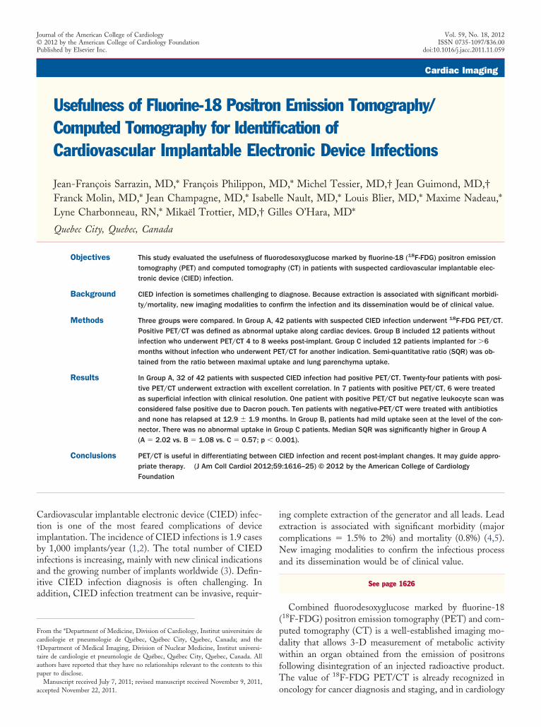
Figure 1 CIED Infection in a Patient With a Deep Pocket Infec
(Upper panels) Attenuation-corrected (AC) and non–attenuation-corrected (NAC) imagephy (PET) in different planes shows abnormal uptake seen posterior to the generatorinfection. (Lower panel) The same abnormality on a fused 18F-FDG PET/computed tom
ependant variable. For continuous data, a 1-way analysis ofariance was fitted to compare groups with heterogeneousariances and whether the model could be reduced to a-way analysis with the same variance across groups wasested. For the time since last intervention and semi-uantitative ratio, values were log transformed to stabilizeariances. Reported p values are on the basis of theseransformations. A posteriori comparisons were performedsing Tukey’s comparison technique. The univariate nor-ality assumptions were verified with the Shapiro-Wilk
est. Sensibility and specificity values of 18F-FDG PET/
nd Positive 18F-FDG PET/CT (Group A)
e fluorodesoxyglucose marked by fluorine-18 (18F-FDG) positron emission tomogra-arrow) and compatible with cardiovascular implantable electronic device (CIED)hy (CT) (yellow arrow). A � anterior, L � left; P � posterior; R � right.
tion a
s of th(black
ograp
1619JACC Vol. 59, No. 18, 2012 Sarrazin et al.May 1, 2012:1616–25 PET/CT and Device Infection
CT, on the basis of the qualitative visual score, were assessedin comparison with the actual gold standard, which is theclinical definition of CIED infection mentioned previously.A receiver operating characteristic (ROC) curve was calcu-lated from the semi-quantitative ratio. A Pearson correla-tion test was performed between 18F-FDG PET/CT resultsand clinical findings at the time of the extraction. Theresults were considered significant with p values �0.05. All
Figure 2 Cardiovascular Implantable Electronic Device InfectioWith an Infected Epicardial Lead and Positive 18F-FDG
(Upper panels) AC and NAC images of the 18F-FDG PET in different planes showspanel) Two images of the same CT transverse section demonstrating abnormal upplain CT for lead localization (upper component) (yellow arrow � epicardial lead).
analyses were conducted using the statistical package SASversion 9.2 (SAS Institute Inc., Cary, North Carolina).
Results
Patient characteristics. Group A included 42 patientswith suspected CIED infection, on the basis of history,physical examination, and initial blood tests. This group
Patient/CT (Group A)
al uptake seen along the course of the epicardial lead (black arrow). (Lowern the lead with 18F-FDG PET fusion (lower component) and the correspondingviations as in Figure 1.
n in aPET
abnormtake oAbbre
opa
f
1620 Sarrazin et al. JACC Vol. 59, No. 18, 2012PET/CT and Device Infection May 1, 2012:1616–25
comprised 28 men and 14 women with a mean age of 62 �17 years, and a mean left ventricular ejection fraction(LVEF) of 44 � 17% (Table 1). All patients underwent18F-FDG PET/CT for risk stratification, and several pa-tients also had a TEE (n � 22). The main presentingsymptom or sign was local wound infection (n � 16),pre-erosion/erosion (n � 13), bacteremia (n � 10), fever ofunknown origin (n � 1), local persistent swelling (n � 1),and chronic wound discomfort (n � 1). Eight patientswithout local signs of device infection met the Duke criteriafor infective endocarditis. Twenty-four patients underwentextraction and 18 patients were treated with antibiotics only.After complete evaluation, 35 patients had confirmedCIED infections. In the remaining 7 patients, 5 patientswere treated successfully for an infection unrelated totheir cardiac device, 1 patient with fever of unknown
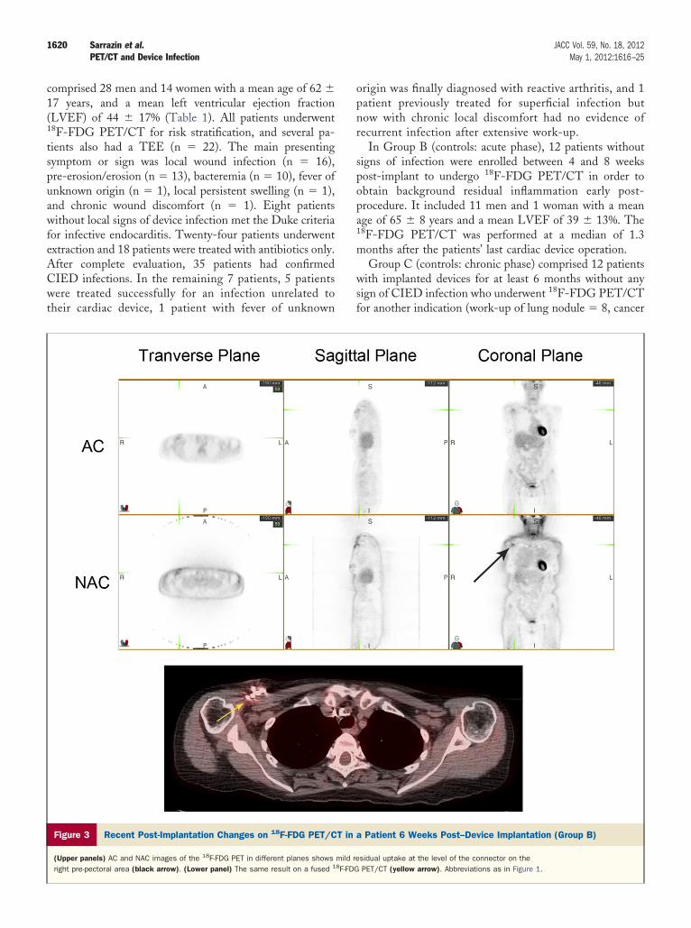
Figure 3 Recent Post-Implantation Changes on 18F-FDG PET/C
(Upper panels) AC and NAC images of the 18F-FDG PET in different planes showsright pre-pectoral area (black arrow). (Lower panel) The same result on a fused 1
origin was finally diagnosed with reactive arthritis, and 1patient previously treated for superficial infection butnow with chronic local discomfort had no evidence ofrecurrent infection after extensive work-up.
In Group B (controls: acute phase), 12 patients withoutsigns of infection were enrolled between 4 and 8 weekspost-implant to undergo 18F-FDG PET/CT in order to
btain background residual inflammation early post-rocedure. It included 11 men and 1 woman with a meange of 65 � 8 years and a mean LVEF of 39 � 13%. The
18F-FDG PET/CT was performed at a median of 1.3months after the patients’ last cardiac device operation.
Group C (controls: chronic phase) comprised 12 patientswith implanted devices for at least 6 months without anysign of CIED infection who underwent 18F-FDG PET/CTor another indication (work-up of lung nodule � 8, cancer
a Patient 6 Weeks Post–Device Implantation (Group B)
sidual uptake at the level of the connector on thePET/CT (yellow arrow). Abbreviations as in Figure 1.
T in
mild re8F-FDG
1
7
w2psoT(p
1621JACC Vol. 59, No. 18, 2012 Sarrazin et al.May 1, 2012:1616–25 PET/CT and Device Infection
staging � 2, chronic cough � 1, pre-transplantation eval-uation � 1). It included 9 men and 3 women with a meanage of 70 � 10 years and a mean LVEF of 50 � 8%.Blood tests. White blood cells count was 8.5 � 2.5 � 109/lin Group A, 6.6 � 1.5 � 109/l in Group B, and 8.9 � 2.4 �09/l in Group C (p � 0.050). Neutrophils count was 6.4 �
2.6 � 109/l in Group A, 4.1 � 1.3 � 109/l in Group B, and.2 � 2.7 � 109/l in Group C (p � 0.016). There was no
significant difference between the median C-reactive pro-tein levels in Group A and Group B (8.9 mg/l, range 1.3 to261.3 mg/l vs. 3.8 mg/l, range 0.3 to 8.6; p � 0.194).
Figure 4 18F-FDG PET/CT in a Patient With Remote Cardiac De
(Upper panels) AC and NAC images of the 18F-FDG PET in different planes showsthe cardiac device system. The black arrow shows the site of the generator. (Low(yellow arrow). Abbreviations as in Figure 1.
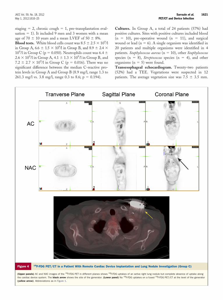
Cultures. In Group A, a total of 24 patients (57%) hadpositive cultures. Sites with positive cultures included blood(n � 10), pre-operative wound (n � 11), and surgical
ound or lead (n � 6). A single organism was identified in0 patients and multiple organisms were identified in 4atients. Staphylococcus aureus (n � 10), other Staphylococcuspecies (n � 8), Streptococcus species (n � 4), and otherrganisms (n � 5) were found.ransesophageal echocardiogram. Twenty-two patients
52%) had a TEE. Vegetations were suspected in 12atients. The average vegetation size was 7.5 � 3.5 mm.
Implantation and Lung Nodule Investigation (Group C)
G uptakes of an active right lung nodule but complete absence of uptake alongel) No 18F-FDG uptakes on a fused 18F-FDG PET/CT at the level of the generator
vice
18F-FDer pan
ivr
C(
twc
wtiec
mpariselectro
1622 Sarrazin et al. JACC Vol. 59, No. 18, 2012PET/CT and Device Infection May 1, 2012:1616–25
The vegetations were seen on the leads in 7 patients, on thevalves in 2 patients, and on both in 3 patients. However,increased 18F-FDG uptake in the same anatomical site wasseen in only 6 patients.18F-FDG PET/CT. Higher doses of 18F-FDG were usedn Group C compared with Group A (370.6 � 27.5 MBqs. 288.8 � 69.6 MBq) (Table 2). The calculated meanadiation dose for an 18F-FDG PET/CT in the context of
CIED infection was 7.0 mSv, which is less than a coronaryangioplasty. In Group A, 32 of 42 patients (76%) withsuspected CIED infection had a positive 18F-FDG PET/
T. Abnormal uptake was visualized around the generatorsn � 18), over the leads (n � 18), in the superficial skin
Patient CharacteristicsTable 1 Patient Characteristics
Group ASuspected CIED
Infection(n � 42)
Age, yrs 62 � 17
Male/female, n 28/14
LVEF, % 44 � 17
CAD 14 (33)
Diabetes mellitus 11 (26)
Warfarin 18 (43)
Corticosteroids use 3 (7)
Type of device
Pacemaker 25 (60)
Defibrillator 17 (40)
Biventricular device 7 (17)
Number of leads 2.2 � 0.8
Time since last intervention, months 11.2 (0.3–101.5
Confirmed infection 35 (83)
Values are mean � SD, n (%), or median (minimum to maximum), unp � 0.001; comparison between Group A and Group C, p � 0.001; co
CAD � coronary artery disease; CIED � cardiovascular implantable
18F-FDG PET/CT ResultsTable 2 18F-FDG PET/CT Results
Group ASuspected CIED
Infection(n � 42)
Dose
MBq 288.8 � 69.6
mCi 7.7 � 1.8
Maximal SUV 4.4 � 1.6
Qualitative visual score
Median 2.0
Lower quartile 1.3
Upper quartile 2.5
Semi-quantitative ratio
Median 2.02
Lower quartile 1.30
Upper quartile 2.98
Values are mean � SD. *Comparison between Group A and Group B, pbetween Group B and Group C, p � 0.001. †Comparison between GC,p � 0.001; comparison between Group B and Group C, p � 0.010.
18
F-FDG PET/CT � fluorodesoxyglucose marked by fluorine-18 positron emimplantable electronic device; SUV � standardized uptake value.issue (n � 13), in the subcutaneous tissue (n � 13), andithin the heart (n � 2). Figure 1 shows an example of
onfirmed CIED infection with a positive 18F-FDGPET/CT in a patient with deep pocket infection and18F-FDG uptake seen posterior to the generator. Figure 2shows another example of CIED infection in a patient withpocket infection as well as an infected epicardial lead. Sixpatients had 18F-FDG uptake limited to superficial tissues
ithout direct contact with the generator or leads, and werereated as superficial skin infection with sustained clinicalmprovement at 9.1 � 6.6 months. One patient with morextensive positive 18F-FDG PET/CT was treated withhronic antibiotic suppressive therapy because of significant
Group BControls
Acute Phase(n � 12)
Group CControls
Chronic Phase(n � 12) p Value
65 � 8 70 � 10 0.189
11/1 9/3 0.315
39 � 13 50 � 8 0.053
6 (50) 6 (50) 0.478
1 (8) 2 (17) 0.388
6 (50) 7 (58) 0.730
0 0 1.000
6 (50) 10 (83) 0.259
6 (50) 2 (17)
3 (25) 0 0.781
2.0 � 0.7 1.8 � 0.5 0.123
1.3 (1.0–2.1) 24.5 (8.0–130.2) �0.001*
0 0 �0.001
herwise specified. *Comparison between Group A and Group B,on between Group B and Group C, p � 0.010.nic device; LVEF � left ventricular ejection fraction.
roup Bontrolste Phase� 12)
Group CControls
Chronic Phase(n � 12) p Value
.0 � 70.8 370.6 � 27.5 �0.001
.3 � 1.6 9.9 � 0.9 0.002
.2 � 1.4 0 �0.001
0.8 0 �0.001*
0.5
1.0
1.08 0.57 �0.001†
0.84 0.40
1.31 0.62
1; comparison between Group A and Group C, p � 0.001; comparisonand Group B, p � 0.005; comparison between Group A and Group
)
less ot
GC
Acu(n
248
7
1
� 0.00roup A
ission tomography and computed tomography; CIED � cardiovascular
tQ
s1
pS
lpi
p
b
D
Mtn4dda
s
iwDp
1623JACC Vol. 59, No. 18, 2012 Sarrazin et al.May 1, 2012:1616–25 PET/CT and Device Infection
comorbidities (elderly woman with severe cachexia). Onepatient with positive 18F-FDG PET/CT but negativeleukocyte scan was considered a false positive due to aDacron pouch surrounding the device. Ten patients withnegative 18F-FDG PET/CT were treated with antibioticsonly and none had relapsed at 12.9 � 1.9 months (initialdiagnoses � local superficial infection in 4, bacteremia in 5,and fever of unknown origin in 1).
In Group B, patients had no or mild uptake only seen atthe level of the connector. Figure 3 shows an example of apatient with presence of mild residual inflammation 4 to 8weeks post–device implantation at the level of the connec-tor. There was no abnormal uptake in any of Group Cpatients. Figure 4 shows an example of a patient undergoinglung nodule investigation with remote cardiac device im-plantation and complete absence of 18F-FDG uptake alonghe cardiac device system.
ualitative visual score and semi-quantitative ratio of18F-FDG PET/CT. The median qualitative visual score wasignificantly higher in Group A (A � 2.0 [lower quartile �.3, upper quartile � 2.5] vs. B � 0.8 [lower quartile � 0.5,
upper quartile � 1.0] vs. C � 0; p � 0.001). The mediansemiquantitative ratio was also significantly higher in GroupA (A � 2.02 [lower quartile � 1.30, upper quartile � 2.98] vs.B � 1.08 [lower quartile � 0.84, upper quartile � 1.31] vs.C � 0.57 [lower quartile � 0.40, upper quartile � 0.62];
� 0.001).ensitivity and specificity of 18F-FDG PET/CT. On the
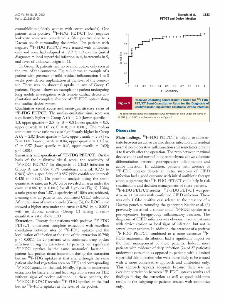
basis of the qualitative visual score, the sensitivity of18F-FDG PET/CT for diagnosis of CIED infection inGroup A was 0.886 (95% confidence interval: 0.723 to0.963) with a specificity of 0.857 (95% confidence interval:0.420 to 0.992). On post-hoc analysis using the semi-quantitative ratio, an ROC curve revealed an area under thecurve at 0.887 (p � 0.001) for all 3 groups (Fig. 5). Usinga ratio greater than 1.87, a specificity of 100% was achieved,meaning that all patients had confirmed CIED infections.After exclusion of acute controls (Group B), the ROC curveshowed a higher area under the curve at 0.961 (p � 0.001)with no chronic controls (Group C) having a semi-quantitative ratio above 1.00.Extraction. Twenty-four patients with positive 18F-FDGPET/CT underwent complete extraction with excellentcorrelation between sites of 18F-FDG uptakes and theocalization of infection at the time of the extraction (0.798;
� 0.001). In 20 patients with confirmed deep pocketnfection during the extraction, 19 patients had significant
18F-FDG uptakes in the same anatomical location; 1patient had pocket tissue induration during the extractionbut no 18F-FDG uptakes at that site, although the same
atient also had vegetation seen on TEE and corresponding18F-FDG uptake on the lead. Finally, 4 patients underwentextraction for bacteremia and lead vegetations seen on TEEwithout signs of pocket infection; in these patients, the18F-FDG PET/CT revealed 18F-FDG uptakes on the lead
ut no 18F-FDG uptakes at the level of the pocket.iscussion
ain findings. 18F-FDG PET/CT is helpful to differen-iate between an active cardiac device infection and residualormal post-operative inflammation still sometimes presentto 8 weeks after the operation. The ratio between maximalevice count and normal lung parenchyma allows adequateifferentiation between post-operative inflammation andctive infection. In addition, patients with absence of
18F-FDG uptakes despite an initial suspicion of CIEDinfection had a good outcome with initial antibiotic therapyalone, suggesting that 18F-FDG PET/CT could help in risktratification and decision management of these patients.
18F-FDG PET/CT results. 18F-FDG PET/CT was pos-tive in 31 patients with confirmed CIED infection. Thereas only 1 false positive case related to the presence of aacron pouch surrounding the generator; Keidar et al. (6)
reviously described a similar mild 18F-FDG uptake as apost-operative foreign-body inflammatory reaction. Thediagnosis of CIED infection was obvious in some patientswith device erosion or local signs of infection, but not inseveral other patients. In addition, the presence of a positive18F-FDG PET/CT combined to a more extensive 18F-FDG anatomical distribution had a significant impact onthe final management of these patients. Indeed, mostpatients with evidence of deep infection (24 of 27 patients)underwent extraction as opposed to patients with a limitedsuperficial skin infection who were more likely to be treatedwith a more conservative approach and antibiotics only.This approach appears adequate because there was anexcellent correlation between 18F-FDG uptakes results andfindings during the extraction as well as good mid-termresults in the subgroup of patients treated with antibiotics
0
0.2
0.4
0.6
0.8
1
0 0.1 0.2 0.3 0.4 0.5 0.6 0.7 0.8 0.9 1
Sensibility
1 - Specificity
Figure 5Receiver-Operating Characteristic Curve for 18F-FDGPET/CT Semi-Quantitative Ratio for the Diagnosis ofCardiovascular Implantable Electronic Device Infection
The receiver-operating characteristic curve revealed an area under the curve at0.887 (p � 0.001). Abbreviations as in Figure 1.
only.
vcowtmsoad
gi
tCw
Fatt
trp
1624 Sarrazin et al. JACC Vol. 59, No. 18, 2012PET/CT and Device Infection May 1, 2012:1616–25
The result of a negative 18F-FDG PET/CT also pro-ided useful additional information. It allowed a successfulonservative management in patients with initial suspicionf CIED infection. In this subgroup of patients, 4 patientsere treated successfully as a limited superficial skin infec-
ion without mid-term recurrence, 4 patients had bactere-ia without evidence of device involvement and treated
uccessfully with intravenous antibiotics without recurrencer need for lead extraction, 1 patient had bacteremia with anbdominal source identified, and 1 patient was finallyiagnosed with reactive arthritis. A negative 18F-FDG
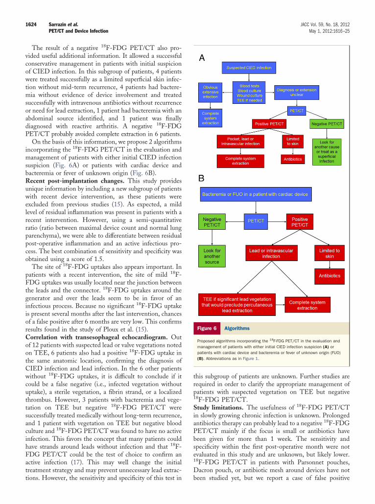
PET/CT probably avoided complete extraction in 6 patients.On the basis of this information, we propose 2 algorithms
incorporating the 18F-FDG PET/CT in the evaluation andmanagement of patients with either initial CIED infectionsuspicion (Fig. 6A) or patients with cardiac device andbacteremia or fever of unknown origin (Fig. 6B).Recent post-implantation changes. This study providesunique information by including a new subgroup of patientswith recent device intervention, as these patients wereexcluded from previous studies (15). As expected, a mildlevel of residual inflammation was present in patients with arecent intervention. However, using a semi-quantitativeratio (ratio between maximal device count and normal lungparenchyma), we were able to differentiate between residualpost-operative inflammation and an active infectious pro-cess. The best combination of sensitivity and specificity wasobtained using a score of 1.5.
The site of 18F-FDG uptakes also appears important. Inpatients with a recent intervention, the site of mild 18F-FDG uptakes was usually located near the junction betweenthe leads and the connector. 18F-FDG uptakes around theenerator and over the leads seem to be in favor of annfectious process. Because no significant 18F-FDG uptake
is present several months after the last intervention, chancesof a false positive after 6 months are very low. This confirmsresults found in the study of Ploux et al. (15).Correlation with transesophageal echocardiogram. Outof 12 patients with suspected lead or valve vegetations notedon TEE, 6 patients also had a positive 18F-FDG uptake inhe same anatomic location, confirming the diagnosis ofIED infection and lead infection. In the 6 other patientsithout 18F-FDG uptakes, it is difficult to conclude if it
could be a false negative (i.e., infected vegetation withoutuptake), a sterile vegetation, a fibrin strand, or a localizedthrombus. However, 3 patients with bacteremia and vege-tation on TEE but negative 18F-FDG PET/CT weresuccessfully treated medically without long-term recurrence,and 1 patient with vegetation on TEE but negative bloodculture and 18F-FDG PET/CT was found to have no activeinfection. This favors the concept that many patients couldhave strands around leads without infection and that 18F-
DG PET/CT could be the test of choice to confirm anctive infection (17). This may well change the initialreatment strategy and may prevent unnecessary lead extrac-
ions. However, the sensitivity and specificity of this test inhis subgroup of patients are unknown. Further studies areequired in order to clarify the appropriate management ofatients with suspected vegetation on TEE but negative
18F-FDG PET/CT.Study limitations. The usefulness of 18F-FDG PET/CTin slowly growing chronic infection is unknown. Prolongedantibiotics therapy can probably lead to a negative 18F-FDGPET/CT mainly if the focus is small or antibiotics havebeen given for more than 1 week. The sensitivity andspecificity within the first post-operative month were notevaluated in this study and are unknown, but likely lower.18F-FDG PET/CT in patients with Parsonnet pouches,Dacron pouch, or antibiotic mesh around devices have not
Figure 6 Algorithms
Proposed algorithms incorporating the 18F-FDG PET/CT in the evaluation andmanagement of patients with either initial CIED infection suspicion (A) orpatients with cardiac device and bacteremia or fever of unknown origin (FUO)(B). Abbreviations as in Figure 1.
been studied yet, but we report a case of false positive
rrpcirm$t
C
imiTtca
1625JACC Vol. 59, No. 18, 2012 Sarrazin et al.May 1, 2012:1616–25 PET/CT and Device Infection
18F-FDG PET/CT in a patient with Dacron in his pocket.Leukocyte scans were not systematically performed in thisstudy; although likely more specific, it has a lower resolutionthan the 18F-FDG PET/CT. Longer follow-up will beequired to ascertain that there is no late infection recur-ence. The 2 proposed algorithms are now being testedrospectively. Although no data are available on theost-effectiveness of this approach, it could be acceptablef it avoids unnecessary lead extractions and devicee-implantations (estimated costs in Canada for a pace-aker are $30,000 and for a defibrillator are $60,000 to
80,000, while the initial 18F-FDG PET/CT cost is lesshan $2,500 in Canada).
onclusions
18F-FDG PET/CT is useful in differentiating betweencardiovascular implantable electronic device infection andrecent post-implantation changes, and to assess theextension of the infectious process. Also, the absence ofabnormal 18F-FDG uptake among asymptomatic patientsmplanted with pacemaker or defibrillator for at least 6
onths suggest that there is no long-term uptake post-mplant, hence a low risk of false positive after such time.his imaging modality is promising for this new indica-
ion. It may guide appropriate therapy and help to limitomplex and high-risk lead extractions to the moreppropriate patients.
Reprint requests and correspondence: Dr. Jean-FrançoisSarrazin, Department of Medicine, Division of Cardiology, Insti-tut universitaire de cardiologie et de pneumologie de Québec,2725, chemin Sainte-Foy, Quebec City, Quebec G1V 4G5,Canada. E-mail: [email protected].
REFERENCES
1. Uslan DZ, Sohail MR, St Sauver JL, et al. Permanent pacemaker andimplantable cardioverter defibrillator infection: a population-basedstudy. Arch Intern Med 2007;167:669–75.
2. Nery PB, Fernandes R, Nair GM, et al. Device-related infectionamong patients with pacemakers and implantable defibrillators: inci-dence, risk factors, and consequences. J Cardiovasc Electrophysiol2010;21:786–90.
3. Voigt A, Shalaby A, Saba S. Rising rates of cardiac rhythm manage-ment device infections in the United States: 1996 through 2003. J Am
Coll Cardiol 2006;48:590–1.4. Wilkoff BL, Love CJ, Byrd CL, et al. Transvenous lead extraction: HeartRhythm Society expert consensus on facilities, training, indications, andpatient management: this document was endorsed by the American HeartAssociation (AHA). Heart Rhythm 2009;6:1085–104.
5. Baddour LM, Epstein AE, Erickson CC, et al., American HeartAssociation Rheumatic Fever, Endocarditis, and Kawasaki DiseaseCommittee; Council on Cardiovascular Disease in Young; Council onCardiovascular Surgery and Anesthesia; Council on CardiovascularNursing; Council on Clinical Cardiology; Interdisciplinary Council onQuality of Care; American Heart Association. Update on cardiovas-cular implantable electronic device infections and their management: ascientific statement from the American Heart Association. Circulation2010;121:458–77.
6. Keidar Z, Engel A, Hoffman A, Israel O, Nitecki S. Prostheticvascular graft infection: the role of 18F-FDG PET/CT. J Nucl Med2007;48:1230–6.
7. Zhuang H, Duarte PS, Pourdehnad M, et al. The promising role of18F-FDG PET in detecting infected lower limb prosthesis implants.J Nucl Med 2001;42:44–8.
8. Vanquickenborne B, Maes A, Nuyts J, et al. The value of 18F-FDGPET for the detection of infected hip prosthesis. Eur J Nucl Med MolImaging 2003;30:705–15.
9. Chen SH, Ho KC, Hsieh PH, Lee MS, Yen TC. Potential clinicalrole of [(18)F]FDG-PET/CT in detecting hip prosthesis infection: astudy in patients undergoing two-stage revision arthroplasty with aninterim spacer. Q J Nucl Med Mol Imaging 2010;54:429–35.
10. Vos FJ, Bleeker-Rovers CP, van Dijk APJ, Oyen WJG. Detection ofpacemaker and lead infection with FDG-PET. Eur J Nucl Med MolImaging 2006;33:1245.
11. Khamaisi M, Medina A, Mazouz B, Bocher M. Imaging coronarysinus infection in pacemaker electrode with [18F]-fluorodeoxyglucosepositron emission tomography. J Cardiovasc Electrophysiol 2008;19:1327–8.
12. Abikhzer G, Turpin S, Bigras JL. Infected pacemaker causing septiclung emboli detected on FDG PET/CT. J Nucl Cardiol 2010;17:514–5.
13. Turpin S, Lambert R, Poirier N. An unusual looking pacemakerinfection imaged with 18F-FDG PET/CT. Eur J Nucl Med MolImaging 2010;37:1438.
14. Bensimhon L, Lavergne T, Hugonnet F, et al. Whole body [(18)F]fluorodeoxyglucose positron emission tomography imaging for thediagnosis of pacemaker or implantable cardioverter defibrillator infec-tion: a preliminary prospective study. Clin Microbiol Infect 2011;17:836–44.
15. Ploux S, Riviere A, Amraoui S, et al. Positron emission tomography inpatients with suspected pacing system infections may play a critical rolein difficult cases. Heart Rhythm 2011;8:1478–81.
16. Sohail MR, Uslan DZ, Khan AH, et al. Management and outcome ofpermanent pacemaker and implantable cardioverter-defibrillator infec-tions. J Am Coll Cardiol 2007;49:1851–9.
17. Downey BC, Juselius WE, Pandian NG, et al. Incidence and signif-icance of pacemaker and implantable cardioverter-defibrillator leadmasses discovered during transesophageal echocardiography. PacingClin Electrophysiol 2011;34:679–83.
Key Words: defibrillator y extraction y infection y pacemaker y PET
scan.Recommended


![PET/ CT [Positron Emission Tomography]](https://img.pdfslide.net/doc/110x75/56d6bf451a28ab30169592f3/pet-ct-positron-emission-tomography.jpg)


