Embed Size (px)
Citation preview

17 Localized Calcification or Ossification in Muscles and
Subcutaneous Tissues

CLINICAL IMAGAGINGAN ATLAS OF DIFFERENTIAL DAIGNOSIS
EISENBERG
DR. Muhammad Bin Zulfiqar PGR-FCPS III SIMS/SHL

• Fig B 17-1 Myositis ossificans. (A) Knee. (B) Elbow.

• Fig B 17-2 Parosteal sarcoma. (A) Frontal and (B) lateral views of the leg demonstrate a broad-based, densely ossified mass extending outward from the distal femur. The characteristic radiolucent line separating the dense mass of tumor bone from the cortex is not seen in this huge lesion.

• Fig B 17-3 Myositis ossificans associated with neurologic disorders. Diffuse osseous deposits in muscles, tendons, and ligaments about the hip in a patient with long-term paralysis.

Fig B 17-4 Extravasation of a calcium gluconate injection. Soft-tissue opacification in a child.
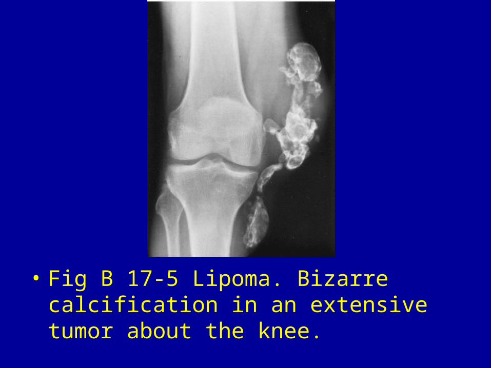
• Fig B 17-5 Lipoma. Bizarre calcification in an extensive tumor about the knee.

• Fig B 17-6 Chondrosarcoma. (A) Prominent dense calcification in a large exostotic chondrosarcoma. (B) Extensive flocculent calcification in the cartilaginous matrix. The arrow points to a small osteochondroma in this patient with multiple hereditary exostoses.

• Fig B 17-7 Extraskeletal osteosarcoma of the posterolateral thigh.30

• Fig B 17-8 Synovioma of the posteromedial thigh. Lateral view shows a large soft-tissue mass with extensive calcific deposits (arrowheads) in it.30

• Fig B 17-9 Ossified surgical scar. Long, linear density on the anterior abdominal wall (arrows).

• Fig B 17-10 Chronic venous stasis. Soft-tissue calcification associated with periosteal reaction about the distal tibia and numerous phleboliths.


![Transcriptional Network Controlling Endochondral Ossification · branous ossification and endochondral ossification.[1] During intramembranous ossification, osteoblasts produce type](https://img.pdfslide.net/doc/110x75/5e8cf0c24763783dcf0d78ef/transcriptional-network-controlling-endochondral-ossification-branous-ossification.jpg)