Embed Size (px)
Citation preview
4 Esophageal Ulceration

CLINICAL IMAGAGINGAN ATLAS OF DIFFERENTIAL DAIGNOSIS
EISENBERG
DR. Muhammad Bin Zulfiqar PGR-FCPS III SIMS/SHL

Fig GI 4-1 Reflux esophagitis. Superficial ulcerations appear as streaks of contrast material superimposed on the flat mucosa of the distal esophagus.

• Fig GI 4-2 Reflux esophagitis. Large penetrating ulcer (arrow).

• Fig GI 4-3 Feline esophagus.6

Fig GI 4-4 Barrett esophagus. Ulcerations (arrow) have developed at a distance from the esophagogastric junction.

• Fig GI 4-5 Candida esophagitis. Multiple ulcers and nodular plaques produce the grossly irregular contour of a shaggy esophagus. This manifestation of far-advanced candidiasis is now infrequent because of earlier and better treatment of the disease.7
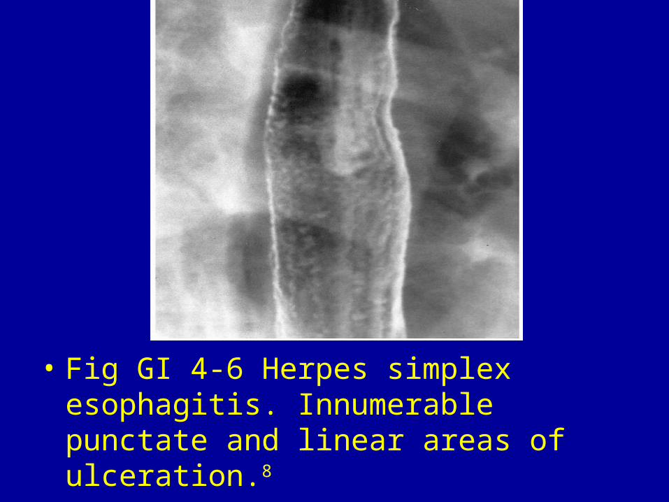
• Fig GI 4-6 Herpes simplex esophagitis. Innumerable punctate and linear areas of ulceration.8

• Fig GI 4-7 Cytomegalovirus. Deep focal ulcer in the distal esophagus (arrows).9

Fig GI 4-8 Tuberculosis. Diffuse mucosal irregularity of the esophagus associated with sinus tracts extending anteriorly into the mediastinum (arrow).10

• Fig GI 4-9 Human immunodeficiency virus. Long, ovoid lesion seen en face in the upper esophagus (black arrow). Note the more distal lesion (white arrows) seen in profile.

• Fig GI 4-10 Squamous carcinoma of the esophagus. On a profile view, the lesion appears as an ulcer crater (arrow) surrounded by a bulging mass projecting into the esophageal lumen.

Fig GI 4-11 Primary ulcerative carcinoma. Characteristic meniscoid ulceration (arrows) surrounded by a tumor mass.

• Fig GI 4-12 Corrosive esophagitis. (A) Dilated, boggy esophagus with ulceration 8 days after the ingestion of a caustic agent. (B) Stricture formation is evident on an esophagram obtained 3 months after the caustic injury.

• Fig GI 4-13 Crohn's esophagitis. Long intramural sinus tract (arrows).11

• Fig GI 4-14 Drug-induced esophagitis. Several focal and linear ulcers (arrows) coalescing in the proximal thoracic esophagus at the level of the aortic arch (AO), related to the ingestion of penicillin tablets for pharyngitis.12

• Fig GI 4-15 Variceal sclerotherapy. Barium esophagram performed 2 weeks after two courses of endoscopic injection sclerotherapy shows diffuse ulceration in the distal third of the esophagus and one intramural sinus tract (arrow).13

• Fig GI 4-16 Intramural pseudodiverticulosis.


![A rare cause of dysphagia due to esophageal intramural ......disease, GERD, and corrosive esophageal injury [4, 11– 14]. Abnormalities in esophageal motility, including, un-coordinated](https://img.pdfslide.net/doc/110x75/60b5b1e8c993b14a95327914/a-rare-cause-of-dysphagia-due-to-esophageal-intramural-disease-gerd-and.jpg)