Embed Size (px)
DESCRIPTION
Citation preview
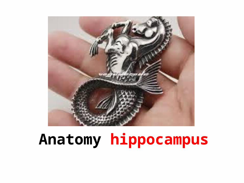
Anatomy hippocampus
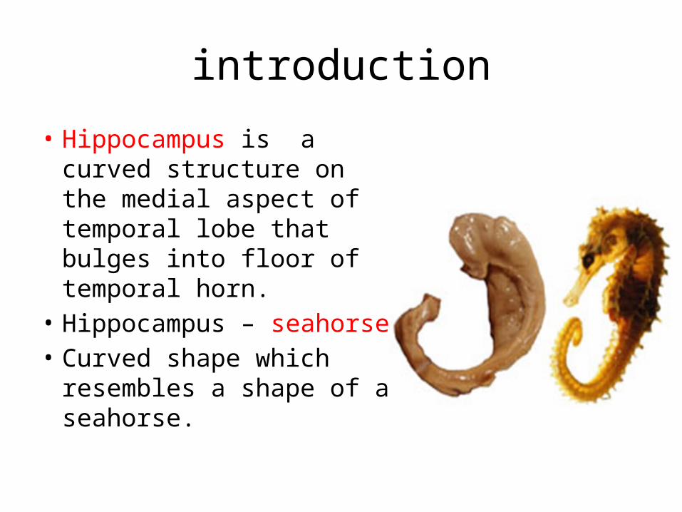
introduction• Hippocampus is a
curved structure on the medial aspect of temporal lobe that bulges into floor of temporal horn.
• Hippocampus – seahorse• Curved shape which
resembles a shape of a seahorse.
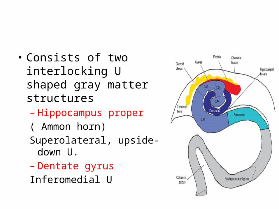
• Consists of two interlocking U shaped gray matter structures– Hippocampus proper ( Ammon horn)Superolateral, upside-
down U.– Dentate gyrusInferomedial U
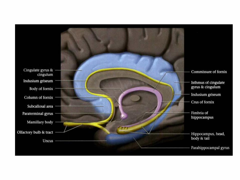
Anatomic divisionsHead ( Pes hippocampus )• most anterior part, oriented transversely• 3 – 4 digitations on superior surface.Body• Cylindrical, oriented parasagittally.Tail• Most posterior, narrows then curves around
splenium to form indusium griseum above cc.
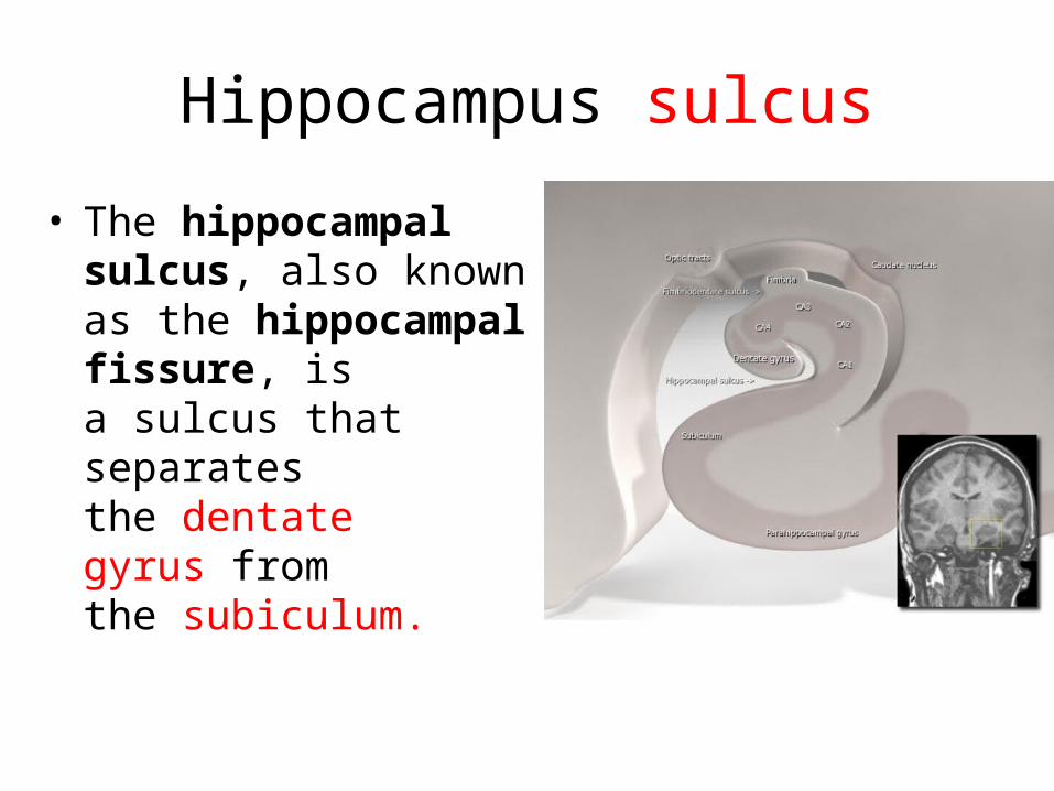
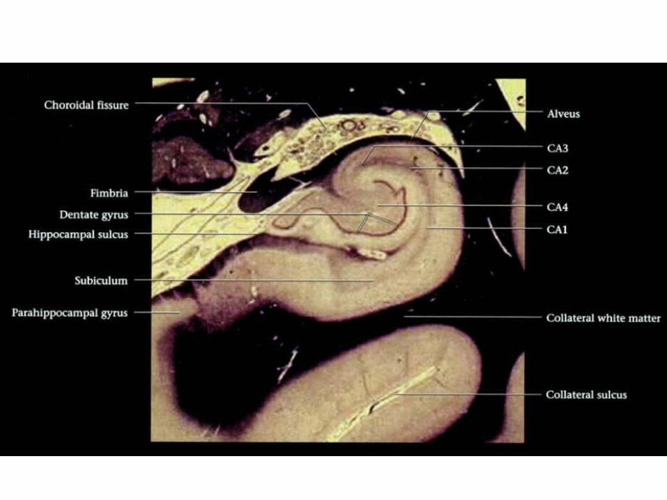
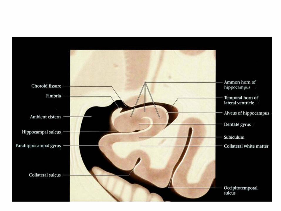
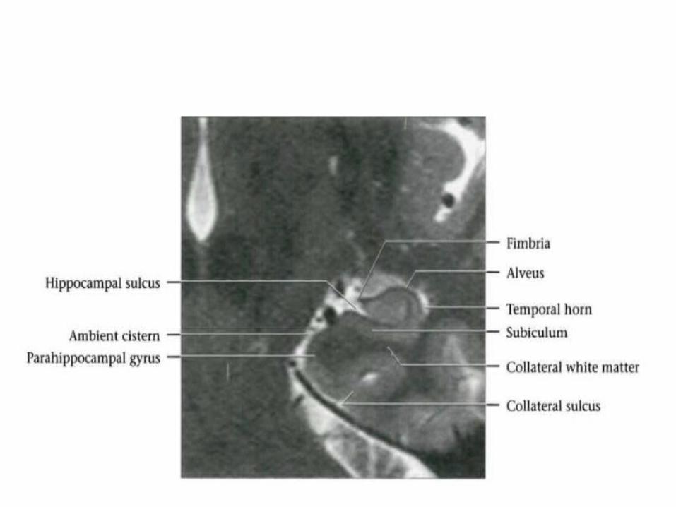
Hippocampus sulcus• The hippocampal
sulcus, also known as the hippocampal fissure, is a sulcus that separates the dentate gyrus from the subiculum.
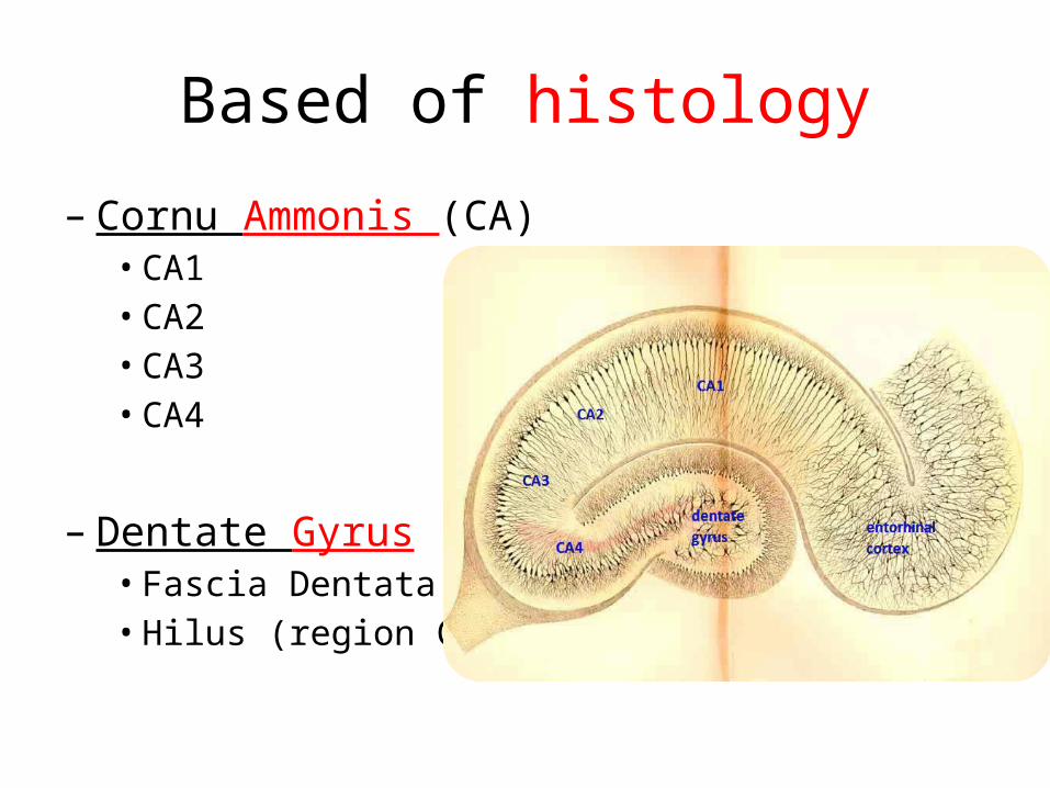
Based of histology– Cornu Ammonis (CA)• CA1• CA2• CA3• CA4
– Dentate Gyrus• Fascia Dentata• Hilus (region CA4)
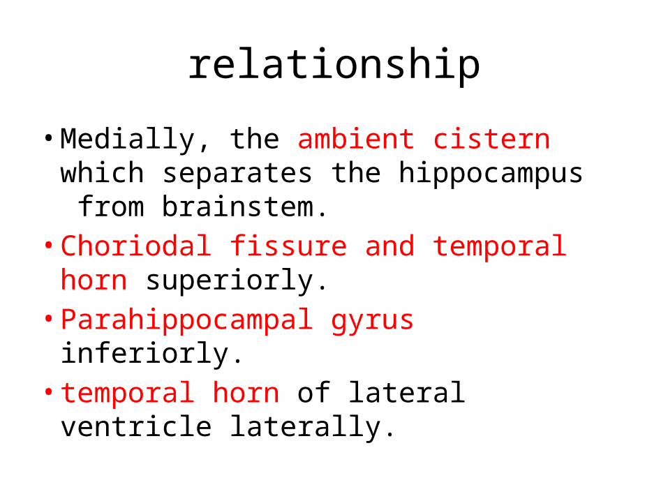
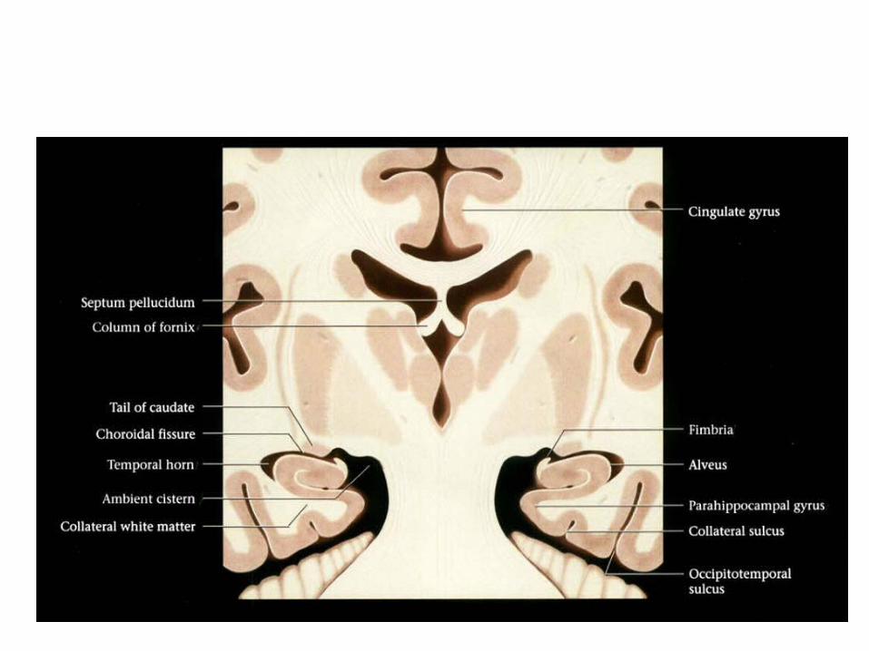
relationship• Medially, the ambient cistern which
separates the hippocampus from brainstem.• Choriodal fissure and temporal
horn superiorly.• Parahippocampal gyrus inferiorly. • temporal horn of lateral ventricle
laterally.
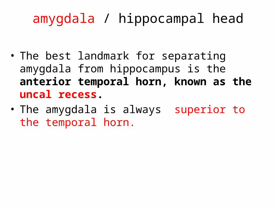
amygdala / hippocampal head
• The best landmark for separating amygdala from hippocampus is the anterior temporal horn, known as the uncal recess.
• The amygdala is always superior to the temporal horn.
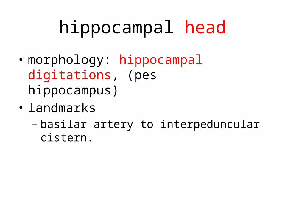
hippocampal head• morphology: hippocampal digitations,
(pes hippocampus) • landmarks– basilar artery to interpeduncular cistern.
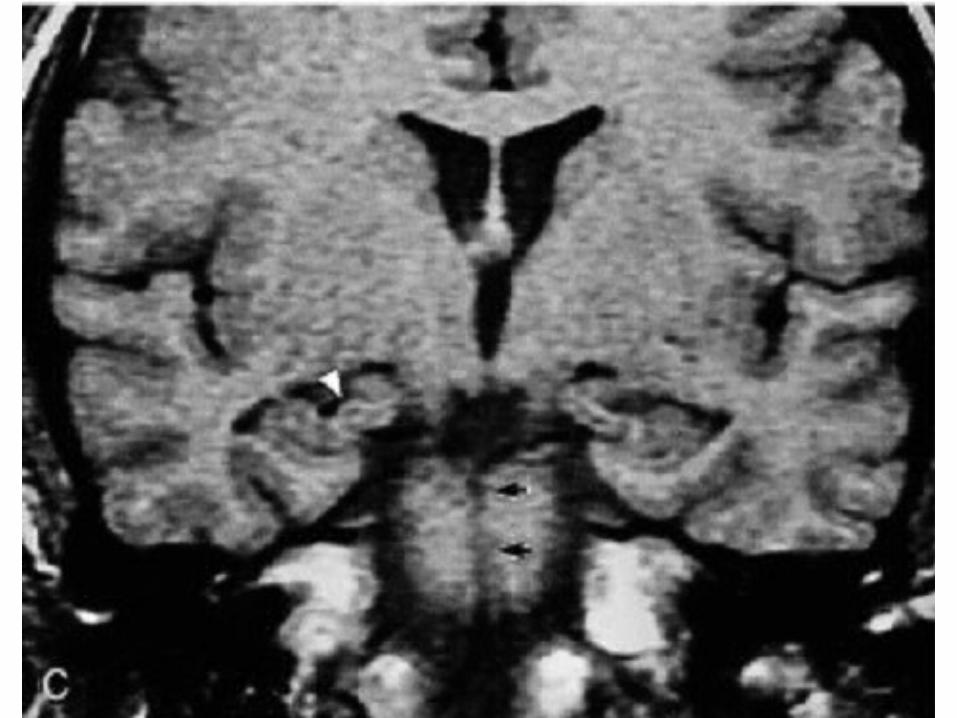
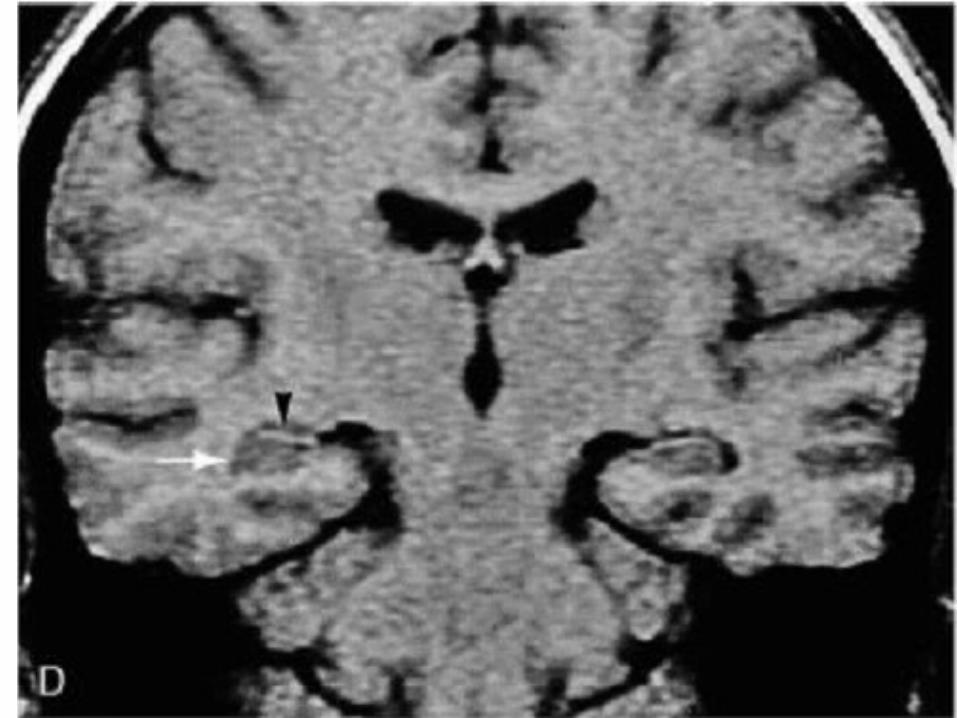
hippocampal body–Morphology: Swiss roll appearance, of
two interlocking U-shaped structures (cornu ammonis and dentate gyrus)
– landmark: • The hippocampal body is oval
and found adjacent to the brainstem• The white matter tracts of the alveus and
fimbria are superior to the hippocampus.
hippocampal tail• morphology: smaller and harder to
describe internal structure.• landmarks– The hippocampal tail is
demonstrated as it ascends posterior to the brainstem.
– from the point at which the fornix can be seen in full profile.
Best choice of imaging• MR in a slightly oblique plane,
perpendicular to long axis of hippocampus.– Coronal T1 weighted– Coronal T2 high resolution– Coronal FLAIR.
MRI imaging
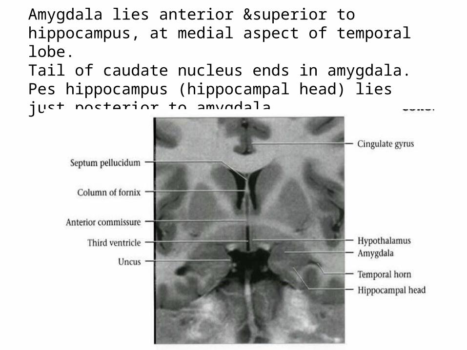
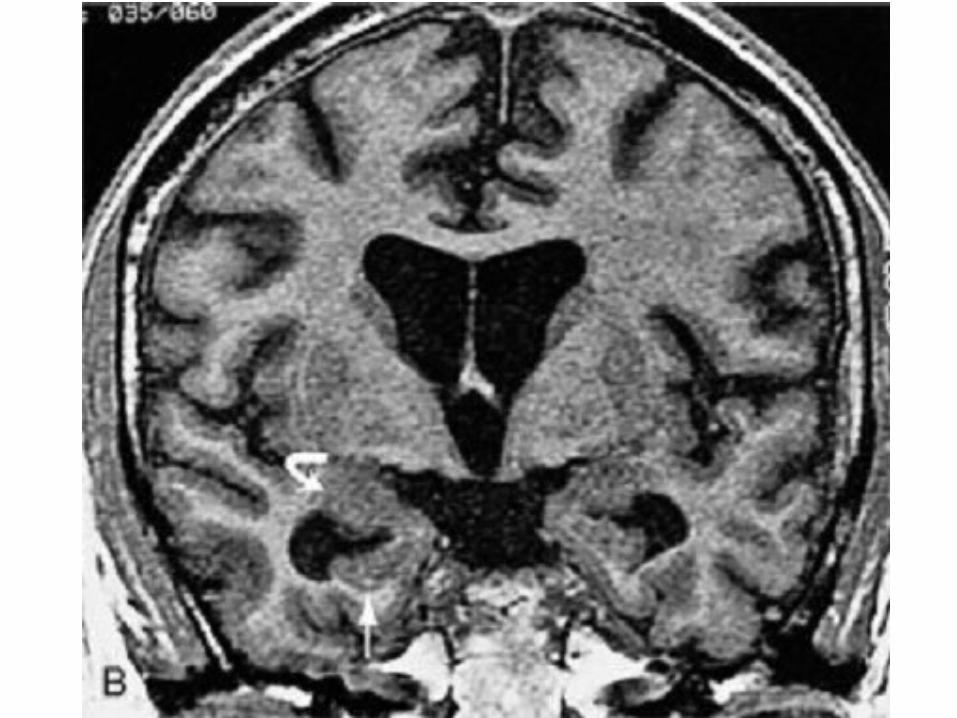
Amygdala lies anterior &superior to hippocampus, at medial aspect of temporal lobe.Tail of caudate nucleus ends in amygdala. Pes hippocampus (hippocampal head) lies just posterior to amygdala.
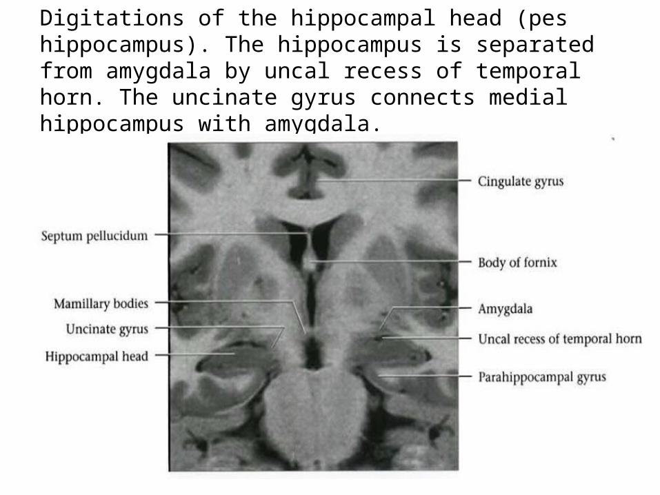
Digitations of the hippocampal head (pes hippocampus). The hippocampus is separated from amygdala by uncal recess of temporal horn. The uncinate gyrus connects medial hippocampus with amygdala.
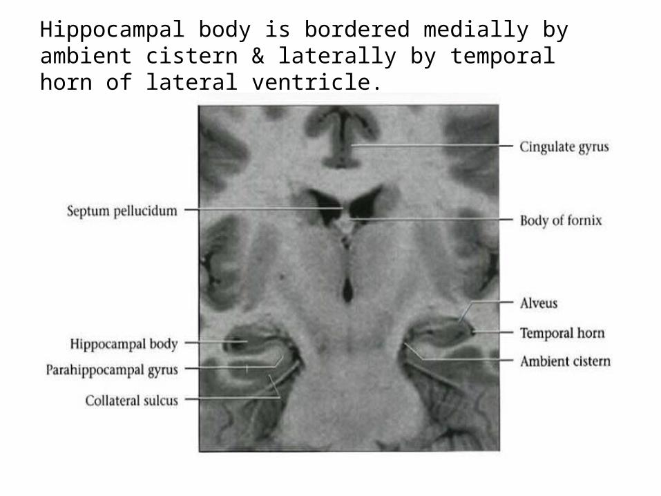
Hippocampal body is bordered medially by ambient cistern & laterally by temporal horn of lateral ventricle.
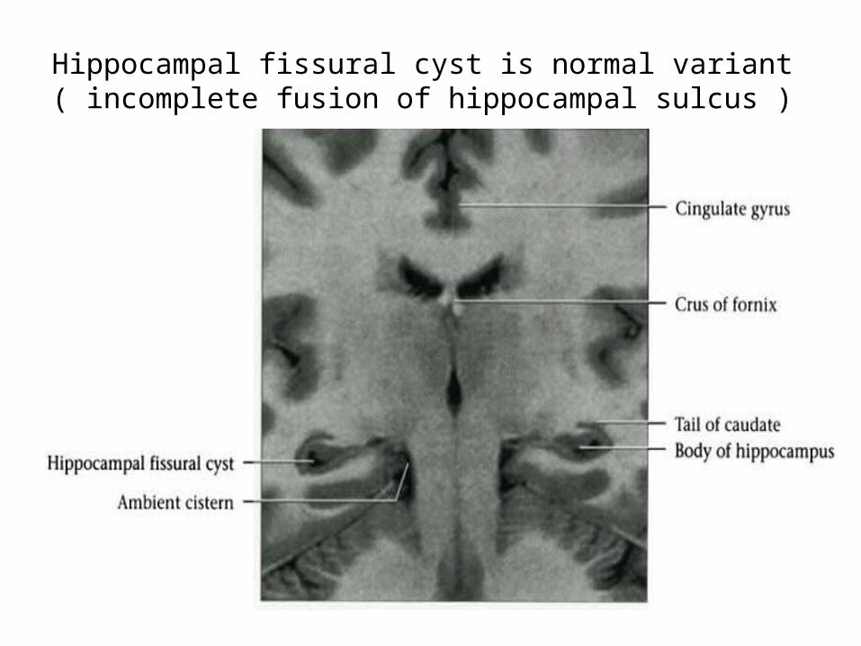
Hippocampal fissural cyst is normal variant ( incomplete fusion of hippocampal sulcus )
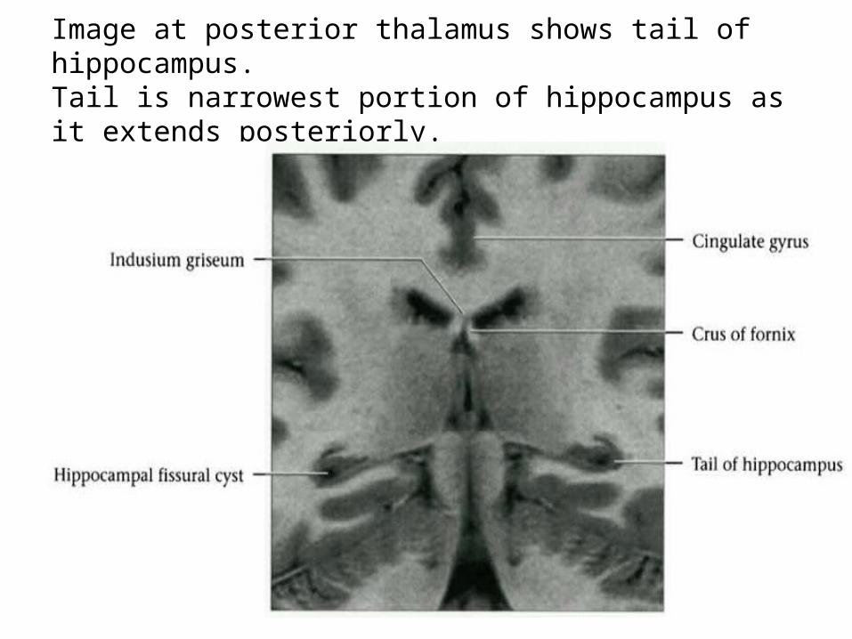
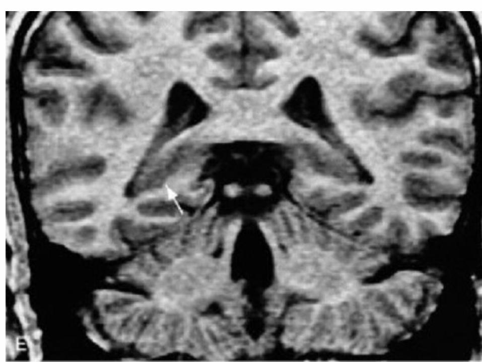
Image at posterior thalamus shows tail of hippocampus.Tail is narrowest portion of hippocampus as it extends posteriorly.
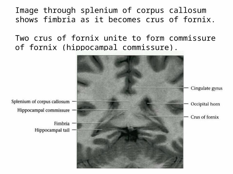
Image through splenium of corpus callosum shows fimbria as it becomes crus of fornix. Two crus of fornix unite to form commissure of fornix (hippocampal commissure).
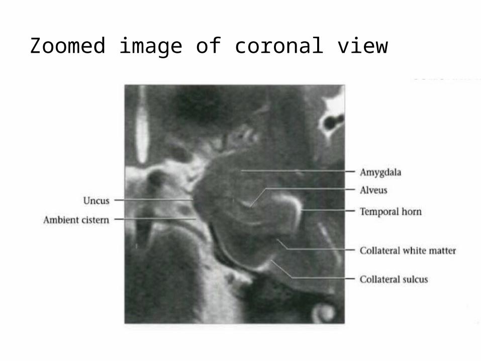
Zoomed image of coronal view
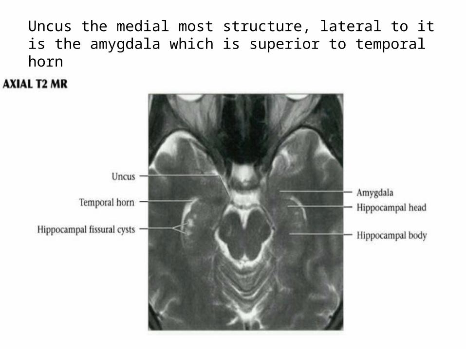
Uncus the medial most structure, lateral to it is the amygdala which is superior to temporal horn
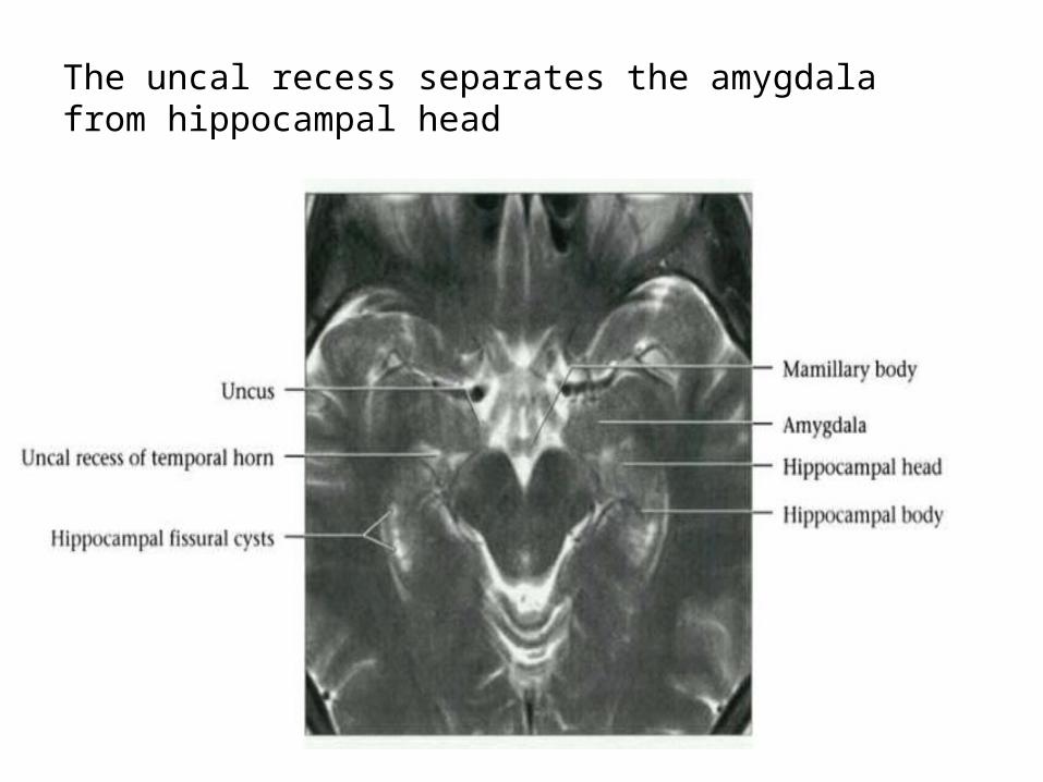
The uncal recess separates the amygdala from hippocampal head
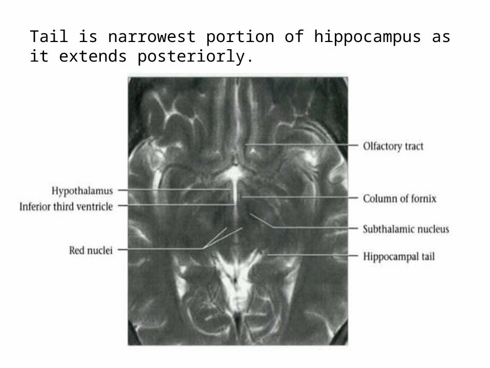
Tail is narrowest portion of hippocampus as it extends posteriorly.
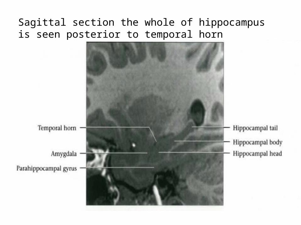
Sagittal section the whole of hippocampus is seen posterior to temporal horn