Embed Size (px)
Citation preview
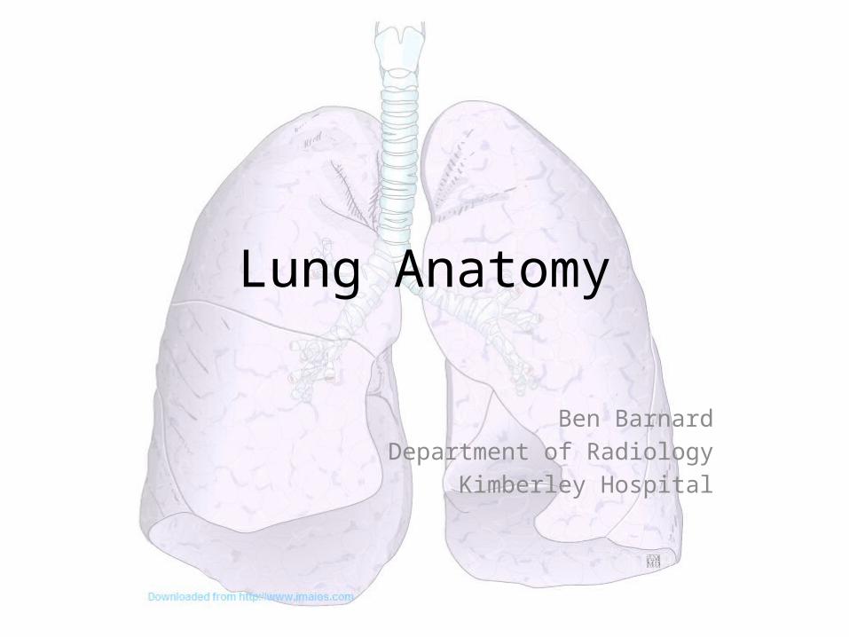
Lung Anatomy
Ben BarnardDepartment of Radiology
Kimberley Hospital
Outline
• Basic morphology• Fissures• Bronchopulmonary segments• Pulmonary vessels• Bronchial vessels• Lymphatics• Lung roots• Pleura
• Radiological features
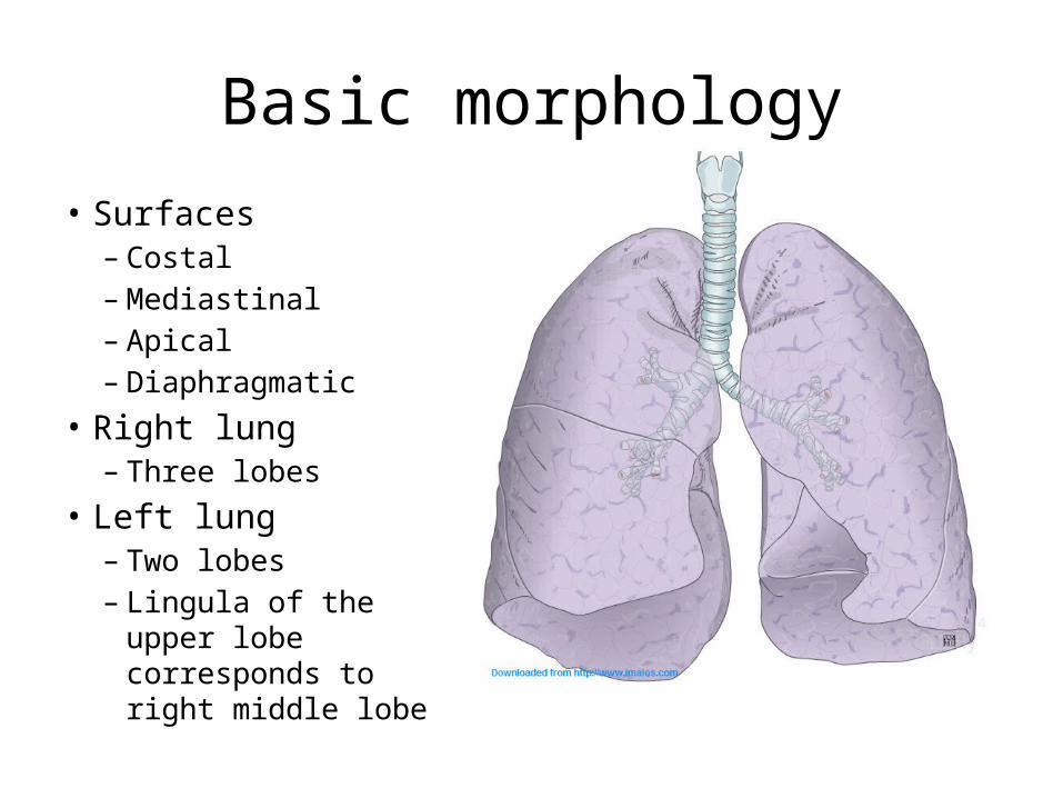
Basic morphology
• Surfaces– Costal– Mediastinal– Apical– Diaphragmatic
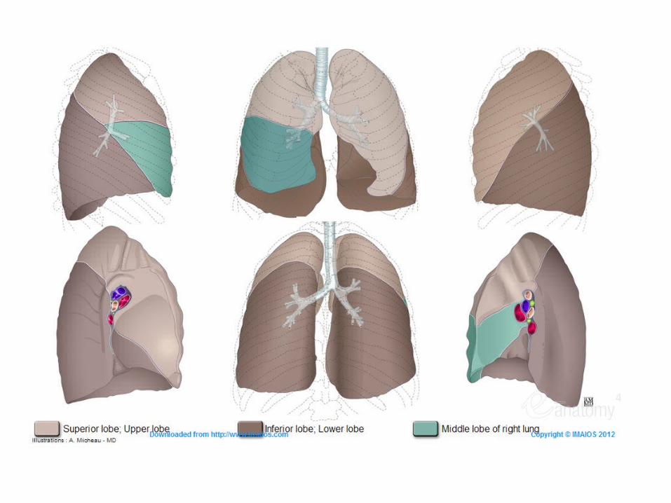
• Right lung– Three lobes
• Left lung– Two lobes– Lingula of the upper
lobe corresponds to right middle lobe
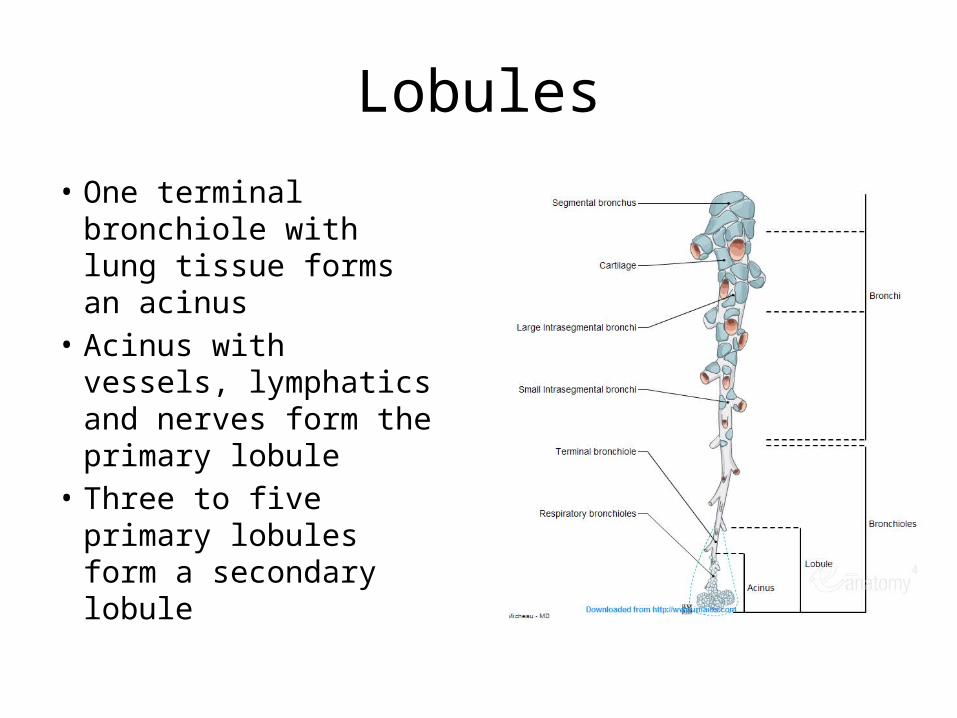
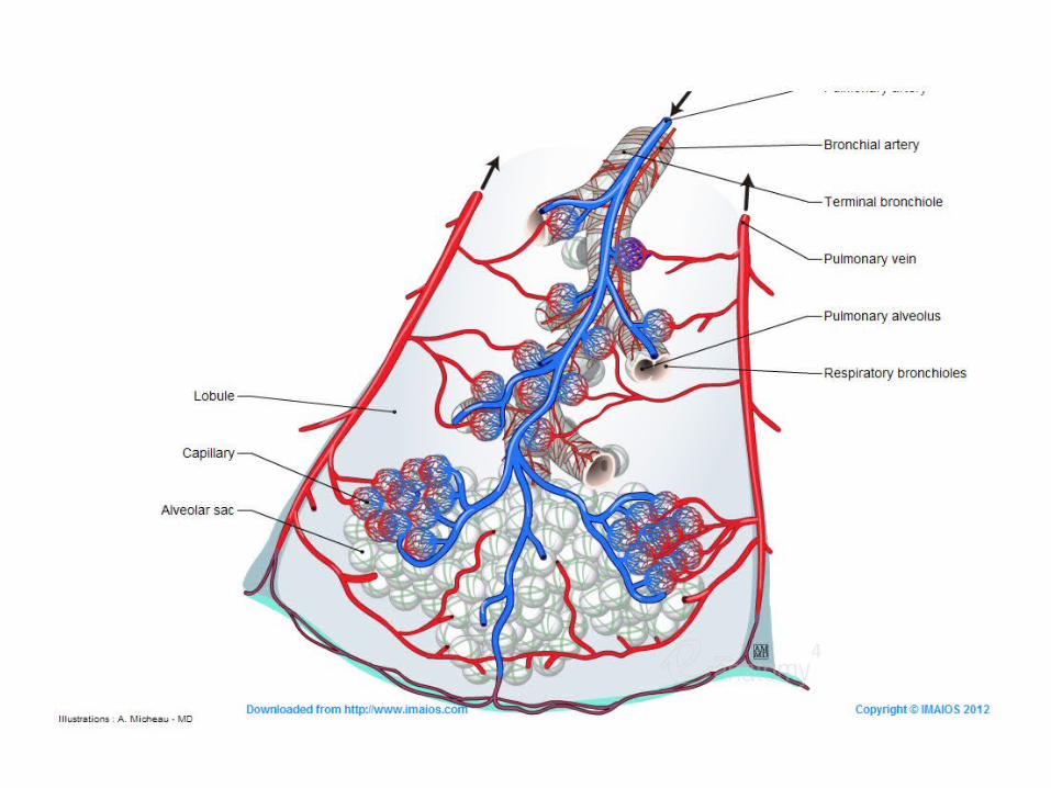
Lobules
• One terminal bronchiole with lung tissue forms an acinus
• Acinus with vessels, lymphatics and nerves form the primary lobule
• Three to five primary lobules form a secondary lobule

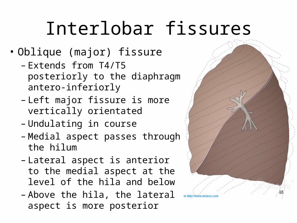
Interlobar fissures• Oblique (major) fissure– Extends from T4/T5 posteriorly to
the diaphragm antero-inferiorly– Left major fissure is more vertically
orientated– Undulating in course– Medial aspect passes through the
hilum– Lateral aspect is anterior to the
medial aspect at the level of the hila and below
– Above the hila, the lateral aspect is more posterior
Interlobar fissures
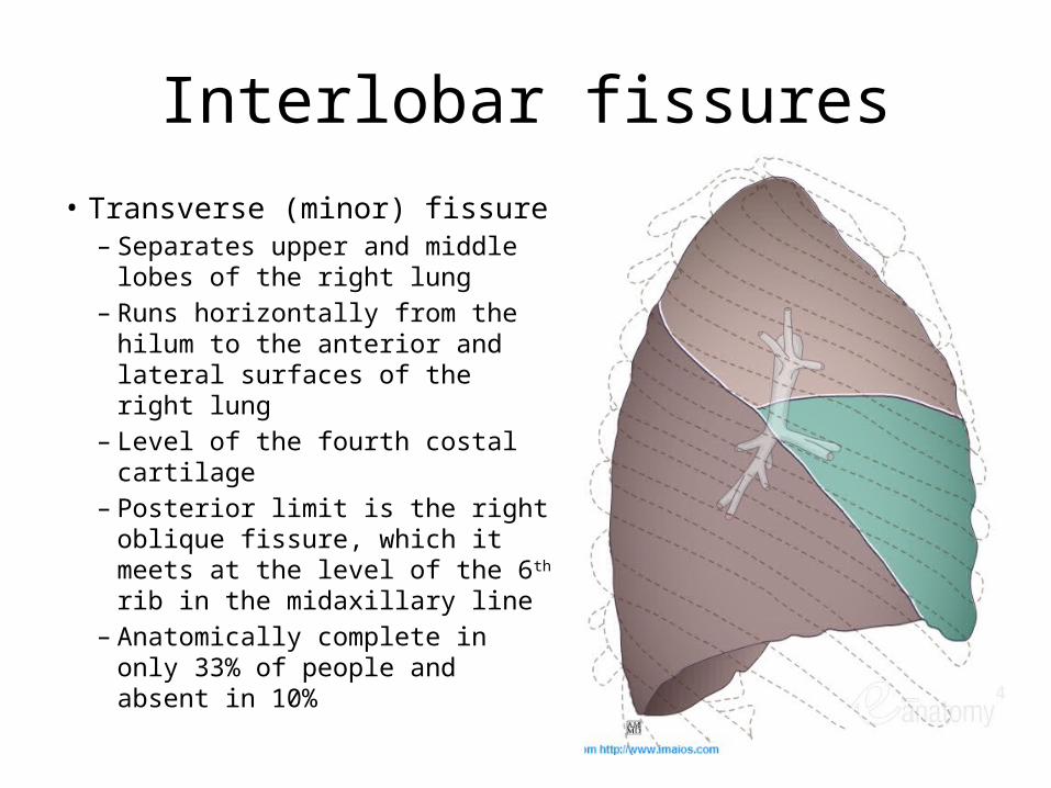
• Transverse (minor) fissure– Separates upper and middle lobes
of the right lung– Runs horizontally from the hilum
to the anterior and lateral surfaces of the right lung
– Level of the fourth costal cartilage– Posterior limit is the right oblique
fissure, which it meets at the level of the 6th rib in the midaxillary line
– Anatomically complete in only 33% of people and absent in 10%
Accessory fissures• Azygos fissure
– Downward invagination of the azygos vein through the apical portion of the right upper lobe
– Four pleural layers – two visceral and two parietal• Superior accessory fissure
– Separates the apical segment of the right lower lobe from the other basal segments
– Lies parallel and inferiorly to the transverse fissure– Passes posteriorly from the right oblique fissure to the posterior surface of the
lung• Inferior accessory fissure
– Separates the medial basal from the other right lower lobe segments– Also called Twining’s line
• Left transverse fissure– Separates the lingula from the rest of the left upper lobe segments– Rarely seen
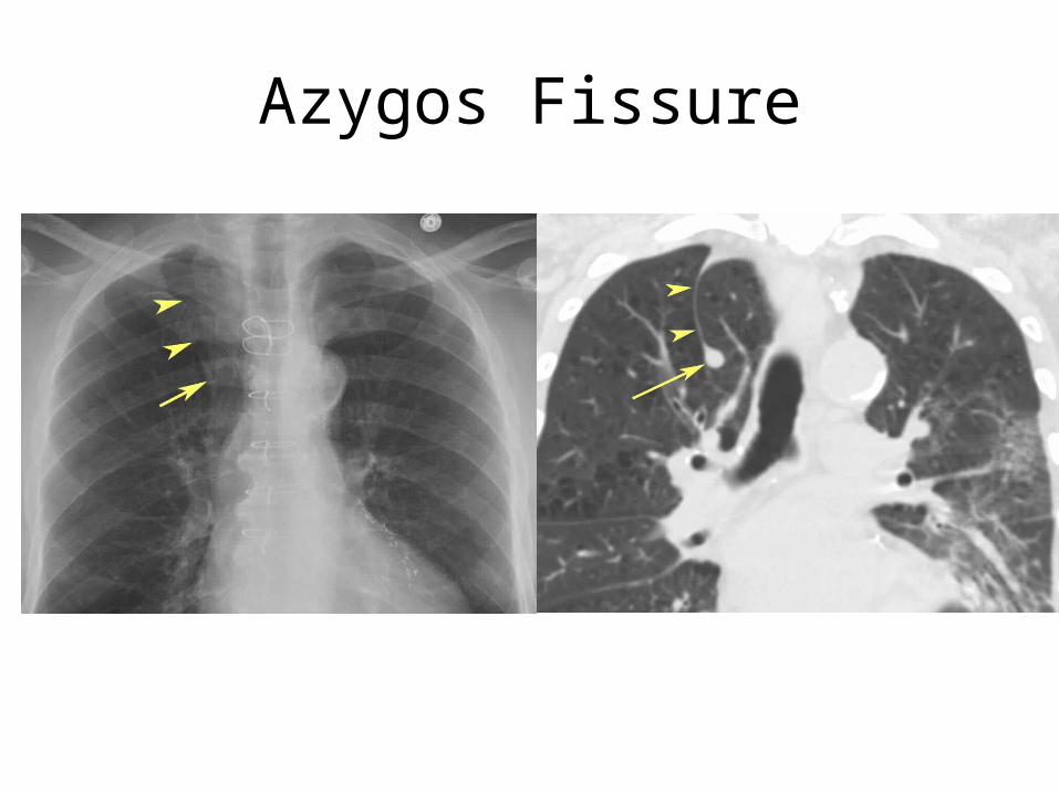
Azygos Fissure
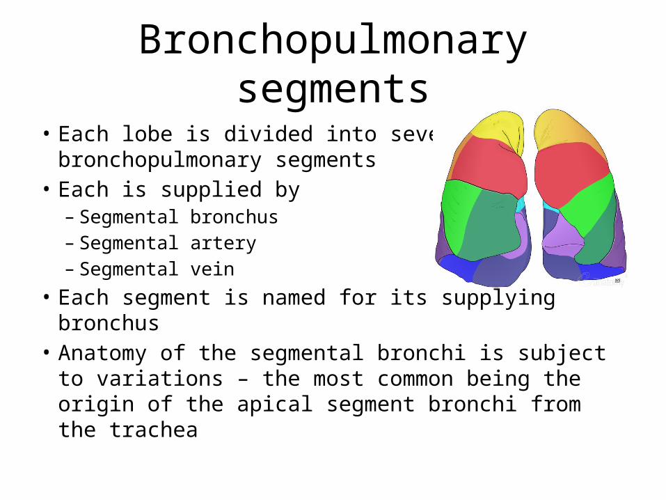
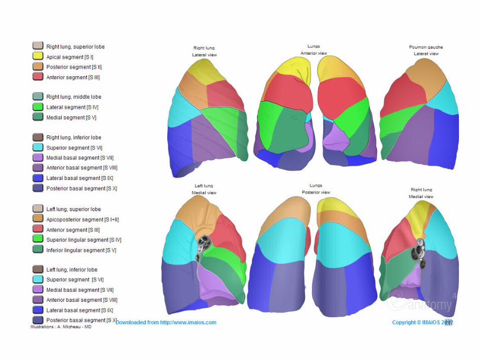
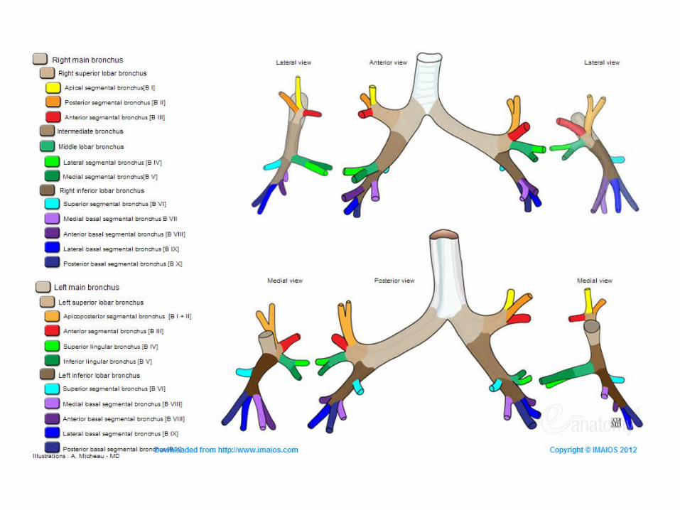
Bronchopulmonary segments
• Each lobe is divided into several bronchopulmonary segments
• Each is supplied by– Segmental bronchus– Segmental artery– Segmental vein
• Each segment is named for its supplying bronchus• Anatomy of the segmental bronchi is subject to
variations – the most common being the origin of the apical segment bronchi from the trachea
Collateral air drift
• Very little connection between segments except via– Pores of Kohn
• Openings in alveolar walls• Connect adjacent alveolar lumens
– Canals of Lambert• Connections between terminal bronchioles and adjacent
alveoli
• Allow gas and fluid transfer between segments but not between lobes
• Ventilation of a segment is thus possible when its segmental bronchus is occluded = collateral air drift
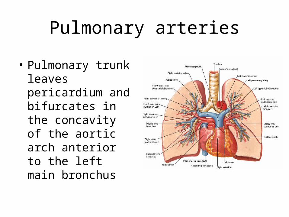
Pulmonary arteries
• Pulmonary trunk leaves pericardium and bifurcates in the concavity of the aortic arch anterior to the left main bronchus
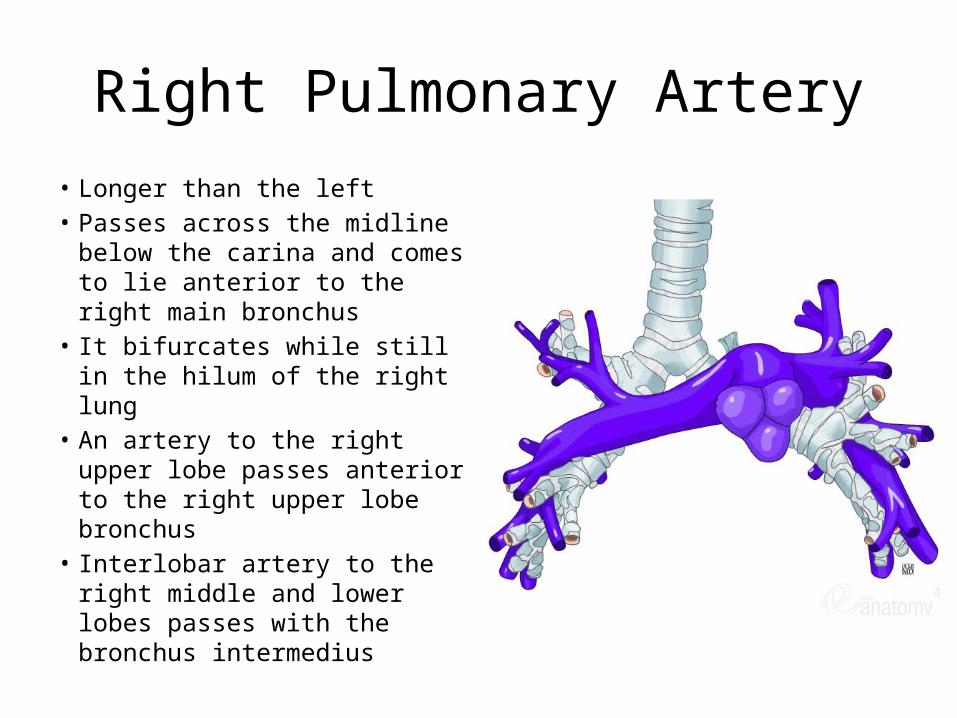
Right Pulmonary Artery
• Longer than the left• Passes across the midline
below the carina and comes to lie anterior to the right main bronchus
• It bifurcates while still in the hilum of the right lung
• An artery to the right upper lobe passes anterior to the right upper lobe bronchus
• Interlobar artery to the right middle and lower lobes passes with the bronchus intermedius
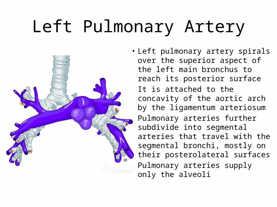
Left Pulmonary Artery• Left pulmonary artery spirals over
the superior aspect of the left main bronchus to reach its posterior surface
• It is attached to the concavity of the aortic arch by the ligamentum arteriosum
• Pulmonary arteries further subdivide into segmental arteries that travel with the segmental bronchi, mostly on their posterolateral surfaces
• Pulmonary arteries supply only the alveoli
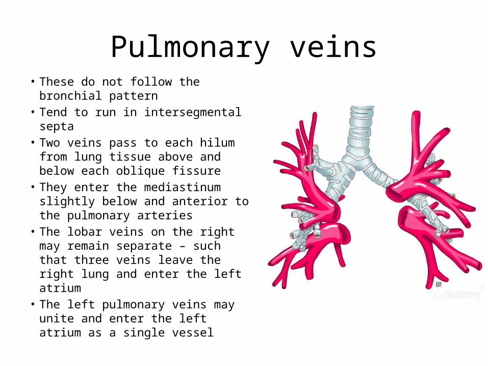
Pulmonary veins• These do not follow the bronchial
pattern• Tend to run in intersegmental septa• Two veins pass to each hilum from
lung tissue above and below each oblique fissure
• They enter the mediastinum slightly below and anterior to the pulmonary arteries
• The lobar veins on the right may remain separate – such that three veins leave the right lung and enter the left atrium
• The left pulmonary veins may unite and enter the left atrium as a single vessel
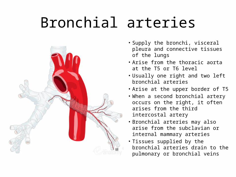
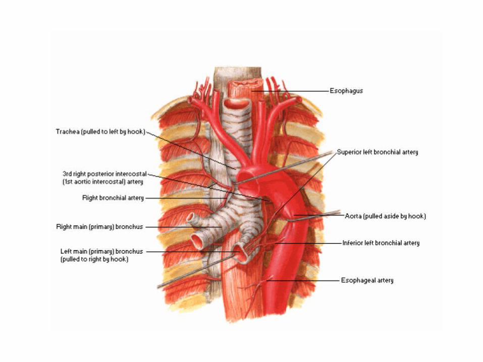
Bronchial arteries• Supply the bronchi, visceral pleura
and connective tissues of the lungs• Arise from the thoracic aorta at the
T5 or T6 level• Usually one right and two left
bronchial arteries• Arise at the upper border of T5• When a second bronchial artery
occurs on the right, it often arises from the third intercostal artery
• Bronchial arteries may also arise from the subclavian or internal mammary arteries
• Tissues supplied by the bronchial arteries drain to the pulmonary or bronchial veins
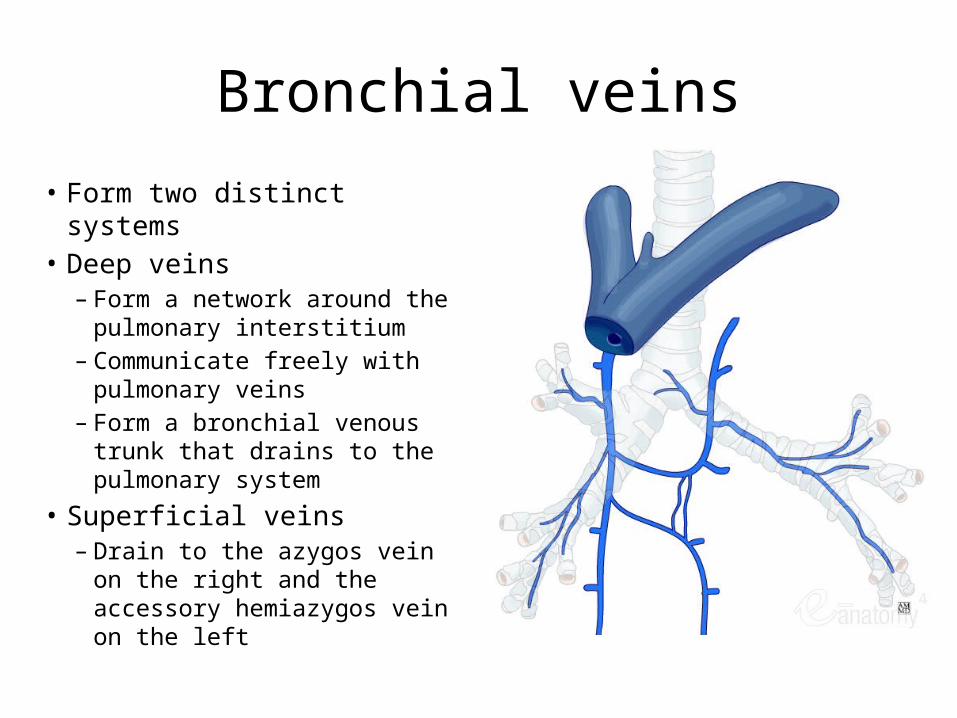
Bronchial veins
• Form two distinct systems• Deep veins
– Form a network around the pulmonary interstitium
– Communicate freely with pulmonary veins
– Form a bronchial venous trunk that drains to the pulmonary system
• Superficial veins– Drain to the azygos vein on
the right and the accessory hemiazygos vein on the left
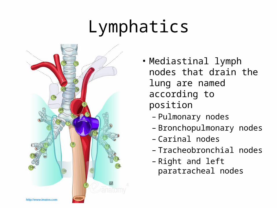
Lymphatics
• Mediastinal lymph nodes that drain the lung are named according to position– Pulmonary nodes– Bronchopulmonary nodes– Carinal nodes– Tracheobronchial nodes– Right and left
paratracheal nodes
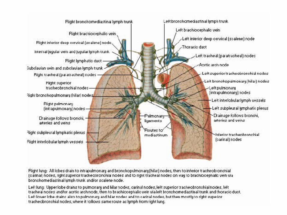
Lymphatics
• Lymph vessels of the lungs are in superficial and deep plexuses• Superficial plexus beneath the pleura drains around the surface
of the lungs and the margins of the fissures to converge at the hila and bronchopulmonary nodes
• Deep channels drain with the pulmonary vessels towards the hila
• Few connections between superficial and deep plexuses except at the hila
• Bronchopulmonary nodes drain to tracheobronchial nodes and paratracheal nodes and then into the bronchomediastinal trunks
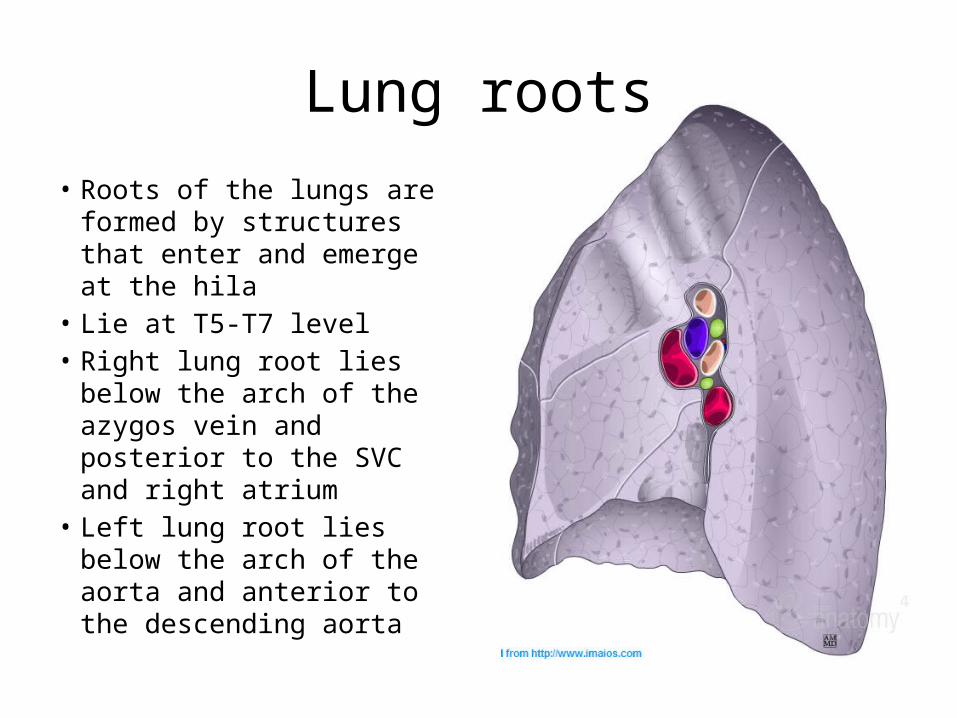
Lung roots
• Roots of the lungs are formed by structures that enter and emerge at the hila
• Lie at T5-T7 level• Right lung root lies below
the arch of the azygos vein and posterior to the SVC and right atrium
• Left lung root lies below the arch of the aorta and anterior to the descending aorta
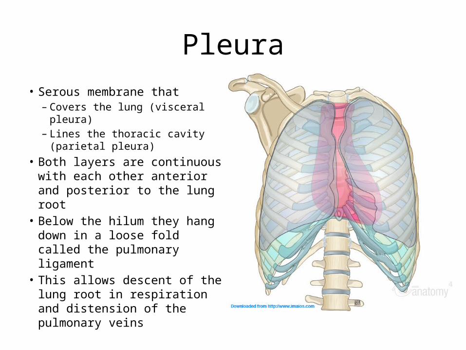
Pleura
• Serous membrane that– Covers the lung (visceral pleura)– Lines the thoracic cavity
(parietal pleura)• Both layers are continuous with
each other anterior and posterior to the lung root
• Below the hilum they hang down in a loose fold called the pulmonary ligament
• This allows descent of the lung root in respiration and distension of the pulmonary veins
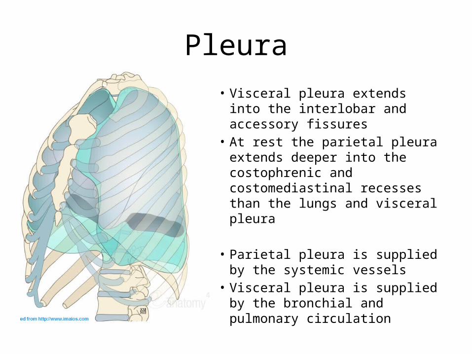
Pleura
• Visceral pleura extends into the interlobar and accessory fissures
• At rest the parietal pleura extends deeper into the costophrenic and costomediastinal recesses than the lungs and visceral pleura
• Parietal pleura is supplied by the systemic vessels
• Visceral pleura is supplied by the bronchial and pulmonary circulation

Imaging of the Lungs
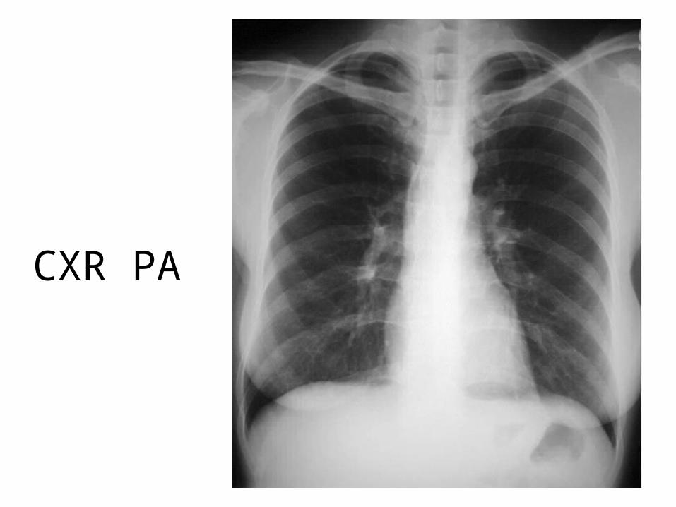
CXR PA
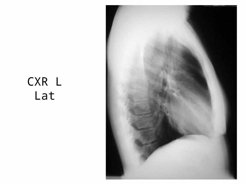
CXR L Lat
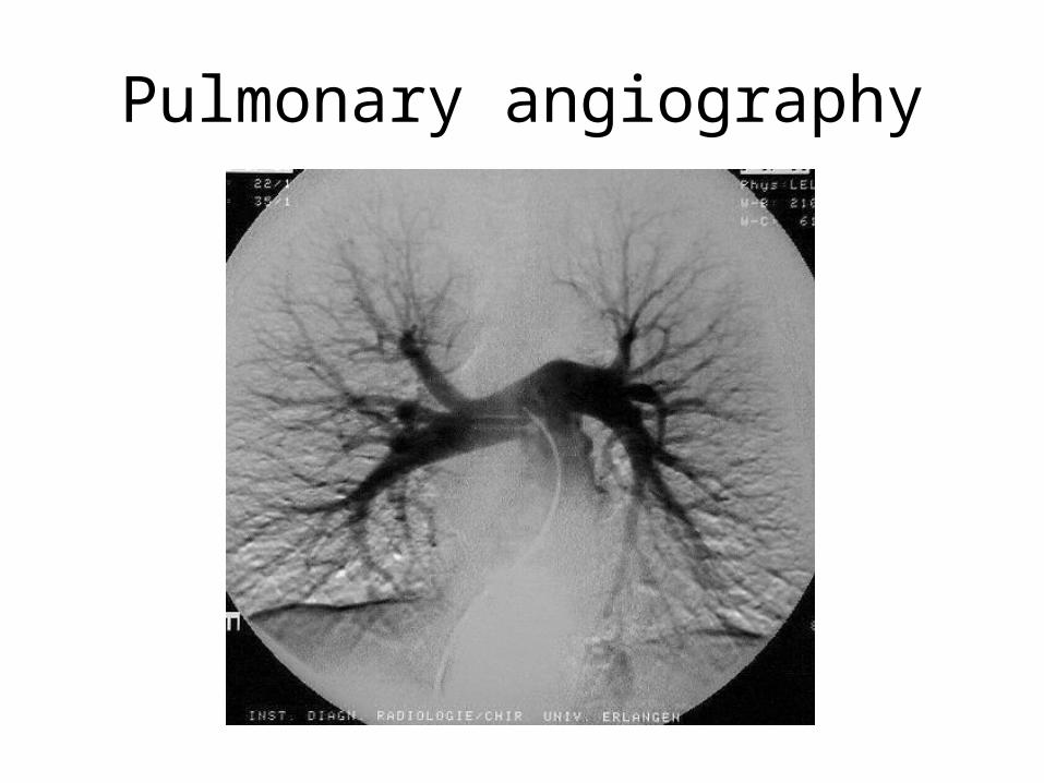
Pulmonary angiography
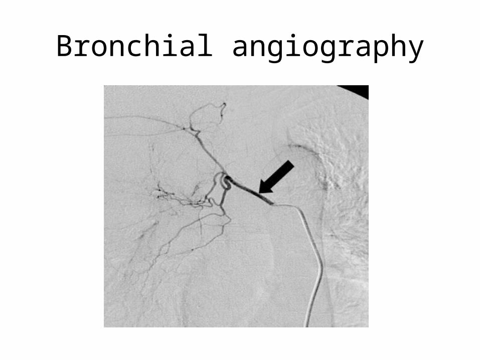
Bronchial angiography
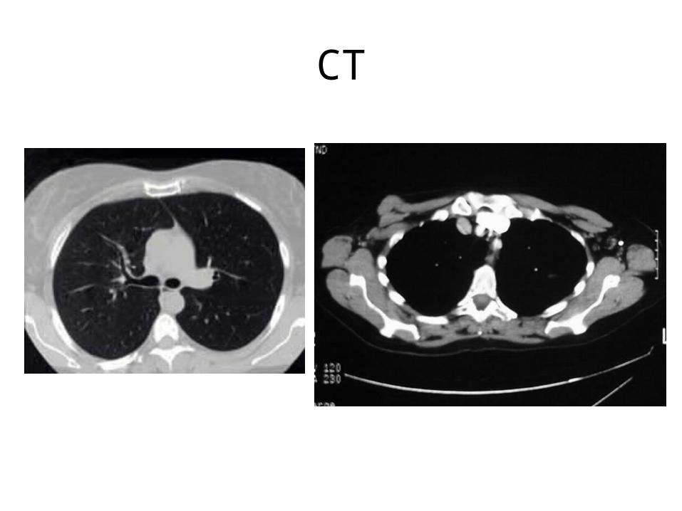
CT
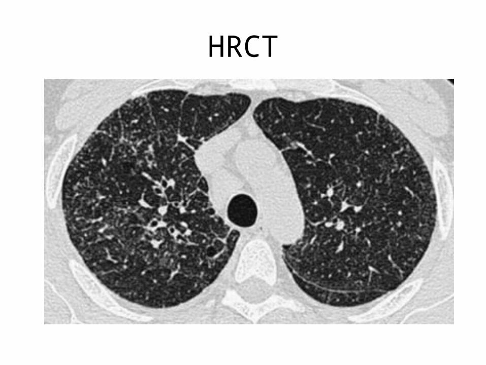
HRCT
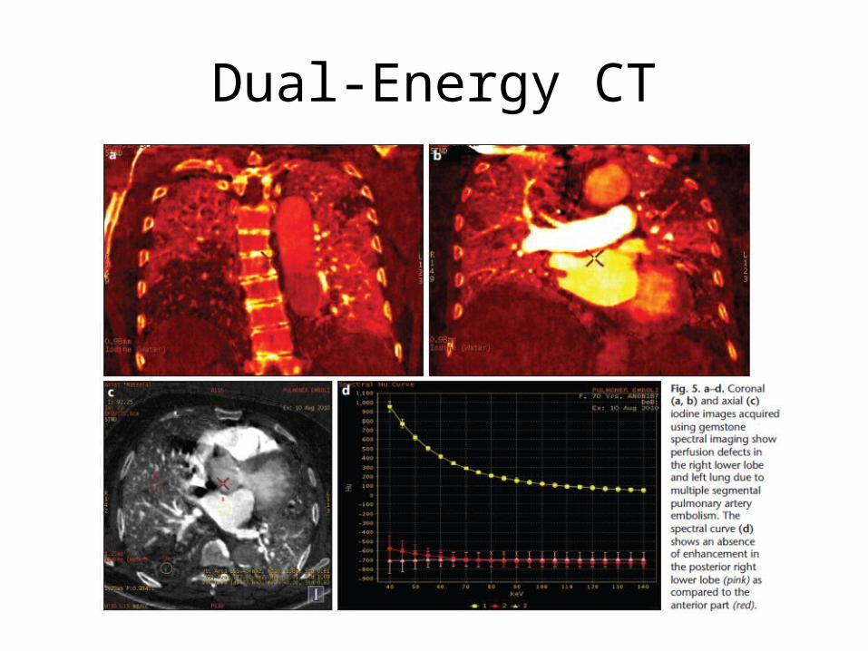
Dual-Energy CT
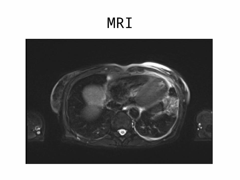
MRI
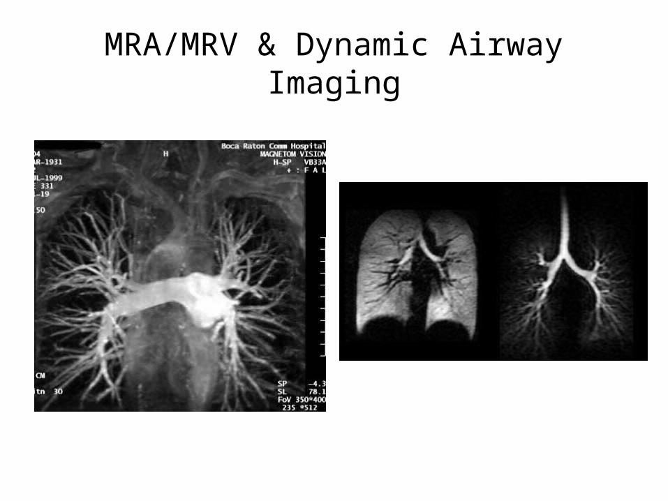
MRA/MRV & Dynamic Airway Imaging
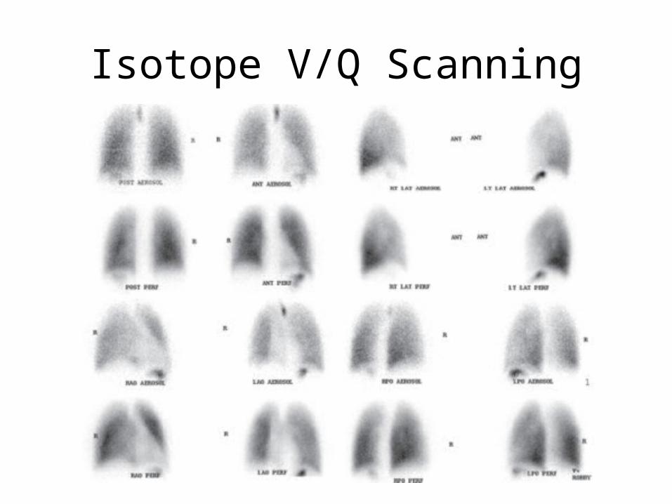
Isotope V/Q Scanning
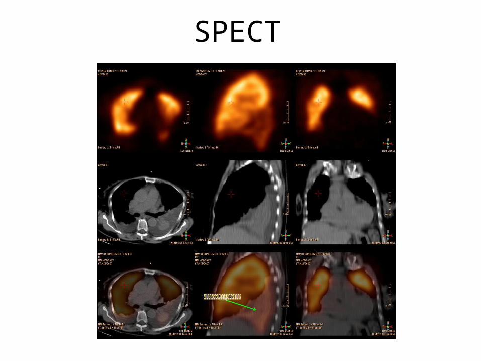
SPECT
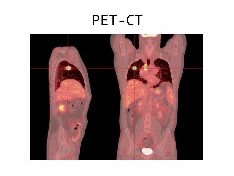
PET-CT
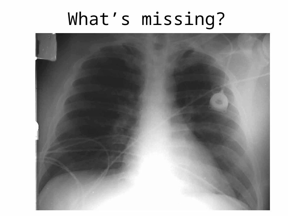
What’s missing?

Thank you
References
• Netter, F.H. (2011). Atlas of Human Anatomy, 5th ed. Philadelphia: Saunders Elsevier
• Ryan, S., McNicholas, M., Eustace, S. (2011). Anatomy for diagnostic imaging, 3rd ed. London: Saunders Elsevier
• Butler, P., Mitchell, A.W.M., Ellis, H. (1999). Applied Radiological Anatomy. Cambridge: Cambridge University Press
• Karcaaltincaba M, Aktas A. Dual-energy CT revisited with multidetector CT: review of principles and clinical applications. Diagn Interv Radiol 2011; 17:181-194