Embed Size (px)
Citation preview
TAHIR HABIB
ZOOLOGY (BIOCHEM AND CELL BIO) FOR
LECTURER ,M.sc ,B.sc,Css
TAHIR HABIB [ZOOLOGY (BIOCHEM AND CELL BIO) FOR LECTURER ,M.SC ,B.SC,CSS]
BUREAUCRATES ACADEMY ,FAIZ M ROAD QUETTA. TAHIR HABIB (03343741995) Page 1
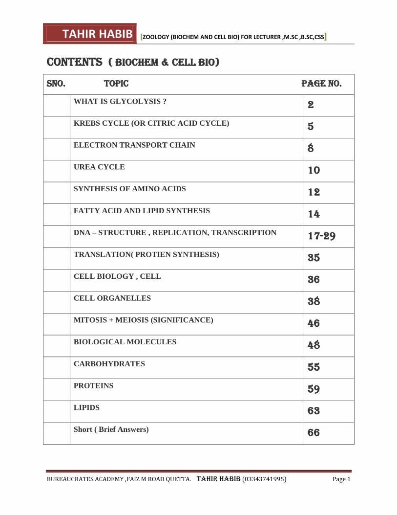
CONTENTS ( BIOCHEM & CELL BIO)
Sno. TOPIC PAGE NO.
WHAT IS GLYCOLYSIS ? 2
KREBS CYCLE (OR CITRIC ACID CYCLE) 5
ELECTRON TRANSPORT CHAIN 8
UREA CYCLE 10
SYNTHESIS OF AMINO ACIDS 12
FATTY ACID AND LIPID SYNTHESIS 14
DNA – STRUCTURE , REPLICATION, TRANSCRIPTION 17-29
TRANSLATION( PROTIEN SYNTHESIS) 35
CELL BIOLOGY , CELL 36
CELL ORGANELLES 38
MITOSIS + MEIOSIS (SIGNIFICANCE) 46
BIOLOGICAL MOLECULES 48
CARBOHYDRATES 55
PROTEINS 59
LIPIDS 63
Short ( Brief Answers) 66
TAHIR HABIB [ZOOLOGY (BIOCHEM AND CELL BIO) FOR LECTURER ,M.SC ,B.SC,CSS]
BUREAUCRATES ACADEMY ,FAIZ M ROAD QUETTA. TAHIR HABIB (03343741995) Page 2
Glycolysis
Glycolysis is the metabolic process that serves as the foundation for both aerobic and anaerobic cellular respiration. In glycolysis, glucose is converted into pyruvate. Glucose is a six- memebered ring molecule found in the blood and is usually a result of the breakdown of carbohydrates into sugars. It enters cells through specific transporter proteins that move it from outside the cell into the cell’s cytosol. All of the glycolytic enzymes are found in the cytosol.
Glycolysis literally means "splitting sugars" and is the process of releasing energy within sugars. In glycolysis, glucose (a six carbon sugar) is split into two molecules of the three-carbon sugar pyruvate. This multi-step process yields two molecules of ATP (free energy containing molecule), two molecules of pyruvate, and two "high energy" electron carrying molecules of NADH. Glycolysis can occur with or without oxygen. In the presence of oxygen, glycolysis is the first stage of cellular respiration. In the abscence of oxygen, glycolysis allows cells to make small amounts of ATP through the process of fermentation. Glycolysis takes place in the cytosol of the cell's cytoplasm. However, the next stage of cellular respiration known as the citric acid cycle, occurs in the matrix of cell mitochondria.
The overall reaction of glycolysis which occurs in the cytoplasm is represented simply as:
C6H12O6 + 2 NAD+ + 2 ADP + 2 P —–> 2 pyruvic acid, (CH3(C=O)COOH + 2 ATP + 2
NADH + 2 H+
Steps 1 and 3 = – 2ATP Steps 7 and 10 = + 4 ATP Net ―visible‖ ATP produced = 2.
Immediately upon finishing glycolysis, the cell must continue respiration in either an aerobic or anaerobic direction; this choice is made based on the circumstances of the particular cell. A cell that can perform aerobic respiration and which finds itself in the presence of oxygen will continue on to the aerobic citric acid cycle in the mitochondria. If a cell able to perform aerobic respiration is in a situation where there is no oxygen (such as muscles under extreme exertion), it will move into a type of anaerobic respiration called homolactic fermentation. Some cells such as yeast are unable to carry out aerobic respiration and will automatically move into a type of anaerobic respiration called alcoholic fermentation.
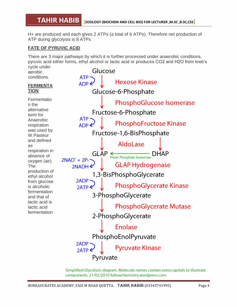
(1) GLYCOLYSIS ( Steps)
Glycolysis is the first and common step in both aerobic and anaerobic respiration. It consists of a complex series of enzymatically catalyzed reactions in which a 6 carbon molecule “Glucose” breaks down into 3 carbon “Pyruvic acid”. These reactions occur in Cytoplasm and doesn’t require oxygen. Following are the different steps of Glycolysis.
(I) PHOSPHORYLATION Phosphorylation is the addition of phosphate groups to the sugar molecules. Glucose is
TAHIR HABIB [ZOOLOGY (BIOCHEM AND CELL BIO) FOR LECTURER ,M.SC ,B.SC,CSS]
BUREAUCRATES ACADEMY ,FAIZ M ROAD QUETTA. TAHIR HABIB (03343741995) Page 3
phosphorylated by a molecule of ATP to form an activated molecule, the glucose 6 phosphate. ATP is converted to ADP.
(II) ISOMERIZATION Glucose -6-phosphate is converted to fructose -6-phosphate, an isomer of it by an enzyme.
(III) SECOND PHOSPHORYLATION Another molecules of ATP is invested which transfers its phosphate group to carbon no.1 of fructose –6-phosphate, forming fructose 1,6-bisphosphate and ADP.
(IV) CLEAVAGE The 6-carbon, fructose 1,6 bisphosphate molecule is break down into 2; three carbon molecules, 3-phosphoglyceraldehyde PGAL and dihydroxyacetone phosphate (DHAP). These two sugar molecules are isomers and are interconvertible. This is the reaction from which glycolysis derives its name. DHAP is converted to its isomer PGAL and then 2 PGAL will be converted to 2 pyruvic acid molecules. Since at this stage 2 ATPs are used, therefore this phase is known as Energy investment phase. In the subsequent reactions, energy is produced therefore this half is also known as Energy yielding phase
(V) DEHYDROGENATION (OXIDATION) In the next step, PGAL is acted upon by an enzyme dehydrogenase along with a co-enzyme nicotine amide adenine dinucleotide (NAD+), which convert PGAL into phosphoglyceric acid PGA or phosphoglycerate by the loss of two hydrogen atoms (2e- + 2H+). These H atoms are captured by NAD+. This is a redox reaction in which PGAL oxidized by removal of electrons and NAD is reduced by the gaining of electrons. Now phosphoglyceric acid PGA picks up phosphate group (Pi) present in cytoplasm and becomes 1,3-bisphosphoglyceric acid (DPGA)
(VI) PHOSPHORYL TRANSFER 1,3-bisphosphoglyceric acid loses its phosphate group to ADP forming ATP and 3-phosphoglyceric acid.
(VII) ISOMERIZATION The PO4 group of PGA, attaches with carbon no,3 changes its position to carbon no.2 forming an isomer 1-phosphoglyceric acid.
(VIII) DEHYDRATION A water molecule is removed from the substrate and forming phosphoenal pyruvate (PEP)
(IX) PHOSPHORYL TRANSFER ADP removes the high energy PO4 from PEP producing ATP and Pyruvic acid. OVERALL REACTION of glycolysis can be summarized as Glucose + 2ADP + 2NAD+ -> 2 Pyruvic acid + 2ATP + 2NADH+ H+ + 2H2O
ENERGY YIELD
Since when PGAL is produced, the cycle is counted twice because DHAP also converts into PGAL and enter the same cycle. 4ATP molecules are produced at Substrate level phosphorylation because PO4 groups are transferred directly to ADP from another molecule. 2 ATP are used in the first phase. Thus there is a net gain of 2 ATPs. 2 NADH+
TAHIR HABIB [ZOOLOGY (BIOCHEM AND CELL BIO) FOR LECTURER ,M.SC ,B.SC,CSS]
BUREAUCRATES ACADEMY ,FAIZ M ROAD QUETTA. TAHIR HABIB (03343741995) Page 4
H+ are produced and each gives 2 ATPs (a total of 6 ATPs). Therefore net production of ATP during glycolysis is 8 ATPs
FATE OF PYRUVIC ACID
There are 3 major pathways by which it is further processed under anaerobic conditions, pyruvic acid either forms, ethyl alcohol or lactic acid or produces CO2 and H2O from kreb’s cycle under aerobic conditions.
FERMENTATION
Fermentation the alternative term for Anaerobic respiration was used by W.Pasteur and defined as respiration in absence of oxygen (air). The production of ethyl alcohol from glucose is alcoholic fermentation and that of lactic acid is lactic acid fermentation
TAHIR HABIB [ZOOLOGY (BIOCHEM AND CELL BIO) FOR LECTURER ,M.SC ,B.SC,CSS]
BUREAUCRATES ACADEMY ,FAIZ M ROAD QUETTA. TAHIR HABIB (03343741995) Page 5
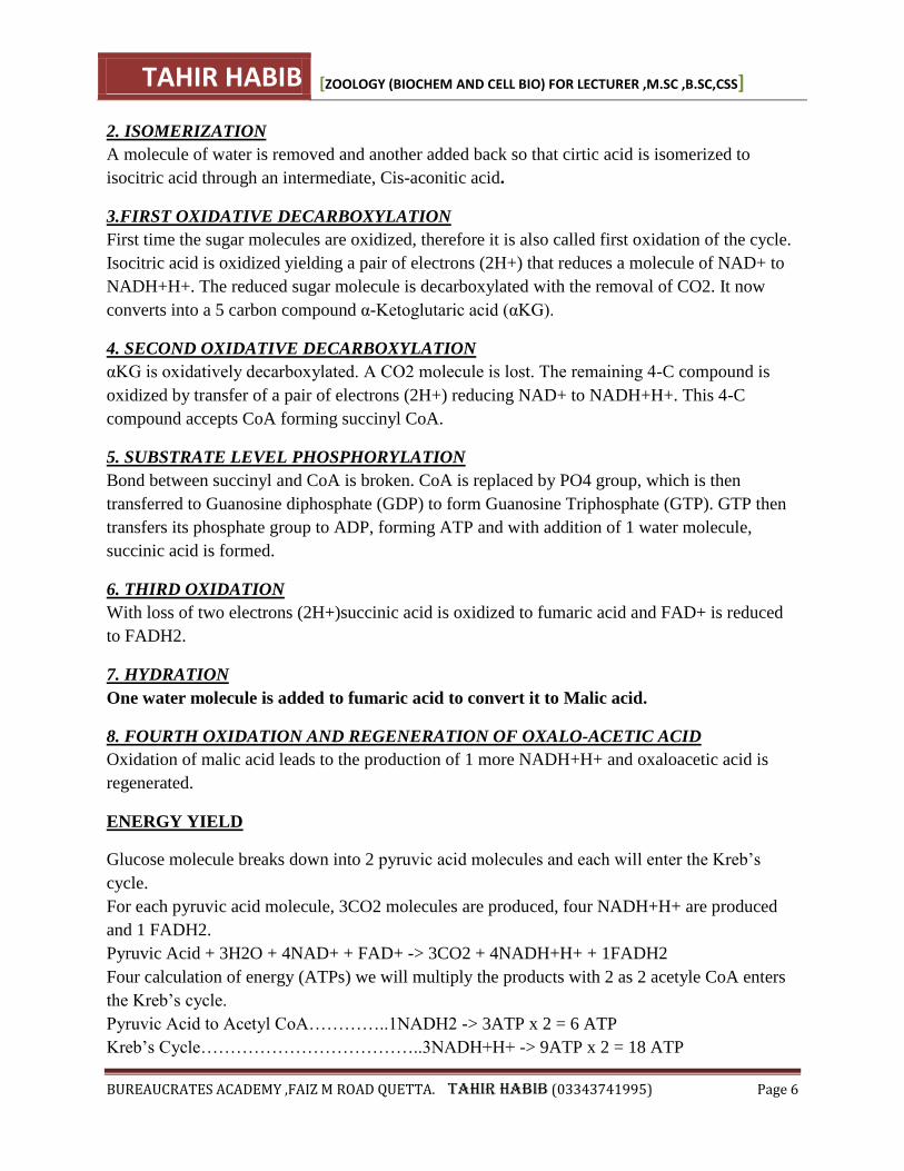
Krebs cycle (or citric acid cycle)
The Krebs cycle (or citric acid cycle) is a part of cellular respiration. Named after Hans Krebs, it
is a series of chemical reactions used by all aerobic organisms to generate energy. Its importance
to many biochemical pathways suggests that it was one of the earliest parts of cellular
metabolism to evolve.
The Krebs cycle comes after the link reaction and provides the hydrogen and electrons needed
for the electron transport chain. It takes place inside mitochondria.
The oxidation of pyruvic acid into CO2 and water is called Krebs cycle. This cycle is also citric
acid cycle because the cycle begins with the formation of citric acid. Citric acid is a carboxylic
acid containing 3 COOH groups. Hence this cycle is also called as tri carboxylic acid cycle or
TCA cycle. This cycle was first described by Kreb's in 1936. This cycle occurs only in the
presence of oxygen. Hence it is an aerobic process. It takes place in the mitochondria.
KREB’S CYCLE (DETaiLD STEpS)
FORMATION OF ACETYL-COA
Before entering the Kreb’s cycle, each molecule of pyruvic acid undergoes oxidative
decarboxylation. During this process one of the three carbons of pyruvic acid molecule is
removed to form CO2 by enzymatic reactions. Simultaneously pyruvic acid is oxidized and a
pair of energy rich Hydrogen atoms are passed on to a H acceptor NAD+ to form NADH+H+.
The remaining 2-carbon component is called acetyle which combines with coenzyme A to form
an activated two carbon compound called acetyle CoA. ―Acetyle CoA connects Kreb’s cycle
with glycolysis.‖ For each molecule of glucose that enters glycoilysis, two molecules of acetyle
CoA produced, which enter in a cyclic series of enzymatically catalyzed reactions known as
Kreb’s Cycle, which occurs in Mitochondria.
Pyruvic acid (3C) + CoA + NAD+ -> Acetyle CoA + CO2 + NADH+H+
SERIES OF REACTIONS IN KREB’S CYCLE
Sir Hans Kreb was working over these cyclical series of reactions therefore the cycle was given
the name as Kreb’s cycle. The first molecule formed during the cycle is citric acid, so it is also
called as ―Citric Acid cycle.‖ This cycle is a multi step process and the steps are given below:
1. FORMATION OF CITRIC ACID
In this first step of the Kreb’s cycle, bond between acetyl and CoA is broken by the addition of
water molecule. The acetyl (2C) reacts with 4 carbon compound (oxalo acetic) acid to form 6-
carbon compound, citric acid, and the CoA is set free. This citric acid possess 3 carboxyl groups,
therefore the cycle is also recommended as Tricarboxylic Acid Cycle (TCA cycle).
TAHIR HABIB [ZOOLOGY (BIOCHEM AND CELL BIO) FOR LECTURER ,M.SC ,B.SC,CSS]
BUREAUCRATES ACADEMY ,FAIZ M ROAD QUETTA. TAHIR HABIB (03343741995) Page 6
2. ISOMERIZATION
A molecule of water is removed and another added back so that cirtic acid is isomerized to
isocitric acid through an intermediate, Cis-aconitic acid.
3.FIRST OXIDATIVE DECARBOXYLATION
First time the sugar molecules are oxidized, therefore it is also called first oxidation of the cycle.
Isocitric acid is oxidized yielding a pair of electrons (2H+) that reduces a molecule of NAD+ to
NADH+H+. The reduced sugar molecule is decarboxylated with the removal of CO2. It now
converts into a 5 carbon compound α-Ketoglutaric acid (αKG).
4. SECOND OXIDATIVE DECARBOXYLATION
αKG is oxidatively decarboxylated. A CO2 molecule is lost. The remaining 4-C compound is
oxidized by transfer of a pair of electrons (2H+) reducing NAD+ to NADH+H+. This 4-C
compound accepts CoA forming succinyl CoA.
5. SUBSTRATE LEVEL PHOSPHORYLATION
Bond between succinyl and CoA is broken. CoA is replaced by PO4 group, which is then
transferred to Guanosine diphosphate (GDP) to form Guanosine Triphosphate (GTP). GTP then
transfers its phosphate group to ADP, forming ATP and with addition of 1 water molecule,
succinic acid is formed.
6. THIRD OXIDATION
With loss of two electrons (2H+)succinic acid is oxidized to fumaric acid and FAD+ is reduced
to FADH2.
7. HYDRATION
One water molecule is added to fumaric acid to convert it to Malic acid.
8. FOURTH OXIDATION AND REGENERATION OF OXALO-ACETIC ACID
Oxidation of malic acid leads to the production of 1 more NADH+H+ and oxaloacetic acid is
regenerated.
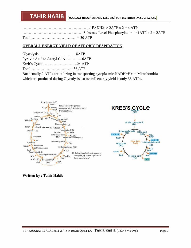
ENERGY YIELD
Glucose molecule breaks down into 2 pyruvic acid molecules and each will enter the Kreb’s
cycle.
For each pyruvic acid molecule, 3CO2 molecules are produced, four NADH+H+ are produced
and 1 FADH2.
Pyruvic Acid + 3H2O + 4NAD+ + FAD+ -> 3CO2 + 4NADH+H+ + 1FADH2
Four calculation of energy (ATPs) we will multiply the products with 2 as 2 acetyle CoA enters
the Kreb’s cycle.
Pyruvic Acid to Acetyl CoA…………..1NADH2 -> 3ATP x 2 = 6 ATP
Kreb’s Cycle………………………………..3NADH+H+ -> 9ATP x 2 = 18 ATP
TAHIR HABIB [ZOOLOGY (BIOCHEM AND CELL BIO) FOR LECTURER ,M.SC ,B.SC,CSS]
BUREAUCRATES ACADEMY ,FAIZ M ROAD QUETTA. TAHIR HABIB (03343741995) Page 7
………………………………………………1FADH2 -> 2ATP x 2 = 4 ATP
………………………………………….Substrate Level Phosphorylation -> 1ATP x 2 = 2ATP
Total………………………………. = 30 ATP
OVERALL ENERGY YIELD OF AEROBIC RESPIRATION
Glycolysis…………………………8ATP
Pyruvic Acid to Acetyl CoA…………..6ATP
Kreb’s Cycle……………………….24 ATP
Total……………………………..38 ATP
But actually 2 ATPs are utilizing in transporting cytoplasmic NADH+H+ to Mitochondria,
which are produced during Glycolysis, so overall energy yield is only 36 ATPs.
Written by : Tahir Habib
TAHIR HABIB [ZOOLOGY (BIOCHEM AND CELL BIO) FOR LECTURER ,M.SC ,B.SC,CSS]
BUREAUCRATES ACADEMY ,FAIZ M ROAD QUETTA. TAHIR HABIB (03343741995) Page 8
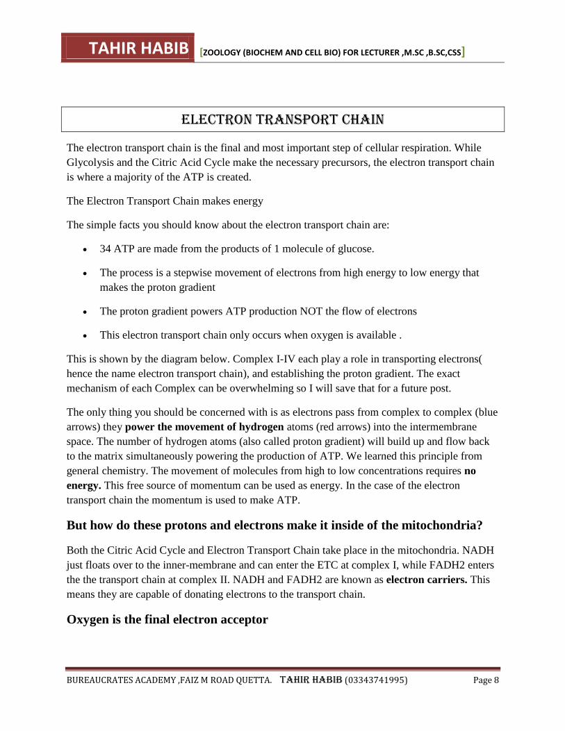
electron transport chain
The electron transport chain is the final and most important step of cellular respiration. While
Glycolysis and the Citric Acid Cycle make the necessary precursors, the electron transport chain
is where a majority of the ATP is created.
The Electron Transport Chain makes energy
The simple facts you should know about the electron transport chain are:
34 ATP are made from the products of 1 molecule of glucose.
The process is a stepwise movement of electrons from high energy to low energy that
makes the proton gradient
The proton gradient powers ATP production NOT the flow of electrons
This electron transport chain only occurs when oxygen is available .
This is shown by the diagram below. Complex I-IV each play a role in transporting electrons(
hence the name electron transport chain), and establishing the proton gradient. The exact
mechanism of each Complex can be overwhelming so I will save that for a future post.
The only thing you should be concerned with is as electrons pass from complex to complex (blue
arrows) they power the movement of hydrogen atoms (red arrows) into the intermembrane
space. The number of hydrogen atoms (also called proton gradient) will build up and flow back
to the matrix simultaneously powering the production of ATP. We learned this principle from
general chemistry. The movement of molecules from high to low concentrations requires no
energy. This free source of momentum can be used as energy. In the case of the electron
transport chain the momentum is used to make ATP.
But how do these protons and electrons make it inside of the mitochondria?
Both the Citric Acid Cycle and Electron Transport Chain take place in the mitochondria. NADH
just floats over to the inner-membrane and can enter the ETC at complex I, while FADH2 enters
the the transport chain at complex II. NADH and FADH2 are known as electron carriers. This
means they are capable of donating electrons to the transport chain.
Oxygen is the final electron acceptor
TAHIR HABIB [ZOOLOGY (BIOCHEM AND CELL BIO) FOR LECTURER ,M.SC ,B.SC,CSS]
BUREAUCRATES ACADEMY ,FAIZ M ROAD QUETTA. TAHIR HABIB (03343741995) Page 9
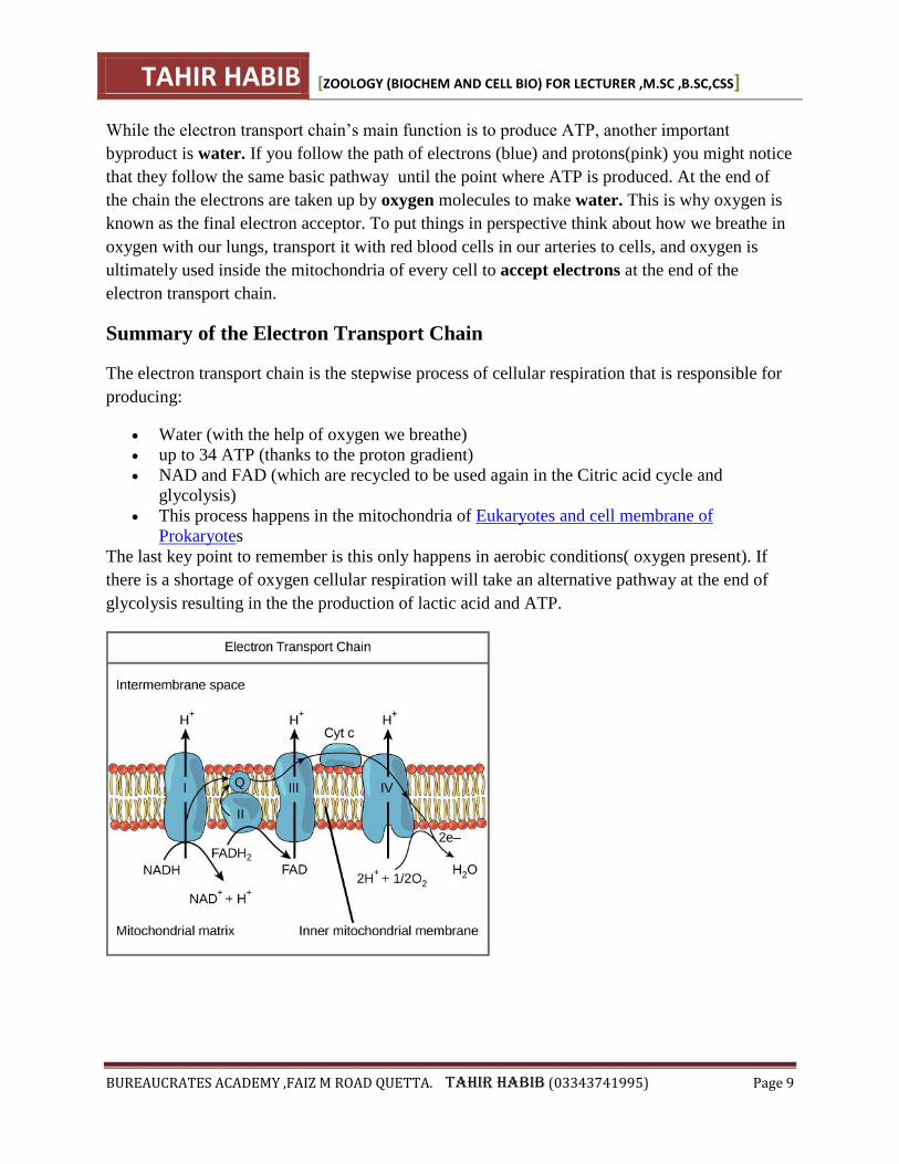
While the electron transport chain’s main function is to produce ATP, another important
byproduct is water. If you follow the path of electrons (blue) and protons(pink) you might notice
that they follow the same basic pathway until the point where ATP is produced. At the end of
the chain the electrons are taken up by oxygen molecules to make water. This is why oxygen is
known as the final electron acceptor. To put things in perspective think about how we breathe in
oxygen with our lungs, transport it with red blood cells in our arteries to cells, and oxygen is
ultimately used inside the mitochondria of every cell to accept electrons at the end of the
electron transport chain.
Summary of the Electron Transport Chain
The electron transport chain is the stepwise process of cellular respiration that is responsible for
producing:
Water (with the help of oxygen we breathe)
up to 34 ATP (thanks to the proton gradient)
NAD and FAD (which are recycled to be used again in the Citric acid cycle and
glycolysis)
This process happens in the mitochondria of Eukaryotes and cell membrane of
Prokaryotes
The last key point to remember is this only happens in aerobic conditions( oxygen present). If
there is a shortage of oxygen cellular respiration will take an alternative pathway at the end of
glycolysis resulting in the the production of lactic acid and ATP.
TAHIR HABIB [ZOOLOGY (BIOCHEM AND CELL BIO) FOR LECTURER ,M.SC ,B.SC,CSS]
BUREAUCRATES ACADEMY ,FAIZ M ROAD QUETTA. TAHIR HABIB (03343741995) Page 10
UREA CYCLE
In humans and mammals, almost 80% of the nitrogen excreted is in the form of urea, which is
produced through a series of reactions occurring in the cytosol and mitochondrial matrix of liver
cells. These reactions are collectively called the urea cycle or the Krebs-Henseleit cycle.
Ammonia is a toxic product of nitrogen metabolism which should be removed from our body.
The urea cycle or ornithine cycle converts excess ammonia into urea in the mitochondria of liver
cells. The urea forms, then enters the blood stream, is filtered by the kidneys and is ultimately
excreted in the urine.
The overall reaction for urea formation from ammonia is as follows:
2 Ammonia + CO2 + 3ATP ---> urea + water + 3 ADP
Steps in the Urea Cycle
The urea cycle is a series of five reactions catalyzed by several key enzymes. The first two
steps in the cycle take place in the mitochondrial matrix and the rest of the steps take place in the
cytosol. Thus the urea cycle spans two cellular compartments of the liver cell.
In the first step of the Krebs-Henseleit cycle, ammonia produced in the mitochondria
is converted to carbamoyl phosphate by an enzyme called carbamoyl phosphate
synthetase I. The reaction can be given as follows:
NH3 + CO2 + 2ATP → carbamoyl phosphate + 2ADP + Pi
The second step involves the transfer of a carbamoyl group from carbamoyl
phosphate to ornithine to form citrulline. This step is catalyzed by the enzyme ornithine
transcarbamoylase (OTC) . The reaction is given as follows:
Carbamoyl phosphate + ornithine → citrulline + Pi
Citrulline thus formed is released into the cytosol for use in the rest of the steps of the cycle.
TAHIR HABIB [ZOOLOGY (BIOCHEM AND CELL BIO) FOR LECTURER ,M.SC ,B.SC,CSS]
BUREAUCRATES ACADEMY ,FAIZ M ROAD QUETTA. TAHIR HABIB (03343741995) Page 11
The third step is catalyzed by an enzyme called argininosuccinate synthetase, which
uses citrulline and ATP to form a citrullyl-AMP intermediate, which reacts with an amino
group from aspartate to produce argininosuccinate. This reaction can be given as follows:
Citrulline + ATP + aspartate → argininosuccinate + AMP + PPi
The fourth step involves the cleavage of argininosuccinate to form fumarate and
arginine. Argininosuccinate lyase is the enzyme catalyzing this reaction, which can be
represented as follows:
Argininosuccinate → arginine + fumarate
In the fifth and last step of the urea cycle, arginine is hydrolyzed to form urea and
ornithine. This is catalyzed by arginase and can be given as follows:
Arginine → urea + ornithine
The overall reaction can be given as follows:
2NH3 + CO2 + 3ATP g urea + 2ADP + AMP + PPi + 2Pi
Significance of the Urea Cycle
The main purpose of the urea cycle is to eliminate toxic ammonia from the body. About 10 to 20
g of ammonia is removed from the body of a healthy adult every day. A dysfunctional urea cycle
would mean excess amount of ammonia in the body, which can lead to hyperammonemia and
related diseases. The deficiency of one or more of the key enzymes catalyzing various reactions
in the urea cycle can cause disorders related to the cycle. Defects in the urea cycle can cause
vomiting, coma and convulsions in new born babies. This is often misdiagnosed as septicemia
and treated with antibiotics in vain. Even 1mm of excess ammonia can cause severe and
irreversible damages.
Diagnosis of Urea Cycle Defects
A blood aminogram is routinely used in the diagnosis of urea cycle disorders. The concentration
of the nitrogen-carrying amino acids, glutamine and alanine, in plasma is elevated in the case of
OTC deficiency. In babies, elevated levels of orotic acid in the urine may be an indicator of OTC
deficiency. Increased levels of blood citrulline and argininosuccinate are also seen in cases of
citrullinemia.
In older children, these disorders may present in the form of growth failure, psychomotor
retardation and behavioral abnormalities. Hence, blood ammonia and urinary orotic acid
monitoring and quantitation are crucial in patients with unexplained neurological symptoms.
TAHIR HABIB [ZOOLOGY (BIOCHEM AND CELL BIO) FOR LECTURER ,M.SC ,B.SC,CSS]
BUREAUCRATES ACADEMY ,FAIZ M ROAD QUETTA. TAHIR HABIB (03343741995) Page 12
SYNTHESIS OF AMINO ACIDS
Synthesis and/or collection of amino acids is critical for cell survival. They not only serve as the
building blocks for proteins but also as starting points for the synthesis of many important
cellular molecules including vitamins and nucleotides.
In most cases bacteria would rather use amino acids in their environment than make them from
scratch. It takes a considerable amount of energy to make the enzymes for the pathway as well as
the energy required to drive some of the reactions of amino acid biosynthesis. The genes that
code for amino acid synthesis enzymes and the enzymes themselves are under tight control and
are only turned on when they are needed.
The amino acids synthesis pathways can be grouped into several logical units. These units reflect
either common mechanisms or the use of common enzymes that synthesize more than one amino
acid. These categories are: simple reactions, branch chain amino acids, aromatic amino acids,
threonine/lysine, serine/glycine, and unique pathways. The aromatic amino acids,
threonine/lysine and serine/glycine pathways have a common beginning and then diverge to form
the amino acid of interest.
Notice that each pathway begins with a central metabolite or something derived from "central
metabolism". Using common compounds instead of synthesizing them from scratch saves energy
and conserves genes since fewer enzymes are needed to code for the pathways.
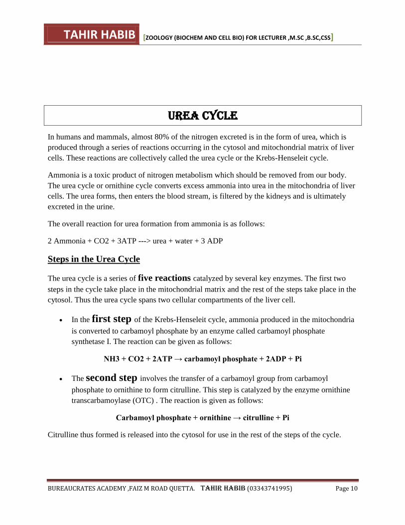
Simple Reactions
glutamine, glutamate, aspartate, asparagine and alanine
In most cases these amino acids can be synthesize by one step reactions from central
metabolites. They are simple in structure and their synthesis is also straight forward.
Glutamate can by synthesized
by the addition of ammonia to -ketoglutarate.
TAHIR HABIB [ZOOLOGY (BIOCHEM AND CELL BIO) FOR LECTURER ,M.SC ,B.SC,CSS]
BUREAUCRATES ACADEMY ,FAIZ M ROAD QUETTA. TAHIR HABIB (03343741995) Page 13
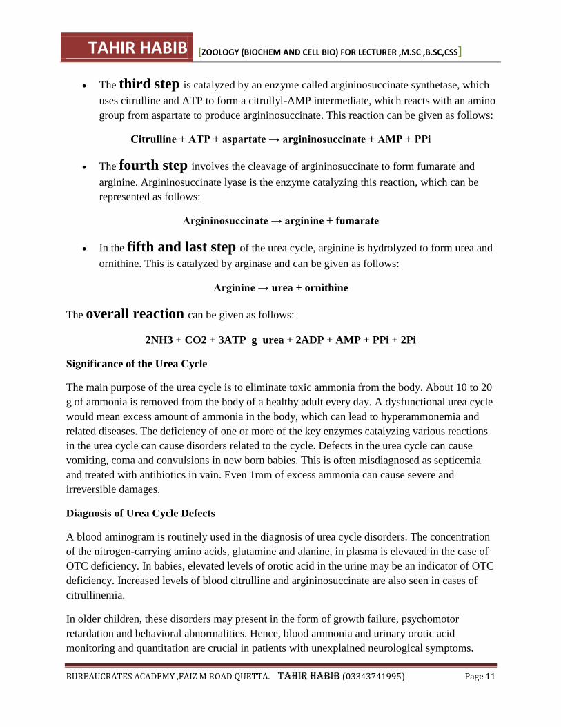
Figure 1 - The synthesis of glutamate.
Glutamine is made by the addition of another ammonia molecule to glutamate.
Figure 2 - Synthesis of glutamine
The rest of the simple reactions involve transfer of the amino group (transamination)
from glutamate or glutamine to a central metabolite to make the required amino acid.
Aspartate is synthesize by the transfer of a ammonia group from glutamate to
oxaloacetate.
Figure 3 - The synthesis of aspartate.
Asparagine is made either by transamination from glutamine or by adding ammonia
directly to aspartate.
or
TAHIR HABIB [ZOOLOGY (BIOCHEM AND CELL BIO) FOR LECTURER ,M.SC ,B.SC,CSS]
BUREAUCRATES ACADEMY ,FAIZ M ROAD QUETTA. TAHIR HABIB (03343741995) Page 14
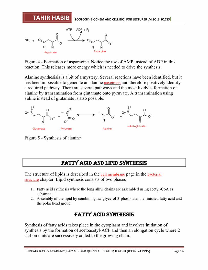
Figure 4 - Formation of asparagine. Notice the use of AMP instead of ADP in this reaction. This releases more energy which is needed to drive the synthesis.
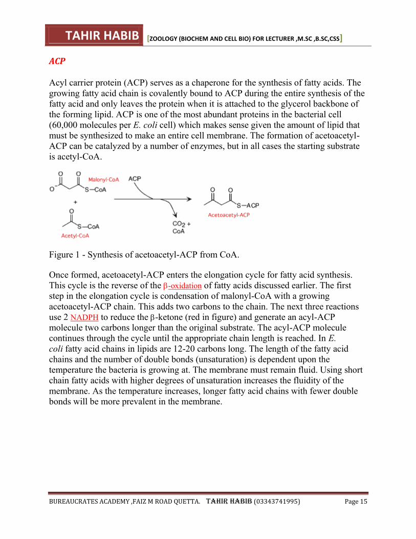
Alanine synthesisis is a bit of a mystery. Several reactions have been identified, but it
has been impossible to generate an alanine auxotroph and therefore positively identify
a required pathway. There are several pathways and the most likely is formation of
alanine by transamination from glutamate onto pyruvate. A transamination using valine instead of glutamate is also possible.
Figure 5 - Synthesis of alanine
Fatty Acid and Lipid Synthesis
The structure of lipids is described in the cell membrane page in the bacterial
structure chapter. Lipid synthesis consists of two phases
1. Fatty acid synthesis where the long alkyl chains are assembled using acetyl-CoA as
substrate.
2. Assembly of the lipid by combining, sn-glycerol-3-phosphate, the finished fatty acid and
the polar head group.
Fatty acid synthesis
Synthesis of fatty acids takes place in the cytoplasm and involves initiation of
synthesis by the formation of acetoacetyl-ACP and then an elongation cycle where 2 carbon units are successively added to the growing chain.
TAHIR HABIB [ZOOLOGY (BIOCHEM AND CELL BIO) FOR LECTURER ,M.SC ,B.SC,CSS]
BUREAUCRATES ACADEMY ,FAIZ M ROAD QUETTA. TAHIR HABIB (03343741995) Page 15
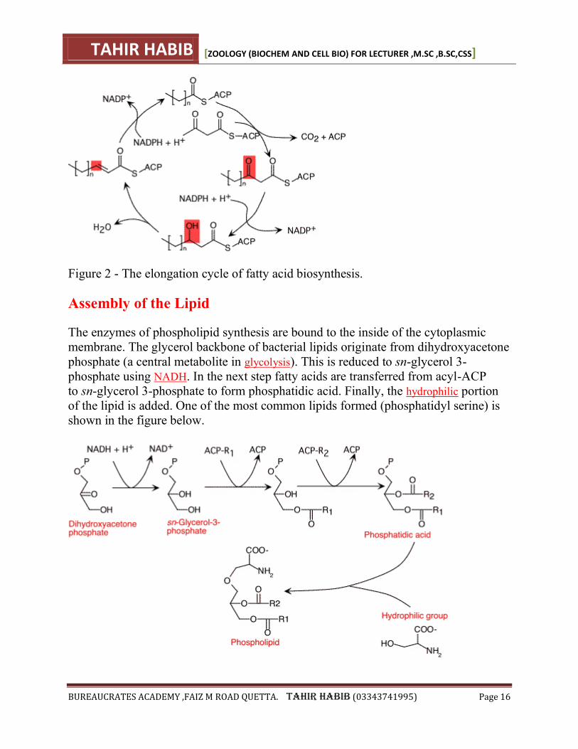
ACP
Acyl carrier protein (ACP) serves as a chaperone for the synthesis of fatty acids. The
growing fatty acid chain is covalently bound to ACP during the entire synthesis of the
fatty acid and only leaves the protein when it is attached to the glycerol backbone of
the forming lipid. ACP is one of the most abundant proteins in the bacterial cell
(60,000 molecules per E. coli cell) which makes sense given the amount of lipid that
must be synthesized to make an entire cell membrane. The formation of acetoacetyl-
ACP can be catalyzed by a number of enzymes, but in all cases the starting substrate
is acetyl-CoA.
Figure 1 - Synthesis of acetoacetyl-ACP from CoA.
Once formed, acetoacetyl-ACP enters the elongation cycle for fatty acid synthesis.
This cycle is the reverse of the -oxidation of fatty acids discussed earlier. The first
step in the elongation cycle is condensation of malonyl-CoA with a growing
acetoacetyl-ACP chain. This adds two carbons to the chain. The next three reactions
use 2 NADPH to reduce the -ketone (red in figure) and generate an acyl-ACP
molecule two carbons longer than the original substrate. The acyl-ACP molecule
continues through the cycle until the appropriate chain length is reached. In E.
coli fatty acid chains in lipids are 12-20 carbons long. The length of the fatty acid
chains and the number of double bonds (unsaturation) is dependent upon the
temperature the bacteria is growing at. The membrane must remain fluid. Using short
chain fatty acids with higher degrees of unsaturation increases the fluidity of the
membrane. As the temperature increases, longer fatty acid chains with fewer double bonds will be more prevalent in the membrane.
TAHIR HABIB [ZOOLOGY (BIOCHEM AND CELL BIO) FOR LECTURER ,M.SC ,B.SC,CSS]
BUREAUCRATES ACADEMY ,FAIZ M ROAD QUETTA. TAHIR HABIB (03343741995) Page 16
Figure 2 - The elongation cycle of fatty acid biosynthesis.
Assembly of the Lipid
The enzymes of phospholipid synthesis are bound to the inside of the cytoplasmic
membrane. The glycerol backbone of bacterial lipids originate from dihydroxyacetone
phosphate (a central metabolite in glycolysis). This is reduced to sn-glycerol 3-
phosphate using NADH. In the next step fatty acids are transferred from acyl-ACP
to sn-glycerol 3-phosphate to form phosphatidic acid. Finally, the hydrophilic portion
of the lipid is added. One of the most common lipids formed (phosphatidyl serine) is
shown in the figure below.
TAHIR HABIB [ZOOLOGY (BIOCHEM AND CELL BIO) FOR LECTURER ,M.SC ,B.SC,CSS]
BUREAUCRATES ACADEMY ,FAIZ M ROAD QUETTA. TAHIR HABIB (03343741995) Page 17
DNA - STRUCTURE
This page, looking at the structure of DNA, is the first in a sequence of pages leading on to how
DNA replicates (makes copies of) itself, and then to how information stored in DNA is used to
make protein molecules. This material is aimed at 16 - 18 year old chemistry students. If you are
interested in this from a biological or biochemical point of view, you may find these pages a
useful introduction before you get more information somewhere else.
Exploring a DNA chain
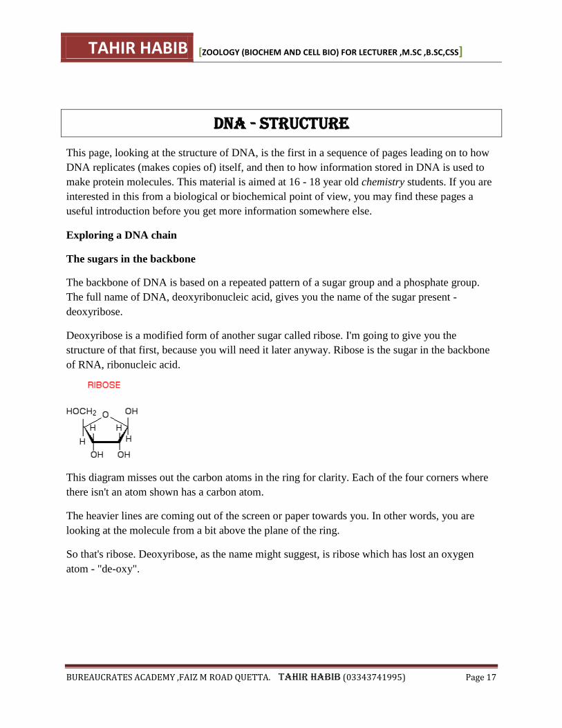
The sugars in the backbone
The backbone of DNA is based on a repeated pattern of a sugar group and a phosphate group.
The full name of DNA, deoxyribonucleic acid, gives you the name of the sugar present -
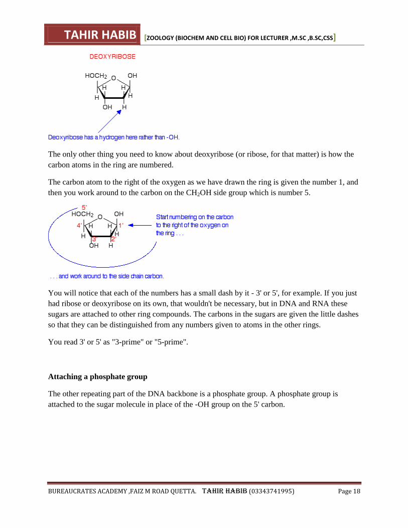
deoxyribose.
Deoxyribose is a modified form of another sugar called ribose. I'm going to give you the
structure of that first, because you will need it later anyway. Ribose is the sugar in the backbone
of RNA, ribonucleic acid.
This diagram misses out the carbon atoms in the ring for clarity. Each of the four corners where
there isn't an atom shown has a carbon atom.
The heavier lines are coming out of the screen or paper towards you. In other words, you are
looking at the molecule from a bit above the plane of the ring.
So that's ribose. Deoxyribose, as the name might suggest, is ribose which has lost an oxygen
atom - "de-oxy".
TAHIR HABIB [ZOOLOGY (BIOCHEM AND CELL BIO) FOR LECTURER ,M.SC ,B.SC,CSS]
BUREAUCRATES ACADEMY ,FAIZ M ROAD QUETTA. TAHIR HABIB (03343741995) Page 18
The only other thing you need to know about deoxyribose (or ribose, for that matter) is how the
carbon atoms in the ring are numbered.
The carbon atom to the right of the oxygen as we have drawn the ring is given the number 1, and
then you work around to the carbon on the CH2OH side group which is number 5.
You will notice that each of the numbers has a small dash by it - 3' or 5', for example. If you just
had ribose or deoxyribose on its own, that wouldn't be necessary, but in DNA and RNA these
sugars are attached to other ring compounds. The carbons in the sugars are given the little dashes
so that they can be distinguished from any numbers given to atoms in the other rings.
You read 3' or 5' as "3-prime" or "5-prime".
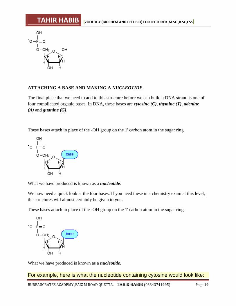
Attaching a phosphate group
The other repeating part of the DNA backbone is a phosphate group. A phosphate group is
attached to the sugar molecule in place of the -OH group on the 5' carbon.
TAHIR HABIB [ZOOLOGY (BIOCHEM AND CELL BIO) FOR LECTURER ,M.SC ,B.SC,CSS]
BUREAUCRATES ACADEMY ,FAIZ M ROAD QUETTA. TAHIR HABIB (03343741995) Page 19
ATTACHING A BASE AND MAKING A NUCLEOTIDE
The final piece that we need to add to this structure before we can build a DNA strand is one of
four complicated organic bases. In DNA, these bases are cytosine (C), thymine (T), adenine
(A) and guanine (G).
These bases attach in place of the -OH group on the 1' carbon atom in the sugar ring.
What we have produced is known as a nucleotide.
We now need a quick look at the four bases. If you need these in a chemistry exam at this level,
the structures will almost certainly be given to you.
These bases attach in place of the -OH group on the 1' carbon atom in the sugar ring.
What we have produced is known as a nucleotide.
For example, here is what the nucleotide containing cytosine would look like:
TAHIR HABIB [ZOOLOGY (BIOCHEM AND CELL BIO) FOR LECTURER ,M.SC ,B.SC,CSS]
BUREAUCRATES ACADEMY ,FAIZ M ROAD QUETTA. TAHIR HABIB (03343741995) Page 20
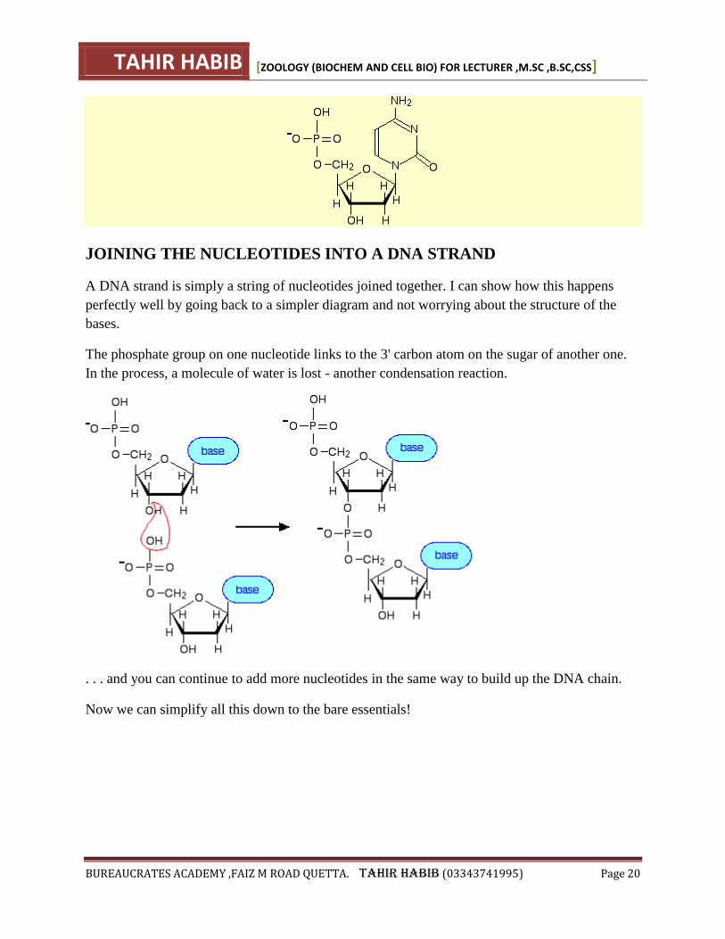
JOINING THE NUCLEOTIDES INTO A DNA STRAND
A DNA strand is simply a string of nucleotides joined together. I can show how this happens
perfectly well by going back to a simpler diagram and not worrying about the structure of the
bases.
The phosphate group on one nucleotide links to the 3' carbon atom on the sugar of another one.
In the process, a molecule of water is lost - another condensation reaction.
. . . and you can continue to add more nucleotides in the same way to build up the DNA chain.
Now we can simplify all this down to the bare essentials!
TAHIR HABIB [ZOOLOGY (BIOCHEM AND CELL BIO) FOR LECTURER ,M.SC ,B.SC,CSS]
BUREAUCRATES ACADEMY ,FAIZ M ROAD QUETTA. TAHIR HABIB (03343741995) Page 21
JOINING THE TWO DNA CHAINS TOGETHER
THE IMPORTANCE OF "BASE PAIRS"
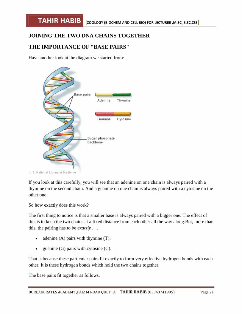
Have another look at the diagram we started from:
If you look at this carefully, you will see that an adenine on one chain is always paired with a
thymine on the second chain. And a guanine on one chain is always paired with a cytosine on the
other one.
So how exactly does this work?
The first thing to notice is that a smaller base is always paired with a bigger one. The effect of
this is to keep the two chains at a fixed distance from each other all the way along.But, more than
this, the pairing has to be exactly . . .
adenine (A) pairs with thymine (T);
guanine (G) pairs with cytosine (C).
That is because these particular pairs fit exactly to form very effective hydrogen bonds with each
other. It is these hydrogen bonds which hold the two chains together.
The base pairs fit together as follows.
TAHIR HABIB [ZOOLOGY (BIOCHEM AND CELL BIO) FOR LECTURER ,M.SC ,B.SC,CSS]
BUREAUCRATES ACADEMY ,FAIZ M ROAD QUETTA. TAHIR HABIB (03343741995) Page 22
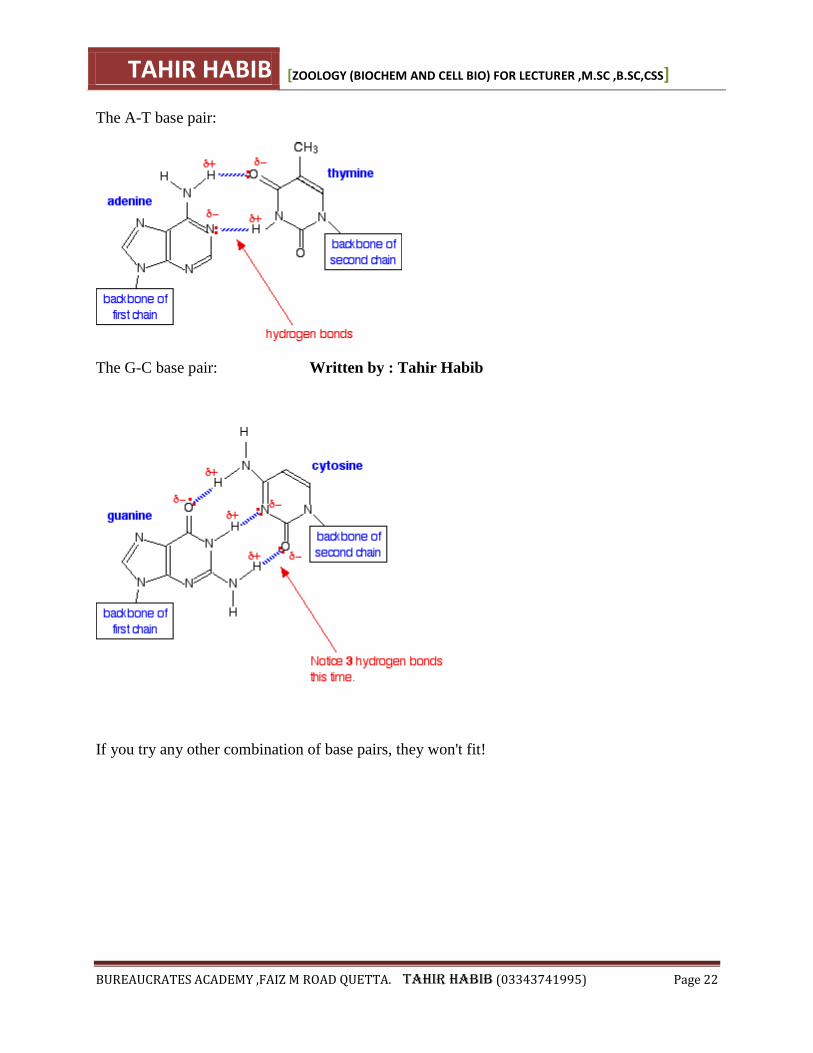
The A-T base pair:
The G-C base pair: Written by : Tahir Habib
If you try any other combination of base pairs, they won't fit!

TAHIR HABIB [ZOOLOGY (BIOCHEM AND CELL BIO) FOR LECTURER ,M.SC ,B.SC,CSS]
BUREAUCRATES ACADEMY ,FAIZ M ROAD QUETTA. TAHIR HABIB (03343741995) Page 23
DNA REPLICATION:
Key points:
DNA replication is semiconservative. Each strand in the double helix acts as a template
for synthesis of a new, complementary strand.
New DNA is made by enzymes called DNA polymerases, which require a template and
a primer (starter) and synthesize DNA in the 5' to 3' direction.
During DNA replication, one new strand (the leading strand) is made as a continuous
piece. The other (the lagging strand) is made in small pieces.
DNA replication requires other enzymes in addition to DNA polymerase, including DNA
primase, DNA helicase, DNA ligase, and topoisomerase.
Introduction
DNA replication, or the copying of a cell's DNA, is no simple task! There are about 6.56.56,
point, 5 \text{billion}billionb, i, l, l, i, o, n base pairs of DNA in your genome, all of which must
be accurately copied when any one of your trillions of cells divides^11start superscript, 1, end
superscript.
The basic mechanisms of DNA replication are similar across organisms. In this article, we'll
focus on DNA replication as it takes place in the bacterium E. coli, but the mechanisms of
replication are similar in humans and other eukaryotes.
Let's take a look at the proteins and enzymes that carry out replication, seeing how they work
together to ensure accurate and complete replication of DNA.
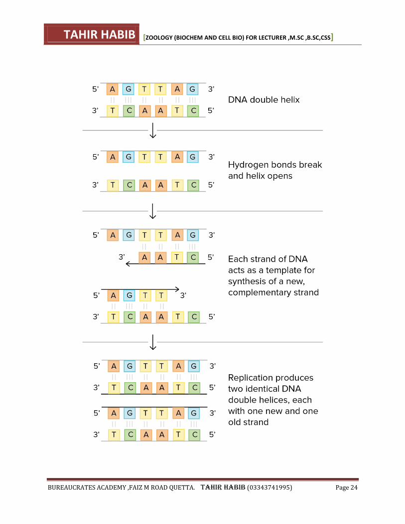
The basic idea
DNA replication is semiconservative, meaning that each strand in the DNA double helix acts as
a template for the synthesis of a new, complementary strand.
This process takes us from one starting molecule to two "daughter" molecules, with each newly
formed double helix containing one new and one old strand.
TAHIR HABIB [ZOOLOGY (BIOCHEM AND CELL BIO) FOR LECTURER ,M.SC ,B.SC,CSS]
BUREAUCRATES ACADEMY ,FAIZ M ROAD QUETTA. TAHIR HABIB (03343741995) Page 24
TAHIR HABIB [ZOOLOGY (BIOCHEM AND CELL BIO) FOR LECTURER ,M.SC ,B.SC,CSS]
BUREAUCRATES ACADEMY ,FAIZ M ROAD QUETTA. TAHIR HABIB (03343741995) Page 25
In a sense, that's all there is to DNA replication! But what's actually most interesting about this
process is how it's carried out in a cell.
Cells need to copy their DNA very quickly, and with very few errors (or risk problem such as
cancer). To do so, they use a variety of enzymes and proteins, which work together to make sure
DNA replication is performed smoothly and accurately.
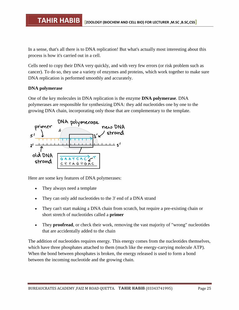
DNA polymerase
One of the key molecules in DNA replication is the enzyme DNA polymerase. DNA
polymerases are responsible for synthesizing DNA: they add nucleotides one by one to the
growing DNA chain, incorporating only those that are complementary to the template.
Here are some key features of DNA polymerases:
They always need a template
They can only add nucleotides to the 3' end of a DNA strand
They can't start making a DNA chain from scratch, but require a pre-existing chain or
short stretch of nucleotides called a primer
They proofread, or check their work, removing the vast majority of "wrong" nucleotides
that are accidentally added to the chain
The addition of nucleotides requires energy. This energy comes from the nucleotides themselves,
which have three phosphates attached to them (much like the energy-carrying molecule ATP).
When the bond between phosphates is broken, the energy released is used to form a bond
between the incoming nucleotide and the growing chain.
TAHIR HABIB [ZOOLOGY (BIOCHEM AND CELL BIO) FOR LECTURER ,M.SC ,B.SC,CSS]
BUREAUCRATES ACADEMY ,FAIZ M ROAD QUETTA. TAHIR HABIB (03343741995) Page 26
Starting DNA replication
How do DNA polymerases and other replication factors know where to begin? Replication
always starts at specific locations on the DNA, which are called origins of replication and are
recognized by their sequence.
E. coli, like most bacteria, has a single origin of replication on its chromosome. The origin is
about 245245245 base pairs long and has mostly A/T base pairs (which are held together by
fewer hydrogen bonds than G/C base pairs), making the DNA strands easier to separate.
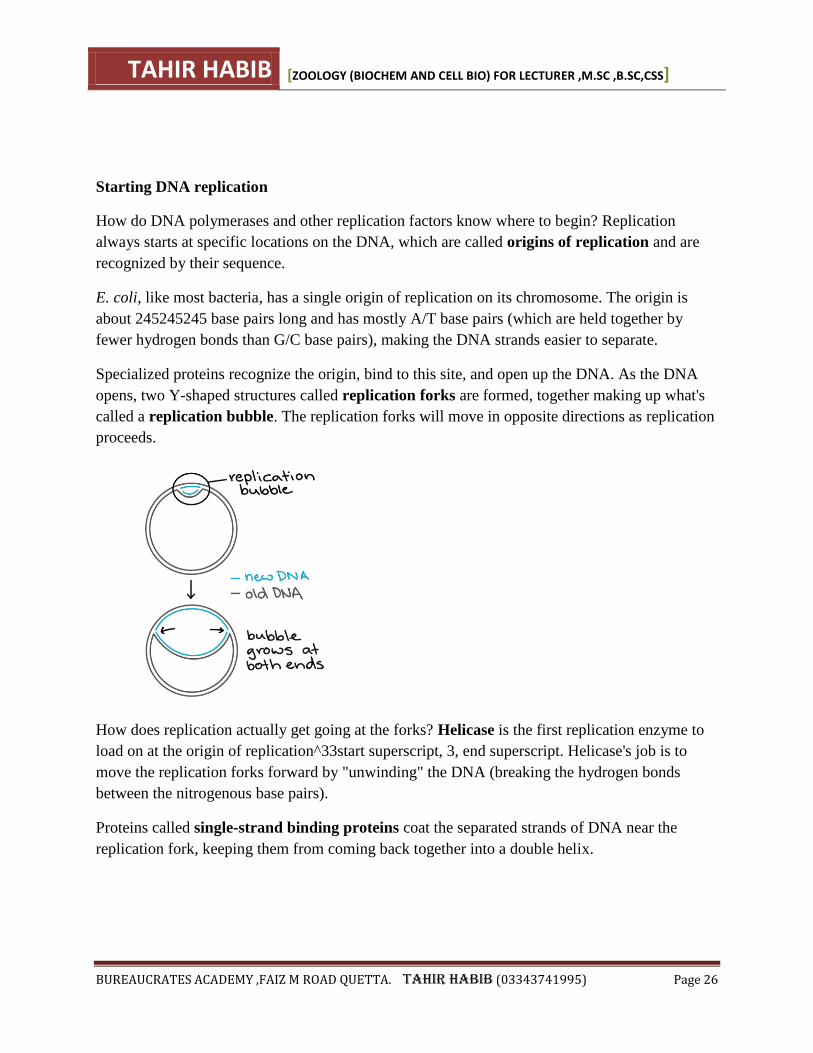
Specialized proteins recognize the origin, bind to this site, and open up the DNA. As the DNA
opens, two Y-shaped structures called replication forks are formed, together making up what's
called a replication bubble. The replication forks will move in opposite directions as replication
proceeds.
How does replication actually get going at the forks? Helicase is the first replication enzyme to
load on at the origin of replication^33start superscript, 3, end superscript. Helicase's job is to
move the replication forks forward by "unwinding" the DNA (breaking the hydrogen bonds
between the nitrogenous base pairs).
Proteins called single-strand binding proteins coat the separated strands of DNA near the
replication fork, keeping them from coming back together into a double helix.
TAHIR HABIB [ZOOLOGY (BIOCHEM AND CELL BIO) FOR LECTURER ,M.SC ,B.SC,CSS]
BUREAUCRATES ACADEMY ,FAIZ M ROAD QUETTA. TAHIR HABIB (03343741995) Page 27
PRIMERS AND PRIMASE
DNA polymerases can only add nucleotides to the 3' end of an existing DNA strand. (They use
the free -OH group found at the 3' end as a "hook," adding a nucleotide to this group in the
polymerization reaction.) How, then, does DNA polymerase add the first nucleotide at a new
replication fork?
Alone, it can't! The problem is solved with the help of an enzyme called primase. Primase
makes an RNA primer, or short stretch of nucleic acid complementary to the template, that
provides a 3' end for DNA polymerase to work on. A typical primer is about five to ten
nucleotides long. The primer primes DNA synthesis, i.e., gets it started.
Once the RNA primer is in place, DNA polymerase "extends" it, adding nucleotides one by one
to make a new DNA strand that's complementary to the template strand.
LEADING AND LAGGING STRANDS
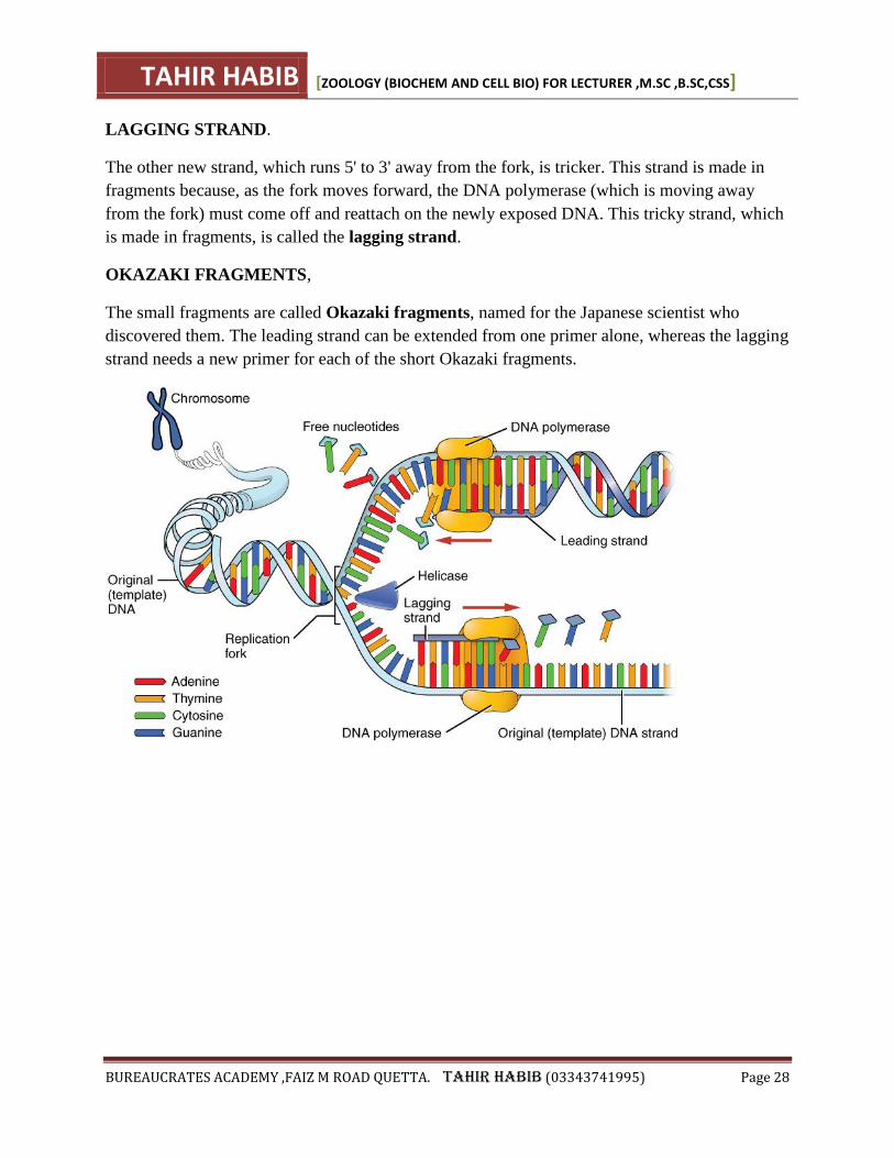
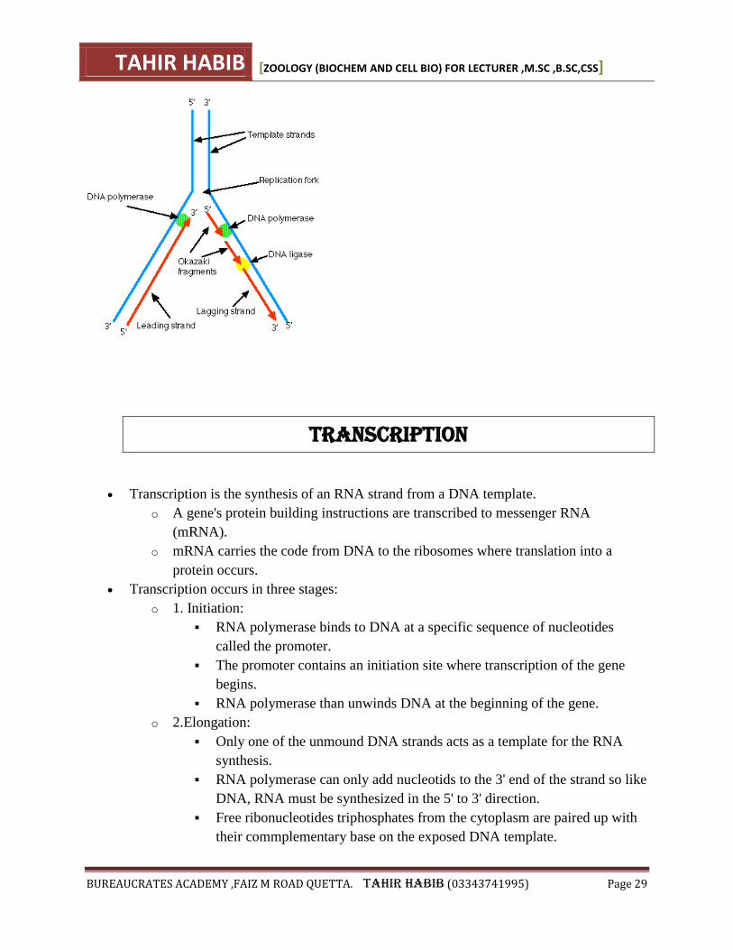
In E. coli, the DNA polymerase that handles most of the synthesis is DNA polymerase III. There
are two molecules of DNA polymerase III at a replication fork, each of them hard at work on one
of the two new DNA strands.
DNA polymerases can only make DNA in the 5' to 3' direction, and this poses a problem during
replication. A DNA double helix is always anti-parallel; in other words, one strand runs in the 5'
to 3' direction, while the other runs in the 3' to 5' direction. This makes it necessary for the two
new strands, which are also antiparallel to their templates, to be made in slightly different ways.
LEADING STRAND.
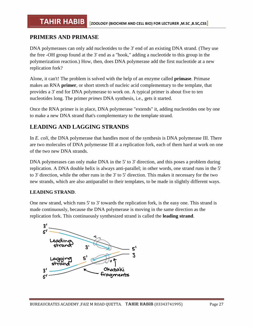
One new strand, which runs 5' to 3' towards the replication fork, is the easy one. This strand is
made continuously, because the DNA polymerase is moving in the same direction as the
replication fork. This continuously synthesized strand is called the leading strand.
TAHIR HABIB [ZOOLOGY (BIOCHEM AND CELL BIO) FOR LECTURER ,M.SC ,B.SC,CSS]
BUREAUCRATES ACADEMY ,FAIZ M ROAD QUETTA. TAHIR HABIB (03343741995) Page 28
LAGGING STRAND.
The other new strand, which runs 5' to 3' away from the fork, is tricker. This strand is made in
fragments because, as the fork moves forward, the DNA polymerase (which is moving away
from the fork) must come off and reattach on the newly exposed DNA. This tricky strand, which
is made in fragments, is called the lagging strand.
OKAZAKI FRAGMENTS,
The small fragments are called Okazaki fragments, named for the Japanese scientist who
discovered them. The leading strand can be extended from one primer alone, whereas the lagging
strand needs a new primer for each of the short Okazaki fragments.
TAHIR HABIB [ZOOLOGY (BIOCHEM AND CELL BIO) FOR LECTURER ,M.SC ,B.SC,CSS]
BUREAUCRATES ACADEMY ,FAIZ M ROAD QUETTA. TAHIR HABIB (03343741995) Page 29
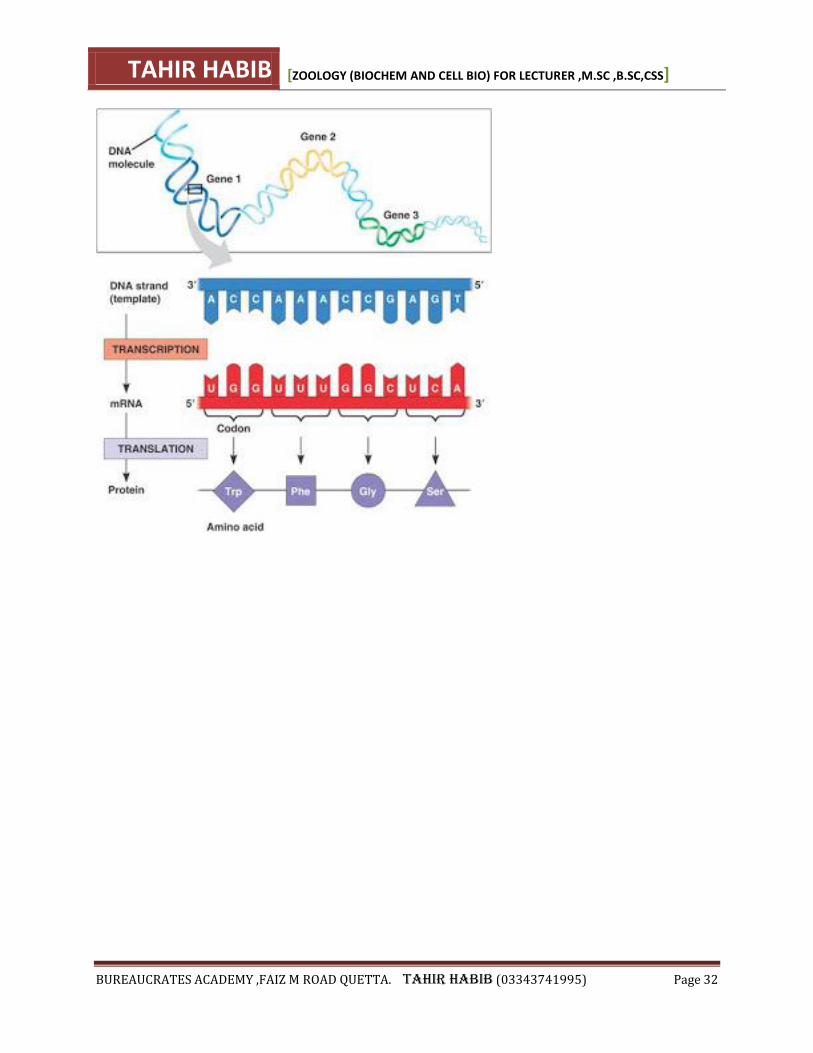
TRANSCRIPTION
Transcription is the synthesis of an RNA strand from a DNA template.
o A gene's protein building instructions are transcribed to messenger RNA
(mRNA).
o mRNA carries the code from DNA to the ribosomes where translation into a
protein occurs.
Transcription occurs in three stages:
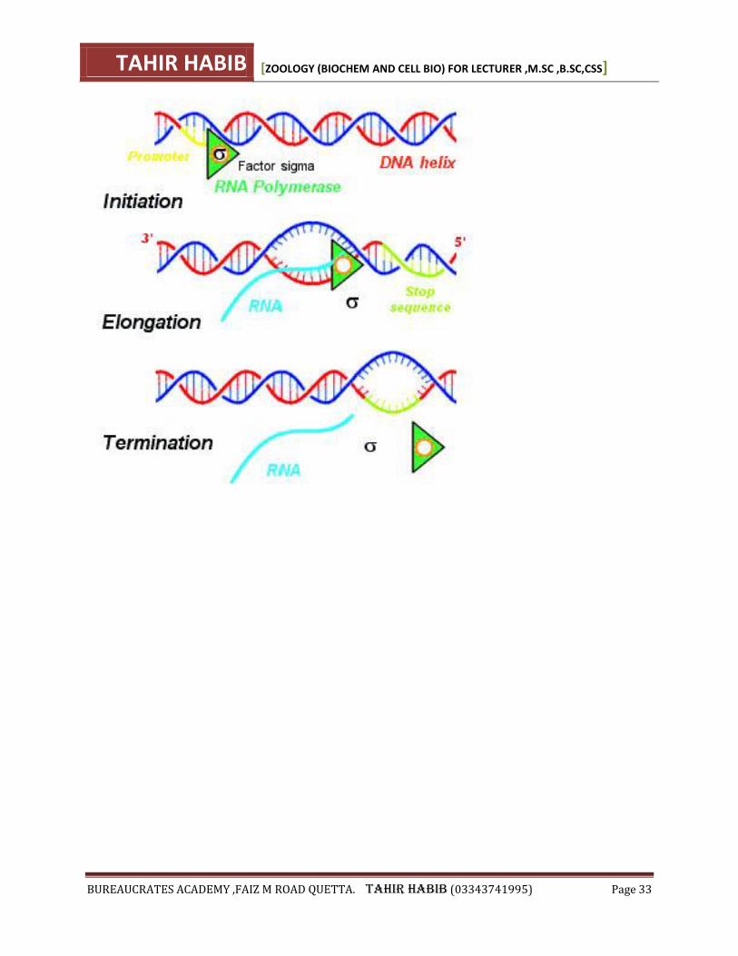
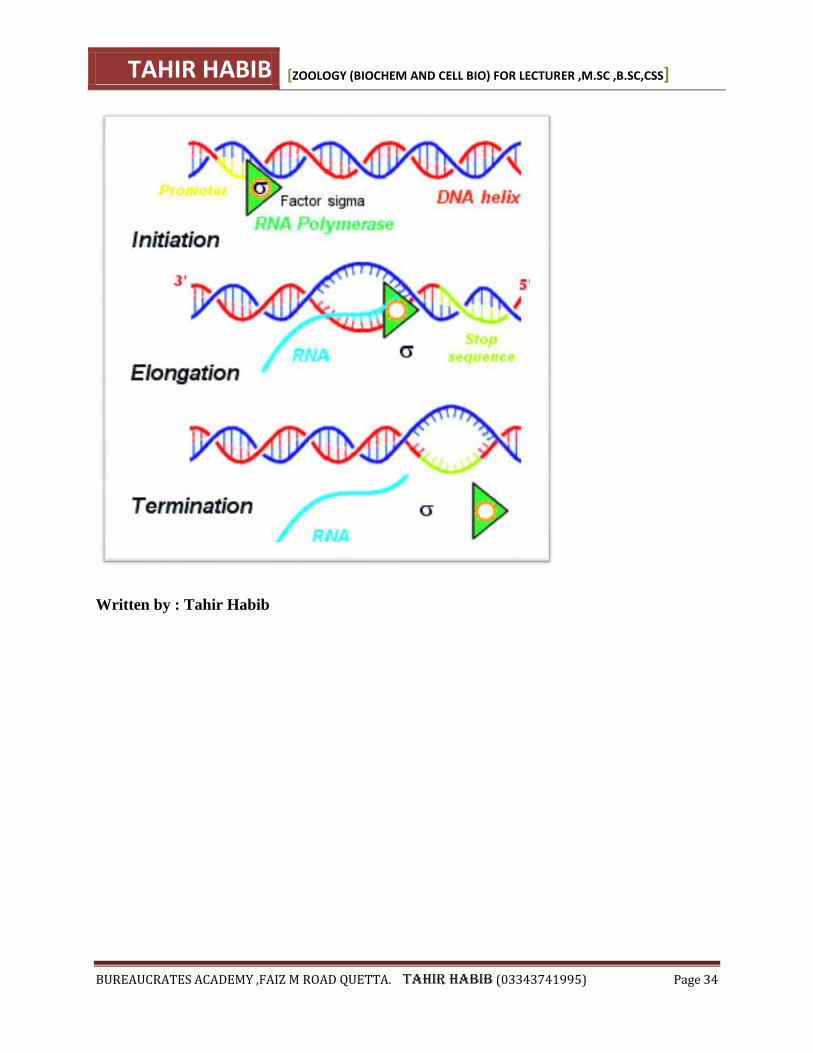
o 1. Initiation:
RNA polymerase binds to DNA at a specific sequence of nucleotides
called the promoter.
The promoter contains an initiation site where transcription of the gene
begins.
RNA polymerase than unwinds DNA at the beginning of the gene.
o 2.Elongation:
Only one of the unmound DNA strands acts as a template for the RNA
synthesis.
RNA polymerase can only add nucleotids to the 3' end of the strand so like
DNA, RNA must be synthesized in the 5' to 3' direction.
Free ribonucleotides triphosphates from the cytoplasm are paired up with
their commplementary base on the exposed DNA template.
TAHIR HABIB [ZOOLOGY (BIOCHEM AND CELL BIO) FOR LECTURER ,M.SC ,B.SC,CSS]
BUREAUCRATES ACADEMY ,FAIZ M ROAD QUETTA. TAHIR HABIB (03343741995) Page 30
RNA polymerase joins the ribonucleoside triphosphates to form an mRNA
strand.
As RNA polymerase advances, the process continues.
The DNA that has been transcribed, re-winds to form a double helix.
o Termination:
RNA polymerase continues to elongate until it reaches the terminator, a
specific sequence of nucleotides that signals the end of transcription.
Transcription stops and mRNA polymerase and the new mRNA transcript
are released from DNA.
The DNA double helix reforms.
The termination sequence usually consists of a series of adjancent
adenines preceded by a nucleotide palindrome.
This gives an RNA molecule that assumes a stem-and loop configuration.
This configuration stops RNA polymerase from transcribing any further.
Transcription
Protein Production faces a number of challenges. Chief amongst these is that proteins are
produced in the cytoplasm of the cell, and DNA never leaves the nucleus. To get around this
problem, DNA creates a messenger molecule to deliver its information outside of the nucleus:
mRNA (messenger RNA). The process of making this messenger molecule is known
as transcription, and has a number of steps:
1. Initiation: The double helix of the DNA is unwound by RNA Polymerase, which docks
on the DNA at a special sequence of bases (promoter)
2. Elongation: RNA Polymerase moves downstream unwinding the DNA. As the double
helix unwinds, ribonucleotide bases (A, C, G and U) attach themselves to the DNA
template strand (the strand being copied) by complementary base pairing.
3. RNA Polymerase catalyses the formation of covalent bonds between the nucleotides. In
the wake of transcription, DNA strands recoil into the double helix.
4. Termination: The RNA transcript is released from the DNA, along with the RNA
polymerase.
The next stage in transcription is the addition of a 5' cap and a poly-A tail. These sections of the
completed RNA molecule are not translated into protein. Instead they:
1. Protect the mRNA from degradation
2. Help the mRNA to leave the nucleus
3. Anchor the mRNA to the ribosome during Translation
At this point a long RNA molecule has been made, but this is not the end of Transcription. The
RNA molecule contains sections that are not needed as part of the protein code that need to be
removed. This is like writing every other paragraph of a novel in wingdings - these sections must
be removed for the story to make sense! While at first the presence of introns seems incredibly
TAHIR HABIB [ZOOLOGY (BIOCHEM AND CELL BIO) FOR LECTURER ,M.SC ,B.SC,CSS]
BUREAUCRATES ACADEMY ,FAIZ M ROAD QUETTA. TAHIR HABIB (03343741995) Page 31
wasteful, a number of genes can give rise to several different proteins, depending on which
sections are treated as exons - this is known as alternative RNA splicing. This allows a relatively
small number of genes to create a much larger number of different proteins. Humans have just
under twice as many genes as a fruit fly, and yet can make many times more protein products.
Sequences not needed to make a protein are called introns; the sequences that are expressed
are called exons. The introns are cut out by various enzymes and the exons are spliced together
to form a complete RNA molecule.
TAHIR HABIB [ZOOLOGY (BIOCHEM AND CELL BIO) FOR LECTURER ,M.SC ,B.SC,CSS]
BUREAUCRATES ACADEMY ,FAIZ M ROAD QUETTA. TAHIR HABIB (03343741995) Page 32
TAHIR HABIB [ZOOLOGY (BIOCHEM AND CELL BIO) FOR LECTURER ,M.SC ,B.SC,CSS]
BUREAUCRATES ACADEMY ,FAIZ M ROAD QUETTA. TAHIR HABIB (03343741995) Page 33
TAHIR HABIB [ZOOLOGY (BIOCHEM AND CELL BIO) FOR LECTURER ,M.SC ,B.SC,CSS]
BUREAUCRATES ACADEMY ,FAIZ M ROAD QUETTA. TAHIR HABIB (03343741995) Page 34
Written by : Tahir Habib
TAHIR HABIB [ZOOLOGY (BIOCHEM AND CELL BIO) FOR LECTURER ,M.SC ,B.SC,CSS]
BUREAUCRATES ACADEMY ,FAIZ M ROAD QUETTA. TAHIR HABIB (03343741995) Page 35
Translation (PROTIEN SYNTHESIS) Once mRNA has left the Nucleus, it is directed to a Ribosome to construct a protein. This
process can be broken down into 6 main stages:
1. Initiation: Ribosome attaches to the mRNA molecule at the start codon. This sequence
(always AUG) signals the start of the gene to be transcribed. The ribosome can enclose
two codons at a time
2. tRNAs (transfer RNAs) act as couriers. There are many types of tRNA, each one
complimentary to the 64 possible codon combinations. Each tRNA is bonded to a specific
amino acid. As AUG is the start codon, the first amino acid to be 'couriered' is always
Methionine.
3. Elongation: The stepwise addition of amino acids to the growing polypeptide chain. The
next amino acid tRNA attaches to the adjacent mRNA codon.
4. The bond holding the tRNA and amino acid together is broken, and a peptide bond is
formed between the adjacent amino acids.
5. As the Ribosome can only cover two codons at a time, it must now shuffle down to cover
a new codon. This releases the first tRNA which is now free to collect another amino
acid. Steps 2-5 repeats along the whole length of the mRNA molecule
6. Termination: As the polypeptide chain elongates, it peels away from the Ribosome.
During this phase, the protein starts to fold into it's specific secondary structure.
Elongation continues (perhaps for hundreds or thousands of amino acids) until the
Ribosome reaches one of three possible Stop codons (UAG, UAA, UGA). At this point
the mRNA dissociates from the ribosome
This seems to be a long, drawn out process, but as always biology finds a work around. mRNA
molecules can be extremely long - long enough for several Ribosomes to work on the same
mRNA strand. This means that a cell can produce lots of copies of the same protein from a single
mRNA molecule.
Post Translational Modifications
Sometimes a protein needs some help to fold into its required tertiary structure. Modifications
can be made after translation by enzymes such as methylation, phosphorylation and
glycosylation. These modifications tend to occur in the Endoplasmic Reticulum, with a few
occurring in the Golgi Body.
Post translational modification can also be used to activate or inactivate proteins. This allows a
cell to stockpile a particular protein, that only becomes active once it is required. This is
particularly important in the case of some hydrolytic enzymes, which would damage the cell if
left to run riot. (The alternative to this is packaging within an organelle such as a Lysosome)
Post-Translation Modifications are the domain of Eukaryotes. Prokaryotes (largely) do not need
any interference to help their proteins to fold into an active form.
TAHIR HABIB [ZOOLOGY (BIOCHEM AND CELL BIO) FOR LECTURER ,M.SC ,B.SC,CSS]
BUREAUCRATES ACADEMY ,FAIZ M ROAD QUETTA. TAHIR HABIB (03343741995) Page 36
Keywords
Amino Acid - the building blocks of proteins; there are 20 different types
Codon - a sequence of three organic bases in a nucleic acid that code for a specific amino acid
Exon - Coding region of eukaryotic gene. Parts of the gene that are expressed
Gene- a length of DNA made up of a number of codons; codes for a specific protein
Intron - Non coding region of a gene that separates exons
Polypeptide - a chain of amino acids joined by a peptide bond
Ribosome - a cellular organelle that functions as a protein-making workbench.
RNA - Ribonucleic Acid; a nucleic acid that acts as a messenger, carrying information from the
DNA to the Ribosomes
Written by : Tahir Habib
CELL ORGANELLES
CELL THEORY
Cells are the basic unit of life.
The Cell Theory states that:
1) All organisms are made up of one or more cells and the products of those cells.
2) All cells carry out life activities ( require energy, grow, have a limited size).
3) New cells arise only from other living cells by the process of cell division.
THE THREE MAIN COMPONENTS OF ANY PLANT OR ANIMAL CELL ARE:
1. PLASMA MEMBRANE/ CELL MEMBRANE
Structure- a bilipid membraneous layer composed of proteins and carbohydrates. It is
fluid like.
Function - the cell membrane separates the cell from its external environment, and is
selectively permeable (controls what gets in and out). It protects the cell and provides
stability.
TAHIR HABIB [ZOOLOGY (BIOCHEM AND CELL BIO) FOR LECTURER ,M.SC ,B.SC,CSS]
BUREAUCRATES ACADEMY ,FAIZ M ROAD QUETTA. TAHIR HABIB (03343741995) Page 37
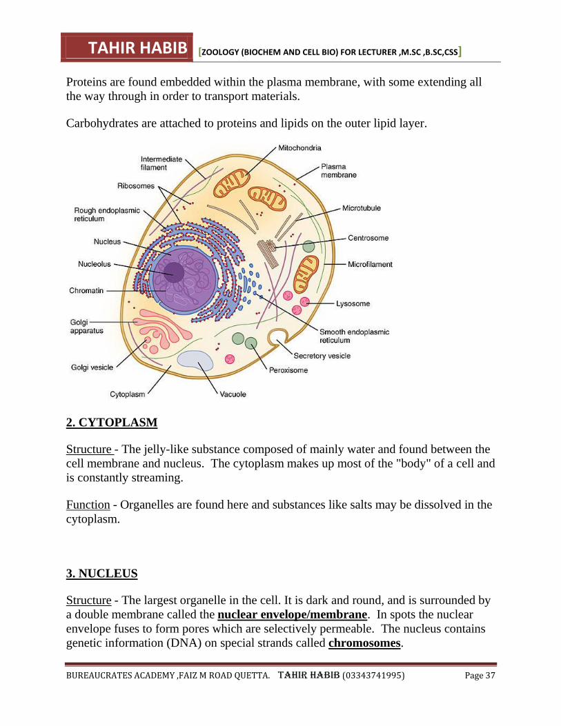
Proteins are found embedded within the plasma membrane, with some extending all
the way through in order to transport materials.
Carbohydrates are attached to proteins and lipids on the outer lipid layer.
2. CYTOPLASM
Structure - The jelly-like substance composed of mainly water and found between the
cell membrane and nucleus. The cytoplasm makes up most of the "body" of a cell and
is constantly streaming.
Function - Organelles are found here and substances like salts may be dissolved in the
cytoplasm.
3. NUCLEUS
Structure - The largest organelle in the cell. It is dark and round, and is surrounded by
a double membrane called the nuclear envelope/membrane. In spots the nuclear
envelope fuses to form pores which are selectively permeable. The nucleus contains
genetic information (DNA) on special strands called chromosomes.
TAHIR HABIB [ZOOLOGY (BIOCHEM AND CELL BIO) FOR LECTURER ,M.SC ,B.SC,CSS]
BUREAUCRATES ACADEMY ,FAIZ M ROAD QUETTA. TAHIR HABIB (03343741995) Page 38
Function - The nucleus is the "control center" of the cell, for cell metabolism and
reproduction.
THE FOLLOWING ORGANELLES ARE FOUND IN BOTH PLANT AND ANIMAL
CELLS.
1. "ER" OR ENDOPLASMIC RETICULUM
The Endoplasmic Reticulum is a network of membranous canals filled with
fluid. They carry materials throughout the cell. The ER is the "transport system" of
the cell.
There are two types of ER: rough ER and smooth ER.
Rough Endoplasmic Reticulum is lined with ribosomes and is rough in appearance
and smooth endoplasmic reticulum contains no ribosomes and is smooth in
appearance.
2. RIBOSOMES
Ribosomes are small particles which are found individually in the cytoplasm and also
line the membranes of the rough endoplasmic reticulum. Ribosomes produce
protein. They could be thought of as "factories" in the cell.
3. GOLGI BODY / APPARATUS
Golgi bodies are stacks of flattened membranous stacks (they look like
pancakes!). The Golgi Body temporarily stores protein which can then leave the cell
via vesiciles pinching off from the Golgi.
4. LYSOSOMES
Lysosomes are small sac-like structures surrounded by a single membrane and
containing strong digestive enzymes which when released can break down worn out
organelles or food. The lysosome is also known as a suicide sac.
5. MITOCHONDRIA
The mitochondria are round "tube-like" organelles that are surrounded by a double
membrane, with the inner membrane being highly folded. the mitochondria are often
TAHIR HABIB [ZOOLOGY (BIOCHEM AND CELL BIO) FOR LECTURER ,M.SC ,B.SC,CSS]
BUREAUCRATES ACADEMY ,FAIZ M ROAD QUETTA. TAHIR HABIB (03343741995) Page 39
referred to as the "powerhouse" of the cell. the mitochondria releases food energy
from food molecules to be used by the cell. This process is called respiration. Some
cells( muscle cells) require more energy than other cells and so would have many
more mitochondria.
6. VACUOLES
Vacuoles are fluid filled organelles enclosed by a membrane. They can store
materials such as food, water, sugar, minerals and waste products.
ANIMAL CELLS ORGANELLES NOT FOUND IN PLANT CELLS:
CILIA AND FLAGELLA
Both cilia and flagella are hair-like organelles which extend from the surface of many
animal cells. the structure is identical in both, except that flagella are longer and
whiplike and cilia are shorter. There are usually only a few flagella on a cell, while
cilia may cover the entire surface of a cell. The function of cilia and flagella ionclude
locomotion for one-celled organisms and to move substances over cell surfaces in
multi-celled organisms.
ORGANELLES AND OTHER FEATURES FOUND ONLY IN PLANT CELLS:
1. CELL WALL
The cell wall is a rigid organelle composed of cellulose and lying just outside the cell
membrane. The cell wall gives the plant cell it's box-like shape. it also protects the
cell. The cell wall contains pores which allow materials to pass to and from the cell
membrane.
2. PLASTIDS
Plastids are double membrane bound organelles. It is in plastids that plants make and
store food. Plastids are found in the cytoplasm and there are two main types:
Leucoplasts - colorless organelles which store starch or other plant nutrients. (
example - starch stored in a potato)
TAHIR HABIB [ZOOLOGY (BIOCHEM AND CELL BIO) FOR LECTURER ,M.SC ,B.SC,CSS]
BUREAUCRATES ACADEMY ,FAIZ M ROAD QUETTA. TAHIR HABIB (03343741995) Page 40
Chromoplasts - contain different colored pigments. The most important type of
chromoplast is the chloroplast, which contains the green pigment chlorophyll. This is
important in the process of photosynthesis.
3. CENTRAL VACUOLE
The central vacuole is a large fluid-filled vacuole found in plants.
List of Functions of Cell Organelles
Many courses in introductory biology include cell biology and require knowledge of
the basic functions of the organelles found in eukaryotic cells. It is useful to be able to
summarize the main functions of each type of organelle in just a few words or
sentences.
The following table of functions of cell organelles is a list of short summary
information for each organelle.
(See the links from some descriptions for further details.)
Membrane-bound Organelles
Organelle Type Main Functions (not necessarily all functions):
1. Nucleus "Control Center" of the cell.
Contains the cell's DNA (genetic information) in the
form of genes.
Re.
Nucleic
Acids
*Sequestration and *replication of
DNA.
*Transcription and *modification
of RNA.
Contains one or more nucleoli (plural, singular
word = nucleolus) whose functions include:
Nucleoli Biosynthesis of ribosomal RNA
(rRNA) and production (assembly)
of ribosomes.
2. Rough Endoplasmic Consists of many interconnected membranous sacs
TAHIR HABIB [ZOOLOGY (BIOCHEM AND CELL BIO) FOR LECTURER ,M.SC ,B.SC,CSS]
BUREAUCRATES ACADEMY ,FAIZ M ROAD QUETTA. TAHIR HABIB (03343741995) Page 41
Reticulum (RER) called cisternae, onto whose external
surface ribosomes are attached (distinguishing RER
from SER on electron micrographs).
Ribosomes Produce polypeptides that are then either
...
inserted into the RER
membrane, or
moved into the lumen (central
region) of the cisternae, or
moved to the Golgi complex and
probably onwards from there.
In lumen
of
cisternae
Produce proteins that are then either ...
retained within vesicles, or
secreted from the cell (via
secretory vesicles - see below).
3. Smooth Endoplasmic
Reticulum (SER)
Consists of many interconnected membranous sacs
called cisternae (withoutribosomes).
Many enzymes are either attached to the surface of
the SER or located within its cisternae. Chemical
reactions within the SER vary with the type and location of cells. E.g.
helps with protein folding and transport of
synthesized proteins
glycosylation - which involves the attachment
of oligosaccharides.
disulfide bond formation and rearrangement -
to stabilize the tertiary and quaternary
structure of many proteins
modification of some drugs e.g. by the
cytochrome P450 enzymes in liver cells.
4. Mitochondria The main function of mitochondria in aerobic cells
is the production of energy by synthesis of ATP.
However, mitochondria also have many other
functions, including e.g.:
Processing and storage of calcium ions
(Ca2+).
TAHIR HABIB [ZOOLOGY (BIOCHEM AND CELL BIO) FOR LECTURER ,M.SC ,B.SC,CSS]
BUREAUCRATES ACADEMY ,FAIZ M ROAD QUETTA. TAHIR HABIB (03343741995) Page 42
Apoptosis, i.e. the process of programmed
cell death
Regulation of cellular metabolism
Synthesis of certain steroids
See also the structure of mitochondria and the
functions of mitochondria.
5. Chloroplasts
(plant cells only)
Chloroplasts are the sites of photosynthesis within
plant cells.
6. Golgi Apparatus The Golgi apparatus modifies, sorts and
packages macromolecules for delivery to other
organelles or secretion from the cell via exocytosis -
see (9.) below.
7. Lysosomes Lysosomes (tiny sacs containing enzymes) are
the main sites of intracellular digestion. They
enable the cell to make use of nutrients. Their
functions can be listed as:
Autophagy - digestion of materials from
within the cell.
Heterophagy - digestion of materials
originating from outside the cell.
Biosynthesis - recycling unwanted products
of chemical reactions to process materials
received from outside the cell.
Lysosomes also destroy the cell - usually after it has
died.
8. Peroxisomes (also called "microbodies"
- smaller than lysosomes
and contain specific
enzymes)
Similar to (but smaller than) lysosomes, the
metabolic functions of peroxisomes include:
Breakdown of fatty acids by beta-oxidation
Breakdown excess purines to urea
Breakdown of toxic compounds e.g. in the
cells of the liver and kidney.
also play a role in the biosynthesis of certain
important molecules incl. cholesterol and (in
liver cells) bile acids derived from
cholesterol.
9. Secretory vesicles Transport and delivery of their contents (e.g.
TAHIR HABIB [ZOOLOGY (BIOCHEM AND CELL BIO) FOR LECTURER ,M.SC ,B.SC,CSS]
BUREAUCRATES ACADEMY ,FAIZ M ROAD QUETTA. TAHIR HABIB (03343741995) Page 43
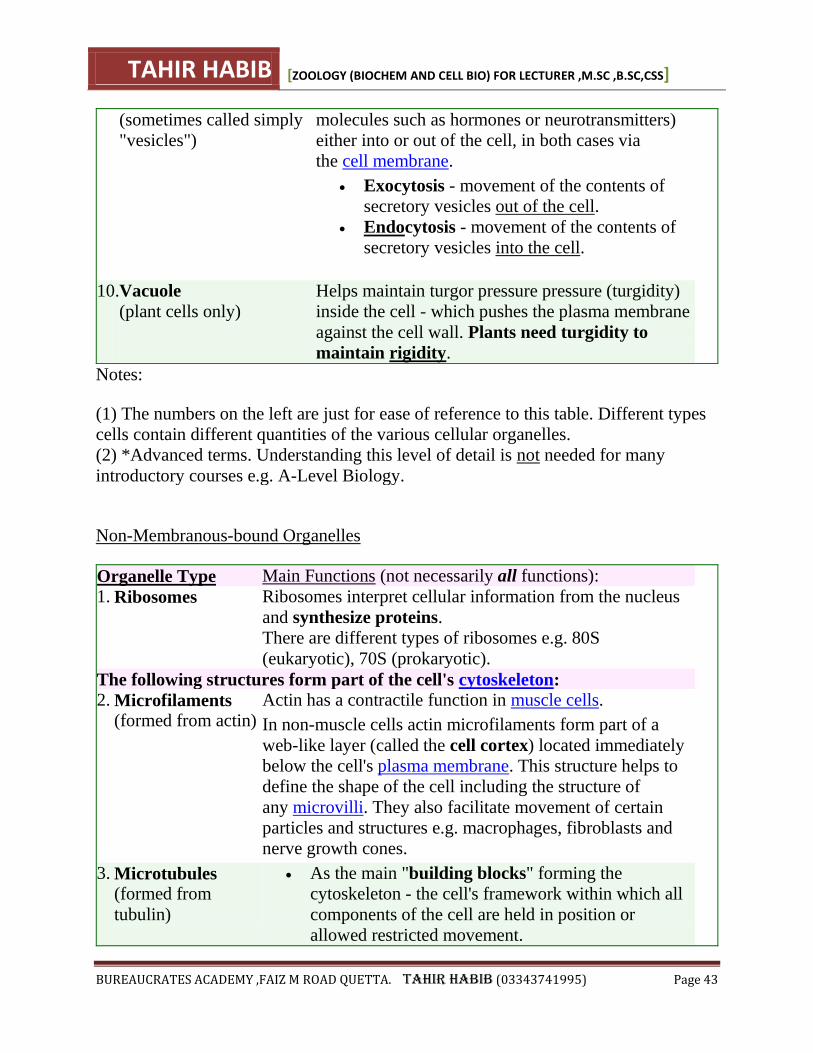
(sometimes called simply
"vesicles")
molecules such as hormones or neurotransmitters)
either into or out of the cell, in both cases via
the cell membrane.
Exocytosis - movement of the contents of
secretory vesicles out of the cell.
Endocytosis - movement of the contents of
secretory vesicles into the cell.
10. Vacuole
(plant cells only)
Helps maintain turgor pressure pressure (turgidity)
inside the cell - which pushes the plasma membrane
against the cell wall. Plants need turgidity to
maintain rigidity.
Notes:
(1) The numbers on the left are just for ease of reference to this table. Different types
cells contain different quantities of the various cellular organelles.
(2) *Advanced terms. Understanding this level of detail is not needed for many
introductory courses e.g. A-Level Biology.
Non-Membranous-bound Organelles
Organelle Type Main Functions (not necessarily all functions):
1. Ribosomes Ribosomes interpret cellular information from the nucleus
and synthesize proteins.
There are different types of ribosomes e.g. 80S
(eukaryotic), 70S (prokaryotic).
The following structures form part of the cell's cytoskeleton: 2. Microfilaments
(formed from actin)
Actin has a contractile function in muscle cells.
In non-muscle cells actin microfilaments form part of a
web-like layer (called the cell cortex) located immediately
below the cell's plasma membrane. This structure helps to
define the shape of the cell including the structure of
any microvilli. They also facilitate movement of certain
particles and structures e.g. macrophages, fibroblasts and
nerve growth cones.
3. Microtubules (formed from
tubulin)
As the main "building blocks" forming the
cytoskeleton - the cell's framework within which all
components of the cell are held in position or
allowed restricted movement.
TAHIR HABIB [ZOOLOGY (BIOCHEM AND CELL BIO) FOR LECTURER ,M.SC ,B.SC,CSS]
BUREAUCRATES ACADEMY ,FAIZ M ROAD QUETTA. TAHIR HABIB (03343741995) Page 44
Movement of materials and structures within cells
e.g. help form the miotic spindle during the
"prophase" part of cell division by mitosis.
For further detail see functions of microtubules.
4. Intermediate
Filaments (formed from
intermediate
filament proteins,
e.g. keratin)
Intermediate filaments are important for maintaining the
mechanical structure of cells. There are different types of
intermediate filaments that can be identified according to
the protein from which they are formed. The different types
of intermediate filaments occur in different types of cells
and therefore provide structural support (to the cell) in
slightly different ways.
E.g. neurofilaments in the axons of neurons are involved in
the radial growth of the axon, so determine its diameter as
well as contributing strength and rigidity to the cell.
5. Junctions "Junctions" are connecting points joining either cells to
other cells, or cells to their basement membrane. See
the diagram of the cytoskeleton.
6. Centrosomes Contain the centrioles, which are involved in the process
of mitosis - see cell-division.
7. Cilia Some eukaryotic cells have cilia (plural, singular word
= cilium) whose function is often to facilitate
either movement of the cell or movement of something
over the surface of cells e.g. fallopian cells move ova
towards the uterus.
8.
Flagella (of
spermatozoa differ
from prokaryotic
flagella)
The main function of the flagellum of a human
spermatozoon (sperm cell) is to enable the sperm to move
close to the oocyte ("egg" cell) and orient itself
appropriately .
Note: The numbers on the left are just for ease of reference to this table. Different
types cells contain different quantities of the various cellular organelles.
The cell membrane is often included in sections about the structure and functions of
cell organelles. However, the cell membrane (also known as the plasma membrane)
is not within the cell but one of the structures that defines the cell - together with the
TAHIR HABIB [ZOOLOGY (BIOCHEM AND CELL BIO) FOR LECTURER ,M.SC ,B.SC,CSS]
BUREAUCRATES ACADEMY ,FAIZ M ROAD QUETTA. TAHIR HABIB (03343741995) Page 45
cell wall in the cases of plant cells and prokaryotic cells. See functions of the cell
membrane.
The above list of functions of organelles shows that many, though not all, membrane-
bound organelles are sites of biochemical reactions, i.e. where chemicals are made (=
"produced", "synthesized" or "biosynthesized") or broken-down (= "degraded')
or changed in some way. Such chemical reactions are examples of metabolic
processes and often form part of metabolic pathways. This is way knowledge of cell
biology is useful when studying metabolism.
TAHIR HABIB [ZOOLOGY (BIOCHEM AND CELL BIO) FOR LECTURER ,M.SC ,B.SC,CSS]
BUREAUCRATES ACADEMY ,FAIZ M ROAD QUETTA. TAHIR HABIB (03343741995) Page 46
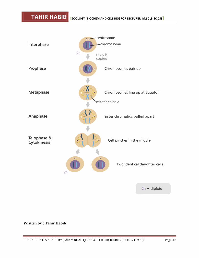
What is mitosis? Mitosis is a process where a single cell divides into two identical daughter cells (cell division).
During mitosis one cell? divides once to form two identical cells.
The major purpose of mitosis is for growth and to replace worn out cells.
If not corrected in time, mistakes made during mitosis can result in changes in
the DNA? that can potentially lead to genetic disorders
?.
Mitosis is divided into five phases:
1. Interphase:
The DNA in the cell is copied in preparation for cell division, this results in two identical
full sets of chromosomes?.
Outside of the nucleus? are two centrosomes, each containing a pair of centrioles, these
structures are critical for the process of cell division.
During interphase, microtubules extend from these centrosomes.
2. Prophase:
The chromosomes condense into X-shaped structures that can be easily seen under a
microscope.
Each chromosome is composed of two sister chromatids, containing identical genetic
information.
The chromosomes pair up so that both copies of chromosome 1 are together, both copies
of chromosome 2 are together, and so on.
At the end of prophase the membrane around the nucleus in the cell dissolves away
releasing the chromosomes.
The mitotic spindle, consisting of the microtubules and other proteins, extends across the
cell between the centrioles as they move to opposite poles of the cell.
3. Metaphase:
The chromosomes line up neatly end-to-end along the centre (equator) of the cell.
The centrioles are now at opposite poles of the cell with the mitotic spindle fibres
extending from them.
The mitotic spindle fibres attach to each of the sister chromatids.
4. Anaphase:
The sister chromatids are then pulled apart by the mitotic spindle which pulls one
chromatid to one pole and the other chromatid to the opposite pole.
5. Telophase:
At each pole of the cell a full set of chromosomes gather together.
A membrane forms around each set of chromosomes to create two new nuclei.
The single cell then pinches in the middle to form two separate daughter cells each
containing a full set of chromosomes within a nucleus. This process is known as
cytokinesis.
TAHIR HABIB [ZOOLOGY (BIOCHEM AND CELL BIO) FOR LECTURER ,M.SC ,B.SC,CSS]
BUREAUCRATES ACADEMY ,FAIZ M ROAD QUETTA. TAHIR HABIB (03343741995) Page 47
Written by : Tahir Habib

TAHIR HABIB [ZOOLOGY (BIOCHEM AND CELL BIO) FOR LECTURER ,M.SC ,B.SC,CSS]
BUREAUCRATES ACADEMY ,FAIZ M ROAD QUETTA. TAHIR HABIB (03343741995) Page 48
The significance of Mitosis are :-
1. It is an equational division through which identical daughter cells are produced having the
same amount and type of genetic constitution as that of the parent cell.
2. It is responsible for growth and development of multi-cellular organisms from a single-celled
zygote.
3. The number of chromosomes remains the same in all the cells produced by this division. Thus,
the daughter cells retain the same characters as those of the parent cell.
4. It helps the cell in maintaining proper size.
5. Mitosis helps in restoring wear and tear in body tissues, replacement of damaged or lost part,
healing of wounds and regeneration of detached parts (as in tail of a lizards).
6. It is a method of multiplication in unicellular organisms.
7. If mitosis remains unchecked, it may result in uncontrolled growth of cells leading to cancer or
tumour.
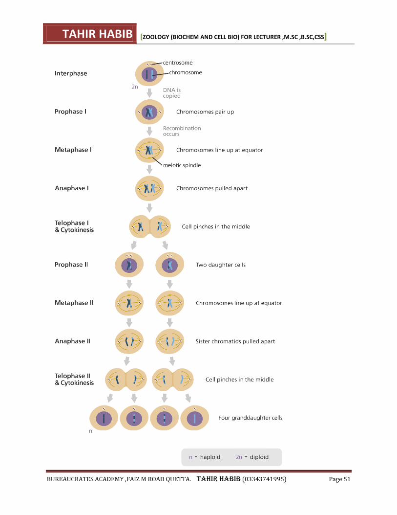
What is meiosis? Meiosis is a process where a single cell divides twice to produce four cells containing half the
original amount of genetic information. These cells are our sex cells – sperm in males, eggs in
females.
During meiosis one cell? divides twice to form four daughter cells.
These four daughter cells only have half the number of chromosomes? of the parent cell –
they are haploid.
Meiosis produces our sex cells or gametes? (eggs in females and sperm in males).
Meiosis can be divided into nine stages. These are divided between the first time the cell divides
(meiosis I) and the second time it divides (meiosis II):
Meiosis I
1. Interphase:
The DNA in the cell is copied resulting in two identical full sets of chromosomes.
Outside of the nucleus? are two centrosomes, each containing a pair of centrioles, these
structures are critical for the process of cell division?.
During interphase, microtubules extend from these centrosomes.
2. Prophase I:
The copied chromosomes condense into X-shaped structures that can be easily seen under
a microscope.
Each chromosome is composed of two sister chromatids containing identical genetic
information.
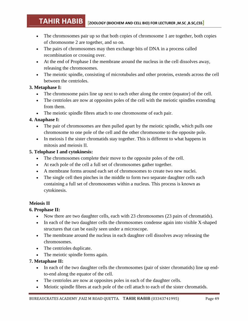
TAHIR HABIB [ZOOLOGY (BIOCHEM AND CELL BIO) FOR LECTURER ,M.SC ,B.SC,CSS]
BUREAUCRATES ACADEMY ,FAIZ M ROAD QUETTA. TAHIR HABIB (03343741995) Page 49
The chromosomes pair up so that both copies of chromosome 1 are together, both copies
of chromosome 2 are together, and so on.
The pairs of chromosomes may then exchange bits of DNA in a process called
recombination or crossing over.
At the end of Prophase I the membrane around the nucleus in the cell dissolves away,
releasing the chromosomes.
The meiotic spindle, consisting of microtubules and other proteins, extends across the cell
between the centrioles.
3. Metaphase I:
The chromosome pairs line up next to each other along the centre (equator) of the cell.
The centrioles are now at opposites poles of the cell with the meiotic spindles extending
from them.
The meiotic spindle fibres attach to one chromosome of each pair.
4. Anaphase I:
The pair of chromosomes are then pulled apart by the meiotic spindle, which pulls one
chromosome to one pole of the cell and the other chromosome to the opposite pole.
In meiosis I the sister chromatids stay together. This is different to what happens in
mitosis and meiosis II.
5. Telophase I and cytokinesis:
The chromosomes complete their move to the opposite poles of the cell.
At each pole of the cell a full set of chromosomes gather together.
A membrane forms around each set of chromosomes to create two new nuclei.
The single cell then pinches in the middle to form two separate daughter cells each
containing a full set of chromosomes within a nucleus. This process is known as
cytokinesis.
Meiosis II
6. Prophase II:
Now there are two daughter cells, each with 23 chromosomes (23 pairs of chromatids).
In each of the two daughter cells the chromosomes condense again into visible X-shaped
structures that can be easily seen under a microscope.
The membrane around the nucleus in each daughter cell dissolves away releasing the
chromosomes.
The centrioles duplicate.
The meiotic spindle forms again.
7. Metaphase II:
In each of the two daughter cells the chromosomes (pair of sister chromatids) line up end-
to-end along the equator of the cell.
The centrioles are now at opposites poles in each of the daughter cells.
Meiotic spindle fibres at each pole of the cell attach to each of the sister chromatids.
TAHIR HABIB [ZOOLOGY (BIOCHEM AND CELL BIO) FOR LECTURER ,M.SC ,B.SC,CSS]
BUREAUCRATES ACADEMY ,FAIZ M ROAD QUETTA. TAHIR HABIB (03343741995) Page 50
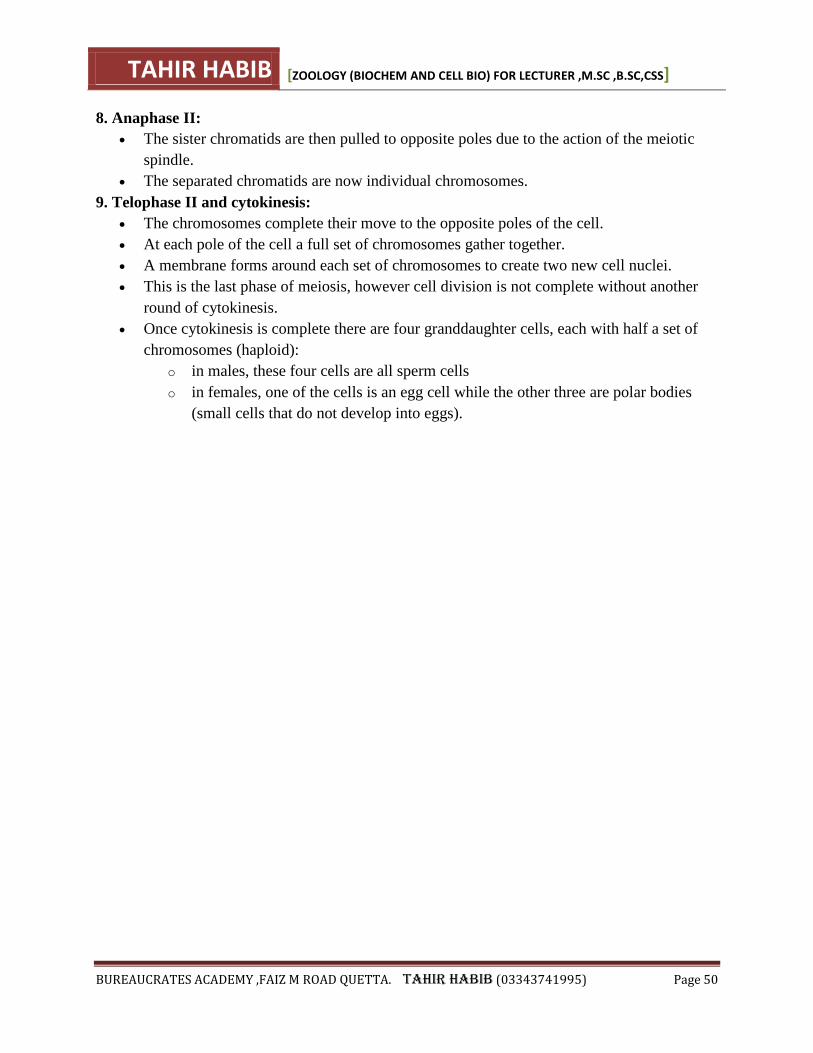
8. Anaphase II:
The sister chromatids are then pulled to opposite poles due to the action of the meiotic
spindle.
The separated chromatids are now individual chromosomes.
9. Telophase II and cytokinesis:
The chromosomes complete their move to the opposite poles of the cell.
At each pole of the cell a full set of chromosomes gather together.
A membrane forms around each set of chromosomes to create two new cell nuclei.
This is the last phase of meiosis, however cell division is not complete without another
round of cytokinesis.
Once cytokinesis is complete there are four granddaughter cells, each with half a set of
chromosomes (haploid):
o in males, these four cells are all sperm cells
o in females, one of the cells is an egg cell while the other three are polar bodies
(small cells that do not develop into eggs).
TAHIR HABIB [ZOOLOGY (BIOCHEM AND CELL BIO) FOR LECTURER ,M.SC ,B.SC,CSS]
BUREAUCRATES ACADEMY ,FAIZ M ROAD QUETTA. TAHIR HABIB (03343741995) Page 51
TAHIR HABIB [ZOOLOGY (BIOCHEM AND CELL BIO) FOR LECTURER ,M.SC ,B.SC,CSS]
BUREAUCRATES ACADEMY ,FAIZ M ROAD QUETTA. TAHIR HABIB (03343741995) Page 52
The significance of meiosis :- Written by : Tahir Habib 1. It maintains the same chromosome n umber in the sexually reproducing organisms. From a
diploid cell, haploid gametes are produced which in turn fuse to form a diploid cell.
2. It restricts the multiplication of chromosome number and maintains the stability of the species.
3. Maternal and paternal genes get exchanged during crossing over. It results in variations among
the offspring.
4. All the four chromatids of a homologous pair of chromosomes segregate and go over
separately to four different daughter cells. This leads to variation in the daughter cells
genetically.
BIOLOGICAL MOLECULES
BIOCHEMISRTY
Biochemistry is a branch of biology, which deals with the study of chemical components and
chemical processes in living organisms.
WATER (H2O)
MAIN CHARACTERISTICS OF WATER
• Chemically it is ―Dihydrogen oxide‖
• It is the most abundant component in living cell.
• Its amount varies approximately from 70 to 90% and life activities occur in the cell due to the
presence of water.
• It is a polar molecule, means that it has a very slightly negative end (the oxygen atom) and a
very slightly positive end (the hydrogen atom).
• Due to its polarity, H2O molecules form hydrogen bonds.
IMPORTANT BIOLOGICAL PROPERTIES OF WATER
(1) BEST SOLVENT
• Water is an excellent solvent for polar substances, when ionic substances dissolved in water,
dissociate into positive and negative ions.
• Non-ionic substances, having charged groups in their molecules, are dispersed in water.
• Because of solvent property of water, almost all reactions in cells occur in aqueous media.
(2) HIGH HEAT CAPACITY
• Water has great ability of absorbing heat due to its high specific heat capacity.
• The specific heat capacity of water is the number of calories required to raise the temperature
TAHIR HABIB [ZOOLOGY (BIOCHEM AND CELL BIO) FOR LECTURER ,M.SC ,B.SC,CSS]
BUREAUCRATES ACADEMY ,FAIZ M ROAD QUETTA. TAHIR HABIB (03343741995) Page 53
of 1g water through 1ºC.
• The thermal stability plays an important role in water based protoplasm of individual’s
metabolic activities.
(3) HIGH HEAT OF VAPORIZATION
• Liquid water requires higher amount of heat energy to change into vapours due to hydrogen
bonding which holds the water molecules together.
• It provides cooling effect to plants when water is transpired, or to animals when water is
respired.
(4) ACT AS AMPHOTERIC MOLECULE
• Water molecule acts both as acid and a base. As acid, it gives up electron to form H+ ion, while
as a base, it gains electron to form OH ions.
H2O ? H+ + OH-
• It acts as buffer and prevents changes in the pH of living body.
(5) PROTECTION
• Water is an effective lubricant that provides protection against damage resulting from friction.
• It also forms a fluid cushion around organs that helps to protect them from trauma.
(6) AS REAGENT /TURGIDITY
• Water acts as a reagent in many processes such as photosynthesis and hydrolysis reactions.
• It also provides turgidity to the cells.
ORGANIC COMPOUNDS
Those compounds containing carbon (other than carbonates) are called organic compounds. E.g:
carbohydrates, Proteins, Lipids and Nucleic acid.
INORGANIC COMPOUNDS
Those compounds, which are without carbon, are called inorganic compounds. E.g: water,
carbondioxide, acids , bases and salts.
MACROMOLECULES
Huge and highly organized molecules which form the structure and carry out the activities of
cells are called ―Macromolecules‖ Macromolecules can be divided into four major groups.
• Proteins
• Carbohydrates
• Lipids
• Nucleic acids.
TAHIR HABIB [ZOOLOGY (BIOCHEM AND CELL BIO) FOR LECTURER ,M.SC ,B.SC,CSS]
BUREAUCRATES ACADEMY ,FAIZ M ROAD QUETTA. TAHIR HABIB (03343741995) Page 54
MONOMERS
Macromolecules are composed of large number of low molecular weight building blocks or
subunits called ―Monomers‖ E.g: Amino-acids (Protein).
CONDENSATION
The process by which two monomers are joined is called ―Condensation‖.
In this process two monomers join together when a hydroxyl(OH) group is removed from one
monomer and a hydrogen (-H) is removed from other monomer.
This type of condensation is called ―Dehydration Synthesis‖ because water is removed
(dehydration ) and a bond is made (synthesis).
HYDROLYSIS
A process during which polymers are broken dawn into their subunits (monomers) by the
addition of H2O called ―Hydrolysis ―. It is just reverse of the condensation.
FUNCTIONAL GROUPS
These are particular group of atoms that behave as a unit and give organic molecules their
physical, chemical properties and solubility in aqueous solution. E.g
• Methyl group (CH3-)
• Hydroxyl or Alcohol group (OH-)
• Carboxylic acid or Organic-acid group (COOH-)
• Amino or Amine group (NH2-)
• Carbonyl group (CO=)
• Sulfhydryl group (SH-)
Written by : Tahir Habib
TAHIR HABIB [ZOOLOGY (BIOCHEM AND CELL BIO) FOR LECTURER ,M.SC ,B.SC,CSS]
BUREAUCRATES ACADEMY ,FAIZ M ROAD QUETTA. TAHIR HABIB (03343741995) Page 55
CARBOHYDRATES
It is a group of organic compounds having carbon, oxygen and hydrogen, in which hydrogen and
oxygen are mostly found in the same ratio as in water i.e. 2:1 and thus called ―Hydrated carbons‖
They are found about 1% by weight and generally called Sugars or saccharides‖ due to their
sweet taste except polysaccharides.
CLASSIFICATION OF CARBOHYDRATES
The carbohydrates can be classified into following groups on the basis of number of monomers.
1. Monosaccharide
2. Oligosaccharides
3. Polysaccharides.
Carbohydrates contain 3 elements:
1. Carbon (C)
2. Hydrogen (H)
3. Oxygen (O)
Carbohydrates are found in one of three forms:
1. Monosaccharides
2. Disaccharides (both sugars)
3. Polysaccharides
Monosaccharides: (1) MONOSACCHARIDES
• These are called ―Simple Sugars‖, because they can not be hydrolysed further into simple
sugars.
• Their general formula is ―Cn H2n On
• They are white crystalline solids with sweet taste and soluble in water.
• They are present in various fruits and vegetables.
E.g: Glucose, Galactose, Fructose and Ribose etc. Monosaccharide can be sub-classified
according to umber of carbon atom present in each molecule. They may be triose, (Glycerose),
tetrose (erythrose), pentose, (ribose), hexone (glucose) or heptose (Glucoheptose) having 3,4,5 ,6
and 7 carbon atoms respectively.
General formula:.
(CH2O)n where n is a number between 3 and 9. They are classified according to the number of
carbon atoms. The monosaccharides you will have to know fall into these categories:
C = 3 = triose
C = 4 = tetrose
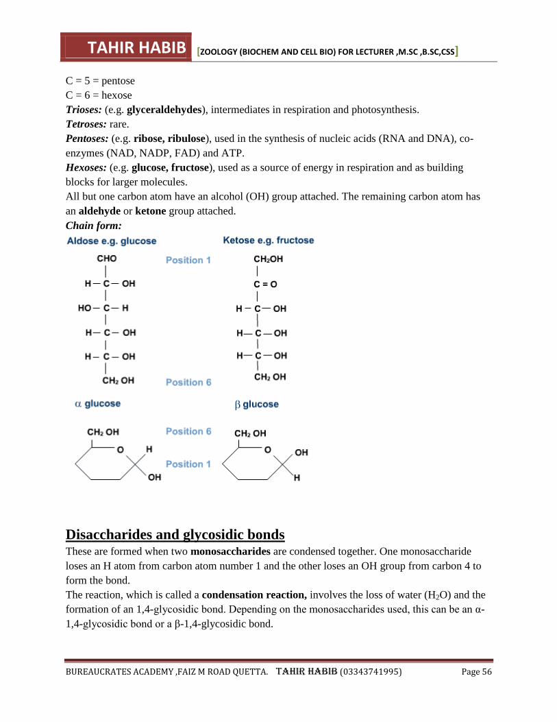
TAHIR HABIB [ZOOLOGY (BIOCHEM AND CELL BIO) FOR LECTURER ,M.SC ,B.SC,CSS]
BUREAUCRATES ACADEMY ,FAIZ M ROAD QUETTA. TAHIR HABIB (03343741995) Page 56
C = 5 = pentose
C = 6 = hexose
Trioses: (e.g. glyceraldehydes), intermediates in respiration and photosynthesis.
Tetroses: rare.
Pentoses: (e.g. ribose, ribulose), used in the synthesis of nucleic acids (RNA and DNA), co-
enzymes (NAD, NADP, FAD) and ATP.
Hexoses: (e.g. glucose, fructose), used as a source of energy in respiration and as building
blocks for larger molecules.
All but one carbon atom have an alcohol (OH) group attached. The remaining carbon atom has
an aldehyde or ketone group attached.
Chain form:
Disaccharides and glycosidic bonds These are formed when two monosaccharides are condensed together. One monosaccharide
loses an H atom from carbon atom number 1 and the other loses an OH group from carbon 4 to
form the bond.
The reaction, which is called a condensation reaction, involves the loss of water (H2O) and the
formation of an 1,4-glycosidic bond. Depending on the monosaccharides used, this can be an α-
1,4-glycosidic bond or a β-1,4-glycosidic bond.
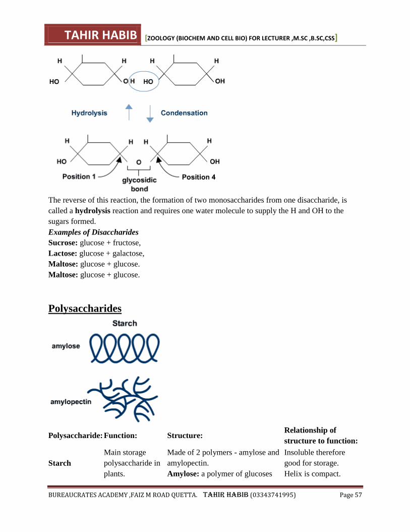
TAHIR HABIB [ZOOLOGY (BIOCHEM AND CELL BIO) FOR LECTURER ,M.SC ,B.SC,CSS]
BUREAUCRATES ACADEMY ,FAIZ M ROAD QUETTA. TAHIR HABIB (03343741995) Page 57
The reverse of this reaction, the formation of two monosaccharides from one disaccharide, is
called a hydrolysis reaction and requires one water molecule to supply the H and OH to the
sugars formed.
Examples of Disaccharides
Sucrose: glucose + fructose,
Lactose: glucose + galactose,
Maltose: glucose + glucose.
Maltose: glucose + glucose.
Polysaccharides
Polysaccharide: Function: Structure: Relationship of
structure to function:
Starch
Main storage
polysaccharide in
plants.
Made of 2 polymers - amylose and
amylopectin.
Amylose: a polymer of glucoses
Insoluble therefore
good for storage.
Helix is compact.
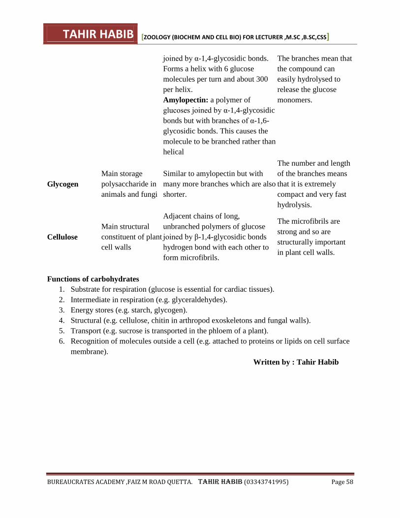
TAHIR HABIB [ZOOLOGY (BIOCHEM AND CELL BIO) FOR LECTURER ,M.SC ,B.SC,CSS]
BUREAUCRATES ACADEMY ,FAIZ M ROAD QUETTA. TAHIR HABIB (03343741995) Page 58
joined by α-1,4-glycosidic bonds.
Forms a helix with 6 glucose
molecules per turn and about 300
per helix.
Amylopectin: a polymer of
glucoses joined by α-1,4-glycosidic
bonds but with branches of α-1,6-
glycosidic bonds. This causes the
molecule to be branched rather than
helical
The branches mean that
the compound can
easily hydrolysed to
release the glucose
monomers.
Glycogen
Main storage
polysaccharide in
animals and fungi
Similar to amylopectin but with
many more branches which are also
shorter.
The number and length
of the branches means
that it is extremely
compact and very fast
hydrolysis.
Cellulose
Main structural
constituent of plant
cell walls
Adjacent chains of long,
unbranched polymers of glucose
joined by β-1,4-glycosidic bonds
hydrogen bond with each other to
form microfibrils.
The microfibrils are
strong and so are
structurally important
in plant cell walls.
Functions of carbohydrates
1. Substrate for respiration (glucose is essential for cardiac tissues).
2. Intermediate in respiration (e.g. glyceraldehydes).
3. Energy stores (e.g. starch, glycogen).
4. Structural (e.g. cellulose, chitin in arthropod exoskeletons and fungal walls).
5. Transport (e.g. sucrose is transported in the phloem of a plant).
6. Recognition of molecules outside a cell (e.g. attached to proteins or lipids on cell surface
membrane).
Written by : Tahir Habib
TAHIR HABIB [ZOOLOGY (BIOCHEM AND CELL BIO) FOR LECTURER ,M.SC ,B.SC,CSS]
BUREAUCRATES ACADEMY ,FAIZ M ROAD QUETTA. TAHIR HABIB (03343741995) Page 59
PROTEINS
These are the complex organic compounds having C, H,O and N as elements but sometimes they
contain P and S also. Due the presence of N they are called ―Nitrogenous Compounds‖ Proteins
constitute more than 50% of dry weight of cell. They are present in all types of cells and in all
parts of the cell.
CHEMICAL COMPOSITION OF PROTEINS
• Proteins are polymers of amino-acids and number of amino-acids varies from a few to 3000 or
even more in different proteins.
• These amino-acids are linked together by specialized bond or linkage called ―peptide linkage‖
• Each proteins has a unique sequence of amino-acids that gives the unique properties to
molecules.
AMINO ACID
It is the basic structural unit of proteins and all amino-acids have an ―Amino group (NH2-) and a
―Carboxyl group (COOH-)‖ attached to the same carbon atom, also known as ―Alpha carbon‖.
The have the general formula as:
1. A hydrogen atom.
2. An amino (NH2) group.
3. A carboxyl group (COOH)
4. ―Something else‖ this is the ―R‖ group.
R
¦
H2N -C - COOH
(Amino group) ¦ (Carboxylic group)
H
―R‖ may be a ―H‖ as in glycine, or CH3 as in alanine, or any other group. So amino acids mainly
differ in the R-group.
POLYPEPTIDES
Amino Acids are linked together to from polypeptides of proteins. The amino group of one
amino acids may react with the carboxyl group of another releasing a molecule of water. E.g:
Glycine and analine may combine to form a dipeptiede
PEPTIDE LINKAGE/ BOND
The linkage between the hydroxyle group of carboxyl group of one amino-acid and the hydrogen
of amino-group of another amino-acid release H2O and C-N link to form a bond called ―Peptide
bond‖.
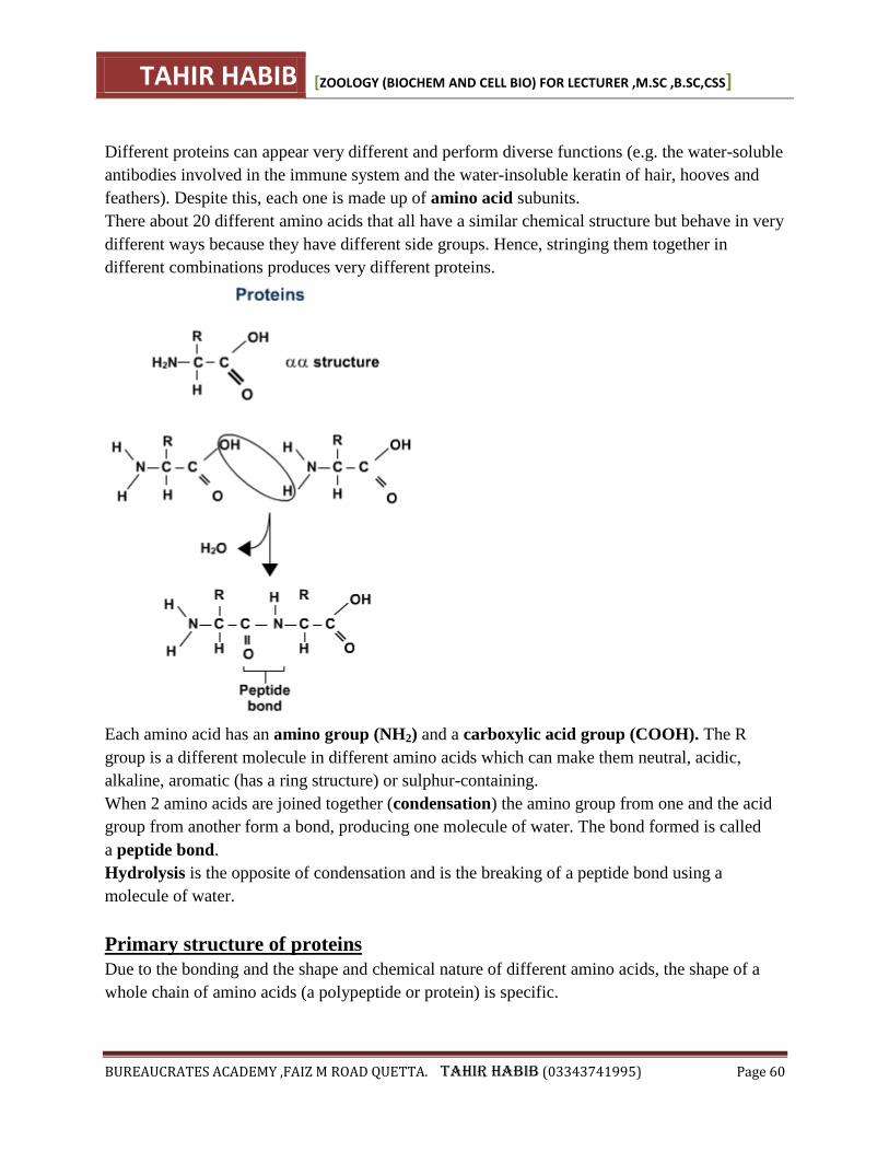
TAHIR HABIB [ZOOLOGY (BIOCHEM AND CELL BIO) FOR LECTURER ,M.SC ,B.SC,CSS]
BUREAUCRATES ACADEMY ,FAIZ M ROAD QUETTA. TAHIR HABIB (03343741995) Page 60
Different proteins can appear very different and perform diverse functions (e.g. the water-soluble
antibodies involved in the immune system and the water-insoluble keratin of hair, hooves and
feathers). Despite this, each one is made up of amino acid subunits.
There about 20 different amino acids that all have a similar chemical structure but behave in very
different ways because they have different side groups. Hence, stringing them together in
different combinations produces very different proteins.
Each amino acid has an amino group (NH2) and a carboxylic acid group (COOH). The R
group is a different molecule in different amino acids which can make them neutral, acidic,
alkaline, aromatic (has a ring structure) or sulphur-containing.
When 2 amino acids are joined together (condensation) the amino group from one and the acid
group from another form a bond, producing one molecule of water. The bond formed is called
a peptide bond.
Hydrolysis is the opposite of condensation and is the breaking of a peptide bond using a
molecule of water.
Primary structure of proteins
Due to the bonding and the shape and chemical nature of different amino acids, the shape of a
whole chain of amino acids (a polypeptide or protein) is specific.
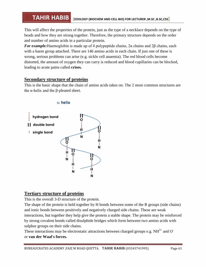
TAHIR HABIB [ZOOLOGY (BIOCHEM AND CELL BIO) FOR LECTURER ,M.SC ,B.SC,CSS]
BUREAUCRATES ACADEMY ,FAIZ M ROAD QUETTA. TAHIR HABIB (03343741995) Page 61
This will affect the properties of the protein, just as the type of a necklace depends on the type of
beads and how they are strung together. Therefore, the primary structure depends on the order
and number of amino acids in a particular protein.
For example:Haemoglobin is made up of 4 polypeptide chains, 2α chains and 2β chains, each
with a haem group attached. There are 146 amino acids in each chain. If just one of these is
wrong, serious problems can arise (e.g. sickle cell anaemia). The red blood cells become
distorted, the amount of oxygen they can carry is reduced and blood capillaries can be blocked,
leading to acute pains called crises.
Secondary structure of proteins
This is the basic shape that the chain of amino acids takes on. The 2 most common structures are
the α-helix and the β-pleated sheet.
Tertiary structure of proteins
This is the overall 3-D structure of the protein.
The shape of the protein is held together by H bonds between some of the R groups (side chains)
and ionic bonds between positively and negatively charged side chains. These are weak
interactions, but together they help give the protein a stable shape. The protein may be reinforced
by strong covalent bonds called disulphide bridges which form between two amino acids with
sulphur groups on their side chains.
These interactions may be electrostatic attractions between charged groups e.g. NH3+
and O-
or van der Waal's forces.
TAHIR HABIB [ZOOLOGY (BIOCHEM AND CELL BIO) FOR LECTURER ,M.SC ,B.SC,CSS]
BUREAUCRATES ACADEMY ,FAIZ M ROAD QUETTA. TAHIR HABIB (03343741995) Page 62
Fibrous proteins are made of long molecules arranged to form fibres (e.g. in keratin). Several
helices may be wound around each other to form very strong fibres. Collagen is another fibrous
protein, which has a greater tensile strength than steel because it consists of three polypeptide
chains coiled round each other in a triple helix. We are largely held together by collagen as it is
found in bones, cartilage, tendons and ligaments.
Globular proteins are made of chains folded into a compact structure. One of the most
important classes are the enzymes. Although these folds are less regular than in a helix, they are
highly specific and a particular protein will always be folded in the same way. If the structure is
disrupted, the protein ceases to function properly and is said to be denatured. An example is
insulin, a hormone produced by the pancreas and involved in blood sugar regulation.
A globular protein based mostly on an α-helix is haemoglobin.
A globular protein based mostly on a β-pleated sheet is the immunoglobulin antibody molecule.
Functions of proteins
1. Virtually all enzymes are proteins.
2. Structural: e.g. collagen and elastin in connective tissue, keratin in skin, hair and nails.
3. Contractile proteins: actin and myosin in muscles allow contraction and therefore
movement.
4. Hormones: many hormones have a protein structure (e.g. insulin, glucagon, growth
hormone).
5. Transport: for example, haemoglobin facilitates the transport of oxygen around the
body, a type of albumin in the blood transports fatty acids.
6. Transport into and out of cells: carrier and channel proteins in the cell membrane
regulate movement across it.
7. Defence: immunoglobulins (antibodies) protect the body against foreign invaders;
fibrinogen in the blood is vital for the clotting process.
TAHIR HABIB [ZOOLOGY (BIOCHEM AND CELL BIO) FOR LECTURER ,M.SC ,B.SC,CSS]
BUREAUCRATES ACADEMY ,FAIZ M ROAD QUETTA. TAHIR HABIB (03343741995) Page 63
LIPIDS
These are naturally occurring compounds, which are insoluble in water but soluble in organic
solvents. They contain carbon, hydrogen and oxygen like carbohydrates rate but in much lesser
ratio of oxygen than carbohydrates. These biomolecules are widely distributed among plants and
animals.
CLASSIFICATION OF LIPIDS
Following are the important groups of lipids.
1. Acylglycerol (fats and oil)
2. Waxes
3. Phospholipids.
4. Terpenoids.
(1) ACYLGLYCEROL (FATS AND OIL)
• These are found in animals and plants, provide energy for different metabolic activates and are
very rich in chemical energy.
• They are composed of glycerol and fatty acids. The most widely spread acylglycerol is triacyl
glycerol, also called triglycerides or natural lipids.
• There are two types of acylgycerol
(A) SATURATED ACYLGLYCEROL
• They contain no double bond.
• They melt at higher temperature than unsatured acylglycerols.
• Lipids containing saturated acylgycerol are solid and known as Saturated lipids.
E.g: Butter and Animal fat. etc.
(B) UNSATURATED ACYLGLYCEROL
• They contain unsaturated fatty acids i.e they contain one or more than one double bond
between carbon atom(C=C-).
• They are liquid at ordinary temperature .
• They are found in plant also called ―Oil‖
E.g: linolin found in cotton seeds etc.
(2) WAXES
Chemically waxes are mixtures of long chain alkanes and alcohols. Ketones and esters of long
chain fathy acids
• Waxes are widespread as protective coatings of fruits and leaves some insects also secrete
wax.
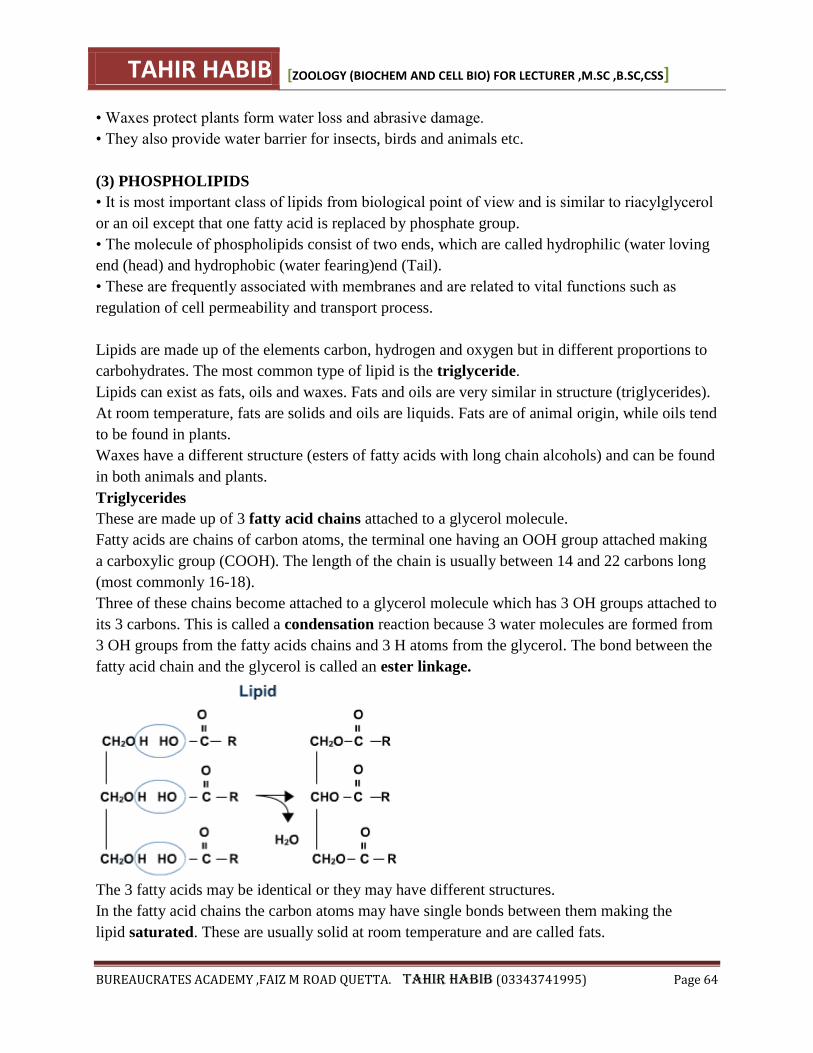
TAHIR HABIB [ZOOLOGY (BIOCHEM AND CELL BIO) FOR LECTURER ,M.SC ,B.SC,CSS]
BUREAUCRATES ACADEMY ,FAIZ M ROAD QUETTA. TAHIR HABIB (03343741995) Page 64
• Waxes protect plants form water loss and abrasive damage.
• They also provide water barrier for insects, birds and animals etc.
(3) PHOSPHOLIPIDS
• It is most important class of lipids from biological point of view and is similar to riacylglycerol
or an oil except that one fatty acid is replaced by phosphate group.
• The molecule of phospholipids consist of two ends, which are called hydrophilic (water loving
end (head) and hydrophobic (water fearing)end (Tail).
• These are frequently associated with membranes and are related to vital functions such as
regulation of cell permeability and transport process.
Lipids are made up of the elements carbon, hydrogen and oxygen but in different proportions to
carbohydrates. The most common type of lipid is the triglyceride.
Lipids can exist as fats, oils and waxes. Fats and oils are very similar in structure (triglycerides).
At room temperature, fats are solids and oils are liquids. Fats are of animal origin, while oils tend
to be found in plants.
Waxes have a different structure (esters of fatty acids with long chain alcohols) and can be found
in both animals and plants.
Triglycerides
These are made up of 3 fatty acid chains attached to a glycerol molecule.
Fatty acids are chains of carbon atoms, the terminal one having an OOH group attached making
a carboxylic group (COOH). The length of the chain is usually between 14 and 22 carbons long
(most commonly 16-18).
Three of these chains become attached to a glycerol molecule which has 3 OH groups attached to
its 3 carbons. This is called a condensation reaction because 3 water molecules are formed from
3 OH groups from the fatty acids chains and 3 H atoms from the glycerol. The bond between the
fatty acid chain and the glycerol is called an ester linkage.
The 3 fatty acids may be identical or they may have different structures.
In the fatty acid chains the carbon atoms may have single bonds between them making the
lipid saturated. These are usually solid at room temperature and are called fats.
TAHIR HABIB [ZOOLOGY (BIOCHEM AND CELL BIO) FOR LECTURER ,M.SC ,B.SC,CSS]
BUREAUCRATES ACADEMY ,FAIZ M ROAD QUETTA. TAHIR HABIB (03343741995) Page 65
If one or more bonds between the carbon atoms are double bonds, the lipid is unsaturated.
These are usually liquid at room temperature and are called oils.
Functions of lipids
1. Storage - lipids are non-polar and so are insoluble in water.
2. High-energy store - they have a high proportion of H atoms relative to O atoms and so
yield more energy than the same mass of carbohydrate.
3. Production of metabolic water - some water is produced as a final result of respiration.
4. Thermal insulation - fat conducts heat very slowly so having a layer under the skin
keeps metabolic heat in.
5. Electrical insulation - the myelin sheath around axons prevents ion leakage.
6. Waterproofing - waxy cuticles are useful, for example, to prevent excess evaporation
from the surface of a leaf.
7. Hormone production - steroid hormones. Oestrogen requires lipids for its formation, as
do other substances such as plant growth hormones.
8. Buoyancy - as lipids float on water, they can have a role in maintaining buoyancy in
organisms.
Phospholipids
A phosphate-base group replaces one fatty acid chain. It makes this part of the molecule (the
head) soluble in water whilst the fatty acid chains remain insoluble in water.
Due to this arrangement, phospholipids form bilayers (the main component of cell and
organelle membranes).
TAHIR HABIB [ZOOLOGY (BIOCHEM AND CELL BIO) FOR LECTURER ,M.SC ,B.SC,CSS]
BUREAUCRATES ACADEMY ,FAIZ M ROAD QUETTA. TAHIR HABIB (03343741995) Page 66
SHORT ANSWERS (BIOCHEM and cell bio)
Q 1. Define Biochemistry? Why study of biochemistry is essential in the field of biology or
in the study of living organisms?
Ans. Biochemistry is a branch of Biology, which deals with the study of chemical components
and the chemical processes in living organisms.
Importance of Biochemistry: A basic knowledge of biochemistry is essential for
understanding anatomy and physiology, because all of the structures of an organism
have biochemicalorganization. Photosynthesis, respiration, digestion, muscle contraction can all
be described in biochemical terms.
Q 2. Name the most important organic and inorganic compounds in living organisms.
Ans. Most important organic compounds in living organisms are carbohydrates, proteins, lipids
and nucleic acids. Among inorganic substances are water carbon dioxide, acids, bases and salts.
Q 3. Compare the chemical composition of bacterial and mammalian cell.
Ans. Chemical Components % Total Cell Weight Bacterial Cell Mammalian Cell
Water 70% 70%
Proteins 15% 15%
Carbohydrates 3% 4%
Lipids 2% 3%
DNA 1% 0.25%
RNA 6% 1.1%
Other organic molecules(Enzymes, hormones, metabolites) 2% 2%
Inorganic ions (Na+, K+. Ca+2, Mg+2, Cl-, (SO-2)4) 1% 1%
Q 4. Differentiate between Metabolism, Catabolism and Anabolism.
Ans. Metabolism: All the chemical reactions taking place within a cell are collectively called
metabolism.
Metabolism = Anabolism + Catabolism
Energy is taken and released simultaneously.
Anabolism: Those reactions in which substances are combined to form complex substances
are called anabolic reactions. Anabolic reactions need energy.
Catabolism: The breakdown of complex molecules into simpler ones, such reactions are called
catabolic reactions. Energy is released during catabolic reactions.
Q 5. Why carbon is considered as the basic element of organic compounds?
Ans. Carbon is considered as the basic element of organic compounds, because it is tetravalent.
TAHIR HABIB [ZOOLOGY (BIOCHEM AND CELL BIO) FOR LECTURER ,M.SC ,B.SC,CSS]
BUREAUCRATES ACADEMY ,FAIZ M ROAD QUETTA. TAHIR HABIB (03343741995) Page 67
Due to its unique properties, carbon occupies the central position in the skeleton of life. It can
react with oxygen, hydrogen, nitrogen, phosphorus and sulphur forming covalent bonds. It is also
important due to following associations.
Carbon-Hydrogen Bond: It is potential source of chemical energy for cellular activities.
Carbon-Oxygen Bond: Carbon Oxygen association in glycosidic linkages provides stability to
complex carbohydrate molecules.
Carbon-Nitrogen Bond: Carbon combines with nitrogen in amino acids linkages to form
peptide bonds and forms proteins which are very important due to their diversity in structure and
functions.
Q 6. What is the percentage of water in bone cells and brain cells of human?
Ans. Human tissue contain about 20 percent water in bone cells and 85 percent water in brain
cells.
Q 7. Water is excellent solvent for polar substances. Justify this statement.
Ans. Due to its polarity, water is an excellent solvent for polar substances. Ionic substances when
dissolves in water, dissociate into positive and negative ions. Non-ionic substances having
charged groups in their molecules are dispersed in water. Almost all the reactions in cell occur in
aqueous media.
Q 8. What is the function of non-polar substances?
Ans. Non-polar organic molecules such as fats, are insoluble in water and help to maintain
membranes which make compartments in the cell.
Q 9. Define specific heat capacity of water.
Ans. The number of calories required to raise the temperature of 1g of water from 15-16C is 1.
This is called specific heat capacity of water.
Q 10. How water works as a temperature stabilizer for living organisms?
Ans. Water has great ability of absorbing heat with minimum of change in its own temperature.
This is because much of the energy is used to break hydrogen bonds. Water thus works as
temperature stabilizer for organisms in the environment and hence protects living material
against sudden thermal changes.
Q 11. How heat of vaporization of water is beneficial in daily life?
Ans. The specific heat of vaporization of water is 574 K cal/kg, which pays an important role in
the regulation of heat produced by oxidation. It also provides cooling effect to plants when water
is transpired, or to animals hen water is respired.
Q 12. What is the concentration of H+ and OH- ions in pure water at 25C?
Ans. At 25C the concentration of each H+ and OH- ions in pure water is about 10-7 mole/litre of
TAHIR HABIB [ZOOLOGY (BIOCHEM AND CELL BIO) FOR LECTURER ,M.SC ,B.SC,CSS]
BUREAUCRATES ACADEMY ,FAIZ M ROAD QUETTA. TAHIR HABIB (03343741995) Page 68
earch.
Q 13. How water act as an effective lubricant?
Ans. Water is effective lubricant that provides protection against damage resulting from friction.
For example, tears protect the surface of eye from the rubbing of eyelids, water also forms a fluid
cushion around organs that helps to protect them from trauma.
Q 14. List some important functions of carbohydrates.
Ans. Carbohydrates occur abundantly in living organisms. They are found in all organisms and
in almost all parts of the ell. Carbohydrates play both structural and functional roles. Simple
carbohydrates are the main source of energy in cells. Some carbohydrates are the main
constituent of cell walls in plants and microorganisms. Cellulose of wood cotton an paper,
starches present cereals, roots tubers, cane sugar and milk sugar are all examples of
carbohydrates.
Q 15. What are carbohydrates? Written by : Tahir Habib
Ans. The word carbohydrate literally means hydrated carbons. They are composed of carbon,
oxygen and hydrogen.
Formula:
Their formula is Cn(H2O)n.
Chemical Definition:
Chemically, carbohydrates are defined as polyhydroxy aldehydes, or complex substances which
no hydrolysis yield polyhydroxy aldehyde or ketone subunits.
Q 16. What is hydrolysis?
Ans. Hydrolysis involves the breakdown of large molecules into smaller ones utilizing water
molecules.
Q 17. Carbohydrates are classified into how many groups?
Ans. Carbohydrates are classified into three groups:
1. Mono saccharides
2. Oligosacchardies
3. polysaccharides
Q 18. What are Mono-saccharides? Give some of its properties and examples.
Ans. Mono saccharides are simple sugars. They are sweet in taste, are easily soluble in water,
and cannot by hydrolyzed into simple sugar. Chemically they are either
polyhydroxy aldehydes or ketones. The sugar with aldehyde group is called also sugar and with
TAHIR HABIB [ZOOLOGY (BIOCHEM AND CELL BIO) FOR LECTURER ,M.SC ,B.SC,CSS]
BUREAUCRATES ACADEMY ,FAIZ M ROAD QUETTA. TAHIR HABIB (03343741995) Page 69
keto group is called keto sugar. Examples are glyceraldehyde. ribose and glucose, etc.
Q 19. How glucose is prepared / produced naturally?
Ans. Glucose is naturally produced in green plants which take carbon dioxide from air and
water from the soil to synthesize glucose. Energy is consumed in this process which is provided
by sunlight. That is why this process is called photosynthesis.
Q 20. How much glucose our blood contains?
Ans. Our blood normally contains 0.08% glucose.
Q 21. How much energy is used to synthesize 10g of glucose?
Ans. 717.6 K cal of solar energy is required for the synthesis of 10g of glucose.
Q 22. What are digosaccharides? Give some examples of important disacchraides.
Ans. These are comparatively less sweet in taste, and less soluble in water. On hydrolysis
oligosacchardies yield from to to ten mono saccharides. The one yielding two mono saccharides
are known as disaccharides, those yielding three are known as trisaccharides and so on. The
covalent bond between two mono saccharides is called glycosidic bond.
Q 23. What are polysaccharides? Name some biologically important polysaccharides.
Ans. Poly saccharides are the most complex and most abundant carbohydrate in nature. They are
usually branched and tasteless. They are formed by several mono saccharides units linked by
glycosidic bonds. Polysaccharides have high molecular weights and are only sparingly soluble in
water. Some biologically important poly saccharides are starch, a gas, glycogen, cellulose,
dextrins, pectin and chitin.
Q 24. Differentiate between amylase and amylopectin.
Ans. Amylose:
1. Amylose starches have unbranched chains of glucose and
2. Are soluble in hot water
Amylopectin:
1. Amylopectin starches have branched chains and
2. Are insoluble in hot or cold water
Q 25. Differentiate between starch, cellulose and glycogen.
Ans. Starch: Is is found in fruits, grains and tubers. It is the main source of carbohydrates for
animals. On hydrolysis, it yields glucose molecules. Starches are of two types, amylose and
amylopectin. Amylose starches have branched chains of glucose and are soluble in hot water.
Amylopectin starches have branched chains and are insoluble in hot or cold water. Give blue
TAHIR HABIB [ZOOLOGY (BIOCHEM AND CELL BIO) FOR LECTURER ,M.SC ,B.SC,CSS]
BUREAUCRATES ACADEMY ,FAIZ M ROAD QUETTA. TAHIR HABIB (03343741995) Page 70
color with iodine.
Cellulose: It is most abundant in nature. Cotton is pure form of cellulose. It is the main
constituent of cell wall of plants and is highly insoluble in water. On hydrolysis, it also yields
glucose molecules. Cellulose gives no color with iodine. It is not digested in human digestive
system. In the herbivores, it is digested because of micro-organism in their digestive tract. These
micro-organisms secrete an enzyme called cellulase for its digestion.
Glycogen: It is also called animal starch. It is the chief form of carbohydrate stored in animal
body. It is abundantly in liver and muscles, though found in all animal cells. It is insoluble in
water and it also yields glucose on hydrolysis. Gives red color with iodine.
Q 26.What are lipids?
Ans. The lipids are heterogenous group of compounds related to fatty acids. They are insoluble
in water but soluble in organic solvents such as ether, alcohol, chloroform and benzene. Lipids
include fats, oils, waxes, cholesterol and related compounds.
Q 27. What is the use of lipids in daily life?
Ans. Lipids as hydrophobic compounds are components of cellular membranes. They are used
to store energy. Some lipids provide insulation against atmospheric heat and cold and also act as
water proof material waxes, in the exoskeleton of insects and cutin, an additional protective layer
on the cuticle of epidermis of some plants organs. e.g., leaves fruits, seeds etc., protect them.
Q 28. Give the classification of lipids.
Ans. Lipids have been classified as acylglycerols, waxes, phospholipids, sphingolipids,
glycolipids, and terpenoids lipids including carotenoids and steroids.
Q 29. What are acylglycerols?
Ans. Acylglycerols are composed of glycerol and fatty acids. The most widely spread
acylglycerol is triacylglycerol also triglycerides or neutral lipids. Chemically acylglycerol can be
defined as esters of fatty acids and alcohol. An ester is the compound produced as the result of a
chemical reaction of an alcohol with an acid and a water molecule is released.
Q 30. What are waxes? What is the importance of waxes?
Ans. Waxes are widespread as protective coatings on fruits and leaves Some insects also secrete
wax. Chemically, waxes are mixtures of long chain alkanes and alcohols, ketones and esters of
long chain fatty acids.
Importance: Waxes protect plants from water loss and abrasive damage. They also provide
barrier for insects, birds and animals such as sheep.
Q 31. What are phospholipids?
TAHIR HABIB [ZOOLOGY (BIOCHEM AND CELL BIO) FOR LECTURER ,M.SC ,B.SC,CSS]
BUREAUCRATES ACADEMY ,FAIZ M ROAD QUETTA. TAHIR HABIB (03343741995) Page 71
Ans. Phospholipids are derivatives of phosphoric acid, which are composed of glycerol, fatty
acids and phosphoric acid. Nitrogenous bases such as choline ethanolamine and serine are
important components of phospholipids. They are widespread in bacteria, animal and plant cells
and are frequently associated with membranes. Phosphatidylcholine is one of the common
phospholipids.
Q 32. What are terpenoids?
Ans. Terpenoids are a very large and important group of compounds which are made up of
simple repeating simple units, isoprenoid units. This unit by condensation in different ways give
rise to compounds such as rubber, carotenoids, steroids terpenes etc.
Q 33. What are proteins?
Ans. Proteins are the most abundant organic compounds to be found in cells and comprise over
50% of their total dry weight.
Q 34. Illustrate the important functions performed by proteins.
Ans.
1. Building Structures of Cell: Proteins build many structures of the cell.
2. As Enzymes: All enzymes are proteins in this way they control the whole metabolism of
the cell.
3. As Hormones: As hormones, proteins regulates metabolic processes.
4. As carriers: Some proteins work as carriers and transport specific substances such as
oxygen, lipids, ions etc.
5. As Antibodies: Some proteins called antibodies, defend the body against pathogens.
6. Blood Clotting Proteins: They prevent loss of blood from the body after an injury.
7. Movement of Organs: Movement of organs and organisms, and movement of
chromosomes during anaphase of cell division are caused by proteins.
Q 35.What are amino acids? Give its general formula.
Ans. Proteins are polymers of amino acid, the compounds containing nitrogen oxygen and
hydrogen. All the amino acids have an amino group (-NH2) and a carboxyl group(-COOH)
attached to the same carbon atom, also known as alpha carbon.
Q 36. How peptide bond is formed between two amino acids?
Ans. The linkage between the hydroxyl group of carboxyl group of one amino acid and the
hydrogen of amino group of another amino acid release H2) and C-N link to form a bond called
peptide bond,. The resultant compound of glycylalanine has two amino acid subunits and is a
dipeptide. A dipeptide has an amino group at one end and a carboxyl group at the other end of
the molecule.
TAHIR HABIB [ZOOLOGY (BIOCHEM AND CELL BIO) FOR LECTURER ,M.SC ,B.SC,CSS]
BUREAUCRATES ACADEMY ,FAIZ M ROAD QUETTA. TAHIR HABIB (03343741995) Page 72
Q 37. Who was the first scientist who determined the sequence of amino acids in a protein
molecule?
Ans. F.Sanger was the first scientist who determined the sequence of amino acids in a protein
molecules. After ten years, of careful work he concluded, that insulin is composed of 51 amino
acids in two chain. One of the chains had 21 amino acids and the other had 30 amino acids and
they were held together by disulphide bridges. Haemoglobin is composed of four chains, two
alpha and two beta chains. Each alpha chain contain 141 amino acids, while beta chain contains
146 amino acids.
Q 38. What is ribosomal RNA?
Ans. It is the major portion of RNA in the cell, and may be up to 80% of the total RNA. It is
strongly associated with the ribosomal protein where 40 to 50% of it is present. It acts as a
machinery for the synthesis of proteins. On the surface of the ribosomal the mRNA and tRNA
molecules interact to translate the information from genes into a specific protein.
Q 39. Why are fats considered as high energy compounds?
Ans. Because of higher proportions of C-H bonds and very low proportion of oxygen, lipids
store double the amount of energy as compared to the same amount of any carbohydrate.
Q 40. How were nucleic acids isolated?
Ans. Nucleic acids were isolated in 1870 by F. Miesches from the nuclei of the pus cells. Due to
their isolation from nuclei and their acidic nature, they were named nucleic acids.
Q 41. Differentiate between a nucleotide and a nucleoside?
Ans. Nucleotide: A nucleoside and a phosphoric acid combine to form a nucleotide.
Nucleoside: The compound formed by combination of a base and pentose sugar is called
nucleoside.
Q 42. What is a gene?
Ans. Gene is a unit of biological inheritance.
Q 43. Which was the first microbe have the genome completely sequenced?
Ans. Haemophilus influenzae is the first microbe to have the genome completely sequenced
and this was published on July 28, 1995.
Q 44. What is transpiration?
Ans. RNA is synthesizes by DNA in a process as transcription.
Q 45. What is messenger RNA?
Ans. it takes the genetic message from the nucleus to the ribosomes in the cytoplasm to form
TAHIR HABIB [ZOOLOGY (BIOCHEM AND CELL BIO) FOR LECTURER ,M.SC ,B.SC,CSS]
BUREAUCRATES ACADEMY ,FAIZ M ROAD QUETTA. TAHIR HABIB (03343741995) Page 73
particular proteins messenger RNA carries the genetic information from DNA to ribosomes,
where amino acids are arranged according to the information in mRNA to form specific protein
molecule. This is a type of a single strand of variable length. Its length depends upon the size of
the gene as well as the protein form which it is taking the message. mRNA is about 3 to 4% of
the total RNA in the cell.
Q 46. What is transfer RNA?
Ans. It comprises about 10 to 20% of the cellular RNA Transfer RNA molecule are small, each
with a chain length of 75 to 90 nucleotides. It is involved in protein synthesis.
Q 47. What are conjugated molecules? Give some examples of it.
Ans. Two different molecules belonging to different categories usually combine together to
form conjugated molecules. For exp: Glycoproteins, glycolipids, nucleoproteins, lipoproteins,
etc.
Q 48. What are nucleohistones? What is their function?
Ans. Nucleic acids have special affinity for basic proteins. Nucleohistones are present in
chromosomes. They play important role in regulation of gene expression.
Q 49. What are lipoproteins? What is their function?
Ans. Lipoproteins are formed by combination of lipids and proteins. They are basic structural
framework of all types pf membranes in the cell.
Q 50. Differentiate between glycoproteins and glycolipids.
Ans. Glycoproteins: Carbohydrate may combine with proteins to form glycoproteins. Most of
the secretions are glycoprotein in nature and they are integral structural components of plasma
membrane.
Glycolipids: Carbohydrate may combine with, lipids to form glycolipids. They are also integral
structural components of plasma membrane.
Q 51. What is the contribution of Erwin Chargaff in biology regarding structure of DNA?
Ans. In 1951 Erwin Chargaff provided data about the ratios of different bases present in the
DNA molecule. This data suggested that adenine and thymine are equal in ratio and so are
guanine and cytosine.
Q 52. Who used the technique of X-ray diffraction to determine structure of DNA?
Ans. Maurice Wilkins and Rosalind Franklin Used the technique of X-ray diffraction to
determine structure of DNA.
Q 53. Who built the scale model of DNA?
TAHIR HABIB [ZOOLOGY (BIOCHEM AND CELL BIO) FOR LECTURER ,M.SC ,B.SC,CSS]
BUREAUCRATES ACADEMY ,FAIZ M ROAD QUETTA. TAHIR HABIB (03343741995) Page 74
Ans. James-D Watson and Francis Crick built the scale model of DNA.
Q 54. Differentiate between purines and pyrimidines?
Ans. Purines: These are double ringed nitrogenous bases. Adenine and guanine are purines.
Pyrimidines: These are single ringed nitrogenous bases. Cytosine, thymine and uracil are
pyrimidines.
Q 55. Which one is more soluble in organic solvent Palmitic acid or Butyric acid? Support
your answer.
Ans. Palmitic acid is more soluble in organic solvents than butyric acid because it has more
carbon atoms that butyric acid and its melting point is also more then butyric acid.
Q 56. Differentiate between saturated and unsaturated.
Ans. Saturated Fatty Acid: Saturated fatty acid contains double bond. These are oils and liquids
at room temperature.
Unsaturated Fatty Acid: Unsaturated fatty acids have no double bonds. They are fats and solid at
room temperature.
Q 57. Describe primary structure of proteins.
Ans. The primary structure comprises the number and sequence of amino acids in a single chain
in a protein molecule. The size of protein molecule is determined by the type of amino acids and
the number of amino acids comprising that particular molecule.
Q 58. What is the secondary structure of proteins?
Ans. The polypeptide chain in secondary do not lie flat. They coil into a helix or some other
configuration. Common secondary structure is alpha-helix which involves spiral formation of the
polypeptide chain. It has 3.6 amino acids in each turn. This structure is kept by the formation of
hydrogen bonds among amino acids molecules. Beta-pleated Sheet is formed by folding back of
the polypeptide.
Q 59. What is tertiary structure? How it is maintained?
Ans. A polypeptide chain bends and folds upon itself forming a globular shape. This is the
proteins tertiary structure. It is maintained by ionic, hydrogen and disulphide bonds.
Q 60. How many type of cell walls are present in different organisms?
Ans. Organism Cell Wall
Bacteria Pepidoglycan and lipopolysaccharides (lipoprotein
complex)
Blue green algae Muramic acid
Fungi Chitin

TAHIR HABIB [ZOOLOGY (BIOCHEM AND CELL BIO) FOR LECTURER ,M.SC ,B.SC,CSS]
BUREAUCRATES ACADEMY ,FAIZ M ROAD QUETTA. TAHIR HABIB (03343741995) Page 75
Algae and other Plants Mainly cellulose
Q 61. Write down the differences between DNA and RNA?
Ans. Feature DNA RNA
Nucleotides Deoxyribonucleotides Ribonucleotides
Pentose Sugar Deoxyribose Ribose
Nitrogenous Bases A.G.C.T A.G.C.U
Physical Structure Double strands Single strand
Location Nuclei and in much lesser Nucleolus, ribosomes, cystosol
amount in mitochondria and in smaller amount in other
and chloroplasts. parts of the cell.
Amount Constant in each cell of species. Variable from cell to cell.
Role Heredity Protein Synthesis
Q 62. Compare four levels of protein molecules.
Ans. Feature Primary Secondary Tertiary Quaternary
Information No and sequence Coiling Bending and Aggregation
of amino acids folding
Bonds Disulphide Hydrogen Ionic, hydrogen, Hydrophobic,
disulphide ionic, hydrogen
Example Insulin, Hb Alpha Helix Single chain of Hb Hb molecule
Q 63. Differentiate between different structure of proteins.
Ans. Feature Fibrous Protein Globular Protein
Shape Fibrils Spherical or ellipsoidal
Structural Organization Secondary Tertiary
Solubility in aqueous media Insoluble Soluble
Crystal Nature Non-crystalline Crystalline
Elasticity Elastic Inelastic
Role Structural Functional
Stability Stable Unstable
Examples Silk fibers, actin, myosin, Enzymes antibodies,
fibrin, keratin hemoglobin, hormones
Q 64. Describe conjugating molecules.
Ans. Components Molecule Role
Carbohydrates + Proteins Glycoproteins Cellular secretions, integral part
of biological membranes.
Carbohydrates + Lipids Glycolipids Integral component of biological
membranes.
TAHIR HABIB [ZOOLOGY (BIOCHEM AND CELL BIO) FOR LECTURER ,M.SC ,B.SC,CSS]
BUREAUCRATES ACADEMY ,FAIZ M ROAD QUETTA. TAHIR HABIB (03343741995) Page 76
Nucleic acid + Histones Nucleohistones Chromosome regulation of gene
Expression.
ADDITIONAL SHORT ANSWERS (FROM PAST PAPER)
1.Purines and Pyrimidines are nitrogenous bases that make up the two different kinds of
nucleotide bases in DNA and RNA. The two-carbon nitrogen ring bases (adenine and guanine)
are purines, while the one-carbon nitrogen ring bases (thymine and cytosine) arepyrimidines.
2.CLONING is the process of producing similar populations of genetically identical
individuals that occurs in nature when organisms such as bacteria, insects or plants reproduce
asexually.
3.Transcription is the first step of gene expression, in which a particular segment of DNA is
copied into RNA (especially mRNA) by the enzyme RNA polymerase. Both DNA and RNA are
nucleic acids, which use base pairs of nucleotides as a complementary language.
4.The LAC OPERON (LACTOSE OPERON) is an operonrequired for the transport
and metabolism oflactose in Escherichia coli and many other enteric bacteria. Although glucose
is the preferred carbon source for most bacteria, thelac operon allows for the effective digestion
oflactose when glucose is not available.
5.PEPTIDE BOND is synthesized when the carboxyl group of one amino acid molecule reacts
with the amino group of the other amino acid molecule, causing the release of a molecule of
water (H2O), hence the process is a dehydration synthesis reaction (also known as a condensation
reaction).
6.The CENTRAL DOGMA: DNA Encodes RNA, RNA Encodes Protein. The central
dogma of molecular biology describes the flow of genetic information in cells from DNA to
messenger RNA (mRNA) to protein. It states that genes specify the sequence of mRNA
molecules, which in turn specify the sequence of proteins
7.GLUCONEOGENESIS (GNG) is a metabolic pathway that results in the generation of
glucose from certain non-carbohydrate carbon substrates.
8.COENZYME: a non-protein compound that is necessary for the functioning of an enzyme.
Examples are: example is thiamine pyrophosphate (TPP), which is tightly bound in
transketolase or pyruvate decarboxylase, while it is less tightly bound in pyruvate
TAHIR HABIB [ZOOLOGY (BIOCHEM AND CELL BIO) FOR LECTURER ,M.SC ,B.SC,CSS]
BUREAUCRATES ACADEMY ,FAIZ M ROAD QUETTA. TAHIR HABIB (03343741995) Page 77
dehydrogenase. Other coenzymes, flavin adenine dinucleotide (FAD), biotin, and lipoamide,
for instance, are covalently bound.
9.RIBONUCLEIC ACID (RNA) is a polymeric molecule essential in various biological roles
in coding,.
Three major TYPES OF RNA are mRNA, or messenger RNA, that serve as temporary copies of
the information found in DNA; rRNA, or ribosomal RNA, that serve as structural components of
protein-making structures known as ribosomes; and finally, tRNA, or transfer RNA, that ferry
amino acids to the ribosome to be assembled
10.TYPES OF AMINO ACIDS: Amino acids bond together to make long chains. Those long
chains ofamino acids are also called proteins. Essential Amino Acids: Histidine, Isoleucine,
Leucine, Lysine, Methionine, Phenylalanine, Threonine, Tryptophan, and Valine.
Nonessential Amino Acids: Alanine, Asparagine, Aspartic Acid, Glutamic Acid.
11.APOPTOSIS: A form of cell death in which a programmed sequence of events leads to the
elimination of cells without releasing harmful substances .
12.In genetics, COMPLEMENTARY DNA (CDNA) is double-stranded DNA synthesized
from a single stranded RNA.
13.CHYLOMICRON: A small fat globule composed of protein and lipid
(fat). Chylomicrons are found in the blood and lymphatic fluid where they serve to transport fat
from its port of entry in the intestine to the liver and to adipose (fat) tissue.
14.HISTONES are highly alkaline proteins found in eukaryotic cell nuclei that package and
order the DNA into structural units called nucleosomes.
15.The TRP OPERON is an operon — a group of genes that are used, or transcribed, together
— that codes for the components for production of tryptophan.
16.DNA LIGASE is a specific type of enzyme, aligase, that facilitates the joining
of DNA strands together by catalyzing the formation of a phosphodiester bond.
17.TERMINATION CODON (STOP CODON):
Any of three mRNA sequences (UGA, UAG, UAA) that do not code for an amino acid and thus
signal the end of protein synthesis. Written by : Tahir Habib