Embed Size (px)
Citation preview

End Show
Slide of 51
Copyright Pearson Prentice Hall
Biology
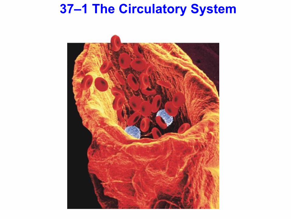
37–1 The Circulatory System
The circulatory system and respiratory system work together to supply cells with the nutrients and oxygen they need to stay alive.

Functions of the Circulatory System
Humans and other vertebrates have closed circulatory systems
In a closed circulatory system, blood is transported within a system of vessels.

The human circulatory system consists of:
•the heart •blood vessels •blood
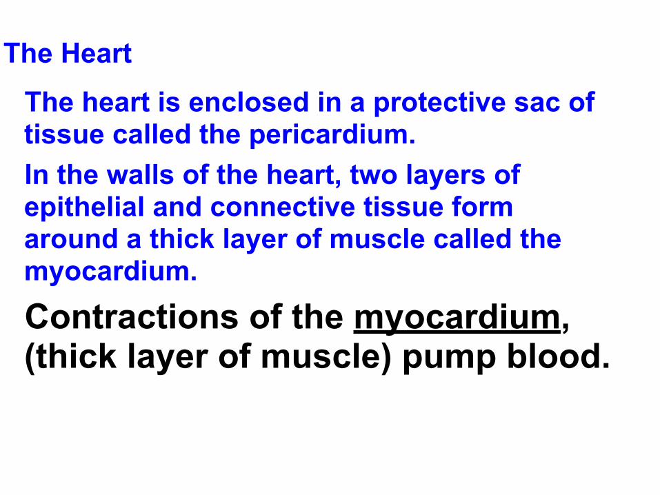
The Heart
The heart is enclosed in a protective sac of tissue called the pericardium.
In the walls of the heart, two layers of epithelial and connective tissue form around a thick layer of muscle called the myocardium.
Contractions of the myocardium, (thick layer of muscle) pump blood.

Structures of the Heart

Large vein that brings oxygen-poor blood from the upper part of the body to the right atrium
Right Atrium
Superior Vena Cava:

Bring oxygen-poor blood from the lungs to the left atrium
Left AtriumPulmonary Veins:

Prevents blood from flowing back into the right ventricle after it has entered the pulmonary artery.
Right Atrium
Pulmonary ArteriesPulmonary Valve:

Prevents blood from flowing back into the right atrium after it has entered the right ventricle
Right Atrium
Tricuspid Valve:

Vein that brings oxygen-poor blood from the lower part of the body to the right atrium.
Right Atrium
Inferior Vena Cava:
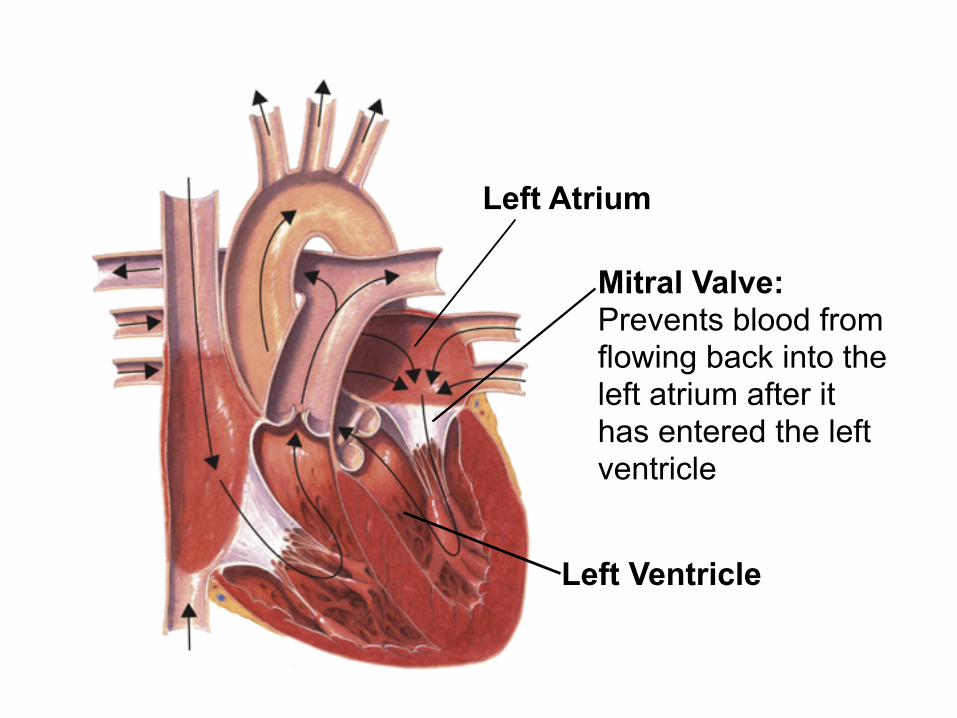
Mitral Valve: Prevents blood from flowing back into the left atrium after it has entered the left ventricle
Left Atrium
Left Ventricle

Aortic Valve: Prevents blood from flowing back into the left ventricle after it has entered the aorta
Left Atrium
Left Ventricle
Aorta

Bring oxygen-poor blood to the right or left lung
Pulmonary Arteries:

Brings oxygen-rich blood from the left ventricle to the body
Aorta:

The septum divides the right side of the heart from the left. It prevents the mixing of oxygen-poor and oxygen-rich blood.

The heart has four chambers—two atria and two ventricles.
There are two chambers on each side of the septum.
The upper chamber, which receives the blood, is the atrium.
The lower chamber, which pumps blood out of the heart, is the ventricle.
Circulation Through the Heart Blood enters the heart through the right and left atria.
As the heart contracts, blood flows into the ventricles and then out from the ventricles to either the body or the lungs.

There are flaps of connective tissue called valves between the atria and the ventricles.
When the ventricles contract, the valves close, which prevents blood from flowing back into the atria.

At the exits from the right and left ventricles, valves prevent blood that flows out of the heart from flowing back in.
Blood leaves the left ventricle, and enters the aorta.
The aorta is one of the blood vessels that carry the blood through the body and back to the heart.

Circulation Through the Body
The heart functions as two separate pumps.

Pulmonary Circulation
Pulmonary circulation pumps blood between the heart and the lungs. RIGHT SIDE OF HEART (smaller)
In the lungs, carbon dioxide leaves the blood and oxygen is absorbed. The oxygen-rich blood returns to the heart.

Systemic Circulation
Systemic circulation pumps blood between the heart and the rest of the body. LEFT SIDE OF heart (larger) This pathway is called systemic circulation.
After returning from the lungs, the oxygen-rich blood is pumped to the rest of the body.

Circulation of Blood through the Body
Capillaries of head and arms
Superior vena cava Aorta
Pulmonary veinCapillaries of
right lungs
Inferior vena cava
Capillaries of abdominal organs and legs
Capillaries of left lung
Pulmonary artery

26

Heartbeat
Each contraction begins in the sinoatrial (SA) node in the right atrium.
Because these cells start the wave of muscle contraction through the heart, they are called the pacemaker.

Sinoatrial (SA) node Conducting fibers
The impulse spreads from the pacemaker (SA node) to a network of fibers in the atria.

Conducting fibers
Atrioventricular (AV) node
The impulse is picked up by a bundle of fibers called the atrioventricular (AV) node and carried to the network of fibers in the ventricles.

When the network in the atria contracts, blood in the atria flows into the ventricles. When the ventricles contract, blood flows out of the heart.

As blood flows through the circulatory system, it moves through three types of blood vessels: •arteries
•capillaries
•veins

Large vessels that carry blood from the heart to the tissues of the body are called arteries. Except for the pulmonary arteries, all arteries carry oxygen-rich blood. Arteries have thick walls.
They contain connective tissue, smooth muscle, and endothelium.


Capillaries
The smallest of the blood vessels are the capillaries.
Their walls are only one cell thick, and most are narrow.
The capillaries bring nutrients and oxygen to the tissues and absorb carbon dioxide and other waste products from them.


Veins
Blood vessels that carry blood back to the heart are veins. Veins have thinner walls than arteries.
The walls of veins contain connective tissue and smooth muscle.

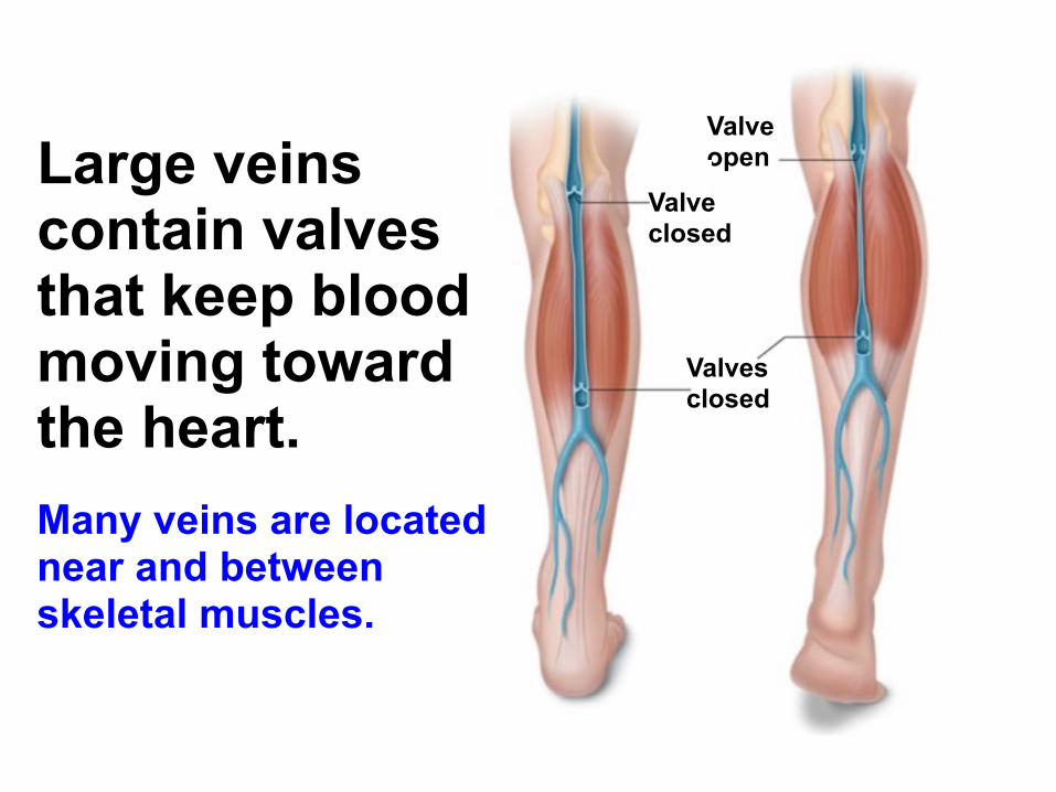
Large veins contain valves that keep blood moving toward the heart. Many veins are located near and between skeletal muscles.
Valve open
Valve closed
Valves closed

Blood Pressure
When the heart contracts, it produces a wave of fluid pressure in the arteries.
The force of the blood on the arteries’ walls is blood pressure.
Blood pressure keeps blood flowing through the body.
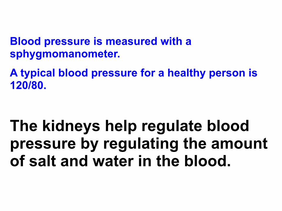
Blood pressure is measured with a sphygmomanometer.
A typical blood pressure for a healthy person is 120/80.
The kidneys help regulate blood pressure by regulating the amount of salt and water in the blood.

Diseases of the Circulatory System
Cardiovascular diseases are among the leading causes of death and disability in the U.S.
Atherosclerosis is a condition in which fatty deposits called plaque build up on the inner walls of the arteries.
High blood pressure is defined as a sustained elevated blood pressure of 140/90 or higher.

Heart Attack and Stroke If one of the coronary arteries becomes blocked, part of the heart muscle may begin to die from a lack of oxygen.
If enough heart muscle is damaged, a heart attack occurs.

If a blood clot gets stuck in a blood vessel leading to the brain, a stroke occurs.
Brain cells die and brain function in that region may be lost.

Circulatory System Health Ways of avoiding cardiovascular disease include:
• getting regular exercise.
• eating a balanced diet.
• avoiding smoking.

37–2 Blood and the Lymphatic System

Blood is a connective tissue that contains both dissolved substances and specialized cells.

The functions of blood include: collecting oxygen from the lungs,
nutrients from the digestive tract, and waste products from tissues.
regulating the body’s internal environment.
helping to fight infections.
forming clots to repair damaged blood vessels.

The human body has 4–6 liters of blood.
About 45% of blood volume is cells.
The other 55% is plasma—a straw-colored fluid.
Plasma is 90% water and 10% dissolved gases, salts, nutrients, enzymes, hormones, waste products, and plasma proteins.

Blood Composition
Whole Blood Sample
Sample Placed in Centrifuge
Plasma
Platelets
White blood cells
Red blood cell
Blood Sample That Has Been Centrifuged

Plasma proteins are divided into three groups:
albumins
globulins
fibrinogen

Albumins and globulins transport substances such as fatty acids, hormones, and vitamins.
Albumins regulate osmotic pressure and blood volume.
Some globulins fight viral and bacterial infections.
Fibrinogen is the protein that clots blood.

Blood Cells
The cellular portion of blood consists of:
• red blood cells • white blood cells • platelets

Red Blood Cells
The most numerous cells in the blood are the red blood cells. Red blood cells transport oxygen.

Red blood cells get their color from hemoglobin.
Hemoglobin is an iron-containing protein, found in red blood cells, that transports oxygen from the lungs to tissues of the body.

Red blood cells look like disks that are thinner in the center.
They are produced in red bone marrow.
They have no nuclei.
They live for about 120 days.
White Blood Cells
White blood cells do not contain hemoglobin.
They are less common than red cells.
White blood cells are produced in bone marrow.
They contain nuclei.
White blood cells may live for days, months, or years.

White blood cells are the “army” of the circulatory system—they guard against infection, fight parasites, attack bacteria.

There are many types of white blood cells.
Phagocytes engulf and digest bacteria and other disease-causing microorganisms.
Some white blood cells release histamines.
Histamines increase blood flow into the affected area, producing redness and swelling.

Lymphocytes produce antibodies.
Antibodies are essential to fighting infection and help to produce immunity to many diseases.

Platelets and Blood Clotting The body has an internal mechanism to slow bleeding and begin healing. Bleeding stops because blood has the ability to form a clot.
Blood clotting is made possible by plasma proteins and platelets.


Blood Clotting Problems
If one of the clotting factors is missing or defective, the clotting process does not work well.
Hemophilia is a genetic disorder that results from a defective protein in the clotting pathway.
Hemophiliacs cannot produce blood clots that are firm enough to stop even minor bleeding.

The Lymphatic System
As blood circulates, some fluid leaks into surrounding tissues.
This helps maintain movement of nutrients and salts from the blood into the tissues.

The lymphatic system collects the fluid that is lost by the blood and returns it back to the circulatory system. The fluid is known as lymph.

The Lymphatic System
Superior vena cava
Thymus
Heart
Thoracic duct
Spleen
Lymph nodes
Lymph vessels

Lymph collects in lymphatic capillaries and flows into larger lymph vessels.
Ducts collect the lymph and return it to the circulatory system through two openings in the superior vena cava.

Along lymph vessels are enlargements called lymph nodes.
Lymph nodes trap disease-causing microorganisms.
When large numbers of microorganisms are trapped in the lymph nodes, the nodes become enlarged.

37-3 The Respiratory System
What Is Respiration? In biology, respiration means two different things.
Cellular respiration is the release of energy from the breakdown of food in the presence of oxygen.
At the organism level, respiration is the process of gas exchange—the release of carbon dioxide and the uptake of oxygen between the lungs and the environment.

The basic function of the human respiratory system is the exchange of oxygen and carbon dioxide between the blood, the air, and tissues.

The respiratory system consists of the:
nose pharynx larynx trachea bronchi lungs

Air entering the respiratory system must be warmed, moistened, and filtered.
In the nose, mucus moistens air and traps particles of dust or smoke and cilia sweep particles and mucus to the throat.
Mucus and particles are either swallowed or spit out.

Epiglottis
Trachea
Nose Pharynx Larynx
Lungs
Bronchus
Mouth
Diaphragm
Bronchioles

76
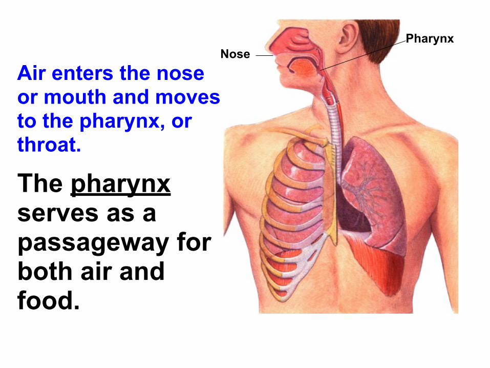
Air enters the nose or mouth and moves to the pharynx, or throat.
The pharynx serves as a passageway for both air and food.
Nose Pharynx

Air moves from the pharynx into the trachea, or windpipe. The epiglottis covers the entrance to the trachea when you swallow.
Epiglottis Trachea

At the top of the trachea is the larynx, which contains two elastic folds of tissue called vocal cords.
Larynx

Air then passes through the trachea into two branches in the chest cavity called bronchi. Each bronchus leads into one of the lungs.
Lungs
Bronchus

In each lung, the bronchus subdivides into smaller bronchi, and then into bronchioles.
Bronchioles

Bronchioles subdivide into millions of tiny air sacs called alveoli.
Alveoli
Bronchiole

Alveoli are grouped in clusters.
A network of capillaries surrounds each alveolus. Pulmonary
vein
Capillaries
Pulmonary artery

Gas Exchange
Gas exchange takes place in the alveoli. Oxygen diffuses from alveoli into the blood.
Capillary
O2

Carbon dioxide in the blood diffuses into the alveolus.
Capillary
O2
CO2
Breathing
Breathing is the movement of air into and out of the lungs.
The force that drives air into the lungs comes from air pressure.

Lungs are sealed in pleural membranes inside the chest cavity.
At the bottom of the cavity is a large, flat muscle known as the diaphragm.

During inhalation, the diaphragm contracts and the rib cage rises up.
This expands the volume of the chest cavity so that fresh air enters.

The chest cavity is sealed, so this creates a partial vacuum inside the cavity.
Atmospheric pressure fills the lungs as air rushes into the breathing passages.

Often exhaling is a passive event.
When the rib cage lowers and the diaphragm relaxes, pressure in the chest cavity is greater than atmospheric pressure.
Air is pushed out of the lungs.
Exhalation
Rib cage lowers
Air Exhaled

How Breathing Is Controlled Breathing is controlled by the medulla oblongata.
The medulla oblongata monitors carbon dioxide in the blood.
As carbon dioxide increases, nerve impulses make the diaphragm contract, bringing air into the lungs.
The higher the carbon dioxide level, the stronger the impulses.

Tobacco and the Respiratory System
Tobacco smoke contains three dangerous substances that affect the body:
• nicotine
• carbon monoxide
• tar
Nicotine is a stimulant that increases heart rate and blood pressure.
Carbon monoxide is a poisonous gas that blocks the transport of oxygen by hemoglobin in the blood.
Nicotine and carbon monoxide paralyze the cilia.
Tar contains compounds that are known to cause cancer.

Smoking reduces life expectancy. Smoking can cause such respiratory diseases as chronic bronchitis, emphysema, and lung cancer.

In chronic bronchitis, the bronchi become swollen and clogged with mucus.
Emphysema is the loss of elasticity in lung tissues.
People with emphysema cannot get enough oxygen to the body tissues or rid the body of excess carbon dioxide.

Smoking is a preventable cause of lung cancer.
Lung cancer is deadly because its cells can spread to other locations.
Smoking is also a major cause of heart disease.

Smoking and the Nonsmoker
Passive smoking is damaging to young children because their lungs are still developing.
Studies show that children of smokers are twice as likely as children of nonsmokers to develop respiratory problems.

Dealing With Tobacco The best way to avoid tobacco-related illness is not to smoke.
If a smoker quits, his or her health can be improved, and some of the damage can be reversed.

End Show
Slide of 51
Copyright Pearson Prentice Hall
37–1
The layer of muscle in the heart that pumps blood through the circulatory system is called the a. myocardium.
b. atrium.
c. ventricle.
d. vena cava.

End Show
Slide of 51
Copyright Pearson Prentice Hall
37–1
Oxygen-poor blood from the body enters the heart through the a. left atrium.
b. left ventricle.
c. right atrium.
d. right ventricle.

End Show
Slide of 51
Copyright Pearson Prentice Hall
37–1
The vein that brings oxygen-poor blood from the upper part of the body to the right atrium is the a. pulmonary vein
b. inferior vena cava.
c. aorta
d. superior vena cava.

End Show
Slide of 51
Copyright Pearson Prentice Hall
37–2
White blood cells that engulf and digest foreign cells are known as a. phagocytes.
b. platelets.
c. antibodies.
d. thrombocytes.

End Show
Slide of 51
Copyright Pearson Prentice Hall
37–2
Blood cells that do not have nuclei and are produced by the red bone marrow are a. red blood cells.
b. lymphocytes.
c. platelets.
d. phagocytes.

End Show
Slide of 51
Copyright Pearson Prentice Hall
37–2
The function of platelets is to a. assist red blood cells in carrying oxygen.
b. destroy viruses and bacteria.
c. initiate the blood clotting process.
d. keep capillaries open so blood can flow freely through.

End Show
Slide of 51
Copyright Pearson Prentice Hall
37–2
The function of lymph nodes is to a. trap bacteria and viruses that cause disease.
b. produce antibodies.
c. manufacture new red and white blood cells.
d. store fat.
End Show
Slide of 51
Copyright Pearson Prentice Hall
37-3
Air passes through the trachea into two large passageways in the chest cavity known as the a. bronchi.
b. alveoli.
c. epiglottis.
d. bronchioles.

End Show
Slide of 51
Copyright Pearson Prentice Hall
37-3
The function of the cilia lining the respiratory surfaces is to a. improve the amount of oxygen and carbon dioxide exchanged in the
lungs.
b. cover the opening of the trachea when you swallow.
c. move air in and out of the lungs.
d. sweep trapped particles and mucus away from the lungs.

End Show
Slide of 51
Copyright Pearson Prentice Hall
37-3
Oxygen diffuses from the alveolus into the blood because a. blood entering the capillaries of the lungs is oxygen-poor.
b. blood entering the capillaries of the lungs is oxygen-rich.
c. air entering the lungs has more carbon dioxide than oxygen.
d. air entering the lungs has less oxygen than is found in the blood.