Embed Size (px)
Citation preview
A.Shajitha
B.Sc Radio Diagnosis Technology
Madras Medical College
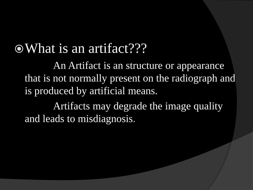
What is an artifact???
An Artifact is an structure or appearance
that is not normally present on the radiograph and
is produced by artificial means.
Artifacts may degrade the image quality
and leads to misdiagnosis.
Sources of artifacts
Patient related
Hardware problems
Software problems
Contrast induced
Contrast induced artifacts
In CT
Streak artifacts.
Edge gradient effect.
Beam hardening artifact.
Aortic root artifact
Beam hardening artifacts
It occurs when the average energy of the x
ray beam passing through the patient
increases.
The beam is hardened.
High energy photons are attenuated less by
the tissue.
This artifact is also called CUPPING
artifact.
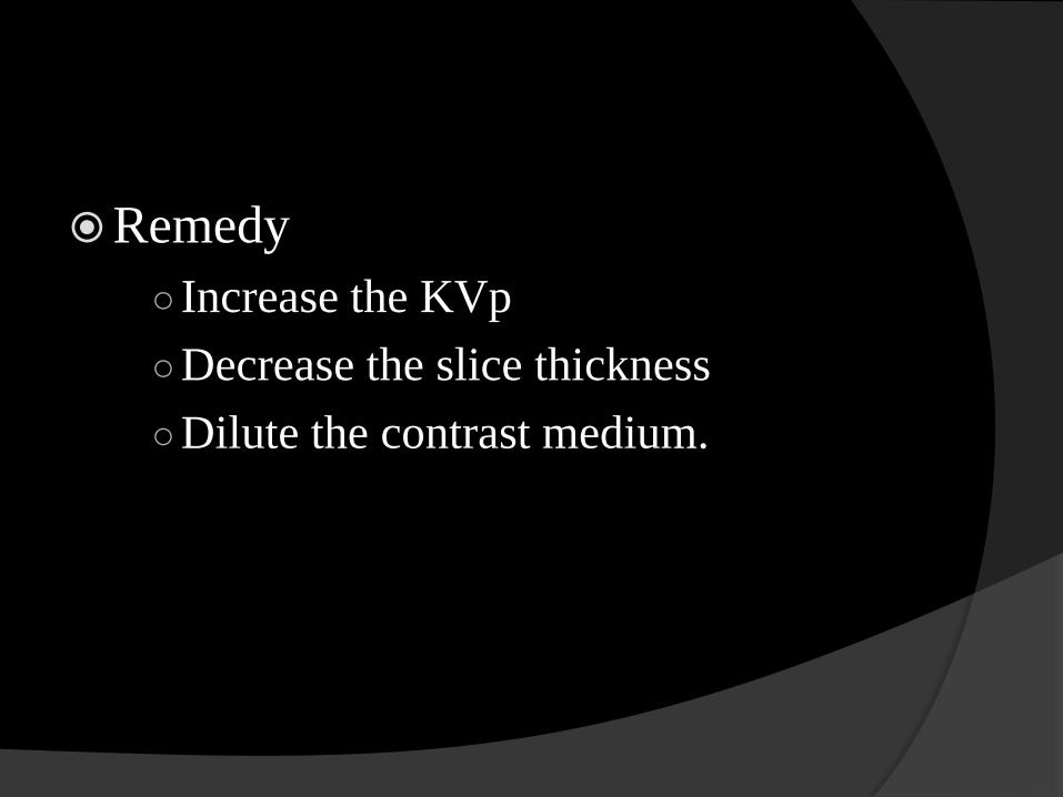
Remedy
○ Increase the KVp
○Decrease the slice thickness
○Dilute the contrast medium.
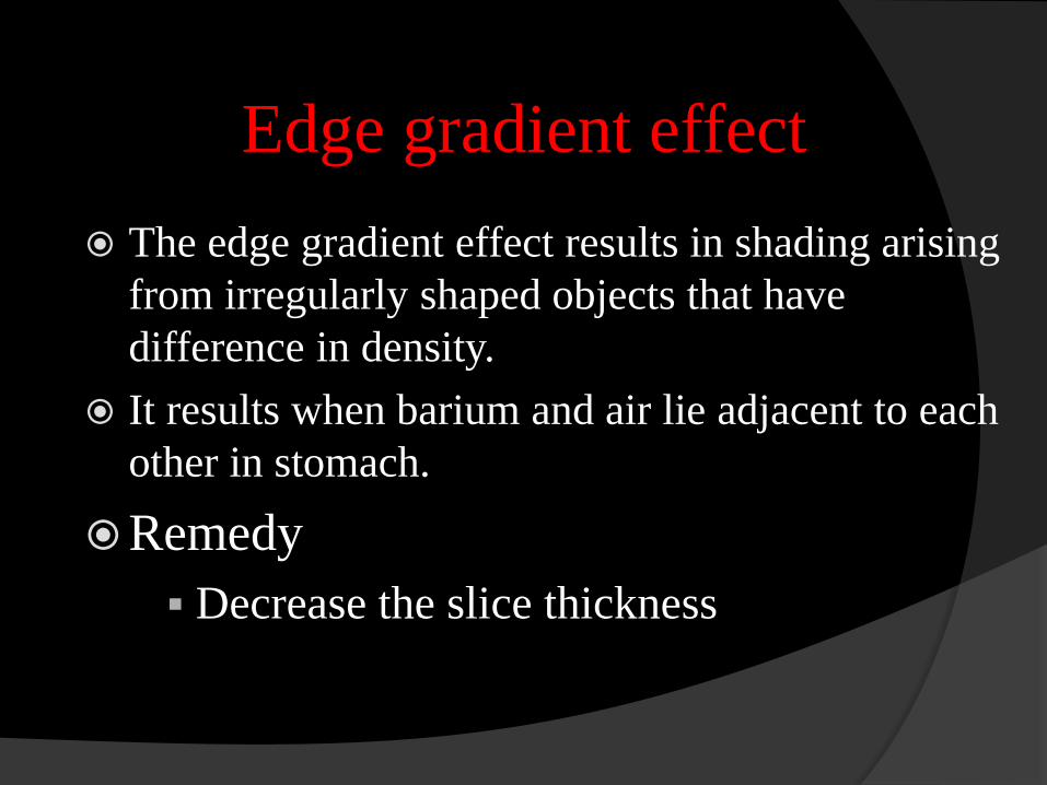
Edge gradient effect
The edge gradient effect results in shading arising
from irregularly shaped objects that have
difference in density.
It results when barium and air lie adjacent to each
other in stomach.
Remedy
Decrease the slice thickness
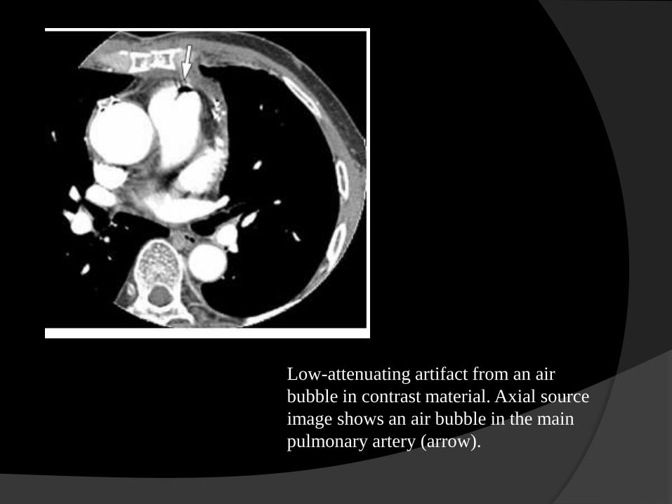
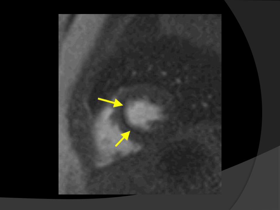
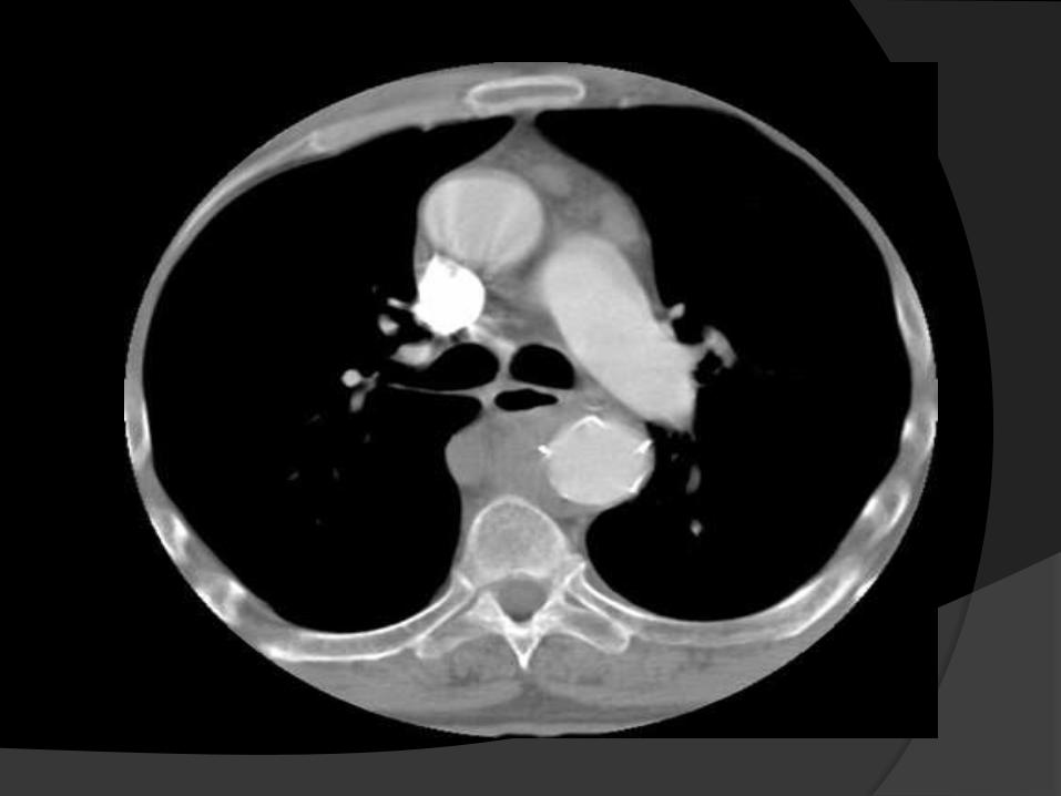
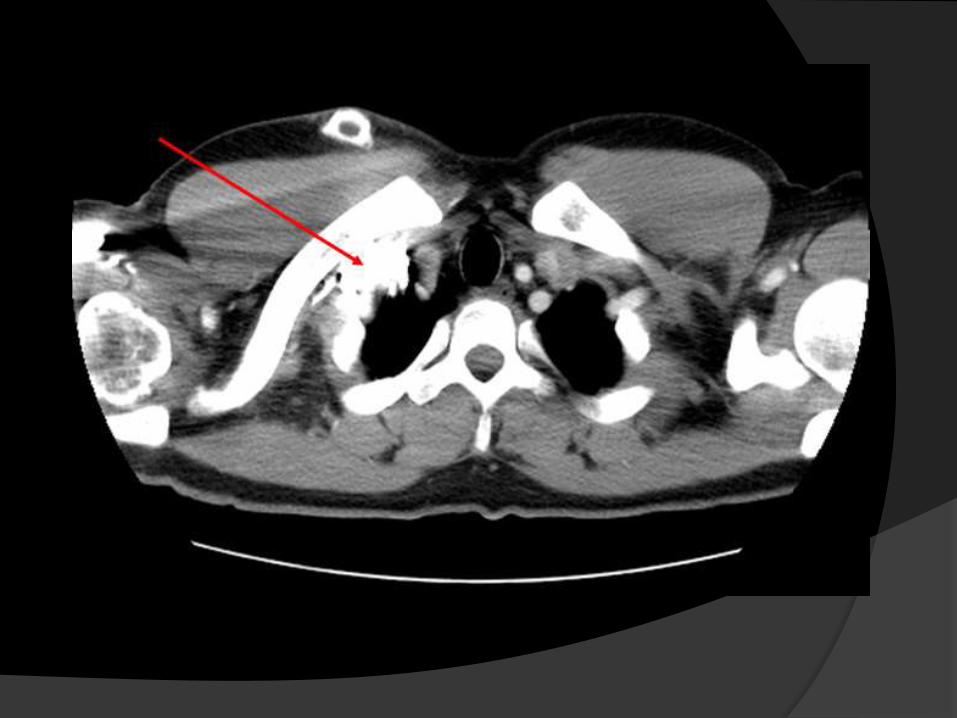
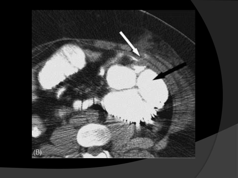
Low-attenuating artifact from an air
bubble in contrast material. Axial source
image shows an air bubble in the main
pulmonary artery (arrow).
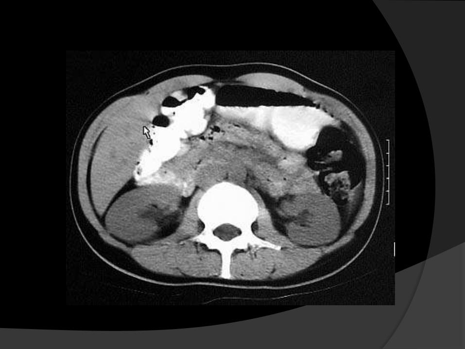
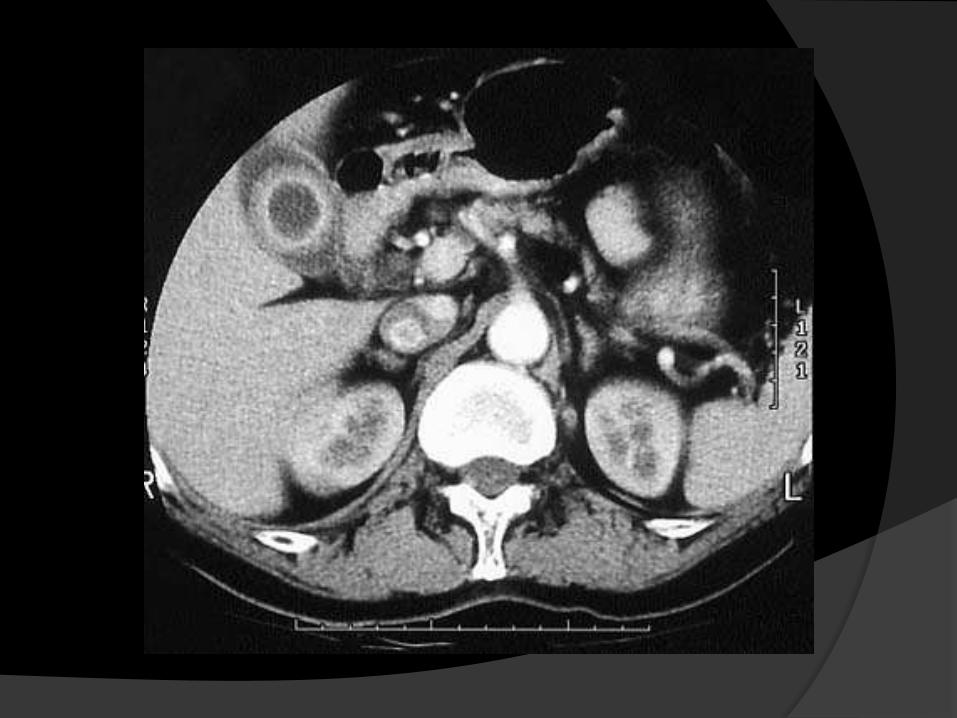
Streak artifact
It occurs when the scan is done with
the barium contrast or
High osmolar contrast medium.
It appears in the image like streaks of
lines obscuring the organ of interest.
This artifact can be reduced by
○Using low osmolar contrast medium.
○Avoid scans if barium studies are
done a week before.
○Using a low HU value oral contrast
such as volumen or water in place of
barium suspension.
Saline flush:
○ Saline flush is used in CT and MRI to
reduce the concentration of contrast
medium.
○ Also reduce the streak artifact.
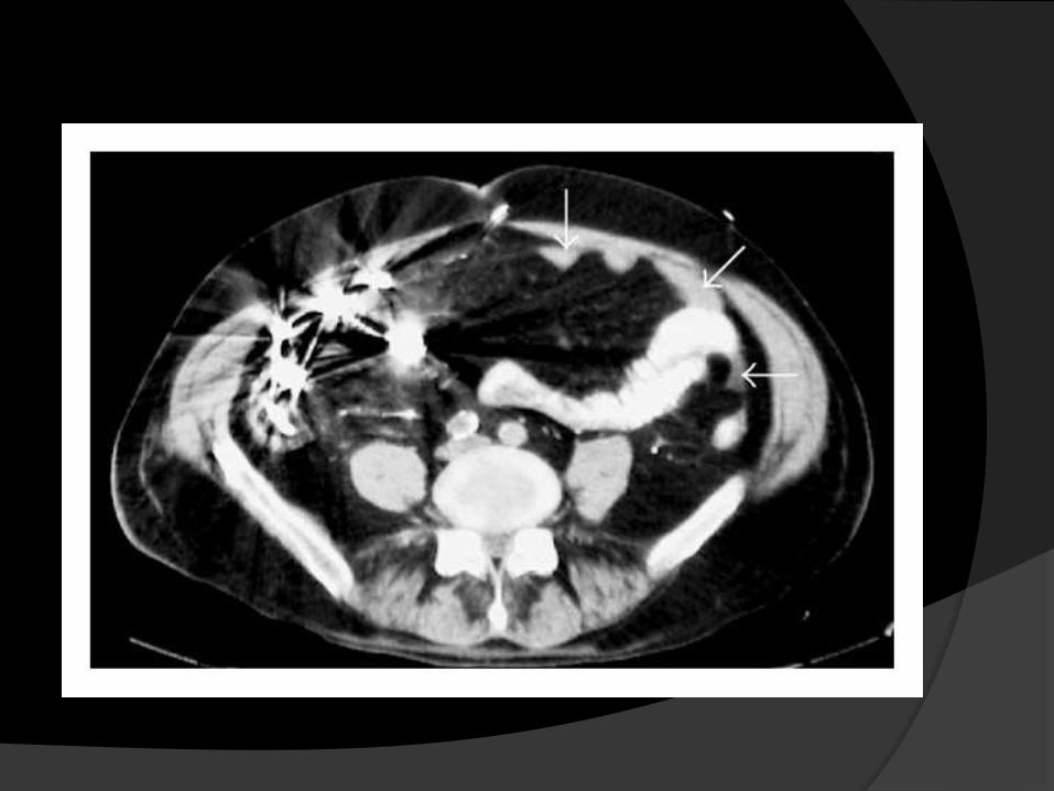
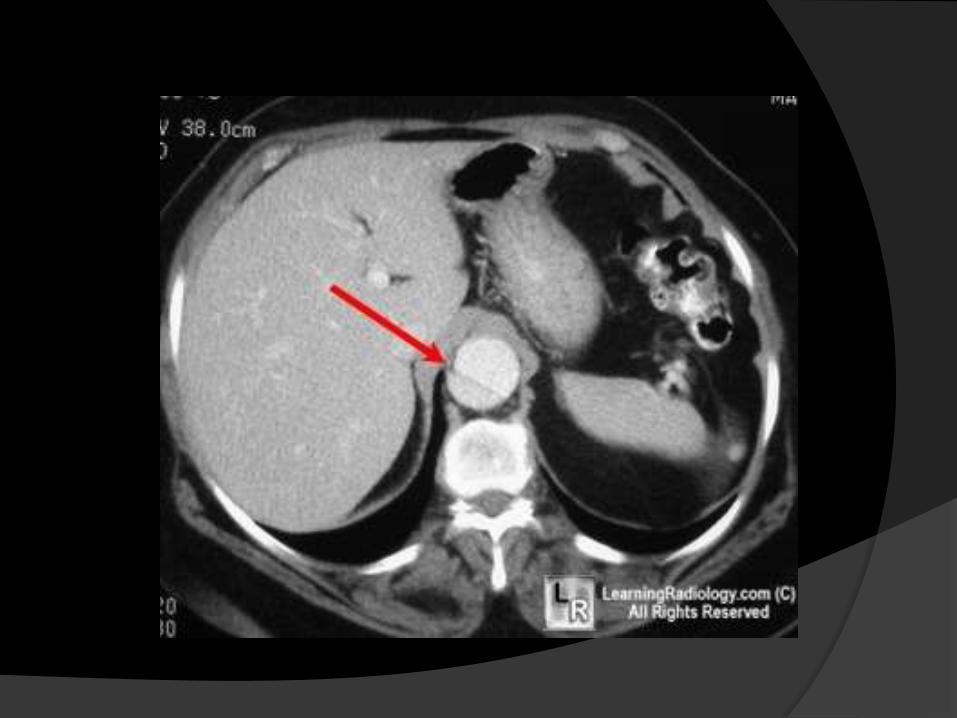
Aortic root artifact
The contrast material in the brachio cephalic
vein or SVC associated with cardiac motion
transmitted to these veins.
It produces artifacts that project over the
ascending aorta and supra aortic arch
branches.
In addition motion in the free wall of the
left ventricle may produce artifacts that
project over the descending thoracic
aorta.
This artifact sometimes resembles a aortic
dissection.
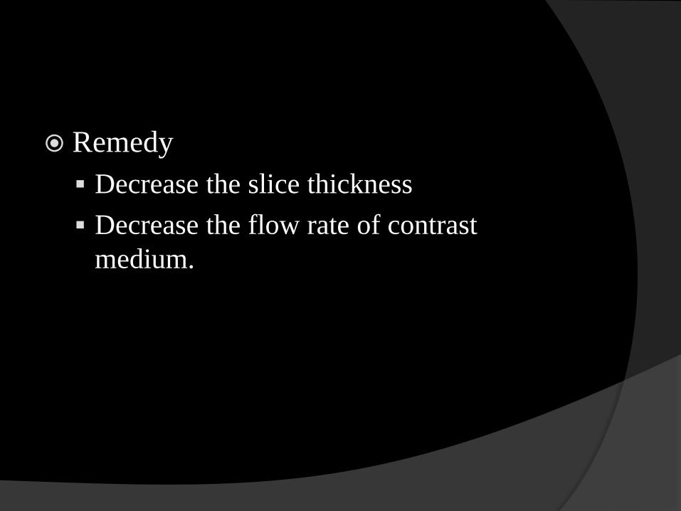
Remedy
Decrease the slice thickness
Decrease the flow rate of contrast
medium.
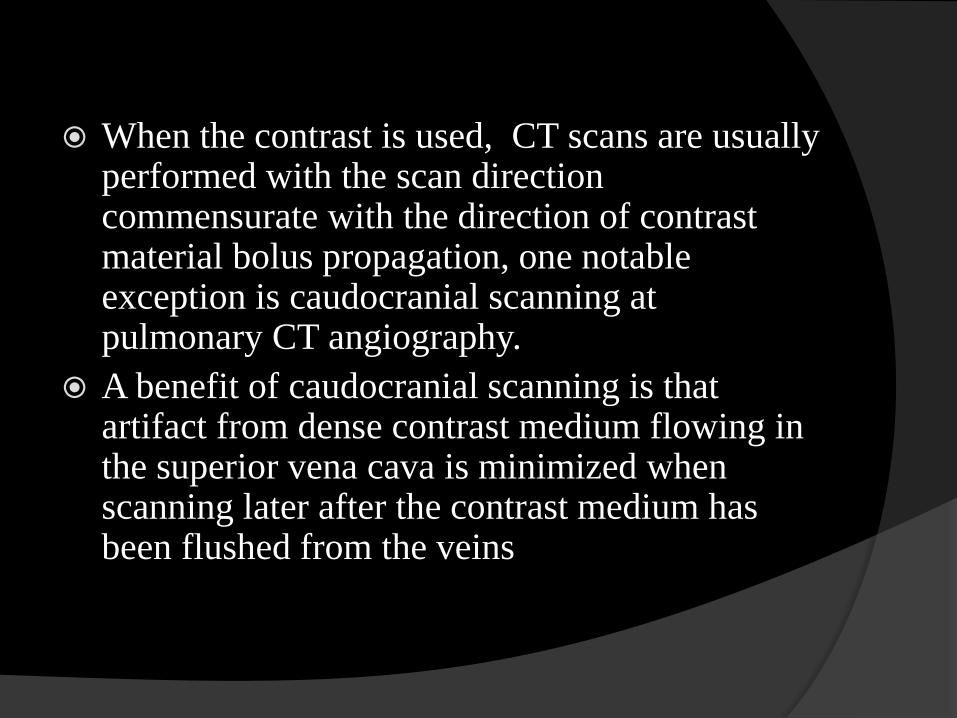
When the contrast is used, CT scans are usually performed with the scan direction commensurate with the direction of contrast material bolus propagation, one notable exception is caudocranial scanning at pulmonary CT angiography.
A benefit of caudocranial scanning is that artifact from dense contrast medium flowing in the superior vena cava is minimized when scanning later after the contrast medium has been flushed from the veins
A similar benefit in artifact reduction was observed at carotid artery CT angiography performed with use of a craniocaudal scan direction.
when the scan direction is opposite to the flow of contrast medium for a long scan, contrast material injection duration may need to be increased to ensure adequate enhancement of the upstream structures.
In MRI
○Susceptibility artifacts
○Dark rim artifact
Susceptibility artifact
It occurs when high concentration of Ferro
magnetic contrast agent is used.
If the contrast is injected excessively
without dilution.
It resembles the metallic susceptibility
artifact.
This artifact can be reduced by diluting the
contrast medium.
In contrast enhanced MR angiography
contrast agents induce susceptibility
artifacts.
This can be avoided by acquiring the full k
space; partial echo sampling can be avoided
or reduced.
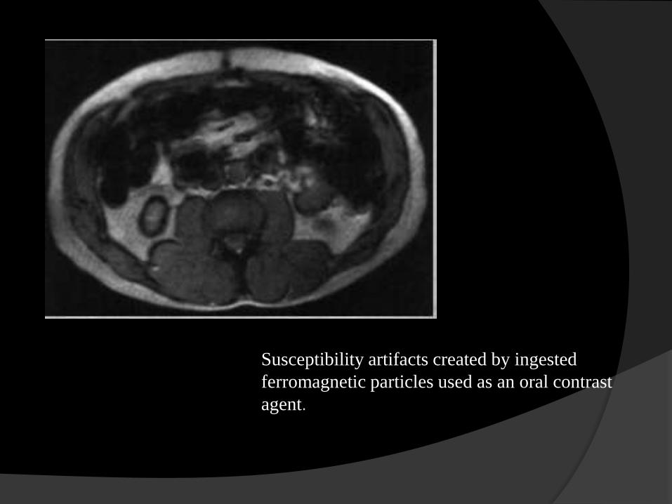
Susceptibility artifacts created by ingested
ferromagnetic particles used as an oral contrast
agent.
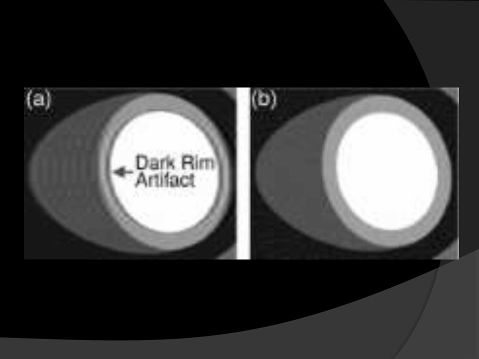
Dark rim artifact
In dynamic contrast enhanced MR imaging,
dark rim artifact appears when gadolinium
contrast bolus appears in the left ventricle.
It appears like a signal loss in the rim of pixels
in the subendocardium at the boundary between
the myocardium and LV blood.
It occurs most frequently in images acquired
with the Steady State Free Precession (SSFP)
pulse sequence