Embed Size (px)
Citation preview

Epithelial Tissue 2Abbas A. A. Shawka

ObjectivesAfter this lecture you should be able to •Know the location and function of the
intercellular adhesions and other junctions.
•Enumerate specialization on the epithelium apical surface cell.

Intercellular adhesions and other junctions
Several membrane-associated structures provide adhesion and communication between cells.
Epithelial cells adhere strongly to neighboring cells and basal laminae, particularly in epithelia subject to friction or other mechanical forces.

Types of Intercellular adhesions and other junctions
1. Tight junction ( Zonulae occludens )2. Adherent junction ( Zonulae adherens )3. Desmosomes ( Macula adherens )4. Gap junctions ( Nexus )5. Hemidesmosomes
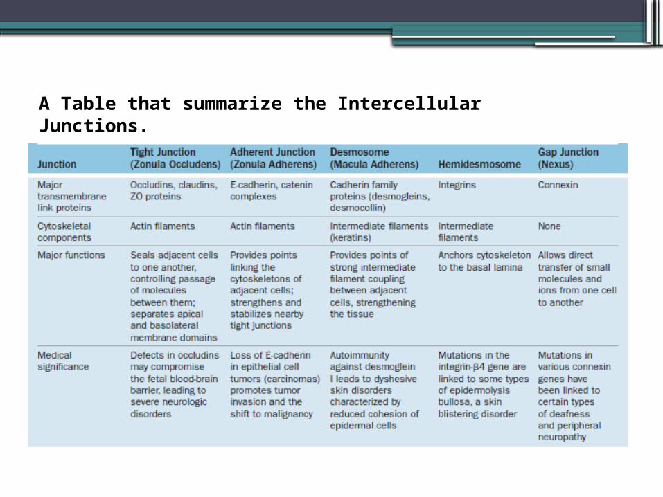
A Table that summarize the Intercellular Junctions.

1. Tight junction ( Zonulae occludens )
Are the most apical of junctions.The term “zonula” indicates that the junction
forms a band completely encircling each cell.
In TEM the adjacent membranes at these junctions appear fused or very tightly apposed.
The seal between the membranes is due to interactions between the transmembrane proteins claudin and occludin of each cell.

Ultrastructural view of the apical region near microvilli (MV) of two epithelial cells, revealing a junctional complex with a tight junction (TJ) or zonula occludens, an adherent junction (AJ) or zonula adherens, and a desmosome(D)associatedwith intermediate filaments (IF).

The intercellular seal of this junctional type ensures that molecules crossing an epithelium in either direction do so by going through the cells (transcellular path) rather than between them (paracellular pathway). Epithelia with one or very few fused sealing strands (eg, proximal renal tubule) are more permeable to water and solutes than are epithelia with numerous fusion sites (eg, the lining of the urinary bladder).

Also Tight junctions prevent membrane proteins at the apical cell surface from moving in the membrane to the basal and lateral surfaces, and vice versa. This produces two membrane domains (apical and basolateral) with different protein populations, which allows the two sides of the epithelium to maintain different receptors and to function differently. Apical cell membranes of epithelia are part of the luminal compartment of a tissue or organ, while the basolateral domains of the epithelial cells are part of a basal compartment that also encompasses the underlying connective tissue.

2- Adherent junction ( Zonulae adherens )
encircles the epithelial cell, usually immediately below the zonula occludens.
This is an adherent junction, firmly anchoring a cell to its neighbors.
Together, the tight and adherent junctions encircling the apical ends of epithelial cells function like the plastic form that holds a six-pack of canned drinks together.

3- Desmosomes ( Macula adherens )
Disc shaped structures at the surface of one cell that are matched with identical structures at an adjacent cell surface.
Because intermediate filaments of the cytoskeleton are very strong, desmosomes provide firm adhesion among the cells.
In nonepithelial cells, the intermediate filaments attached to desmosomes are composed of other proteins, such as desmin or vimentin.

pemphigus vulgaris disease

4- Gap junctions ( Nexus )
Gap junctions mediate communication rather than adhesion or occlusion between cells
Gap junctions consist of aggregated transmembrane protein complexes that form circular patches in the plasma membrane
Gap junctions permit intercellular exchange of molecules with small (<1.5 nm) diameters. Some molecules mediating signal transduction, such as cyclic nucleotides and ions, move rapidly through gap junctions, allowing cells in many tissues to act in a coordinated manner rather than as independent units. For example, in heart and visceral muscles gap junctions help produce rhythmic contractions.

Gap junction “ cryofracture”

5- Hemidesmosomes The basal domain of an epithelial cell
attaches to the subjacent basal lamina by Hemidesmosome.
They resemble Desmosome, Except the connect the cells to its basal lamina.

Hemidesmosome

Specializations of the apical cell surfaceThe apical ends of many tall or cuboidal
epithelial cells face an organ’s lumen and often have specialized projecting structures.
Function of these projections
A) either to increase the apical surface area for absorption
B) or to move substances along the epithelial surface.

Types of these apical specializations1) Microvilli 2) Steriocilia 3) Cilia

1- Microvilli•in epithelial cells specialized for
absorption, the apical surfaces present an array of projections called microvilli.
• In cells such as those lining the small intestine apical surfaces are densely covered with uniform microvilli, which are visible as a brush or striated border on these cells.
•The average microvillus is about 1 μm long and 0.1 μm wide.

Celiac Disease

The diagram shows a few microfilaments in a microvillus, with various actin-binding proteins important for F-actin assembly, capping, cross-linking, and movement. Like microfilaments in other regions of the cytoskeleton, those of microvilli are highly dynamic, with treadmilling and various myosin-based interactions. Myosin motors import various microvillicomponents along the actin filaments.
Dr John Heuser, Washington UniversitySchool of Medicine, St. Louis, MO

TEM of microvilli sectioned longitudinally and transversely (inset) reveals the microfilament arrays that form the core of these projections. The terminal web (TW) of the cytoskeleton is also seen. The glycocalyx (G) extending from glycoproteins and glycolipids of the microvilli plasmalemma contains certain enzymes for late stages of macromolecule digestion. X15,000.

2- steriocilia•Stereocilia are a much less common type
of apical process, restricted to absorptive epithelial cells lining the epididymis.
•and the proximal part of ductus deferens in the male reproductive system
•stereocilia are typically much longer and much less motile than microvilli, and may show distal branching along their length

At the apical ends of the tall epithelial cells lining organssuch as the epididymis (shown here) are numerous very long stereocilia, which increase the surface area availablefor absorption. Stereocilia are much longer than microvilliand often have distal branching. X400. H&E.

3- cila• Motile cilia are found only in epithelia, where they
are abundant on the apical domains of many cuboidal or columnar cells.
• Typical cilia are 5-10 μm long and 0.2 μm in diameter,
• Epithelial cilia exhibit rapid beating patterns of movement that propel a current of fluid and suspended matter in one direction over the epithelium. Ciliary motion occurs through successive changes in the conformation of the axoneme, in which a large variety of accessory proteins make each cilium relatively stiff, but elastic

By light microscopy cilia (C) on the columnar cells appear as a wave of long projections interrupted by nonciliated, mucus-secreting goblet cells (G). X400
SEM of the apical surfaces of thisepithelium shows the density of the cilia (C) and the scatteredgoblet cells (G). X300.

Quiz•------------ is immotile process of an
epithelial cell.•What is the function of ( Cilia,
Microvilli ) ?• --------------- provide nutrition continuity
between cells. • -------------- is the most apical of junctions.•What is the different between Desmosome
and Hemidesmosome ?