Embed Size (px)
Citation preview
Gestational Trophoblastic Disease
Sittichoke Mahasukontachat, MD
Objective study
• Define spectrum of Gestational Trophoblastic Disease (GTD)
• Review types of hydatidiform molar pregnancies, diagnosis, treatment, and follow up
• Review types of gestational trophoblastic neoplasia, diagnosis, classification, treatment, and follow up
• Review expectations for future childbearing

Gestational Trophoblastic Disease• Gestational trophoblastic disease (GTD) is the
term used to describe the heterogeneous group of interrelated lesions that arise from abnormal proliferation of placental trophoblasts.
• Benign lesions – Hydatidiform moles (complete and partial )
• Malignant lesions – Invasive mole, placental-site trophoblastic
tumor(PSTT), and choriocarcinoma.
Berek JS, editor. Berek and Novak’s Gynecology. Philadelphia: Lippincott Williams & Wilkins; 2012. pp. 1458.

Hydatidiform Mole
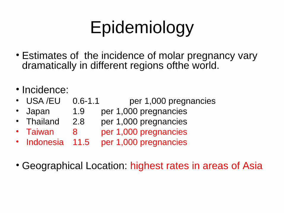
• Estimates of the incidence of molar pregnancy vary dramatically in different regions ofthe world.
• Incidence:• USA /EU 0.6-1.1 per 1,000 pregnancies• Japan 1.9 per 1,000 pregnancies• Thailand 2.8 per 1,000 pregnancies• Taiwan 8 per 1,000 pregnancies• Indonesia 11.5 per 1,000 pregnancies
• Geographical Location: highest rates in areas of Asia
Epidemiology
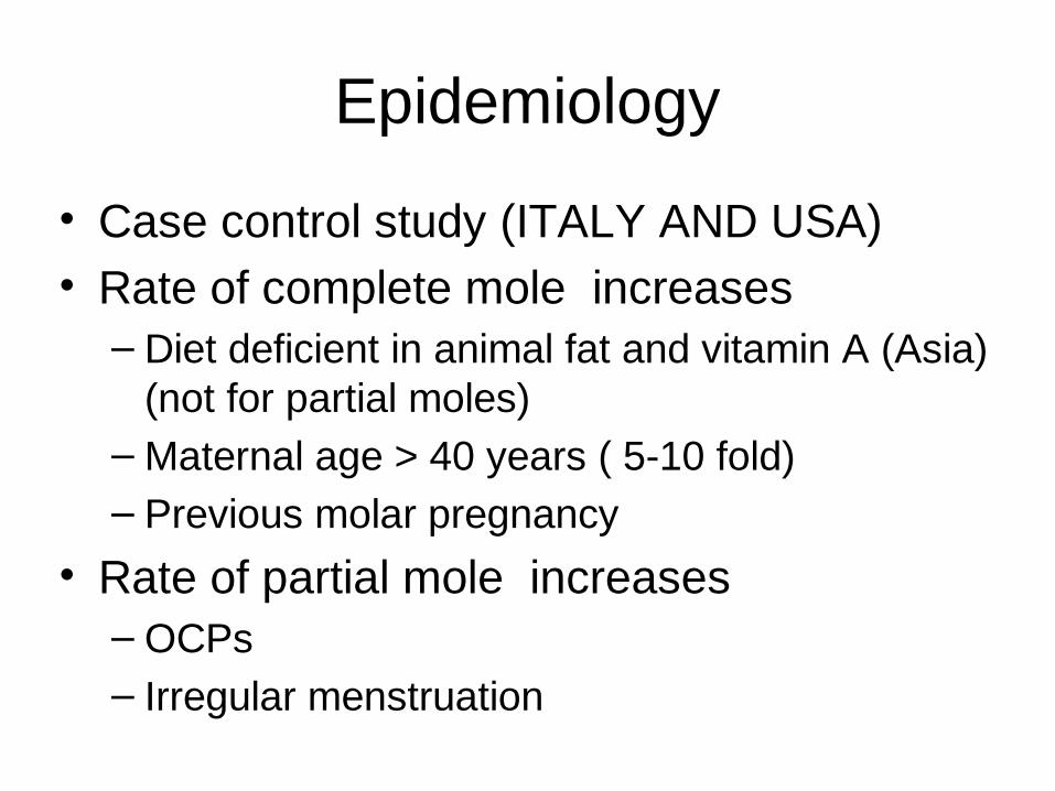
• Case control study (ITALY AND USA)• Rate of complete mole increases– Diet deficient in animal fat and vitamin A (Asia)
(not for partial moles)–Maternal age > 40 years ( 5-10 fold)– Previous molar pregnancy
• Rate of partial mole increases– OCPs– Irregular menstruation
Epidemiology

Pathogenesis and Cytogenetics of HM
Genetic ConstitutionDiploid Triploid/ teraploid
Patho-genesis
4%Fertilization of an empty ovum by two sperms“Diandric dispermy”
90%Triploidfertilization of a normal ovum by two sperms
“Dispermic triploidy”
96%Fertilization of an empty ovum by one sperms that undergoes duplication“Diandric diploidy”
10%Tetraploidfertilization of a normal ovum by three sperms
“Dispermic triploidy”
Karyotype46XX69XXX69YXX69YYX
46XX46XY
Complete Partial

Emptyovum
EndoreduplicationEndoreduplication
Emptyovum
Emptyovum
23X
23X
23X
23X
23Y
23X
46XX
46XY
46YY
46XX
HeterozygousHeterozygous
HomozygousHomozygous
Non-viable gameteNon-viable gamete
Complete mole

Partial mole
23X
23X
23Y
23Y
23X
69XXX
23Y
23X
23X
69XXX
23X
69XXX
69YYY
Non-viable gameteNon-viable gamete

Complete VS Partial Mole
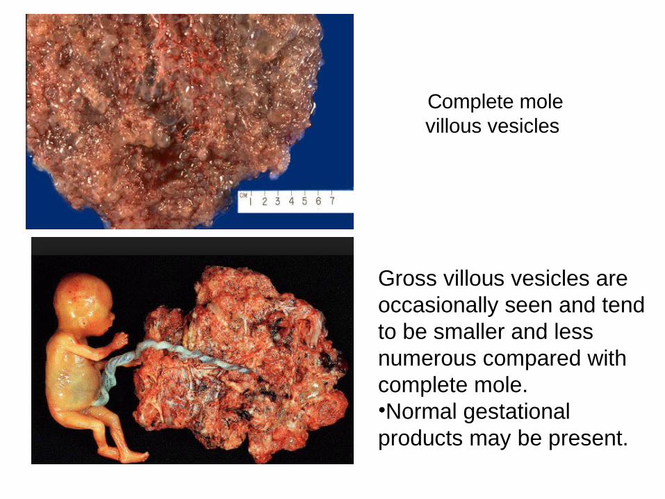
Complete molevillous vesicles
Gross villous vesicles are occasionally seen and tend to be smaller and less numerous compared with complete mole. •Normal gestational products may be present.

The microscopic of complete mole:
•Hyperplasia of trophoblastic cells•Hydropic swelling of all villi•Vessels are usually absent
•Partial mole •Hydropic villi and trophoblastic hyperplasia are less conspicuous in partial mole. •Fetal tissues like erythrocyte may be found. •Scalloping of chorionic villi and trophoblastic stromal inclusions.
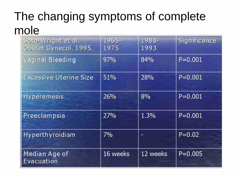
The changing symptoms of complete mole

Diagnosis of Hydatidiform Mole
• Passage of vesicular tissue• Serum quant -hCG elevated β (Can be > 1,000,000 IU/L)• Enlarged uterus• Ultrasound: multiple echoes, cystic dilatations • “snowstorm pattern”• Fetus vs. No fetus• Ectopic hydatidiform moles
Ultrasonography
• Ultrasonography is a reliable and sensitive technique for the diagnosis of complete molar pregnancy. Because the chorionic villi exhibit diffuse hydropic swelling, complete moles produce a characteristic vesicular ultrasonographic pattern as soon as in the first-trimester

Complete Mole
Snow-storm appearanceOrVesicular ultrasonoghaphic pattern

Partial mole• Ultrasonography finding: – Focal cystic spaces in the placental tissues
and an increase in the transverse diameter of the gestational sac (Ratio of T A-P : >1.5)
– PPV 90%.
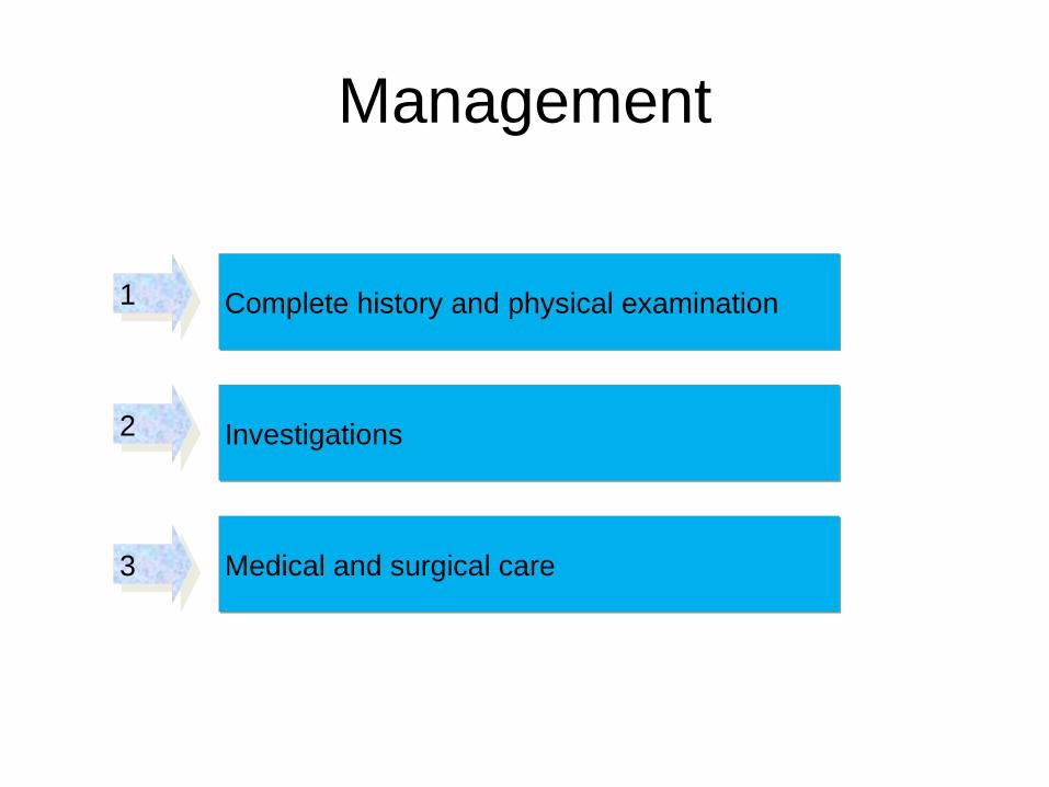
Management
Complete history and physical examination
Investigations
Medical and surgical care
1
3
2

• History and physcal examination:– Should aim to rule out the classic
symptoms and signs that would lead to a diagnosis of:• severe anemia • dehydration• preeclampsia• thyrotoxicosis
• The patient should be stabilized hemodynamically
Complete history and physical examination1

• Investigations:– Laboratory:
Pre-evacuation hCGComplete blood countElectrolytes, BUN, creatinine Liver function tests Thyroid function tests
– Imaging:Pelvic ultrasoundChest x-ray
Investigations2

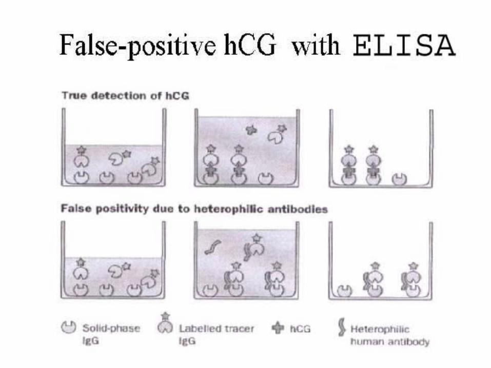
Phantom hCG
• Pseudohypergonadotropinemia is a result of the presence of heterophilic antibodies in serum giving rise to a falsely elevated hCG. •If there is discrepancy with the clinical presentation, hCG levels should be measured again with a different immunoassay. •The other alternative is to measure the urine hCG level because heterophilic antibodies are not excreted into the urine.
Hook effect
•Can occur with a falsely low serum hCG level. When the serum hCG level is too high, there are not enough antibodies in the solution to bind the hCG molecules. •Much of them are being washed away without being measured. •If very high hCG level is suspected, the laboratory should be informed and the serum should be diluted before measurement.
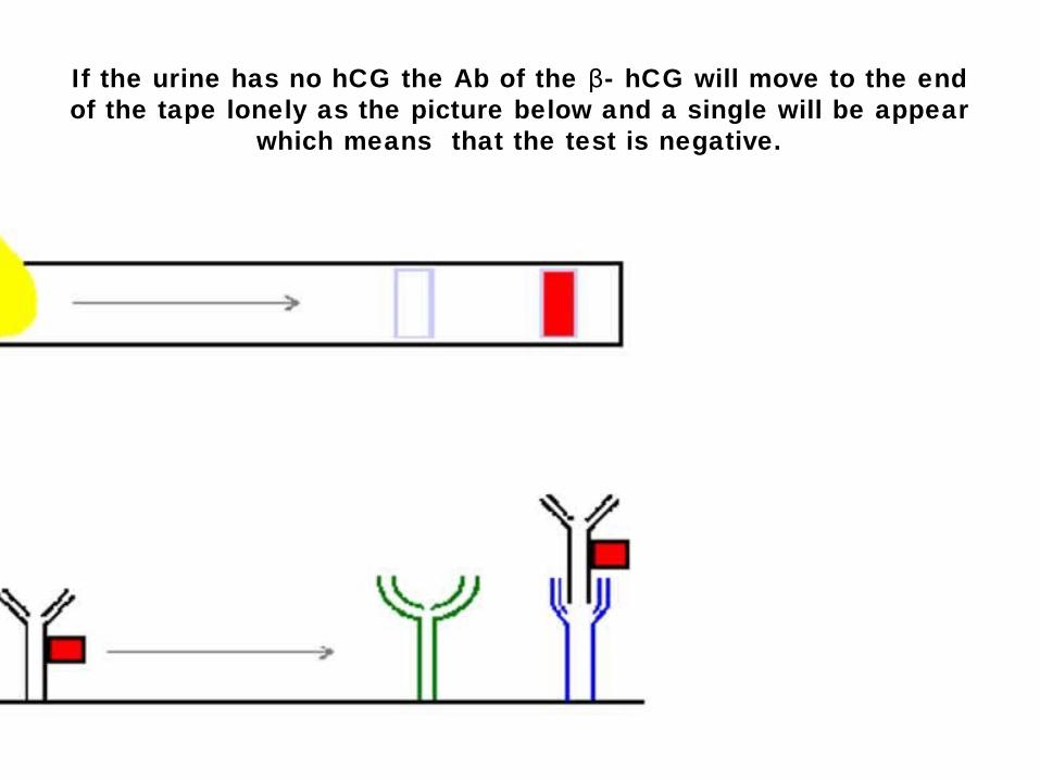
If the urine has no hCG the Ab of the - hCG will move to the end βof the tape lonely as the picture below and a single will be appear
which means that the test is negative.

If some hCG exist in the urine tow line will be appeared and the test result is positive.

The home kit tests are not safe from hook effect !, which produce a false negative result . When the concentration of hormone is very high the hook effect will happen as the mechanism shown below .

• Medical care:– Correction of:
Anemia Dehydration Hyperthyroidismhypertension
Medical and surgical care3

Surgical care:
Suction curettageSuction curettage
HysterectomyHysterectomy
•The method of choice
•Performed with the mole in situ. •The ovaries may be preserved at the time of surgery•Large ovarian cysts may be decompressed by aspiration.•Not prevent metastasis

Suction Curettage• 1. Oxytocin infusion—Before the induction of
anesthesia.• 2. Cervical dilation• 3. Suction curettage• The use of a12-mm cannula is strongly advised
to facilitate evacuation.• Uterus > 14 weeks of gestation, one hand
should be placed on top of the fundus, and the uterus should be massaged to stimulate uterine contraction and reduce the risk of perforation.
• 4. Sharp curettage—Remove any residual molar tissue.

Prophylactic ChemotherapyProphylactic Chemotherapy
• Controvery .Controvery .
• Prevented metastasisPrevented metastasis
• Reduced the incidence and morbidity of local Reduced the incidence and morbidity of local uterine invasionuterine invasion
• Single course of actinomycin D at time of Single course of actinomycin D at time of evacuationevacuation
• Useful in the management of high-risk Useful in the management of high-risk complete mole, especially when hCG complete mole, especially when hCG assessments for follow up are unavailable or assessments for follow up are unavailable or unreliableunreliable
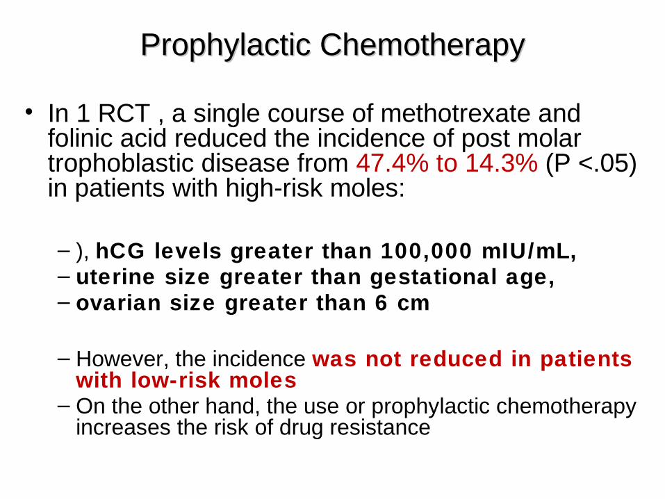
• In 1 RCT , a single course of methotrexate and folinic acid reduced the incidence of post molar trophoblastic disease from 47.4% to 14.3% (P <.05) in patients with high-risk moles:
– ), hCG levels greater than 100,000 mIU/mL,– uterine size greater than gestational age, – ovarian size greater than 6 cm
– However, the incidence was not reduced in patients with low-risk moles
– On the other hand, the use or prophylactic chemotherapy increases the risk of drug resistance
Prophylactic ChemotherapyProphylactic Chemotherapy

Follow up• 1. Human Chorionic Gonadotropin (hCG)
– Monitored with weekly of hCG levels until these levels are normal for 3 consecutive weeks
– Followed by monthly until levels are normal for 6 consecutive months

2. Contraception
• Patients should be used effective contraception during the entire interval of hCG follow up
• Oral contraceptive may be used safely
• IUD should not inserted until the patient achieves a normal hCG level.
Follow up (2)
Gestational Trophoblastic Neoplasia (GTN)
Diagnosis
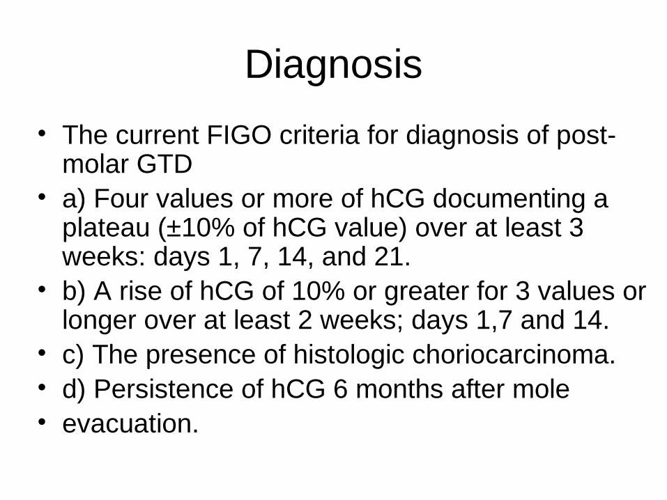
• The current FIGO criteria for diagnosis of post-molar GTD
• a) Four values or more of hCG documenting a plateau (±10% of hCG value) over at least 3 weeks: days 1, 7, 14, and 21.
• b) A rise of hCG of 10% or greater for 3 values or longer over at least 2 weeks; days 1,7 and 14.
• c) The presence of histologic choriocarcinoma.• d) Persistence of hCG 6 months after mole• evacuation.
36
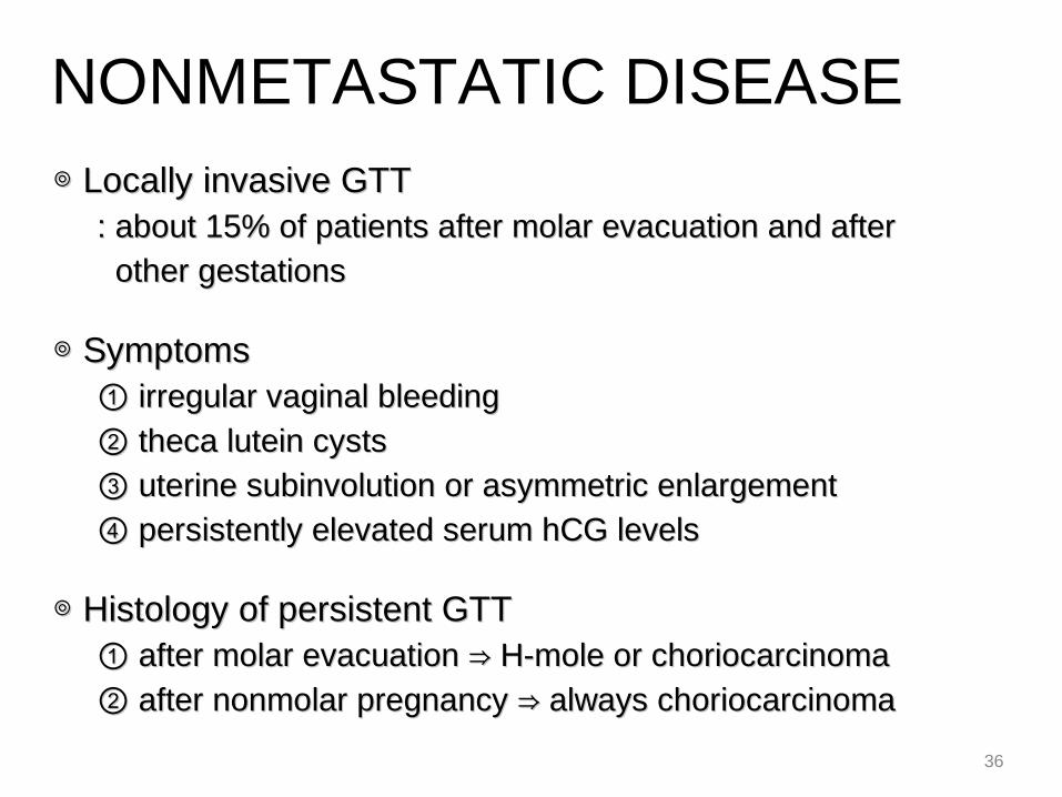
◎ ◎ Locally invasive GTTLocally invasive GTT : about 15% of patients after molar evacuation and after: about 15% of patients after molar evacuation and after other gestationsother gestations
◎ ◎ SymptomsSymptoms ① ① irregular vaginal bleedingirregular vaginal bleeding ② ② theca lutein cyststheca lutein cysts ③ ③ uterine subinvolution or asymmetric enlargementuterine subinvolution or asymmetric enlargement ④ ④ persistently elevated serum hCG levelspersistently elevated serum hCG levels
◎ ◎ Histology of persistent GTTHistology of persistent GTT ① ① after molar evacuation H-mole or choriocarcinoma⇒after molar evacuation H-mole or choriocarcinoma⇒ ② ② after nonmolar pregnancy always choriocarcinoma⇒after nonmolar pregnancy always choriocarcinoma⇒
NONMETASTATIC DISEASE
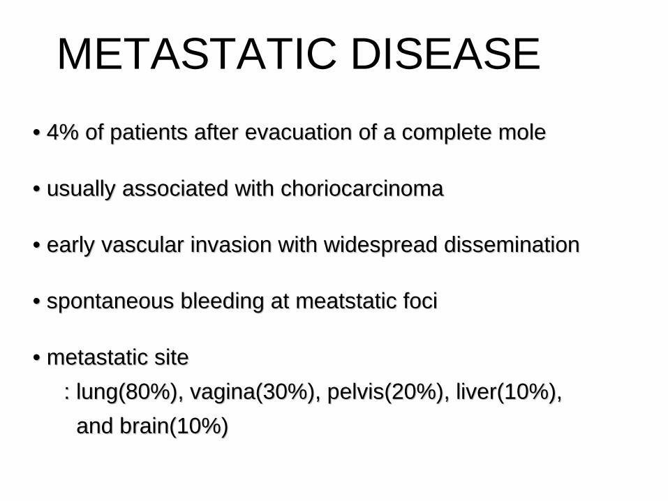
• • 4% of patients after evacuation of a complete mole4% of patients after evacuation of a complete mole
• • usually associated with choriocarcinomausually associated with choriocarcinoma
• • early vascular invasion with widespread disseminationearly vascular invasion with widespread dissemination
• • spontaneous bleeding at meatstatic focispontaneous bleeding at meatstatic foci
• • metastatic sitemetastatic site
: lung(80%), vagina(30%), pelvis(20%), liver(10%),: lung(80%), vagina(30%), pelvis(20%), liver(10%),
and brain(10%)and brain(10%)
METASTATIC DISEASE

38
1. Pulmonary1. Pulmonary • • lung involvement is visible on chest x-ray of 80% oflung involvement is visible on chest x-ray of 80% of patients with metastatic GTTpatients with metastatic GTT “① “① snowstorm” patternsnowstorm” pattern ② ② discrete rounded densitydiscrete rounded density ③ ③ pleural effusionpleural effusion ④ ④ an embolic pattern by pulmonary arterial occlusionan embolic pattern by pulmonary arterial occlusion • • Sign : chest pain, cough, hemoptysis, dyspnea,Sign : chest pain, cough, hemoptysis, dyspnea, or asymptomatic lesionor asymptomatic lesion
2. Vaginal2. Vaginal • • occurs in 30% of patients with metastatic tumoroccurs in 30% of patients with metastatic tumor • • highly vascular, may bleed vigorously if sample is takenhighly vascular, may bleed vigorously if sample is taken for biopsyfor biopsy • • Sign : irregular bleeding, purulent dischargeSign : irregular bleeding, purulent discharge
METASTATIC DISEASE
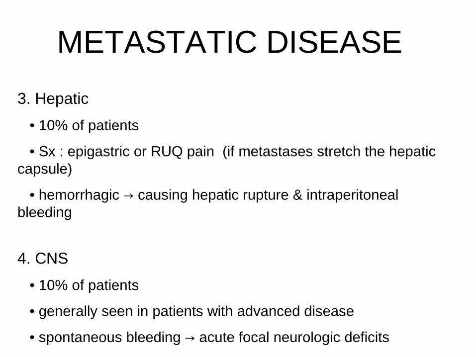
3. Hepatic
• 10% of patients
• Sx : epigastric or RUQ pain (if metastases stretch the hepatic capsule)
• hemorrhagic causing hepatic rupture & intraperitoneal →bleeding
4. CNS
• 10% of patients
• generally seen in patients with advanced disease
• spontaneous bleeding acute focal neurologic deficits→
METASTATIC DISEASE
40
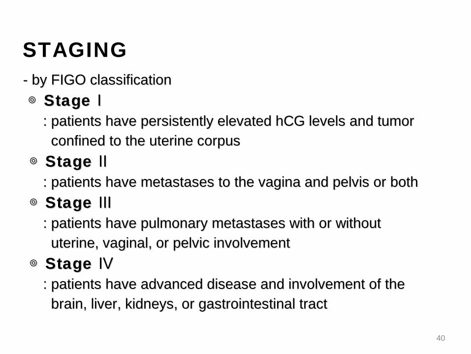
- by FIGO classification- by FIGO classification
◎ ◎ Stage ⅠStage Ⅰ : patients have persistently elevated hCG levels and tumor: patients have persistently elevated hCG levels and tumor confined to the uterine corpusconfined to the uterine corpus
◎ ◎ Stage ⅡStage Ⅱ : patients have metastases to the vagina and pelvis or both: patients have metastases to the vagina and pelvis or both
◎ ◎ Stage ⅢStage Ⅲ : patients have pulmonary metastases with or without: patients have pulmonary metastases with or without uterine, vaginal, or pelvic involvementuterine, vaginal, or pelvic involvement
◎ ◎ Stage ⅣStage Ⅳ : patients have advanced disease and involvement of the: patients have advanced disease and involvement of the brain, liver, kidneys, or gastrointestinal tractbrain, liver, kidneys, or gastrointestinal tract
STAGING
41
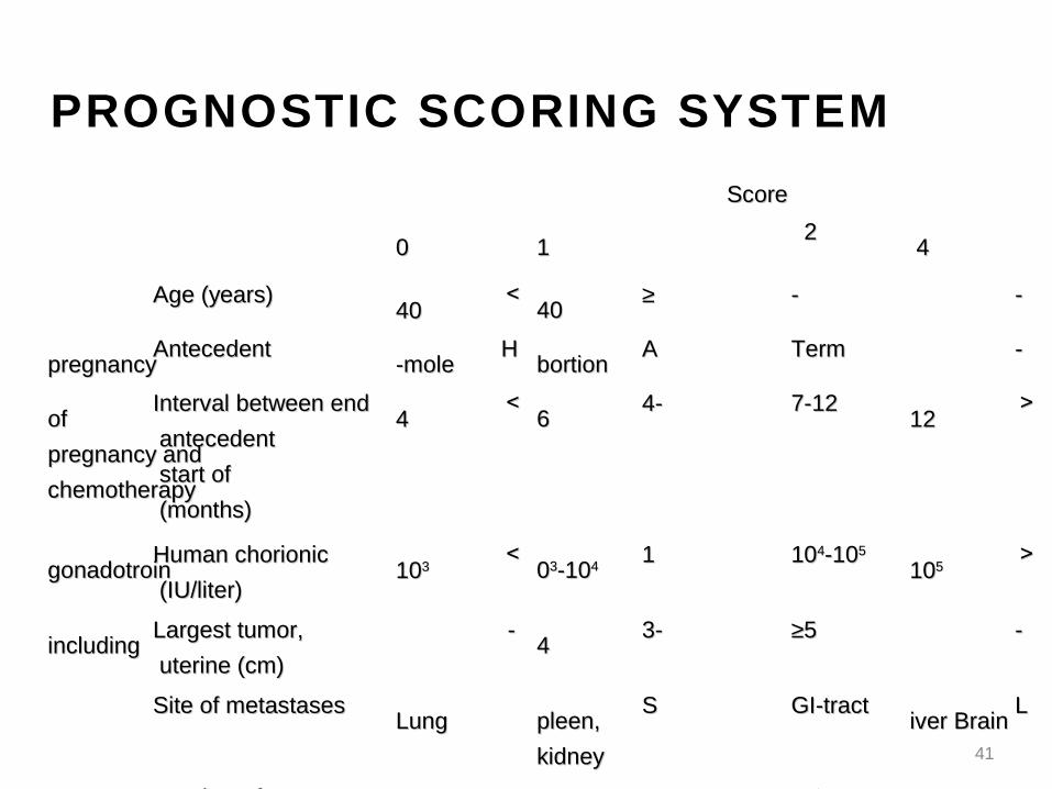
PROGNOSTIC SCORING SYSTEM
ScoreScore
00
11
22 44
Age (years)Age (years) <<4040
≥≥4040
-- --
Antecedent Antecedent pregnancypregnancy
HH-mole-mole
AAbortionbortion
TermTerm --
Interval between end Interval between end of of
antecedent antecedent pregnancy andpregnancy and
start of start of chemotherapychemotherapy
(months)(months)
<<44
4-4-66
7-127-12 >>1212
Human chorionic Human chorionic gonadotroingonadotroin
(IU/liter)(IU/liter)
<<101033
110033-10-1044
101044-10-1055 >>101055
Largest tumor, Largest tumor, includingincluding
uterine (cm)uterine (cm)
-- 3-3-44
≥≥55 --
Site of metastasesSite of metastases LungLung
SSpleen,pleen,
kidneykidney
GI-tract GI-tract LLiver Brainiver Brain
Number of Number of metastasesmetastases
-- 1-1-44
5-85-8 >>88
Prior chemotherapyPrior chemotherapy -- -- SingleSingle ≥≥2 drugs2 drugs

42
◎ ◎ to consider other variables to consider other variables
to predict the drug resistanceto predict the drug resistance
to assist in selecting appropriate to assist in selecting appropriate chemotherapychemotherapy
◎ ◎ higher than 7higher than 7
: categorized as high risk requires intensive : categorized as high risk requires intensive combination chemotherapycombination chemotherapy
Ex � STAGE III:8Ex � STAGE III:8
PROGNOSTIC SCORING SYSTEM
43
◎ ◎ All patients with persistent GTT should undergo a carefulAll patients with persistent GTT should undergo a careful pretreatment evaluationpretreatment evaluation ① ① complete history and physical examcomplete history and physical exam ② ② measurement of serum hCG level measurement of serum hCG level ③ ③ hepatic, thyroid, and renal function testhepatic, thyroid, and renal function test ④ ④ determination of baseline peripheral WBC & platelet countdetermination of baseline peripheral WBC & platelet count
◎ ◎ The metastatic workupThe metastatic workup ① ① chest radiograph or CT scanchest radiograph or CT scan ② ② ultrasonography or CT scan of the abdomen & pelvisultrasonography or CT scan of the abdomen & pelvis ③ ③ CT or MRI scan of the headCT or MRI scan of the head
◎ ◎ When the pelvic exam & chest radiographic findings areWhen the pelvic exam & chest radiographic findings are negative, metastatic involvement of other sites is uncommonnegative, metastatic involvement of other sites is uncommon
DIAGNOSTIC EVALUATION
44
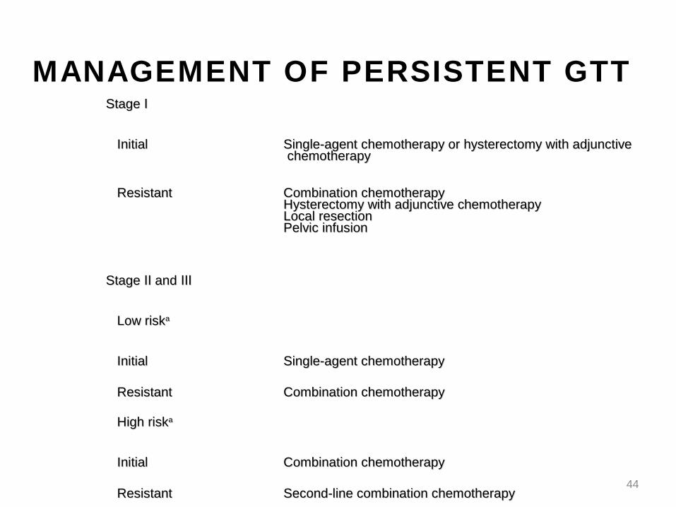
MANAGEMENT OF PERSISTENT GTTStage ⅠStage Ⅰ
InitialInitial Single-agent chemotherapy or hysterectomy with adjunctiveSingle-agent chemotherapy or hysterectomy with adjunctive chemotherapychemotherapy
ResistantResistant Combination chemotherapyCombination chemotherapyHysterectomy with adjunctive chemotherapyHysterectomy with adjunctive chemotherapyLocal resectionLocal resectionPelvic infusionPelvic infusion
Stage and Ⅱ ⅢStage and Ⅱ Ⅲ
Low riskLow riskaa
InitialInitial Single-agent chemotherapySingle-agent chemotherapy
ResistantResistant Combination chemotherapyCombination chemotherapy
High riskHigh riskaa
InitialInitial Combination chemotherapyCombination chemotherapy
ResistantResistant Second-line combination chemotherapySecond-line combination chemotherapy
Stage ⅣStage Ⅳ
InitialInitial Combination chemotherapyCombination chemotherapy
BrainBrain Whole-heat irradiation (3,000 cGy)Whole-heat irradiation (3,000 cGy)Craniotomy to manage complicationsCraniotomy to manage complications
LiverLiver Resection to manage complicationsResection to manage complications
ResistantResistantaa Second-line combination chemotherapySecond-line combination chemotherapyHepatic arterial infusionHepatic arterial infusion
45
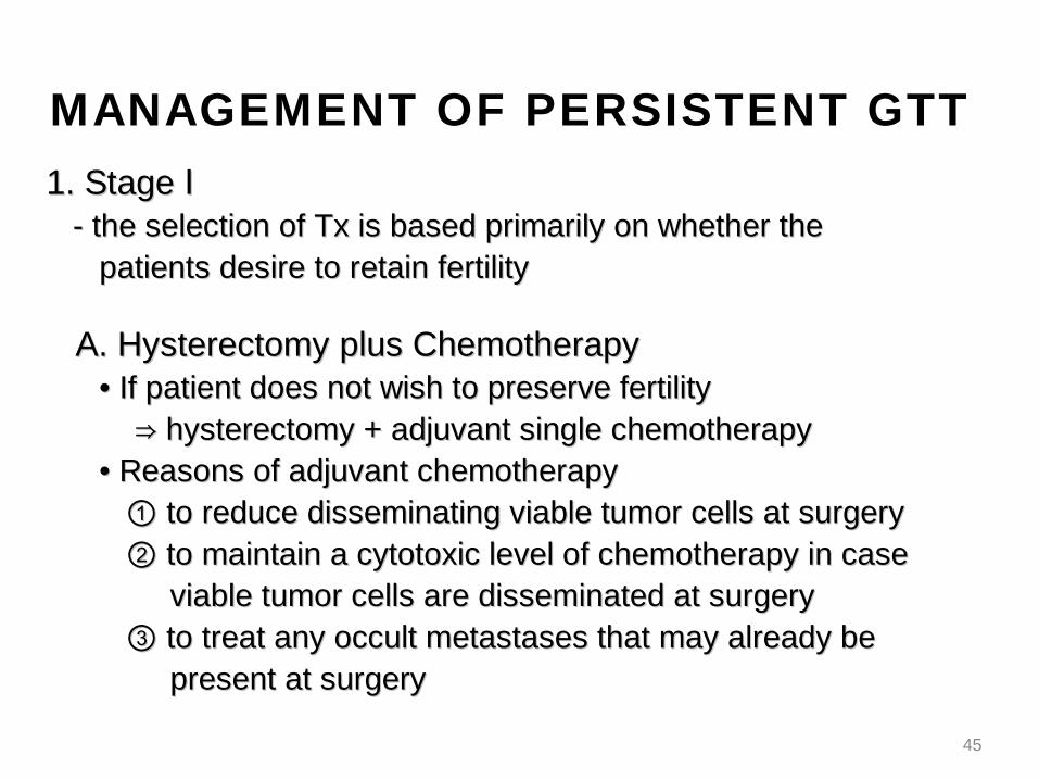
1. Stage Ⅰ1. Stage Ⅰ - the selection of Tx is based primarily on whether the- the selection of Tx is based primarily on whether the patients desire to retain fertilitypatients desire to retain fertility
A. Hysterectomy plus ChemotherapyA. Hysterectomy plus Chemotherapy • • If patient does not wish to preserve fertilityIf patient does not wish to preserve fertility ⇒ ⇒ hysterectomy + adjuvant single chemotherapyhysterectomy + adjuvant single chemotherapy • • Reasons of adjuvant chemotherapyReasons of adjuvant chemotherapy ① ① to reduce disseminating viable tumor cells at surgeryto reduce disseminating viable tumor cells at surgery ② ② to maintain a cytotoxic level of chemotherapy in caseto maintain a cytotoxic level of chemotherapy in case viable tumor cells are disseminated at surgeryviable tumor cells are disseminated at surgery ③ ③ to treat any occult metastases that may already beto treat any occult metastases that may already be present at surgerypresent at surgery
MANAGEMENT OF PERSISTENT GTT
46
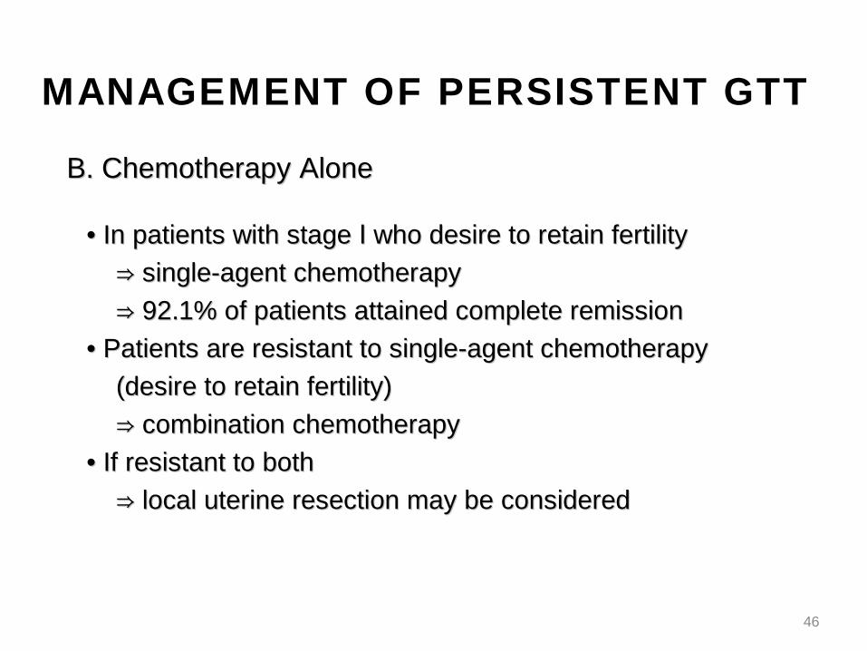
B. Chemotherapy AloneB. Chemotherapy Alone
• • In patients with stage who desire to retain fertilityⅠIn patients with stage who desire to retain fertilityⅠ ⇒ ⇒ single-agent chemotherapysingle-agent chemotherapy
⇒ ⇒ 92.1% of patients attained complete remission92.1% of patients attained complete remission
• • Patients are resistant to single-agent chemotherapy Patients are resistant to single-agent chemotherapy
(desire to retain fertility)(desire to retain fertility)
⇒ ⇒ combination chemotherapycombination chemotherapy
• • If resistant to bothIf resistant to both
⇒ ⇒ local uterine resection may be consideredlocal uterine resection may be considered
MANAGEMENT OF PERSISTENT GTT

47
2. Stage and Ⅱ Ⅲ2. Stage and Ⅱ Ⅲ - low-risk : single agent chemoTx- low-risk : single agent chemoTx - high-risk : combination chemoTx- high-risk : combination chemoTx
A. Vaginal and Pelvic MetastasisA. Vaginal and Pelvic Metastasis • • vaginal metastases may bleed profuselyvaginal metastases may bleed profusely ( highly vascular and friable)∵( highly vascular and friable)∵ ⇒ ⇒ controlled by packing or by wide local excisioncontrolled by packing or by wide local excision
B. Pulmonary MetastasisB. Pulmonary Metastasis • • persistent viable pulmonary metastasis despite intensivepersistent viable pulmonary metastasis despite intensive chemotherapychemotherapy ⇒ ⇒ thoracotomy may be attempted to excise thethoracotomy may be attempted to excise the resistant focusresistant focus
MANAGEMENT OF PERSISTENT GTT
48
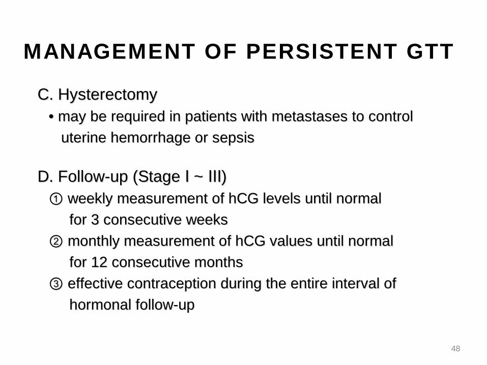
C. HysterectomyC. Hysterectomy • • may be required in patients with metastases to controlmay be required in patients with metastases to control
uterine hemorrhage or sepsisuterine hemorrhage or sepsis
D. Follow-up (Stage ~ )Ⅰ ⅢD. Follow-up (Stage ~ )Ⅰ Ⅲ ① ① weekly measurement of hCG levels until normalweekly measurement of hCG levels until normal
for 3 consecutive weeksfor 3 consecutive weeks
② ② monthly measurement of hCG values until normalmonthly measurement of hCG values until normal
for 12 consecutive monthsfor 12 consecutive months
③ ③ effective contraception during the entire interval ofeffective contraception during the entire interval of
hormonal follow-uphormonal follow-up
MANAGEMENT OF PERSISTENT GTT
49
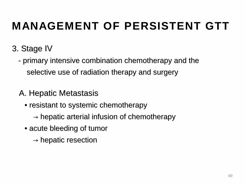
3. Stage Ⅳ3. Stage Ⅳ - primary intensive combination chemotherapy and the - primary intensive combination chemotherapy and the
selective use of radiation therapy and surgeryselective use of radiation therapy and surgery
A. Hepatic MetastasisA. Hepatic Metastasis
• • resistant to systemic chemotherapyresistant to systemic chemotherapy
→ → hepatic arterial infusion of chemotherapyhepatic arterial infusion of chemotherapy
• • acute bleeding of tumoracute bleeding of tumor
→ → hepatic resectionhepatic resection
MANAGEMENT OF PERSISTENT GTT

50
B. Cerebral MetastasisB. Cerebral Metastasis • • If diagnosed, whole-brain irradiation (3,000 cGy in If diagnosed, whole-brain irradiation (3,000 cGy in 1010 fractions) can be instituted promptlyfractions) can be instituted promptly • • conurrent use of combination chemotherapy and conurrent use of combination chemotherapy and brainbrain irradiationirradiation → → spontaneous cerebral hemorrhage ↓spontaneous cerebral hemorrhage ↓
C. Follow-up (Stage )ⅣC. Follow-up (Stage )Ⅳ ① ① weekly determination of hCG levels until they weekly determination of hCG levels until they areare normal 3 consecutive weeksnormal 3 consecutive weeks ② ② monthly determination of hCG levels until they monthly determination of hCG levels until they areare normal for 24 consecutive monthsnormal for 24 consecutive months • • increased risk of late recurrenceincreased risk of late recurrence → → prolonged gonadotropin follow-up is requiredprolonged gonadotropin follow-up is required
MANAGEMENT OF PERSISTENT GTT
51
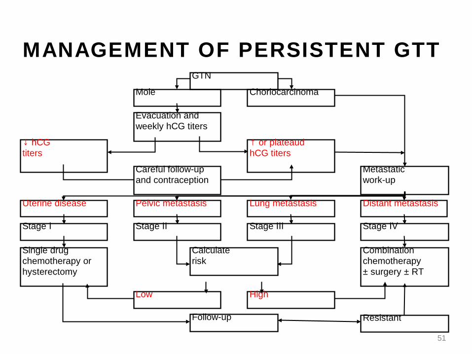
MANAGEMENT OF PERSISTENT GTT
Evacuation andweekly hCG titers
GTN
Mole
Metastaticwork-up
↓ hCGtiters
↑ or plateaudhCG titers
Choriocarcinoma
Distant metastasisUterine disease
Careful follow-upand contraception
Lung metastasis
Single drugchemotherapy orhysterectomy
Pelvic metastasis
Calculaterisk
Combinationchemotherapy± surgery ± RT
Low High
Follow-up Resistant
Stage ⅣStage Ⅰ Stage ⅢStage Ⅱ

52
# Chemotherapy

53
◎ ◎ Nonmetastatic and low-risk GTTNonmetastatic and low-risk GTT ⇒ ⇒ actinomycin D (Act-D) or MTX has achieved comparable actinomycin D (Act-D) or MTX has achieved comparable and excellent remission ratesand excellent remission rates
◎ ◎ ProtocolsProtocols ① ① Act-D can be given every other week as a 5-day regimenAct-D can be given every other week as a 5-day regimen or in a pulsatile fashionor in a pulsatile fashion ② ② use of MTX was the sameuse of MTX was the same
◎ ◎ Methotrexate with folic acid (MTX-FA)Methotrexate with folic acid (MTX-FA) ① ① the preferred single agent in the Tx of GTTthe preferred single agent in the Tx of GTT ② ② remission ratesremission rates - 90.2% in stage Ⅰ- 90.2% in stage Ⅰ - 68.2% in low-risk stages & Ⅱ Ⅲ- 68.2% in low-risk stages & Ⅱ Ⅲ ③ ③ after Tx with MTX-FA, thrombocytopenia, granulocytopeniaafter Tx with MTX-FA, thrombocytopenia, granulocytopenia and hepatotoxicity developed in only 1.6%, 5.9% and 14.1%and hepatotoxicity developed in only 1.6%, 5.9% and 14.1% of patientsof patients
SINGLE-AGENT TREATMENT
54
1. Triple Therapy1. Triple Therapy
◎ ◎ etoposide, MTX, Act-D, cyclophosphamideetoposide, MTX, Act-D, cyclophosphamide
and vincristine (EMA-CO)and vincristine (EMA-CO)
- had complete remission in patients with metastasis and - had complete remission in patients with metastasis and aa
high-risk score (76~94%)high-risk score (76~94%)
- remission occurred in 13 of 15 patients (86%) with brain- remission occurred in 13 of 15 patients (86%) with brain
metastasismetastasis
◎ ◎ EMA-CO regimenEMA-CO regimen
① ① the preferred primary Tx in patients with metastasis the preferred primary Tx in patients with metastasis and aand a
high-risk prognostic scorehigh-risk prognostic score
② ② generally well toleratedgenerally well tolerated
③ ③ seldom has to be suspended because of toxicityseldom has to be suspended because of toxicity
COMBINATION CHEMOTHERAPY

55
2. Duration of therapy2. Duration of therapy
◎ ◎ Combination chemotherapy should be given as often Combination chemotherapy should be given as often asas
toxicity permits until the patients achieves threetoxicity permits until the patients achieves three
consecutive normal hCG levelsconsecutive normal hCG levels
◎ ◎ After normal hCG levels are attained, at least twoAfter normal hCG levels are attained, at least two
additional course are administered to reduce the additional course are administered to reduce the riskrisk
of relapseof relapse
COMBINATION CHEMOTHERAPY

Subsequent Pregnancies• Patients with hydatidiform moles can
anticipate normal reproduction in the future.• Patient has had a molar pregnancy, either
partial or complete, increased risk of having a molar gestation in subsequent conceptions.
• After one molar pregnancy, the risk of having molar disease in a future gestation is about1% to 1.5%.
• 1.Perform pelvic ultrasonographic examination during the first trimester to confirm normal gestational development.
• 2.Obtain an hCG measurement 6 weeks after completion of the pregnancy to exclude occult trophoblastic neoplasia.

58
# Surgery

Role of surgery
•Remove resistant or persistent disease in the uterus or metastatic sites •Decrease the uterine tumor load in case of limited metastasis •Control profuse tumor hemorrhage •Treat infection •Relieve symptoms like bowel or urinary obstruction due to the large tumor bulk.

Thank you