Embed Size (px)
Citation preview

CONCEPTS OF BIOLOGY Chapter 1 INTRODUCTION TO BIOLOGY
PowerPoint Image Slideshow
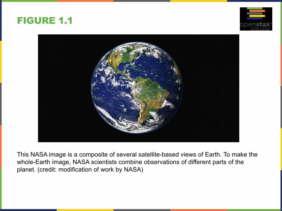
FIGURE 1.1
This NASA image is a composite of several satellite-based views of Earth. To make the
whole-Earth image, NASA scientists combine observations of different parts of the
planet. (credit: modification of work by NASA)
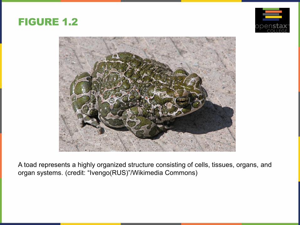
FIGURE 1.2
A toad represents a highly organized structure consisting of cells, tissues, organs, and
organ systems. (credit: “Ivengo(RUS)”/Wikimedia Commons)
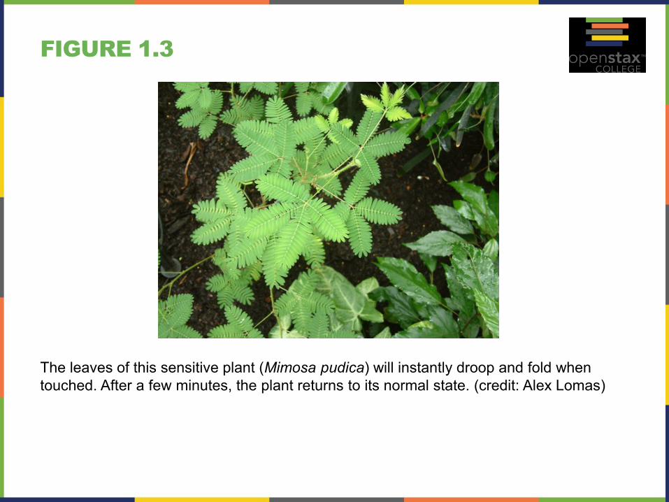
FIGURE 1.3
The leaves of this sensitive plant (Mimosa pudica) will instantly droop and fold when
touched. After a few minutes, the plant returns to its normal state. (credit: Alex Lomas)

FIGURE 1.4
Although no two look alike, these kittens have inherited genes from both parents and
share many of the same characteristics. (credit: Pieter & Renée Lanser)
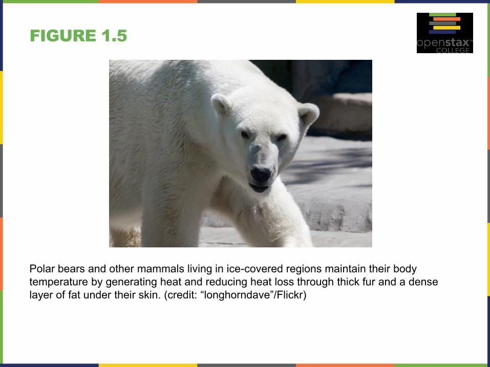
FIGURE 1.5
Polar bears and other mammals living in ice-covered regions maintain their body
temperature by generating heat and reducing heat loss through thick fur and a dense
layer of fat under their skin. (credit: “longhorndave”/Flickr)
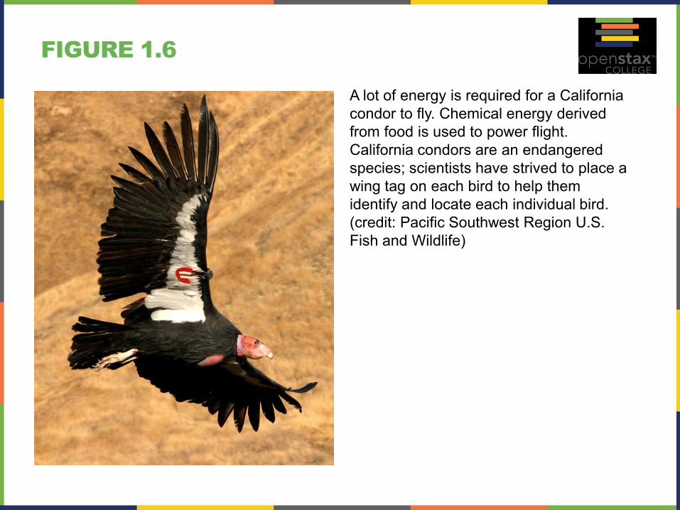
FIGURE 1.6
A lot of energy is required for a California
condor to fly. Chemical energy derived
from food is used to power flight.
California condors are an endangered
species; scientists have strived to place a
wing tag on each bird to help them
identify and locate each individual bird.
(credit: Pacific Southwest Region U.S.
Fish and Wildlife)
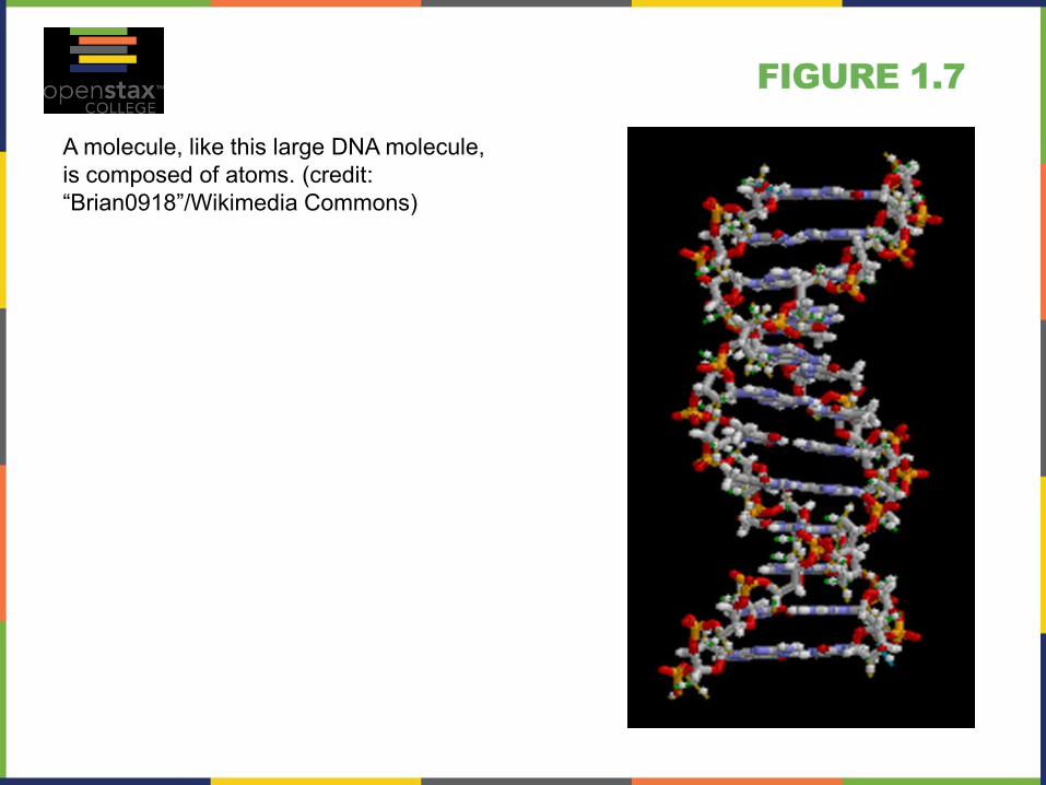
FIGURE 1.7
A molecule, like this large DNA molecule,
is composed of atoms. (credit:
“Brian0918”/Wikimedia Commons)
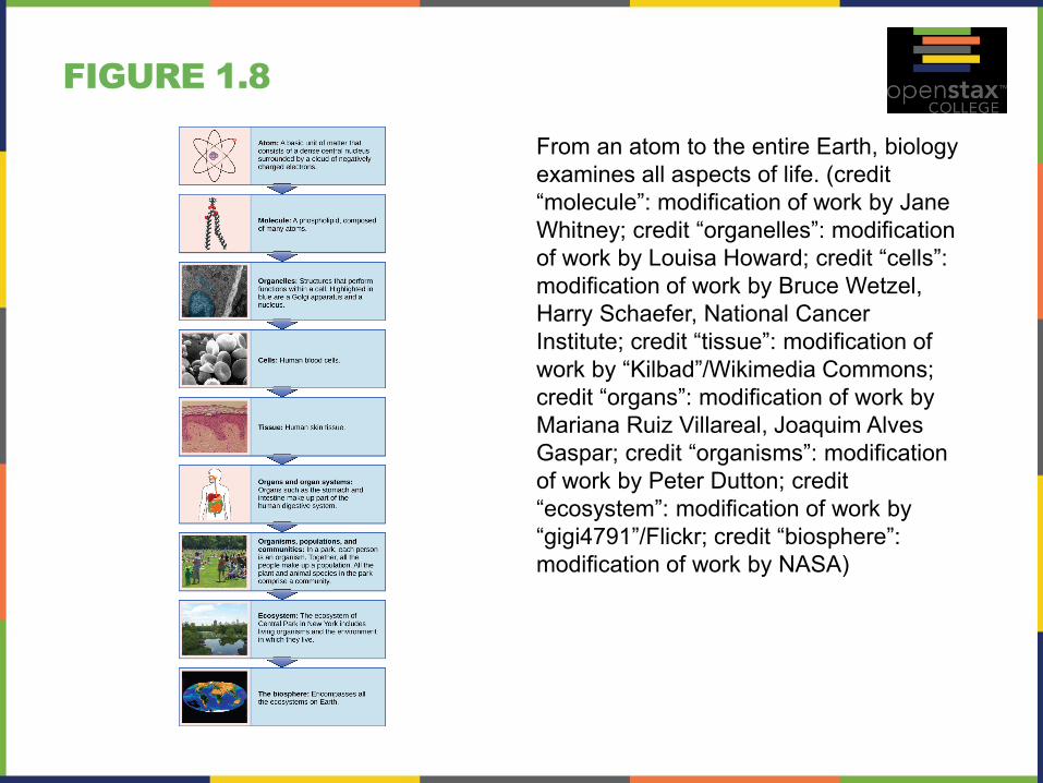
FIGURE 1.8
From an atom to the entire Earth, biology
examines all aspects of life. (credit
“molecule”: modification of work by Jane
Whitney; credit “organelles”: modification
of work by Louisa Howard; credit “cells”:
modification of work by Bruce Wetzel,
Harry Schaefer, National Cancer
Institute; credit “tissue”: modification of
work by “Kilbad”/Wikimedia Commons;
credit “organs”: modification of work by
Mariana Ruiz Villareal, Joaquim Alves
Gaspar; credit “organisms”: modification
of work by Peter Dutton; credit
“ecosystem”: modification of work by
“gigi4791”/Flickr; credit “biosphere”:
modification of work by NASA)
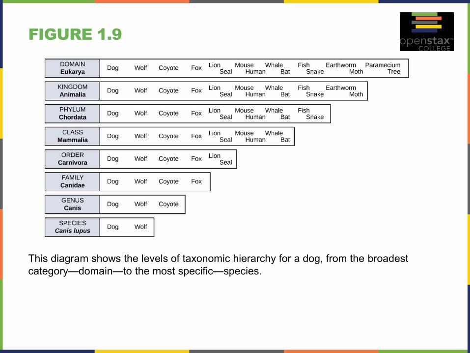
FIGURE 1.9
This diagram shows the levels of taxonomic hierarchy for a dog, from the broadest
category—domain—to the most specific—species.
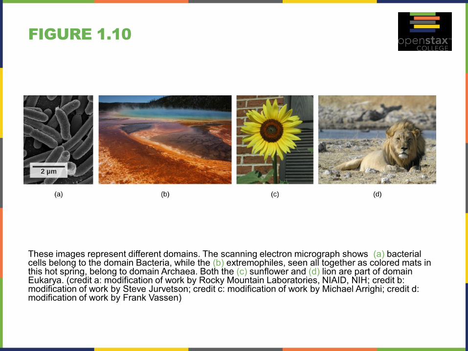
FIGURE 1.10
These images represent different domains. The scanning electron micrograph shows (a) bacterial cells belong to the domain Bacteria, while the (b) extremophiles, seen all together as colored mats in this hot spring, belong to domain Archaea. Both the (c) sunflower and (d) lion are part of domain Eukarya. (credit a: modification of work by Rocky Mountain Laboratories, NIAID, NIH; credit b: modification of work by Steve Jurvetson; credit c: modification of work by Michael Arrighi; credit d: modification of work by Frank Vassen)
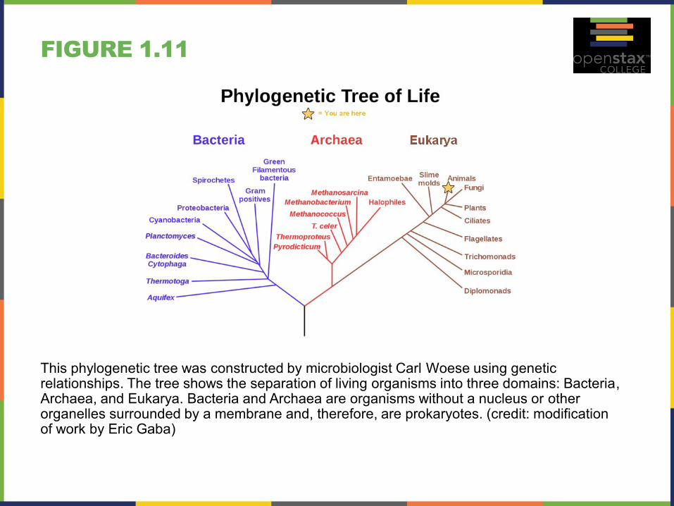
FIGURE 1.11
This phylogenetic tree was constructed by microbiologist Carl Woese using genetic relationships. The tree shows the separation of living organisms into three domains: Bacteria, Archaea, and Eukarya. Bacteria and Archaea are organisms without a nucleus or other organelles surrounded by a membrane and, therefore, are prokaryotes. (credit: modification of work by Eric Gaba)
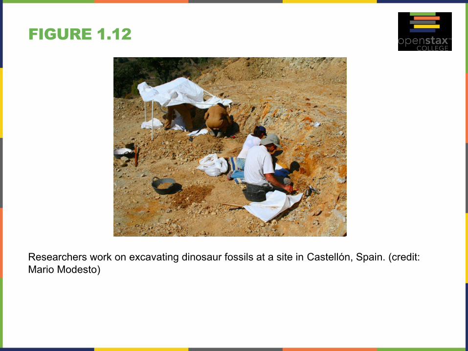
FIGURE 1.12
Researchers work on excavating dinosaur fossils at a site in Castellón, Spain. (credit:
Mario Modesto)
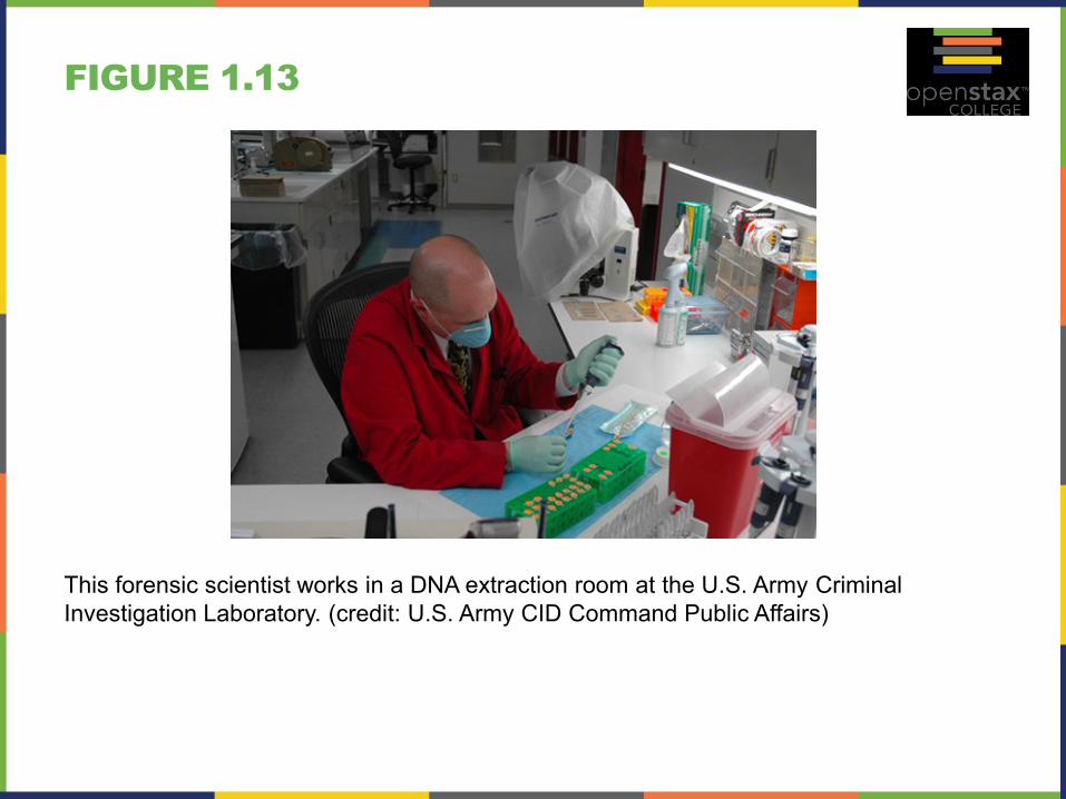
FIGURE 1.13
This forensic scientist works in a DNA extraction room at the U.S. Army Criminal
Investigation Laboratory. (credit: U.S. Army CID Command Public Affairs)
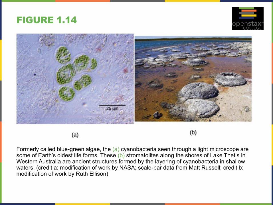
FIGURE 1.14
Formerly called blue-green algae, the (a) cyanobacteria seen through a light microscope are some of Earth’s oldest life forms. These (b) stromatolites along the shores of Lake Thetis in Western Australia are ancient structures formed by the layering of cyanobacteria in shallow waters. (credit a: modification of work by NASA; scale-bar data from Matt Russell; credit b: modification of work by Ruth Ellison)
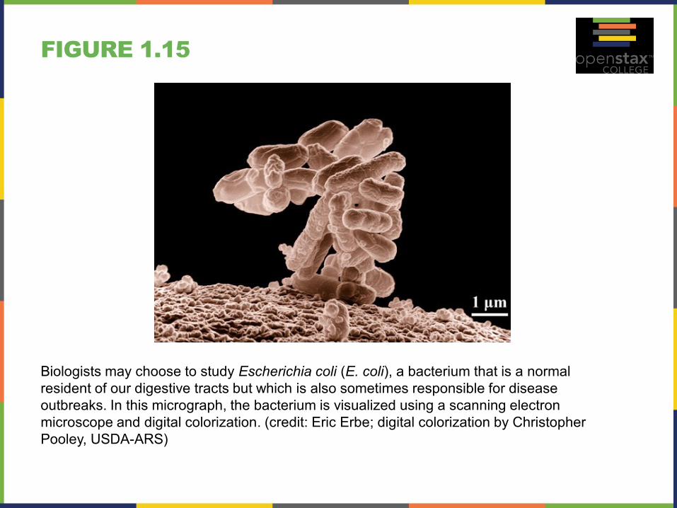
FIGURE 1.15
Biologists may choose to study Escherichia coli (E. coli), a bacterium that is a normal
resident of our digestive tracts but which is also sometimes responsible for disease
outbreaks. In this micrograph, the bacterium is visualized using a scanning electron
microscope and digital colorization. (credit: Eric Erbe; digital colorization by Christopher
Pooley, USDA-ARS)
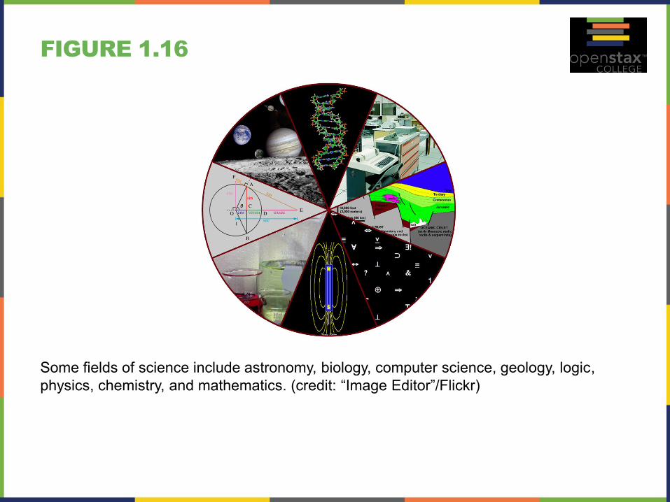
FIGURE 1.16
Some fields of science include astronomy, biology, computer science, geology, logic,
physics, chemistry, and mathematics. (credit: “Image Editor”/Flickr)

FIGURE 1.17
Sir Francis Bacon is credited with being
the first to document the scientific
method.
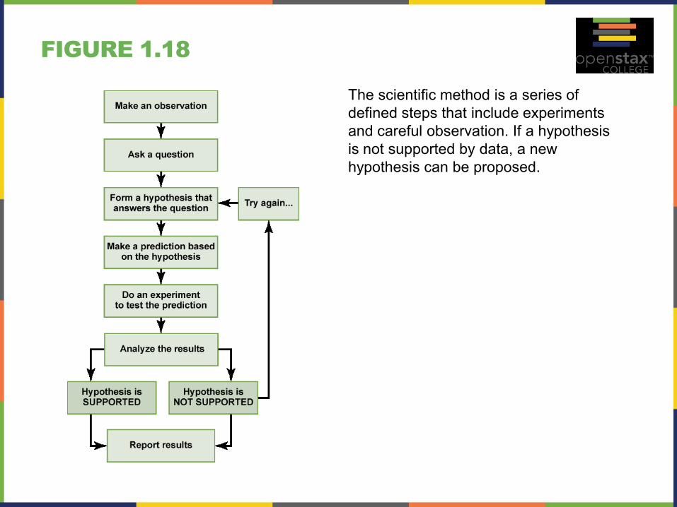
FIGURE 1.18
The scientific method is a series of
defined steps that include experiments
and careful observation. If a hypothesis
is not supported by data, a new
hypothesis can be proposed.
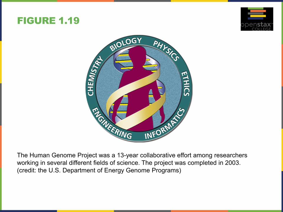
FIGURE 1.19
The Human Genome Project was a 13-year collaborative effort among researchers
working in several different fields of science. The project was completed in 2003.
(credit: the U.S. Department of Energy Genome Programs)

This PowerPoint file is copyright 2011-2013, Rice University. All
Rights Reserved.