Embed Size (px)
Citation preview
Overview of Respiration andOverview of Respiration andRespiratory MechanicsRespiratory Mechanics
Dr Shihab Khogali
Ninewells Hospital & Medical School, University of Dundee
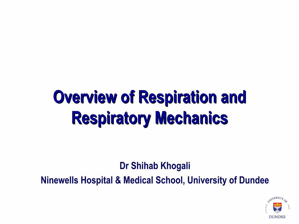
This lecture is the first of four-linked lectures …in this lecture:
Understand what is meant by the terms “internal respiration” and “external respiration”
Know the four steps of external respiration
Understand Ventilation - the first step of external respiration
What is This LectureAbout?
See blackboard for detailed learning objectives
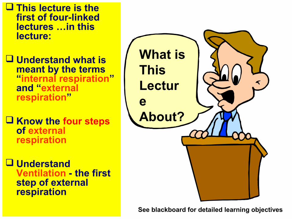
Know that gases move from higher to lower pressure, with the Boyle’s Law.
Understand the respiratory mechanics and the relationship between atmospheric, intra-alveolar, and intrapleural pressures.
understand the significance of transmural pressure gradient. Know that peumothorax abolishes the transmural pressure gradient.
Understand that inspiration is an active process and that normal resting expiration is a passive process.
Know the inspiratory muscles and the accessory muscles of respiration (link with anatomy).
Describe the role and importance of pulmonary surfactant, with the Law of Laplace and alveolar stability.
Know the lung volumes and capacities. Understand the changes in dynamic lung volumes in obstructive and restrictive lung disease.
Know the factors which influence airway resistance.
Define the compliance of lungs and thorax.
Understand what is meant by the term work of breathing.
Understand ventilation (Step 1 of external respiration).
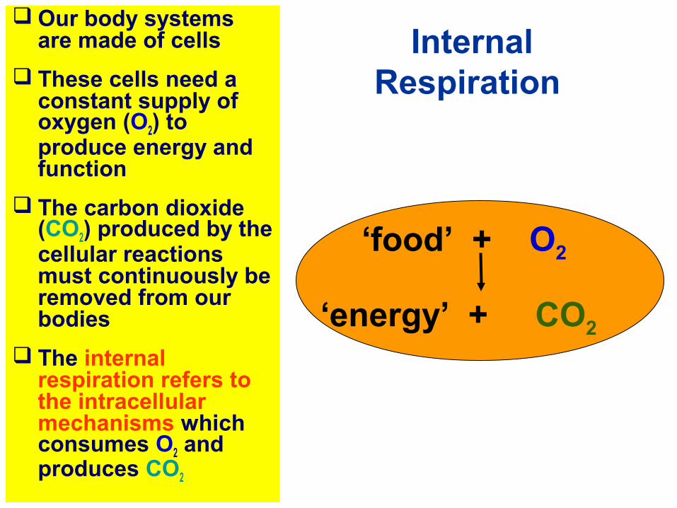
Our body systems are made of cells
These cells need a constant supply of oxygen (O2) to produce energy and function
The carbon dioxide (CO2) produced by the cellular reactions must continuously be removed from our bodies
The internal respiration refers to the intracellular mechanisms which consumes O2 and produces CO2
‘food’ + O2
‘energy’ + CO2
Internal Respiration
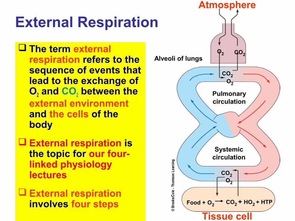
The term external respiration refers to the sequence of events that lead to the exchange of O2 and CO2 between the external environment and the cells of the body
External respiration is the topic for our four-linked physiology lectures
External respiration involves four steps
Atmosphere
Tissue cell
Alveoli of lungs
Pulmonarycirculation
Systemiccirculation
CO2O2
Food + O2 CO2 + HO2 + HTP
O2
CO2
CO2
O2
External Respiration
Atmosphere
Tissue cell
Alveoli of lungs
Pulmonarycirculation
Systemiccirculation
CO2O2
Food + O2 CO2 + HO2 + ATP
O2
CO2
CO2
O2
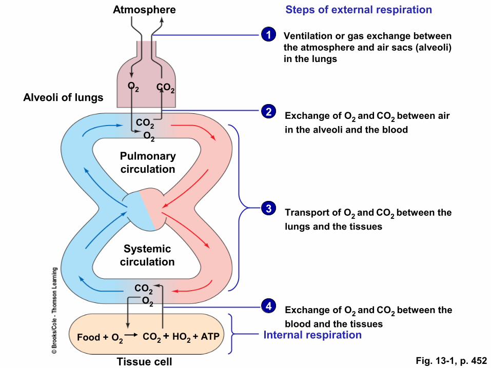
1
Steps of external respiration
Ventilation or gas exchange betweenthe atmosphere and air sacs (alveoli)in the lungs
Exchange of O2 and CO2 between air
in the alveoli and the blood
Transport of O2 and CO2 between the
lungs and the tissues
Exchange of O2 and CO2 between the
blood and the tissuesInternal respiration
2
3
4
Fig. 13-1, p. 452
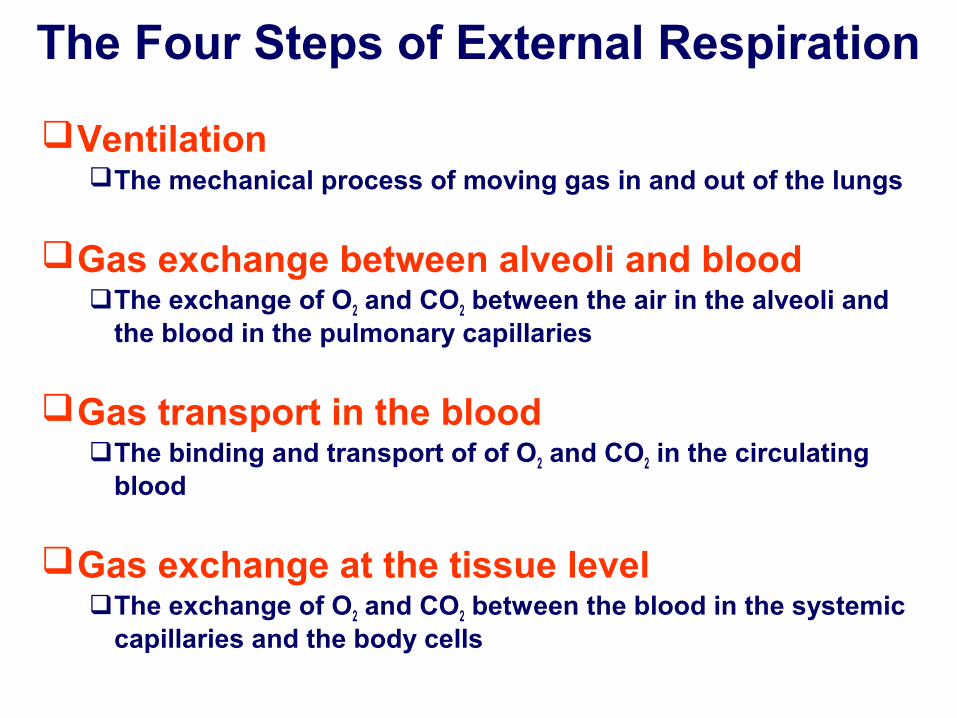
The Four Steps of External Respiration
VentilationThe mechanical process of moving gas in and out of the lungs
Gas exchange between alveoli and bloodThe exchange of O2 and CO2 between the air in the alveoli and
the blood in the pulmonary capillaries
Gas transport in the bloodThe binding and transport of of O2 and CO2 in the circulating
blood
Gas exchange at the tissue levelThe exchange of O2 and CO2 between the blood in the systemic
capillaries and the body cells

The Respiratory System
The Cardiovascular System
The Haematology System
Atmosphere
Tissue cell
Alveoli of lungs
Pulmonarycirculation
Systemiccirculation
CO2O2
Food + O2 CO2 + HO2 + HTP
O2
CO2
CO2
O2
Three body systems are involved in external respiration
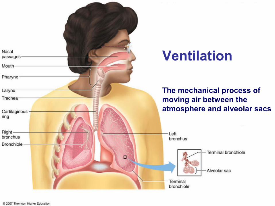
The mechanical process of moving air between the atmosphere and alveolar sacs
Ventilation
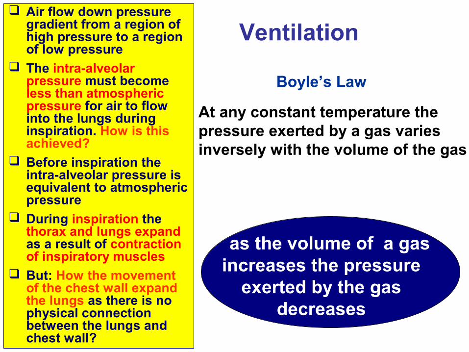
Air flow down pressure gradient from a region of high pressure to a region of low pressure
The intra-alveolar pressure must become less than atmospheric pressure for air to flow into the lungs during inspiration. How is this achieved?
Before inspiration the intra-alveolar pressure is equivalent to atmospheric pressure
During inspiration the thorax and lungs expand as a result of contraction of inspiratory muscles
But: How the movement of the chest wall expand the lungs as there is no physical connection between the lungs and chest wall?
as the volume of a gas increases the pressure
exerted by the gas decreases
Boyle’s Law
At any constant temperature the pressure exerted by a gas varies inversely with the volume of the gas
Ventilation
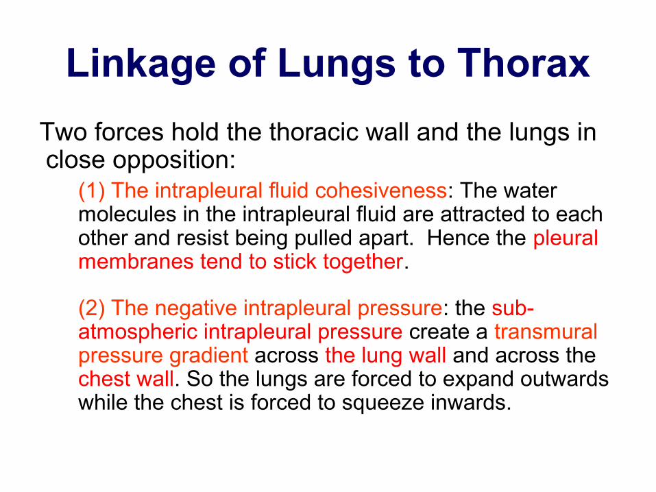
Linkage of Lungs to Thorax
Two forces hold the thoracic wall and the lungs in close opposition:
(1) The intrapleural fluid cohesiveness: The water molecules in the intrapleural fluid are attracted to each other and resist being pulled apart. Hence the pleural membranes tend to stick together.
(2) The negative intrapleural pressure: the sub-atmospheric intrapleural pressure create a transmural pressure gradient across the lung wall and across the chest wall. So the lungs are forced to expand outwards while the chest is forced to squeeze inwards.

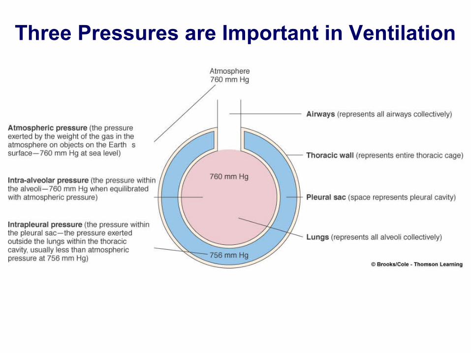
Three Pressures are Important in Ventilation
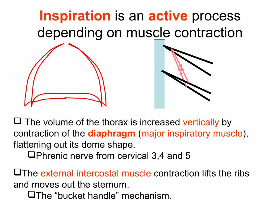
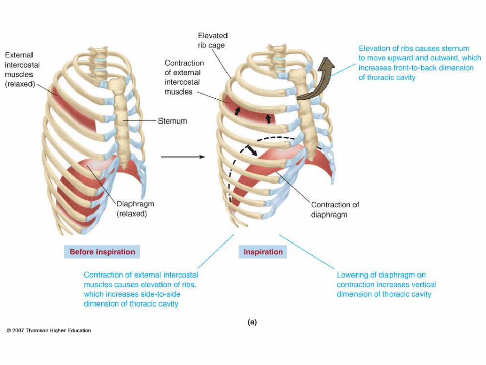
Inspiration is an active process depending on muscle contraction
The volume of the thorax is increased vertically by contraction of the diaphragm (major inspiratory muscle), flattening out its dome shape.
Phrenic nerve from cervical 3,4 and 5
The external intercostal muscle contraction lifts the ribs and moves out the sternum.
The “bucket handle” mechanism.
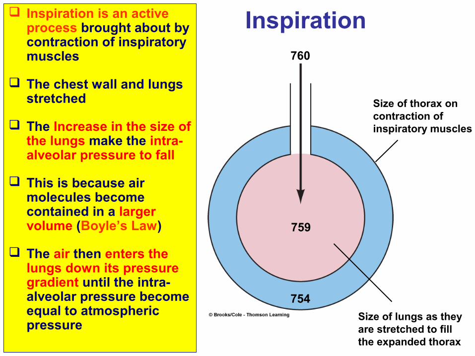
Inspiration is an active process brought about by contraction of inspiratory muscles
The chest wall and lungs stretched
The Increase in the size of the lungs make the intra-alveolar pressure to fall
This is because air molecules become contained in a larger volume (Boyle’s Law)
The air then enters the lungs down its pressure gradient until the intra-alveolar pressure become equal to atmospheric pressure
Inspiration
759
Size of thorax oncontraction ofinspiratory muscles
Size of lungs as theyare stretched to fillthe expanded thorax
754
760
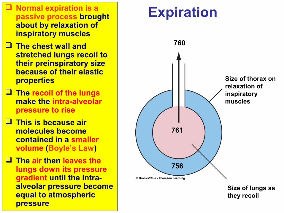
Normal expiration is a passive process brought about by relaxation of inspiratory muscles
The chest wall and stretched lungs recoil to their preinspiratory size because of their elastic properties
The recoil of the lungs make the intra-alveolar pressure to rise
This is because air molecules become contained in a smaller volume (Boyle’s Law)
The air then leaves the lungs down its pressure gradient until the intra-alveolar pressure become equal to atmospheric pressure
761
Size of thorax onrelaxation ofinspiratory muscles
Size of lungs asthey recoil
756
760
Expiration
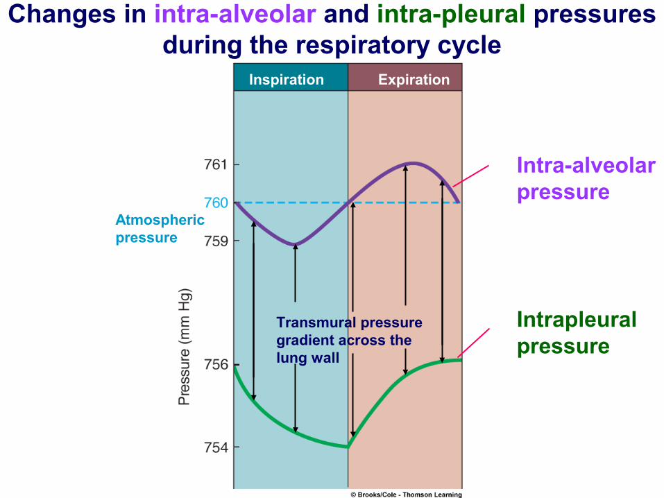
Inspiration Expiration
Atmosphericpressure
Intra-alveolarpressure
Intrapleuralpressure
Transmural pressuregradient across thelung wall
Changes in intra-alveolar and intra-pleural pressures during the respiratory cycle
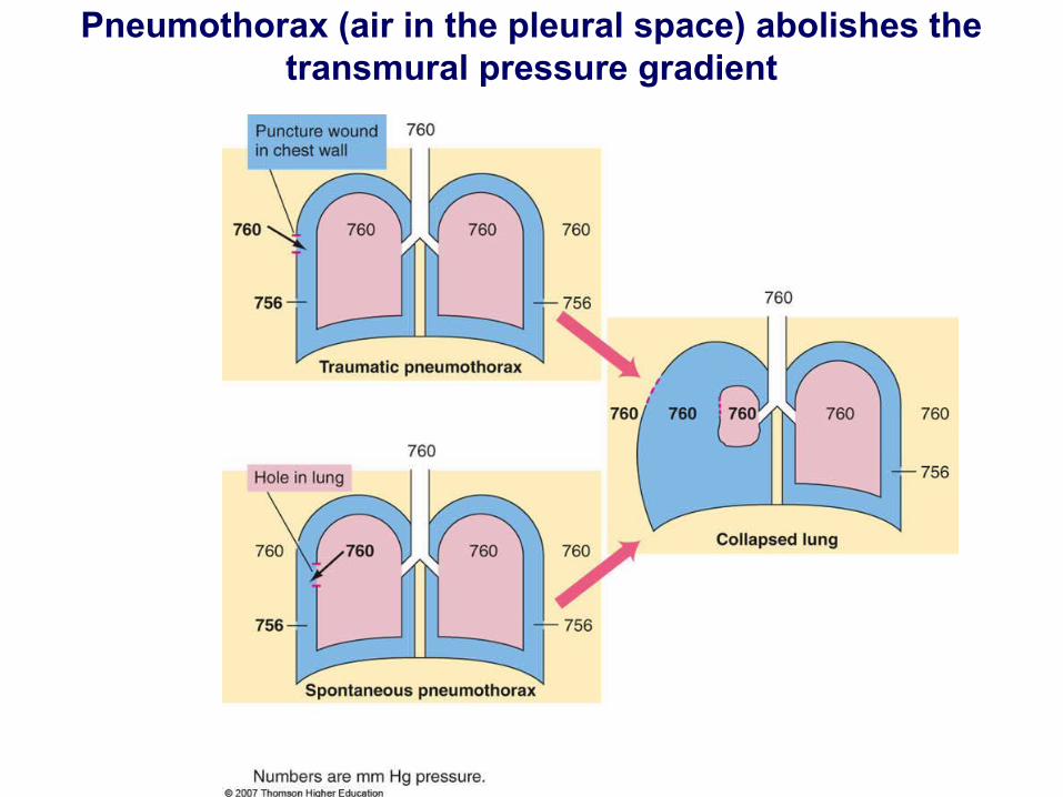
Pneumothorax (air in the pleural space) abolishes the transmural pressure gradient
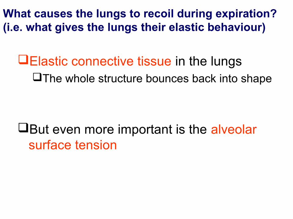
What causes the lungs to recoil during expiration?(i.e. what gives the lungs their elastic behaviour)
Elastic connective tissue in the lungsThe whole structure bounces back into shape
But even more important is the alveolar surface tension
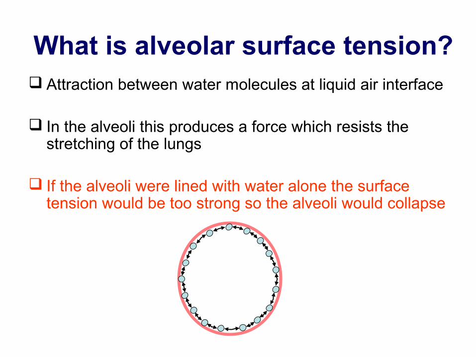
What is alveolar surface tension? Attraction between water molecules at liquid air interface
In the alveoli this produces a force which resists the stretching of the lungs
If the alveoli were lined with water alone the surface tension would be too strong so the alveoli would collapse
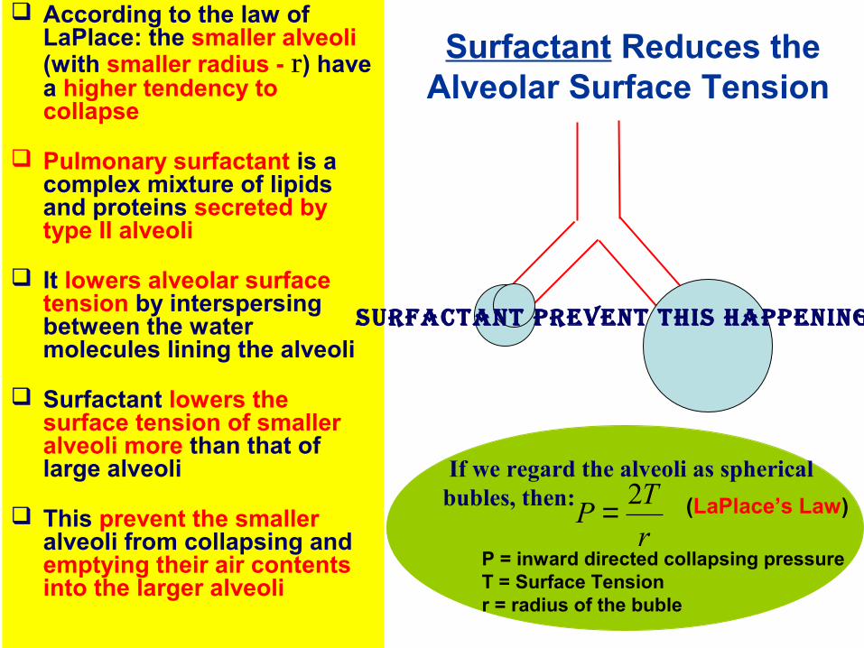
According to the law of LaPlace: the smaller alveoli (with smaller radius - r) have a higher tendency to collapse
Pulmonary surfactant is a complex mixture of lipids and proteins secreted by type II alveoli
It lowers alveolar surface tension by interspersing between the water molecules lining the alveoli
Surfactant lowers the surface tension of smaller alveoli more than that of large alveoli
This prevent the smaller alveoli from collapsing and emptying their air contents into the larger alveoli
Surfactant Reduces the Alveolar Surface Tension
If we regard the alveoli as spherical bubles, then:
r
TP
2=P = inward directed collapsing pressureT = Surface Tensionr = radius of the buble
(LaPlace’s Law)
Surfactant prevent thiS happening
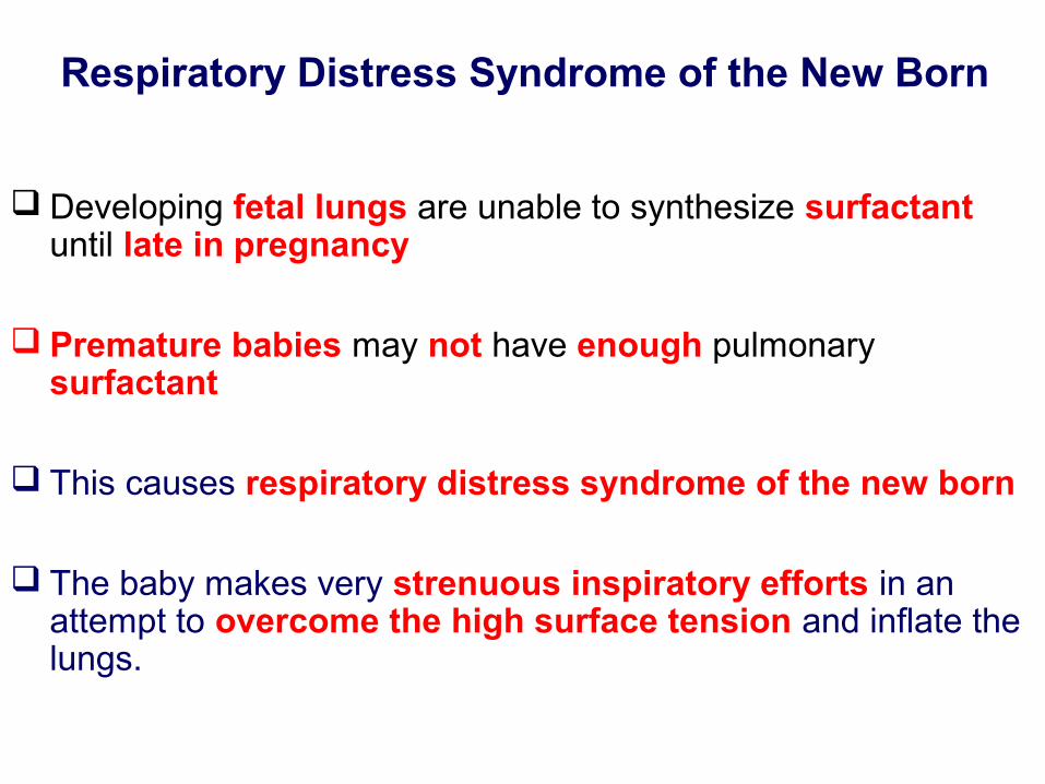
Respiratory Distress Syndrome of the New Born
Developing fetal lungs are unable to synthesize surfactant until late in pregnancy
Premature babies may not have enough pulmonary surfactant
This causes respiratory distress syndrome of the new born
The baby makes very strenuous inspiratory efforts in an attempt to overcome the high surface tension and inflate the lungs.
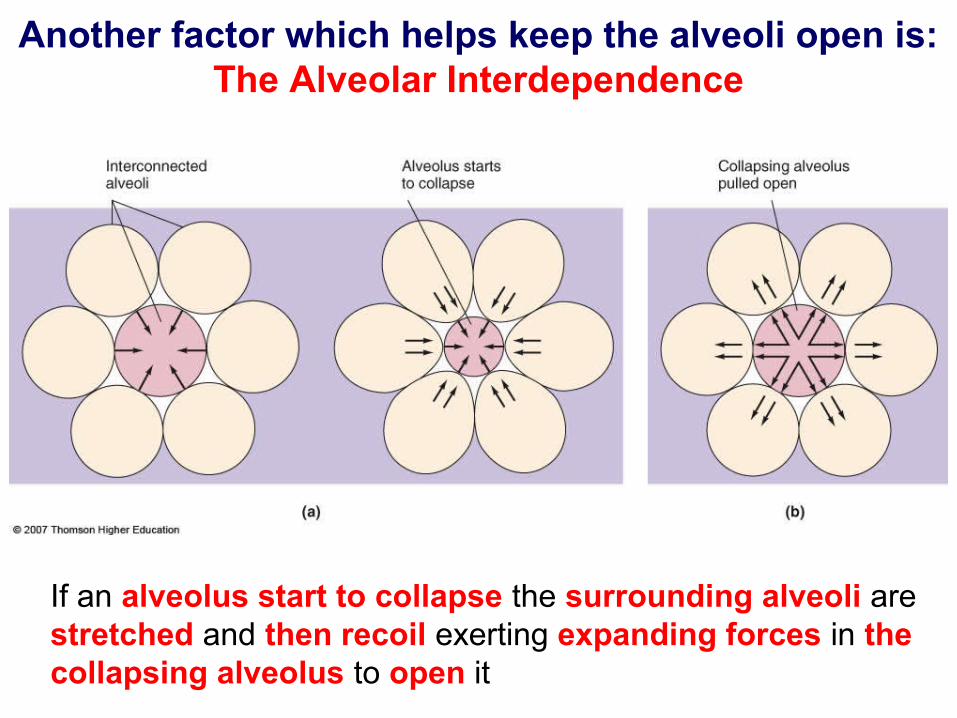
Another factor which helps keep the alveoli open is: The Alveolar Interdependence
If an alveolus start to collapse the surrounding alveoli are stretched and then recoil exerting expanding forces in the collapsing alveolus to open it
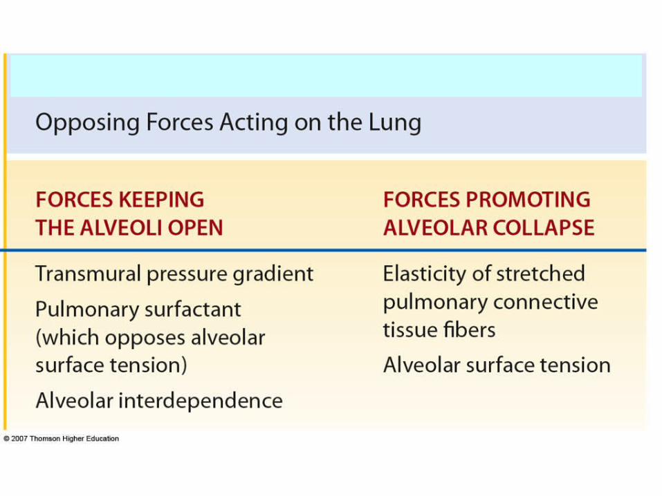
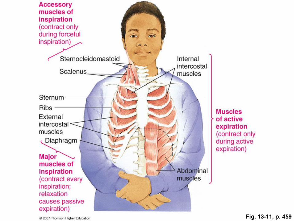
Fig. 13-11, p. 459
See Practical Class and Online Tutorial
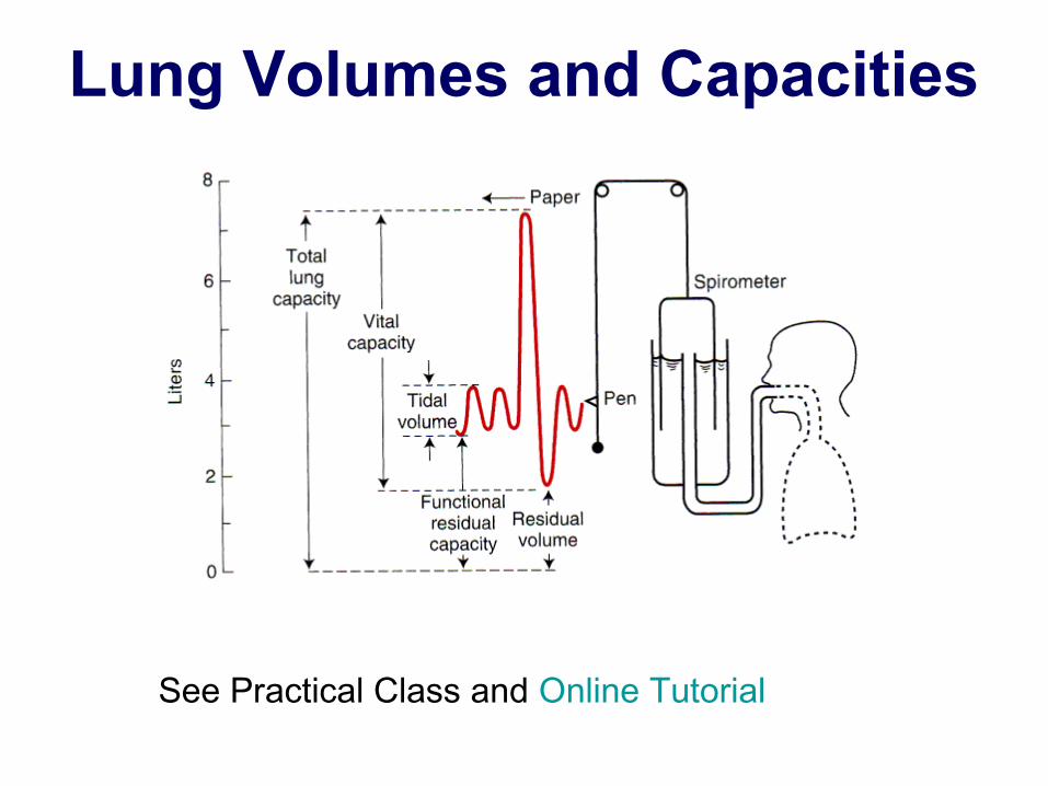
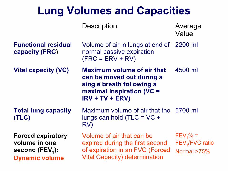
Lung Volumes and Capacities
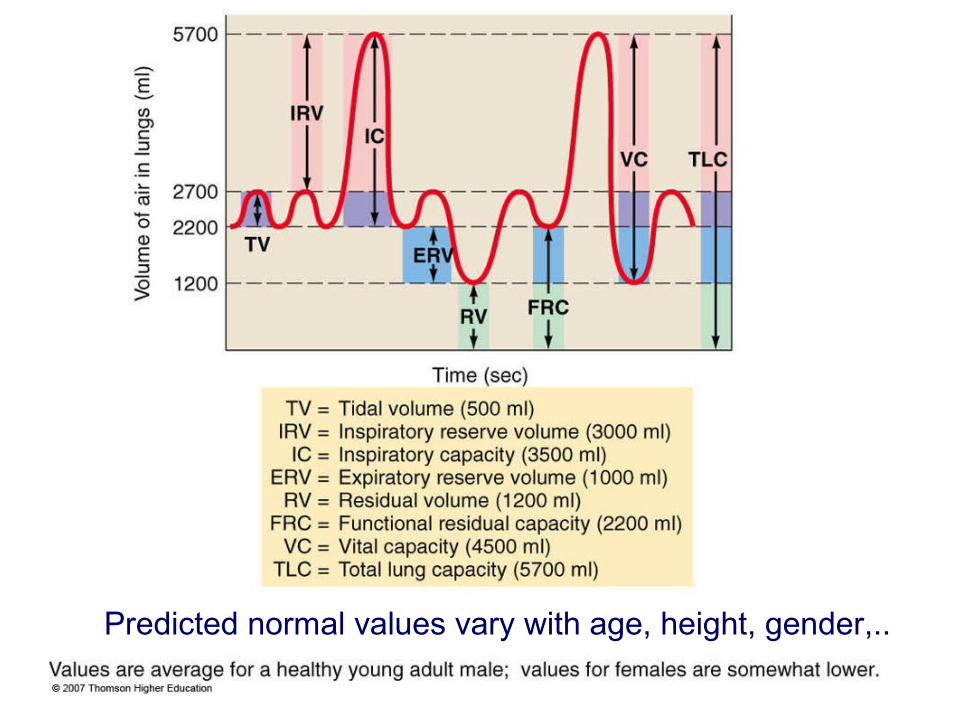
Predicted normal values vary with age, height, gender,..
Lung Volumes and CapacitiesDescription Average
Value
Tidal volume (TV)
Volume of air entering or leaving lungs during a single breath
500 ml
Inspiratory reserve volume (IRV)
Extra volume of air that can be maximally inspired over and above the typical resting tidal volume
3000 ml
Inspiratory capacity (IC)
Maximum volume of air that can be inspired at the end of a normal quiet expiration (IC =IRV + TV)
3500 ml
Expiratory reserve volume (ERV)
Extra volume of air that can be actively expired by maximal contraction beyond the normal volume of air after a resting tidal volume
1000 ml
Residual volume (RV)
Minimum volume of air remaining in the lungs even after a maximal expiration
1200 ml
Lung Volumes and CapacitiesDescription Average
Value
Functional residual capacity (FRC)
Volume of air in lungs at end of normal passive expiration (FRC = ERV + RV)
2200 ml
Vital capacity (VC) Maximum volume of air that can be moved out during a single breath following a maximal inspiration (VC = IRV + TV + ERV)
4500 ml
Total lung capacity (TLC)
Maximum volume of air that the lungs can hold (TLC = VC + RV)
5700 ml
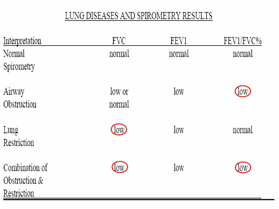
Forced expiratory volume in one second (FEV1): Dynamic volume
Volume of air that can be expired during the first second of expiration in an FVC (Forced Vital Capacity) determination
FEV1% = FEV1/FVC ratio
Normal >75%
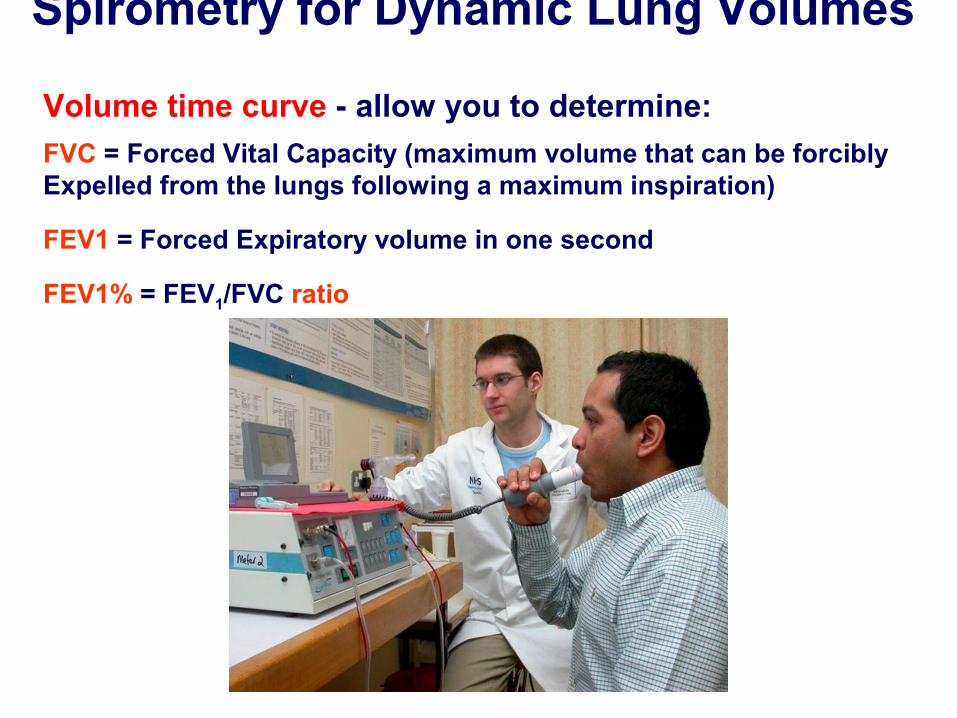
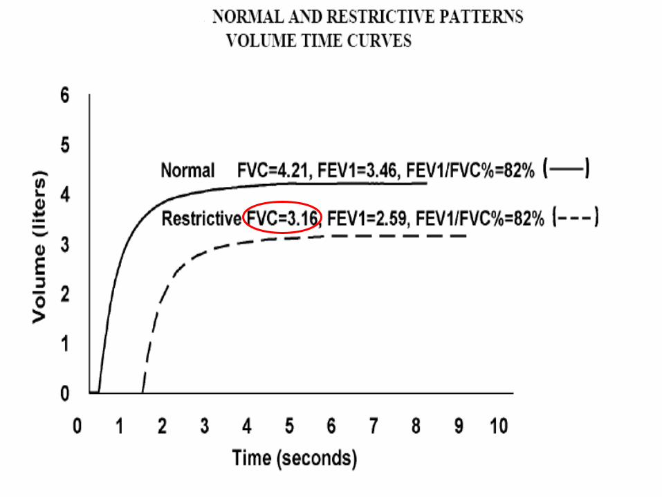
Volume time curve - allow you to determine:
FVC = Forced Vital Capacity (maximum volume that can be forcibly Expelled from the lungs following a maximum inspiration)
FEV1 = Forced Expiratory volume in one second
FEV1% = FEV1/FVC ratio
Spirometry for Dynamic Lung Volumes
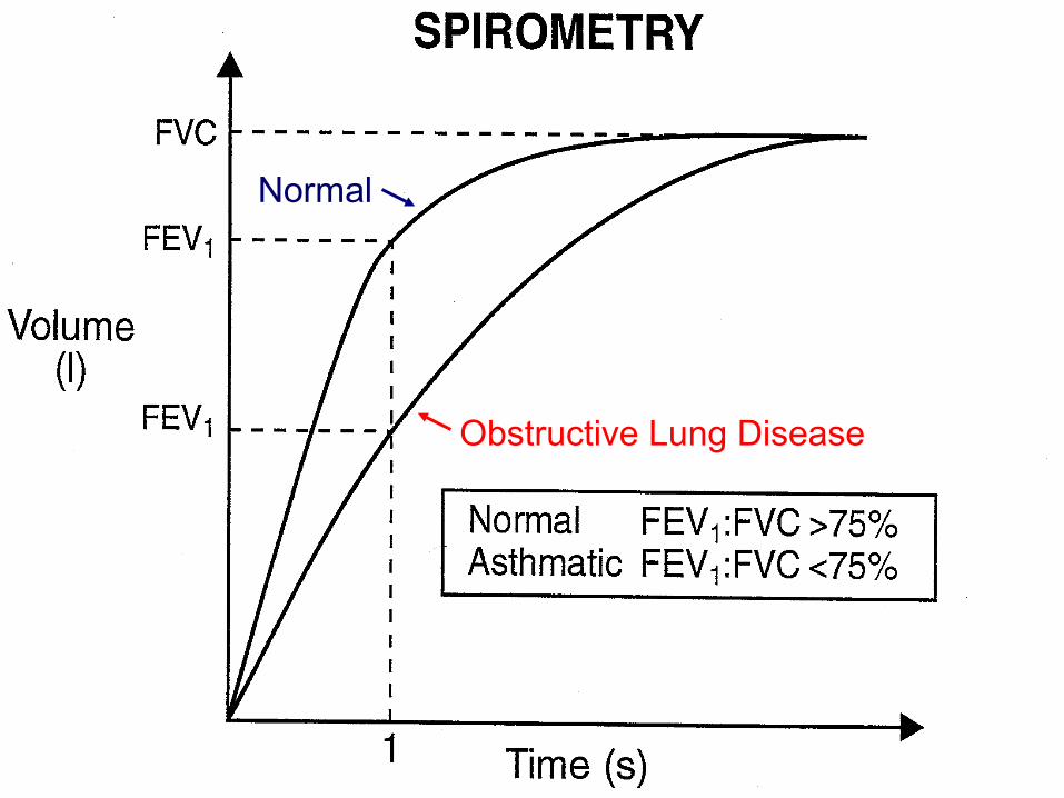
Normal
Obstructive Lung Disease
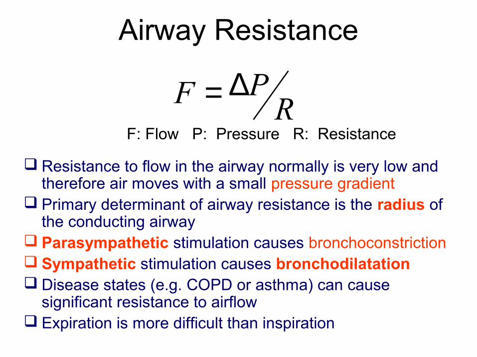
Airway Resistance
Resistance to flow in the airway normally is very low and therefore air moves with a small pressure gradient
Primary determinant of airway resistance is the radius of the conducting airway
Parasympathetic stimulation causes bronchoconstriction Sympathetic stimulation causes bronchodilatation Disease states (e.g. COPD or asthma) can cause
significant resistance to airflow Expiration is more difficult than inspiration
RPF ∆=
F: Flow P: Pressure R: Resistance
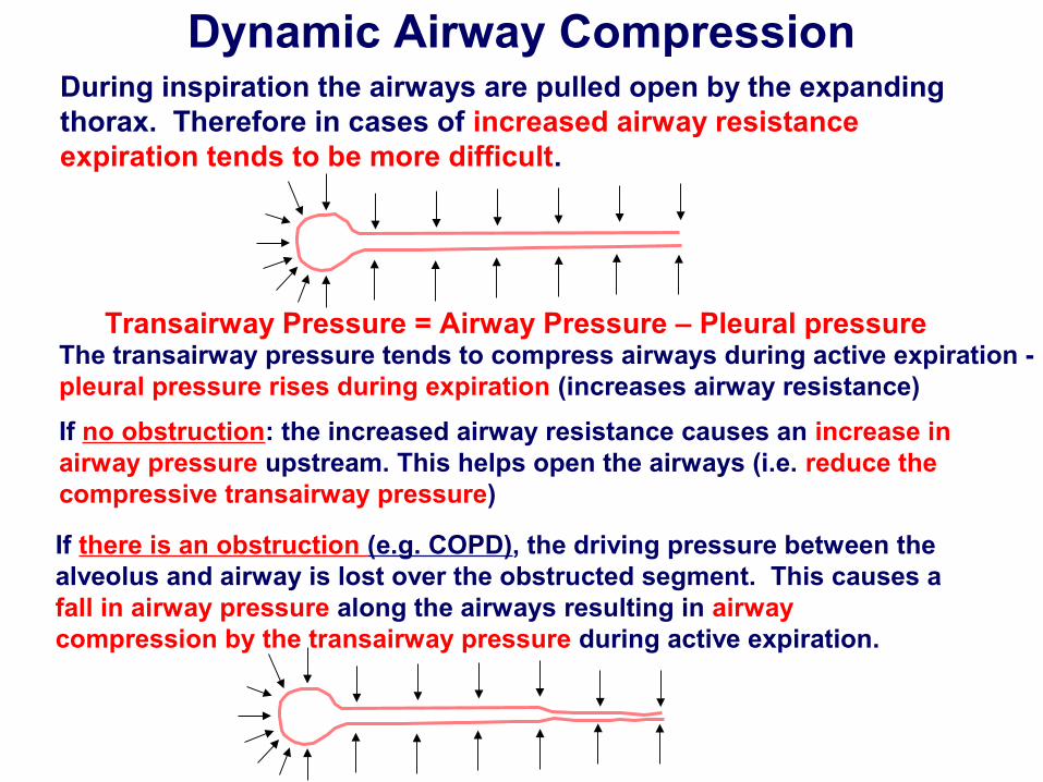
Dynamic Airway Compression
If there is an obstruction (e.g. COPD), the driving pressure between the alveolus and airway is lost over the obstructed segment. This causes a fall in airway pressure along the airways resulting in airway compression by the transairway pressure during active expiration.
During inspiration the airways are pulled open by the expanding thorax. Therefore in cases of increased airway resistance expiration tends to be more difficult.
The transairway pressure tends to compress airways during active expiration -pleural pressure rises during expiration (increases airway resistance)
If no obstruction: the increased airway resistance causes an increase in airway pressure upstream. This helps open the airways (i.e. reduce thecompressive transairway pressure)
Transairway Pressure = Airway Pressure – Pleural pressure
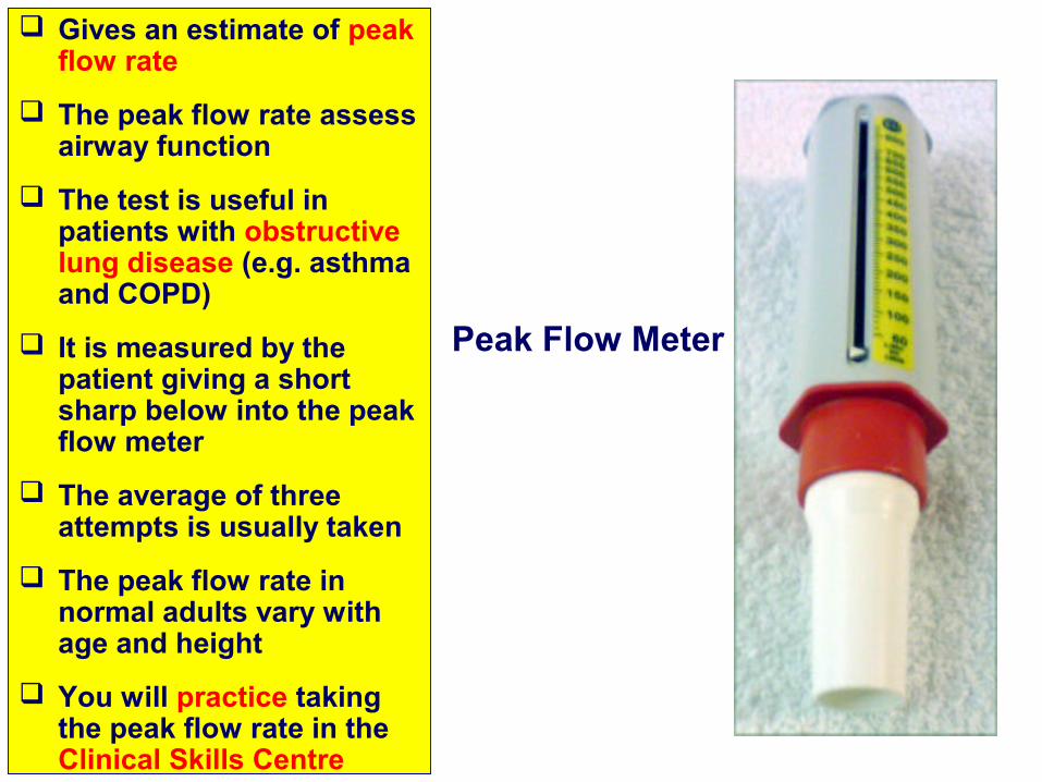
Gives an estimate of peak flow rate
The peak flow rate assess airway function
The test is useful in patients with obstructive lung disease (e.g. asthma and COPD)
It is measured by the patient giving a short sharp below into the peak flow meter
The average of three attempts is usually taken
The peak flow rate in normal adults vary with age and height
You will practice taking the peak flow rate in the Clinical Skills Centre
Peak Flow Meter
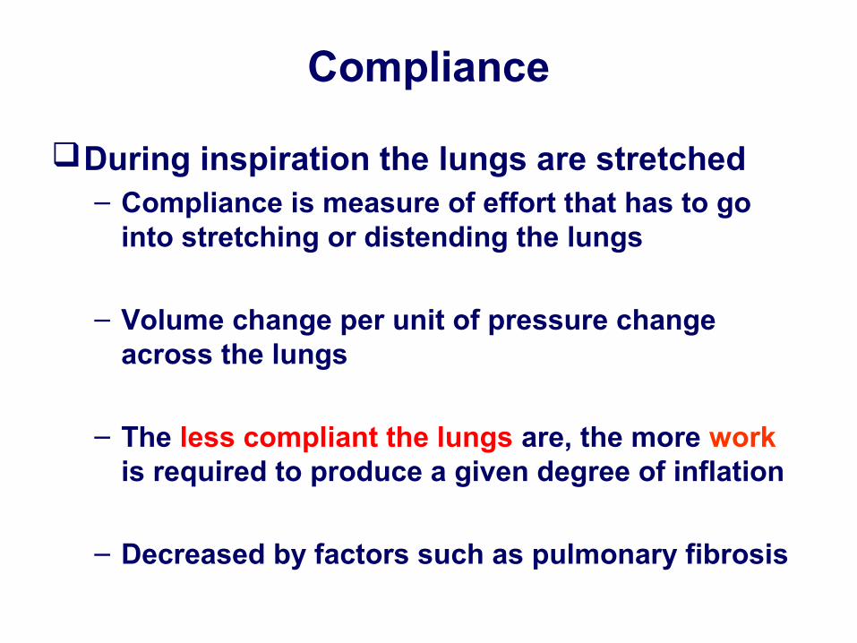
Compliance
During inspiration the lungs are stretched– Compliance is measure of effort that has to go
into stretching or distending the lungs
– Volume change per unit of pressure change across the lungs
– The less compliant the lungs are, the more work is required to produce a given degree of inflation
– Decreased by factors such as pulmonary fibrosis
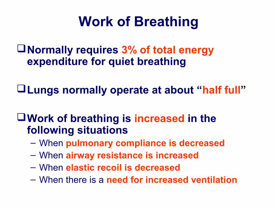
Work of Breathing
Normally requires 3% of total energy expenditure for quiet breathing
Lungs normally operate at about “half full”
Work of breathing is increased in the following situations– When pulmonary compliance is decreased– When airway resistance is increased– When elastic recoil is decreased– When there is a need for increased ventilation