Embed Size (px)
Citation preview
Dr Amit KumarAssistant ProfessorDGCN COVAS, Palampur
In order that changes in outline, position,and opacity be appreciated, it is essentialthat the radiologist be familiar with theradiologic appearance of normal structuresthat is, radiologic anatomy.
If one is unfamiliar with the normalappearance, one cannot appreciateaberrations from it.
Because almost any structure can be rotatedthrough 360°, it would be virtuallyimpossible to become familiar with all thepossible projections that could be producedfrom any given organ.
Consequently, standard projections of eachpart of the body are used. These usuallyconsist of two projections made at rightangles to one another so that a threedimensional impression is gained of thestructure under study. Agreed terms areused to describe the standard projections.
The terminology used here is thatsuggested by the Nomenclature Committeeof the American College of VeterinaryRadiology.
The committee recommended thatveterinary anatomic directional termsshould be those listed in the NominaAnatomica Veterinaria.
Radiographic projections are described bythe direction in which the central ray of theprimary beam penetrates the body part ofinterest-from the point of entrance to thepoint of exit.

The meanings to be ascribed to the different terms areas follows:
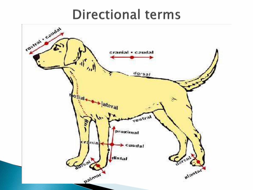
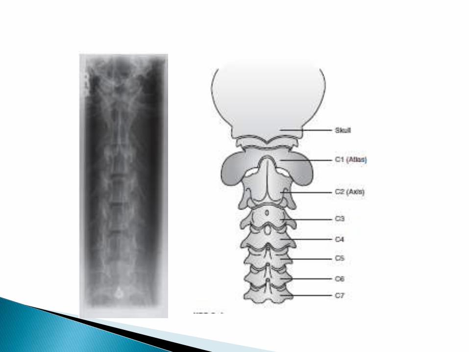
Dorsal: The upper aspect of the head, neck, trunk,tail, and cranial (anterior) aspects of the limbs fromthe antebrachiocarpal (radiocarpal) and tarsocruralarticulations distally (downward). Dorsal also meanstoward the back or vertebrae.
Ventral: The lower aspect of the head, neck, trunk,and tail. Ventral also means toward the lower aspectof the animal.
Cranial: A directional term that describes parts of theneck, trunk, and tail positioned toward the head fromany given point. Cranial also describes those aspectsof the limb above the antebrachiocarpal andtarsocrural joints that face toward the head.
Rostral. Describes parts of the head positionedtoward the nares from any given point on the head.
Caudal: A directional term that describes parts of the head,neck, and trunk positioned toward the tail from any givenpoint. Caudal also describes those aspects of the limbsabove the antebrachiocarpal and tarsocrural articulationsthat face toward the tail.
Palmar: This term is used instead of caudal when describingthe forelimb from the antebrachiocarpal articulation distally.
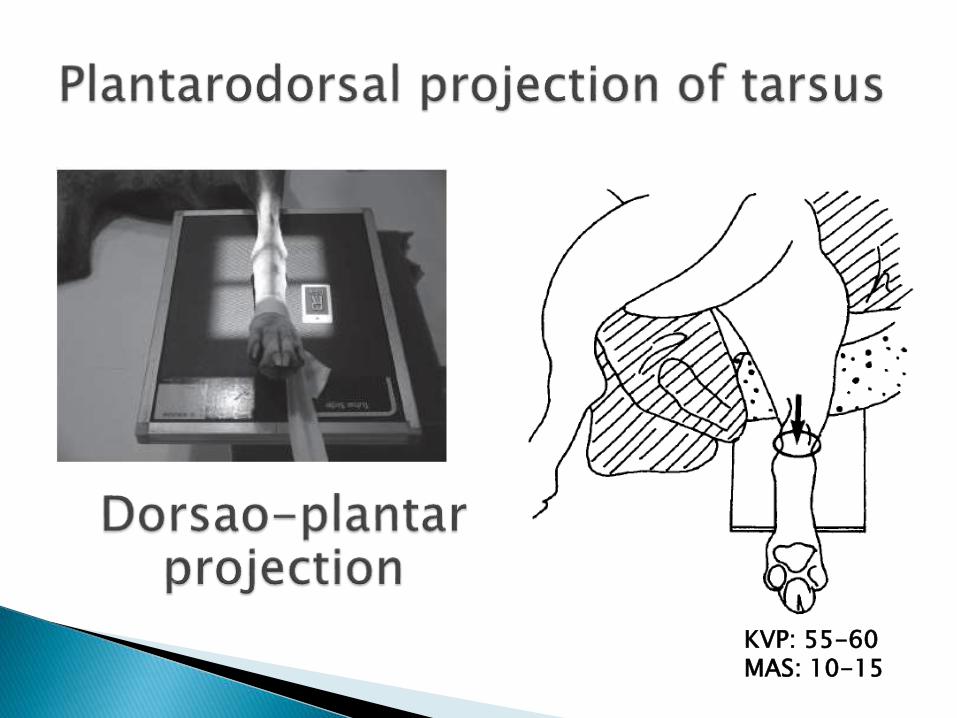
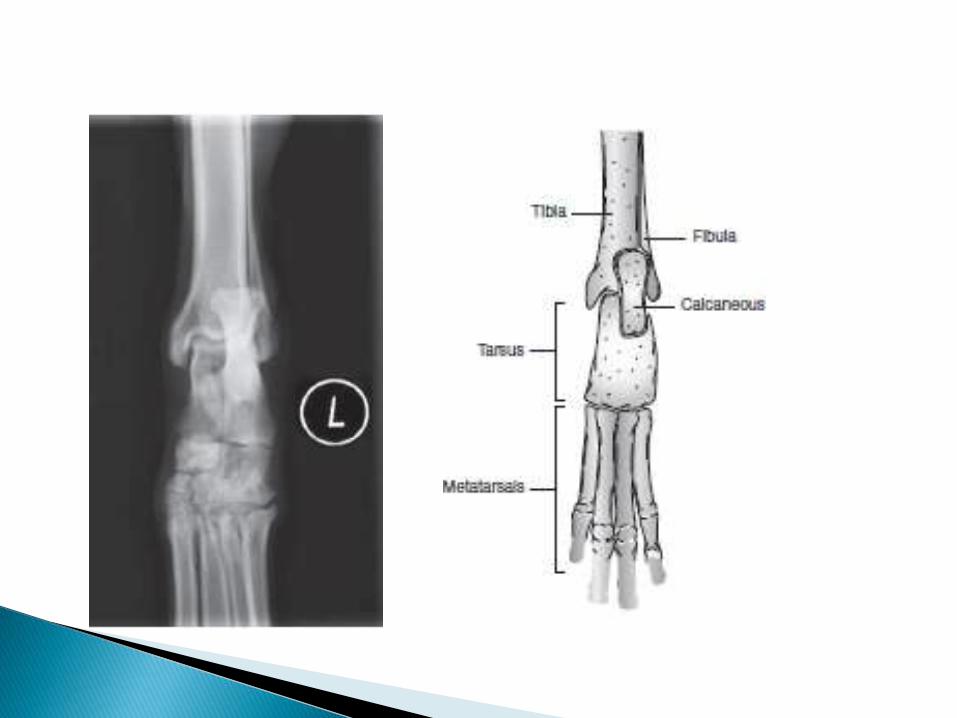
Plantar: This term is used instead of caudal when describingthe hindlimb from the tarsocrural articulation distally.
Proximal: Describes nearness to the point of origin of astructure.
Distal: Describes remoteness (farther away) from the pointof origin of a structure.
Superior and Inferior: These terms are used to describe theupper and lower dental arcades.
Recumbent: The animal is lying down when the radiographis made. Most radiographs of the dog and cat are made withthe animal in the recumbent position, and this positionshould be presumed unless the contrary is stated. The termdecubitus is used when a horizontal beam is employed.
The direction of the x-ray beam is described from itspoint of entry into the body to its point of exit.
For example, a right-left lateral recumbent projectionmeans that the animal is lying on its left side, and thex-ray beam enters the body through the right side andexits through the left side.
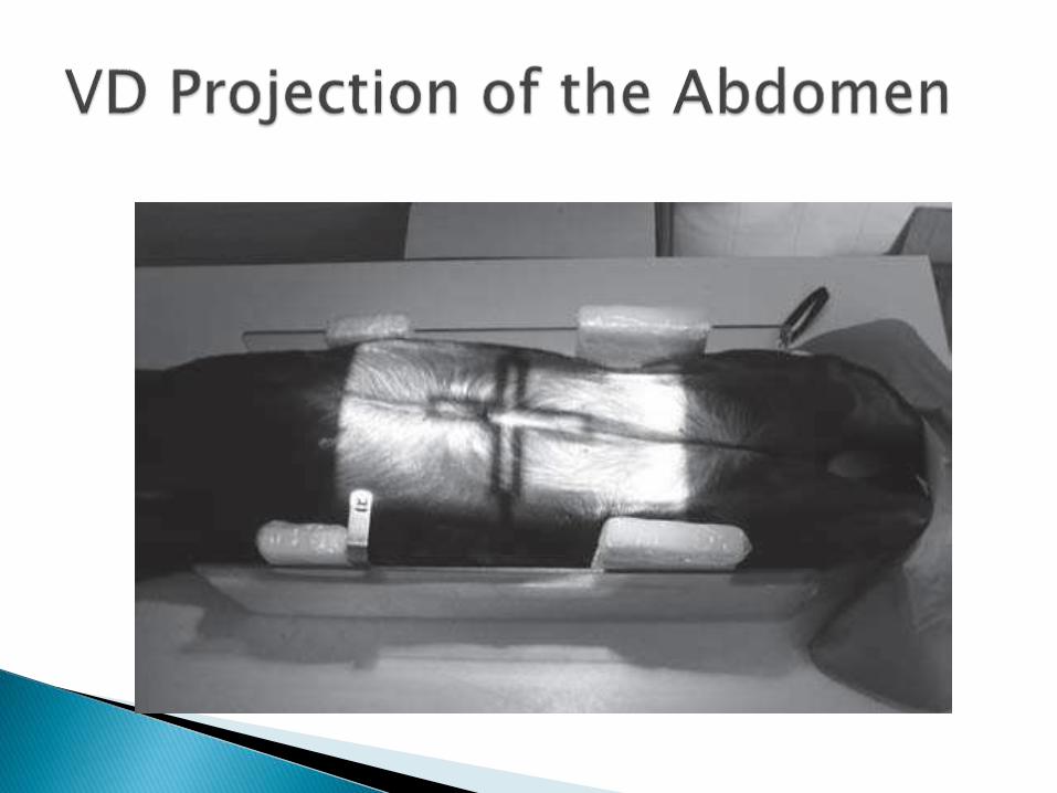
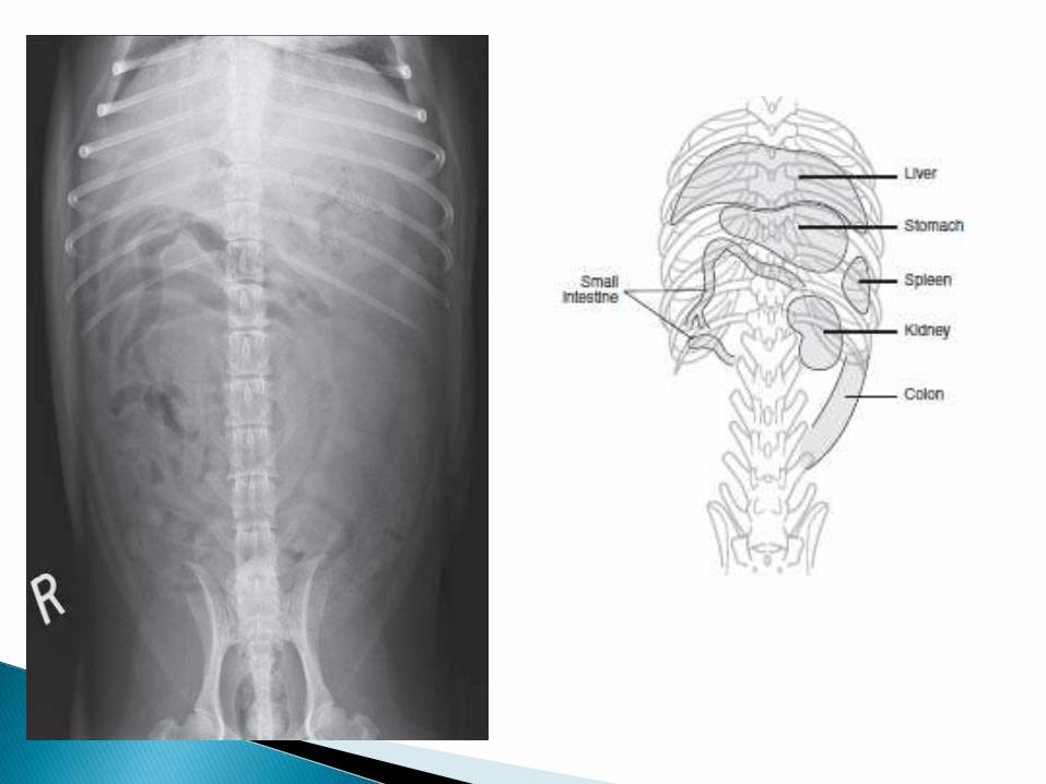
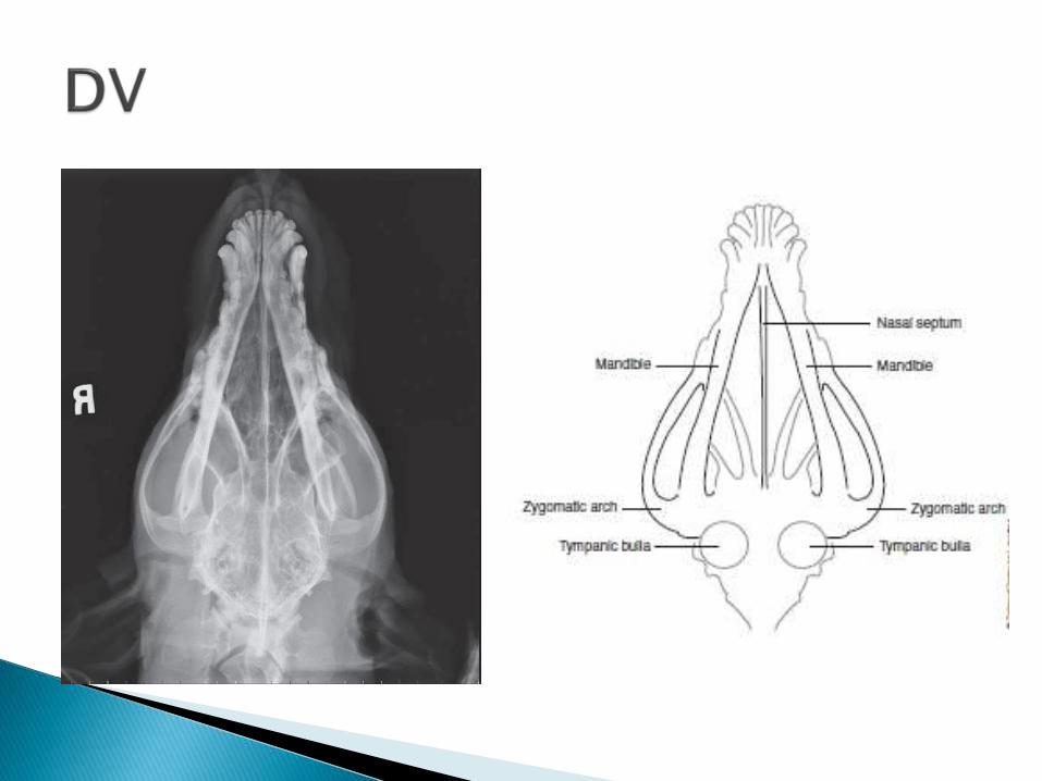
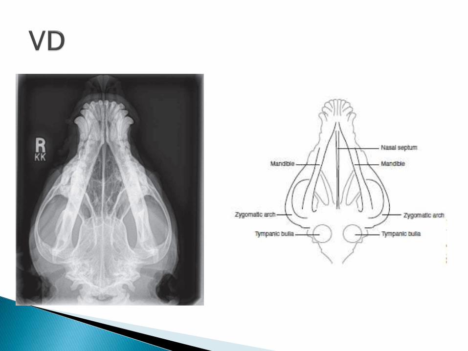
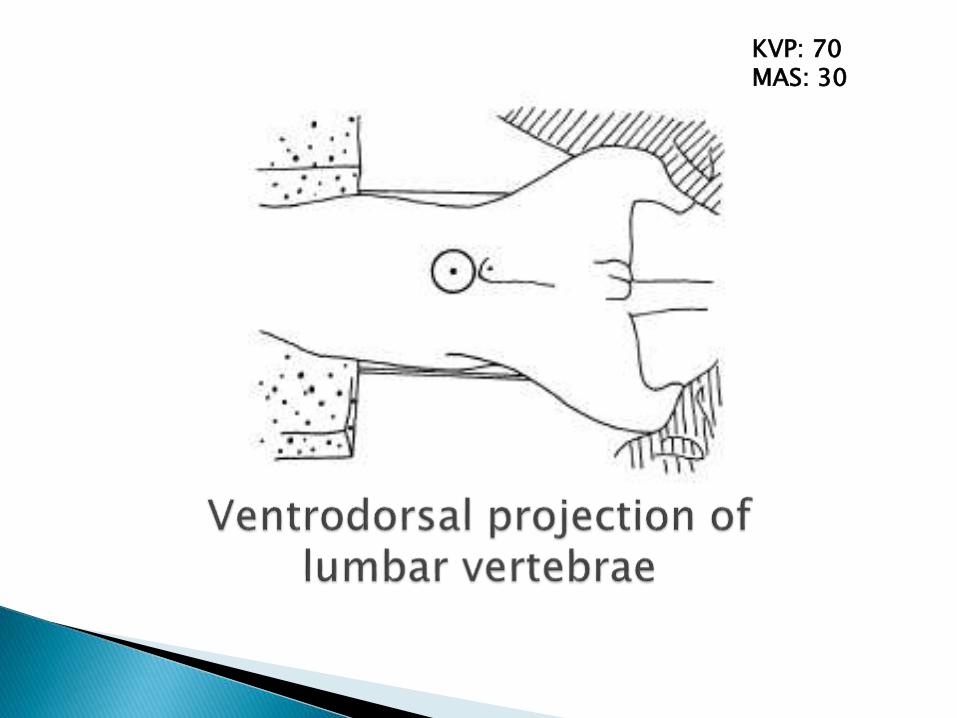
A ventrodorsal (VD) projection means that the x-raybeam enters the body ventrally and exits dorsally toreach the film. A dorsoventral (DV) projection indicatesthe opposite.
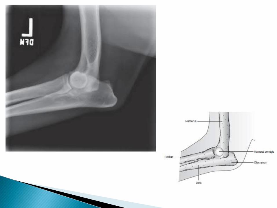
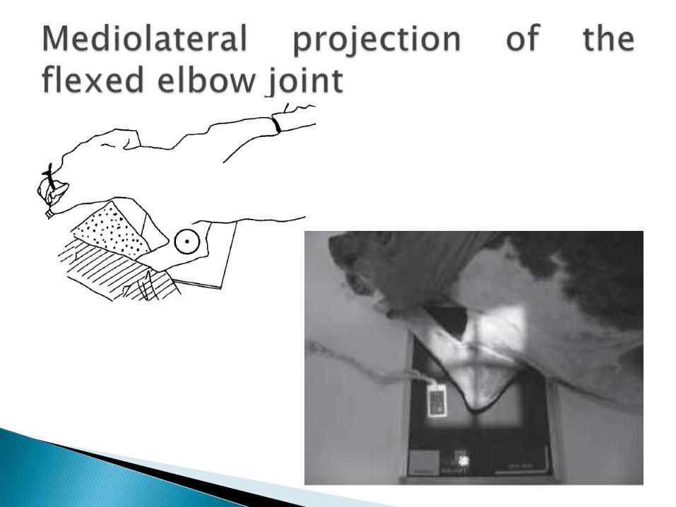
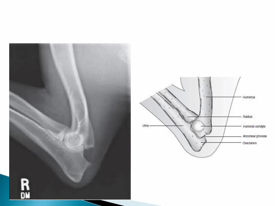
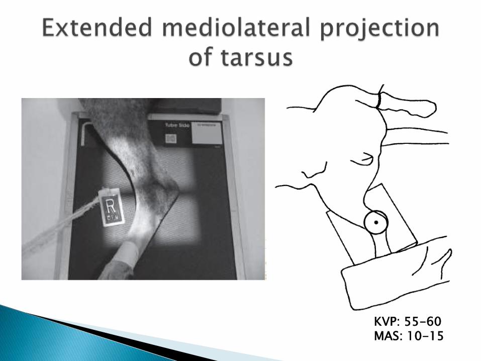
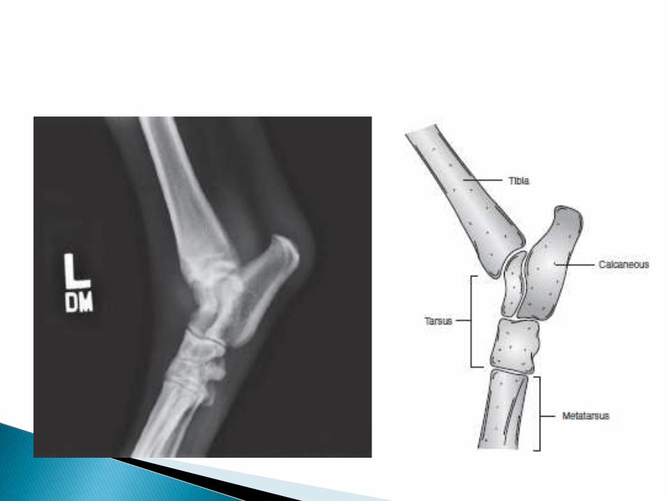
Mediolateral means the x-ray beam enters a limb fromthe medial side and exits on the lateral side. Most so-called lateral radiographs of the limbs are taken in amediolateral direction. In a lateromedial projection, thex-ray beam enters a limb from the lateral side andexits on the medial side.
Appropriate safety measures should be adoptedirrespective of beam direction, and special care isneeded when horizontal beams are in use.
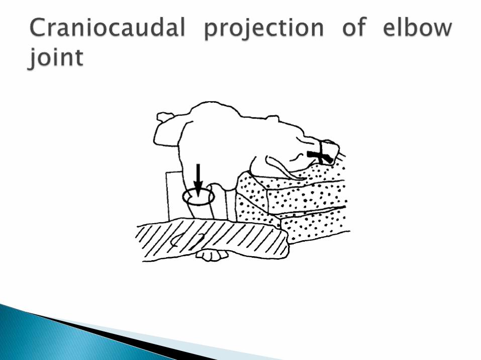
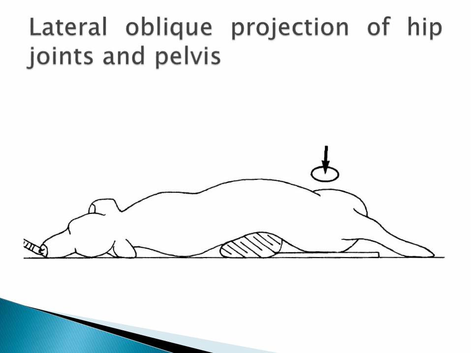
Standard projections are taken at right angles to oneanother and usually are made in the routineexamination of a part of the body. The most commonare the dorsoventral, ventrodorsal, lateral,mediolateral, craniocaudal, dorsopalmar, anddorsoplantar. An oblique projection is made at anangle, somewhere between the standard projections.In the case of oblique projections, in addition tostating the anatomic points of entry and exit of thex-ray beam, the angle of obliquity may be given. Thisinformation enables studies to be repeated withaccuracy. Thus, L50D-RVO is read as left 50° dorsal-right ventral oblique. It means that an oblique studywas made with the beam entering the body on theleft side dorsally at an angle of 50° toward the backand exiting on the right side ventrally.
Lesion-orientated studies are sometimes employedusing tangential (skyline) or nonstandard projections.A lesion-orientated oblique projection is one thatprofiles a lesion.
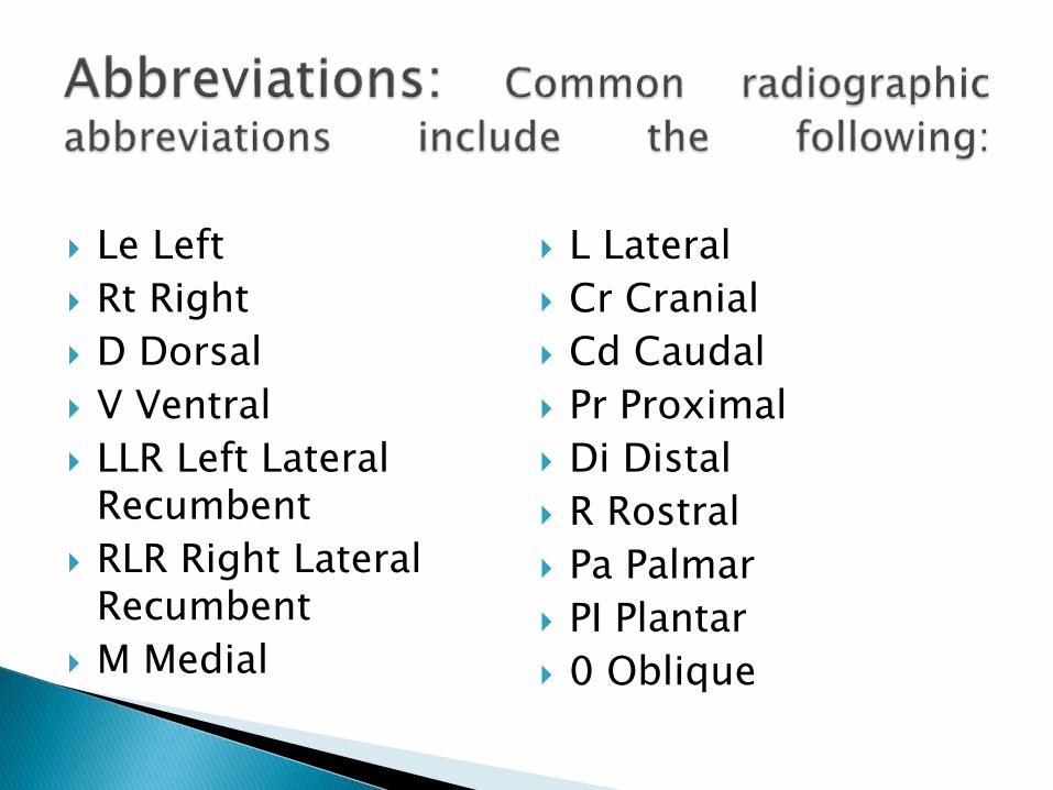
Le Left
Rt Right
D Dorsal
V Ventral
LLR Left Lateral Recumbent
RLR Right Lateral Recumbent
M Medial
L Lateral
Cr Cranial
Cd Caudal
Pr Proximal
Di Distal
R Rostral
Pa Palmar
PI Plantar
0 Oblique

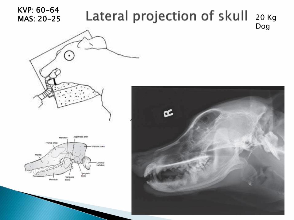
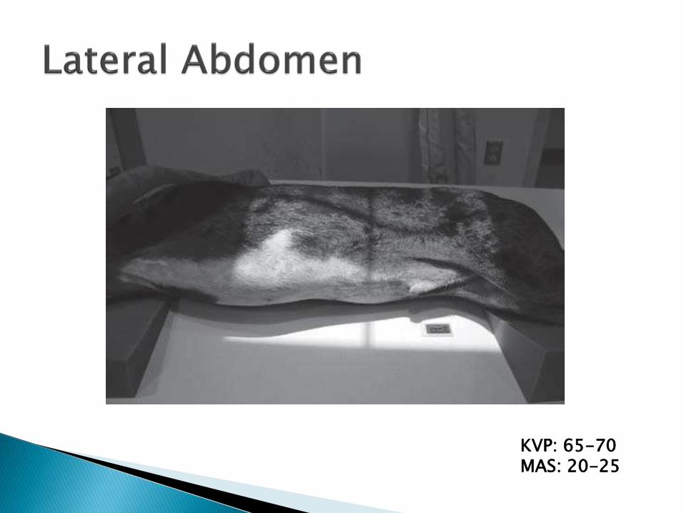
KVP: 60-64 MAS: 20-25 20 Kg
Dog
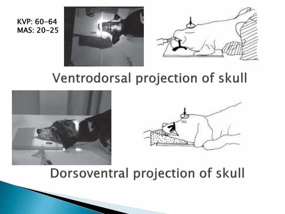
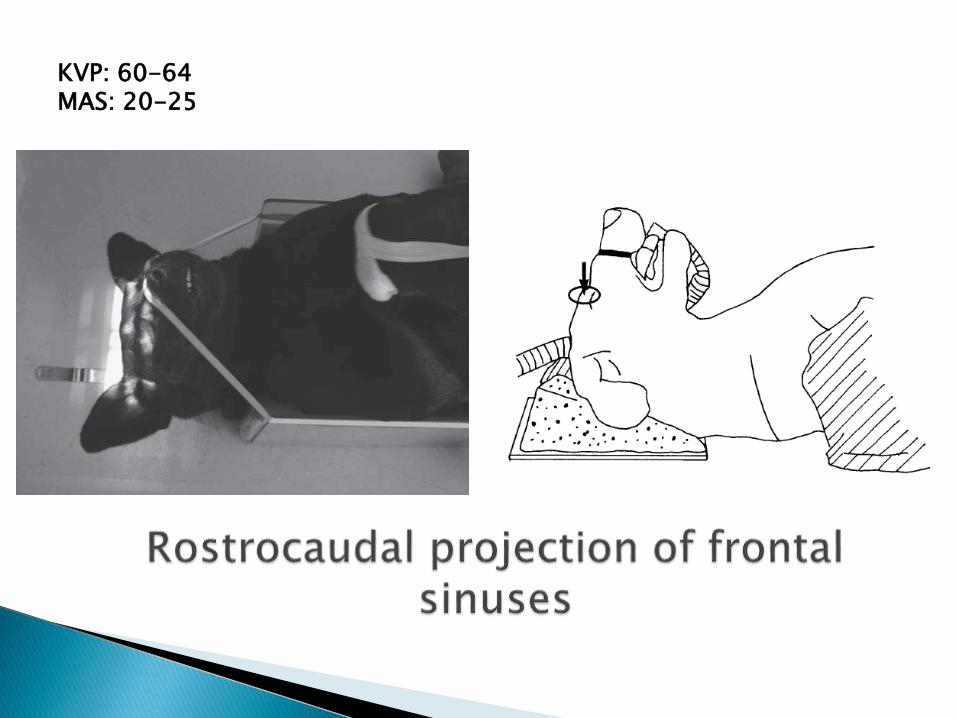
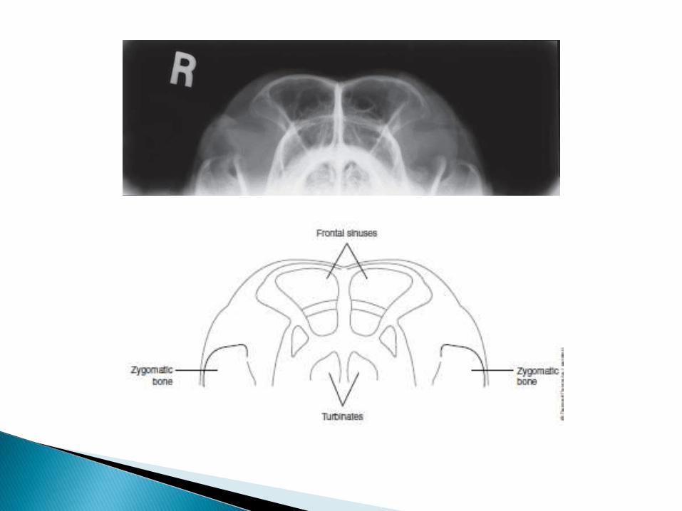
KVP: 60-64 MAS: 20-25
KVP: 60-64 MAS: 20-25
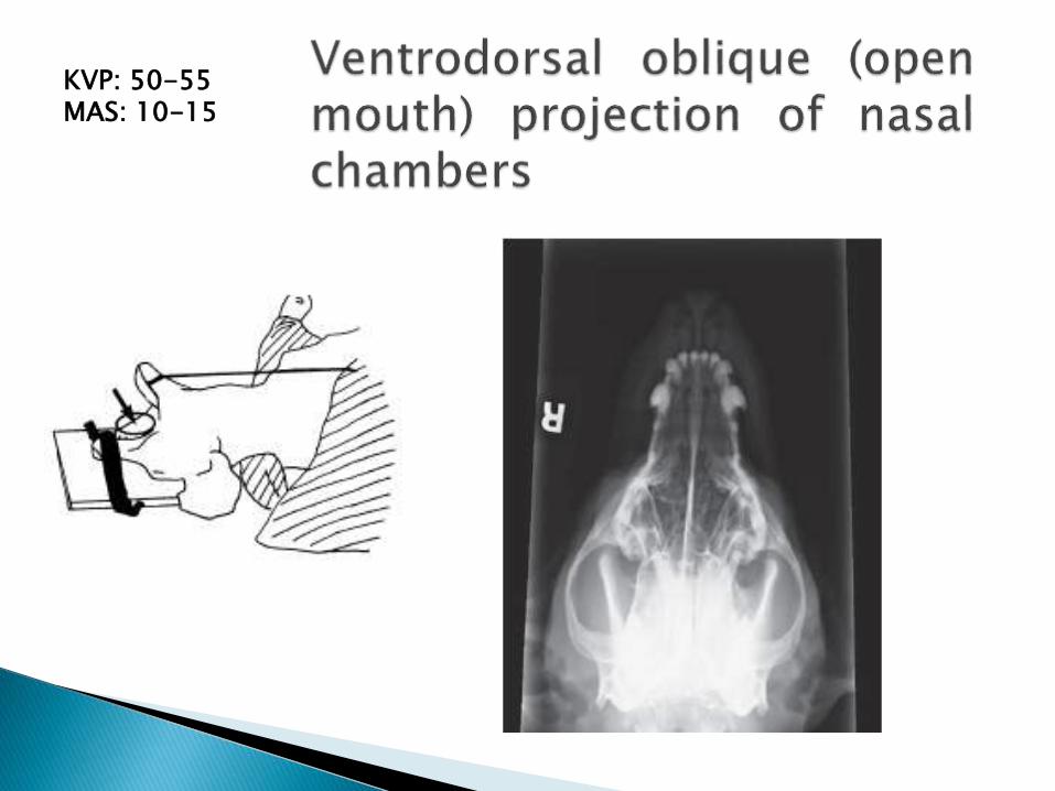
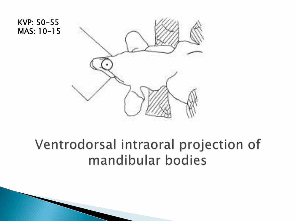
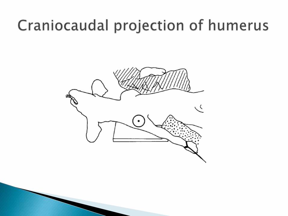
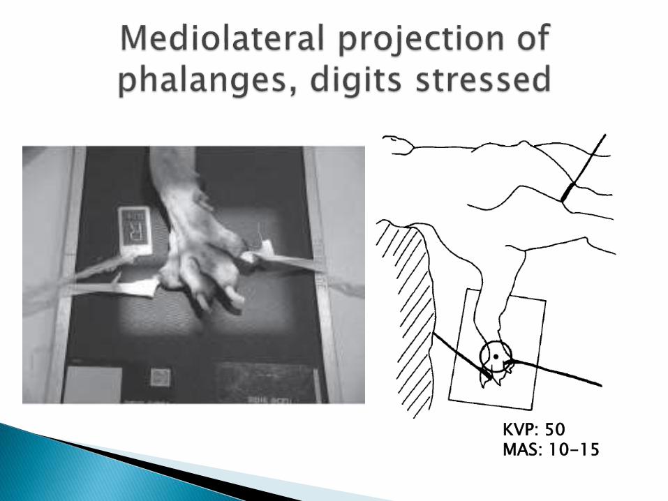
KVP: 50-55 MAS: 10-15
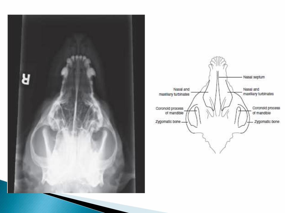

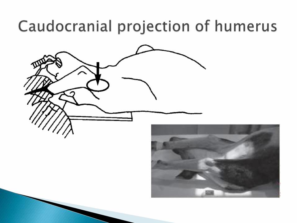
KVP: 50-55 MAS: 10-15
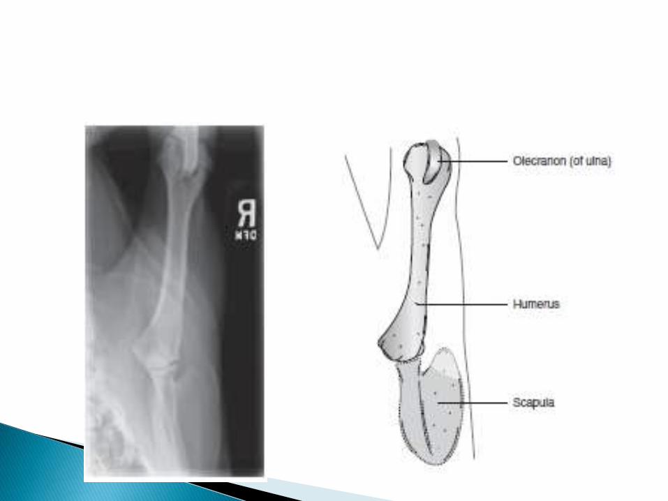
KVP: 50-55 MAS: 10-15
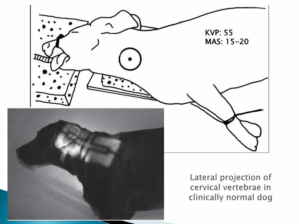
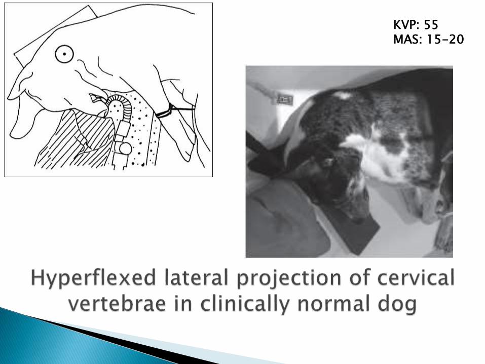
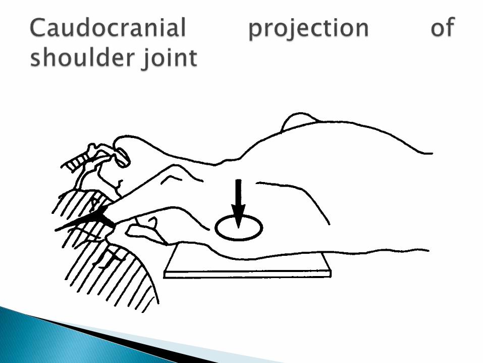
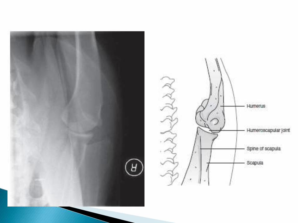
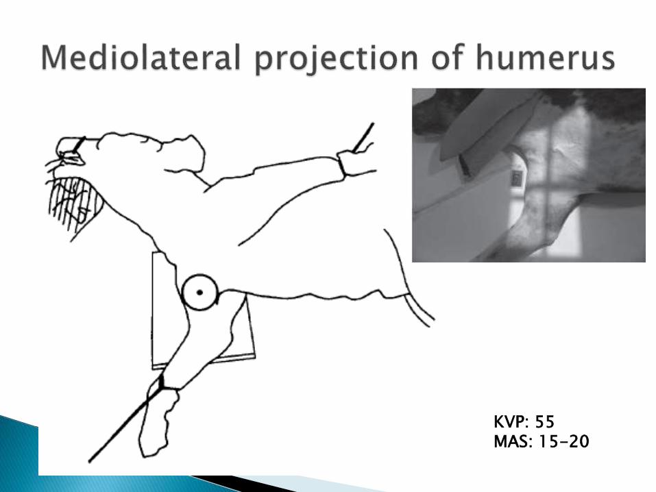
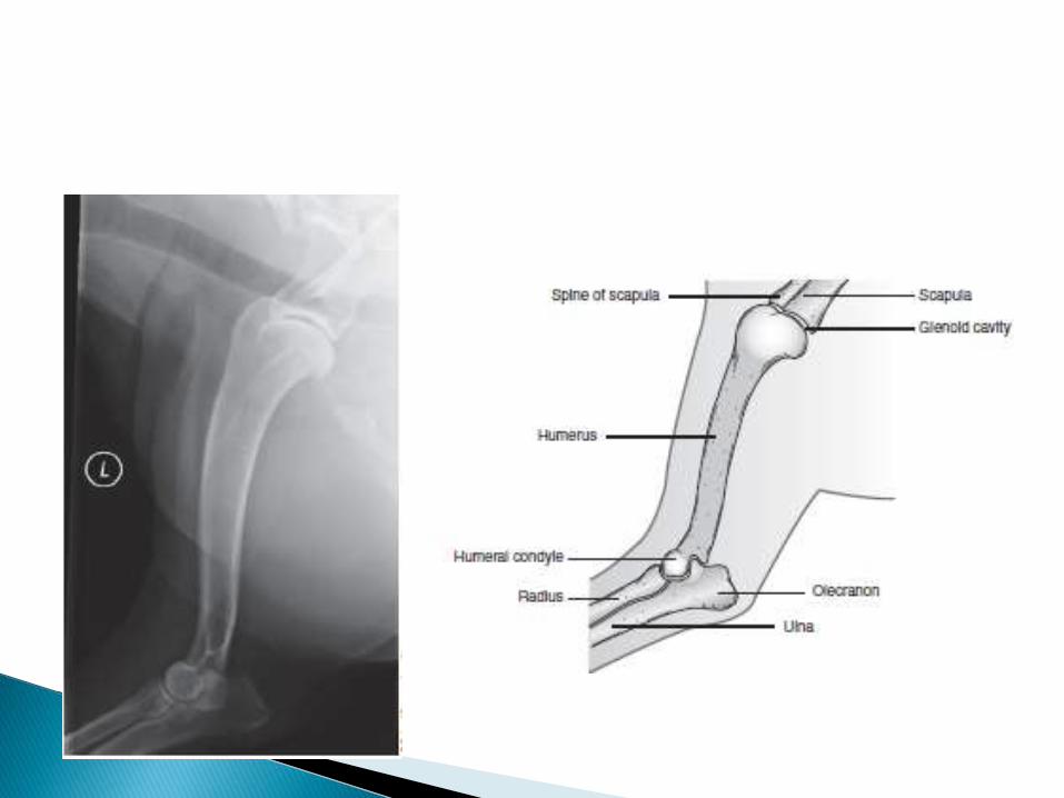
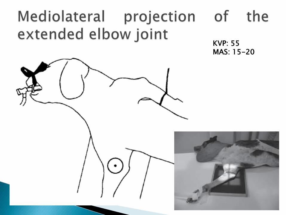
KVP: 55MAS: 15-20

KVP: 55MAS: 15-20
KVP: 55MAS: 15-20
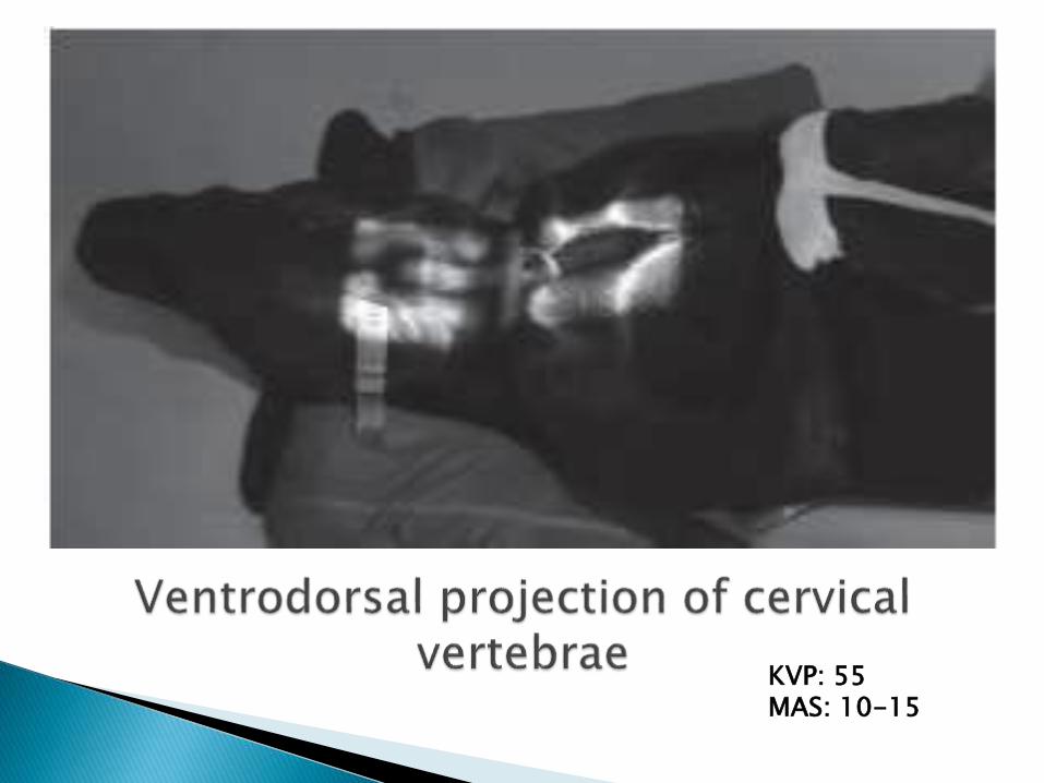
KVP: 55MAS: 10-15
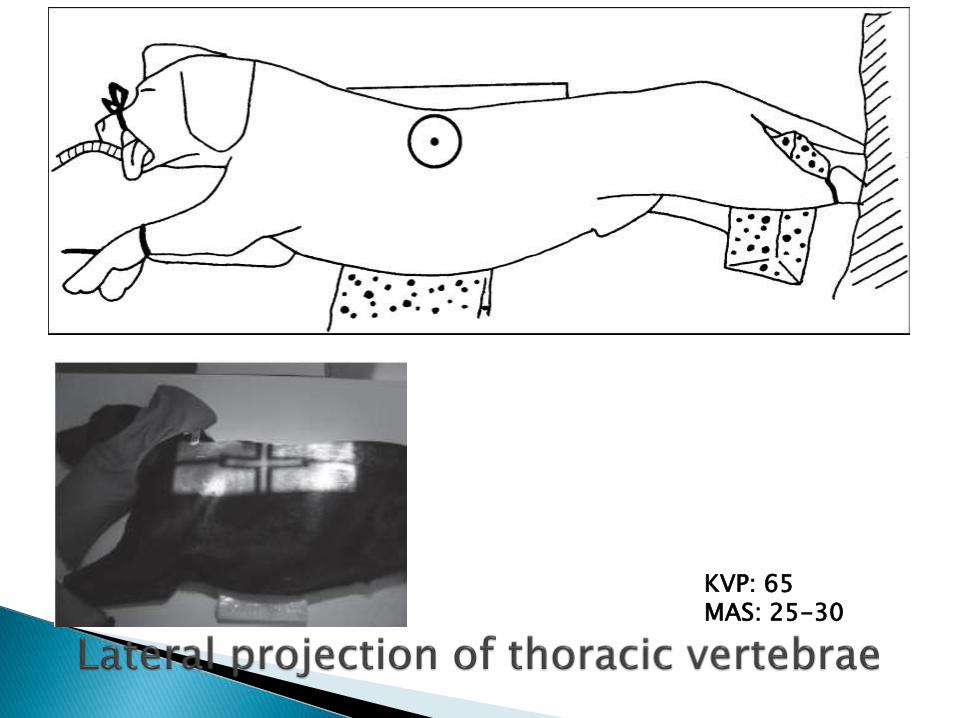
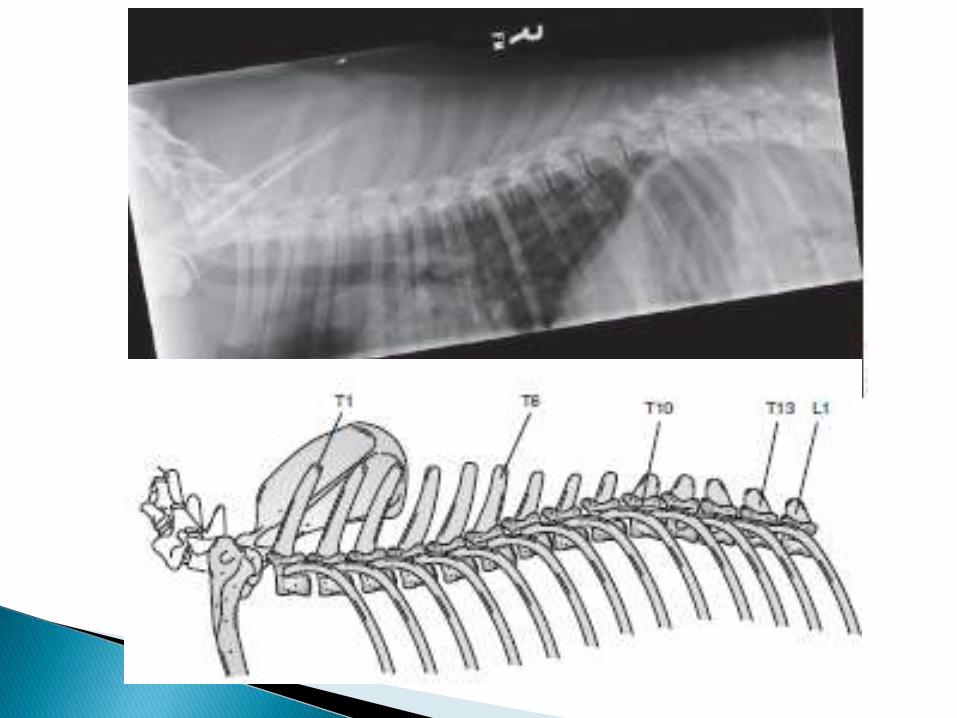
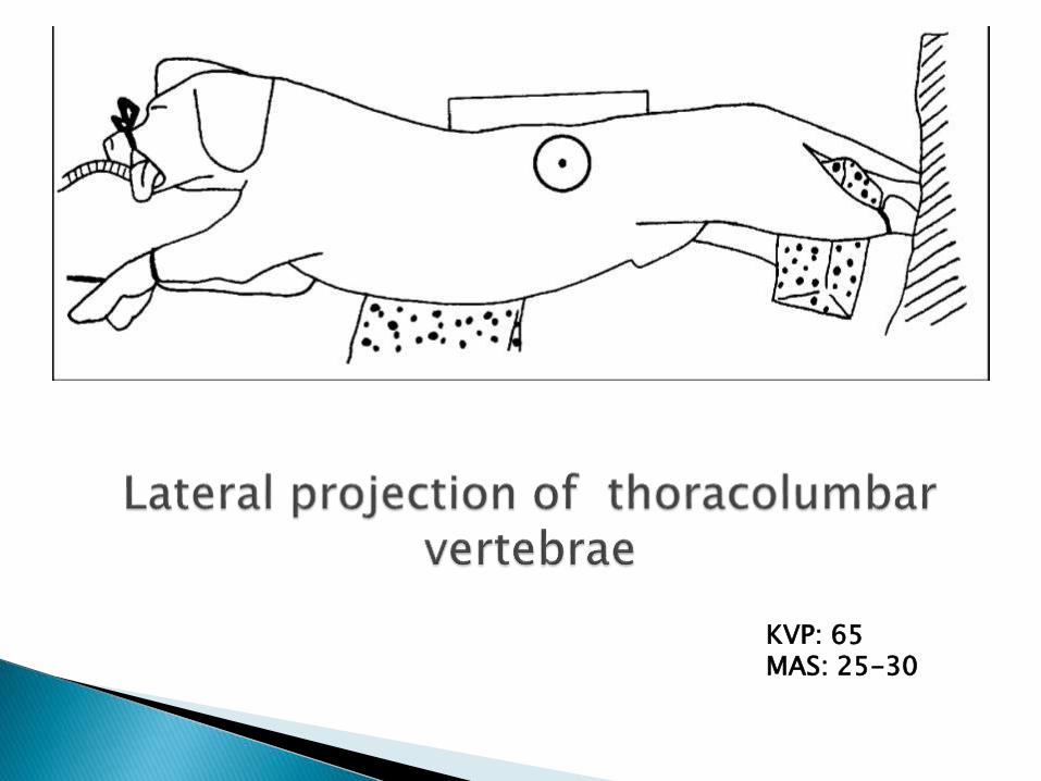
KVP: 65MAS: 25-30

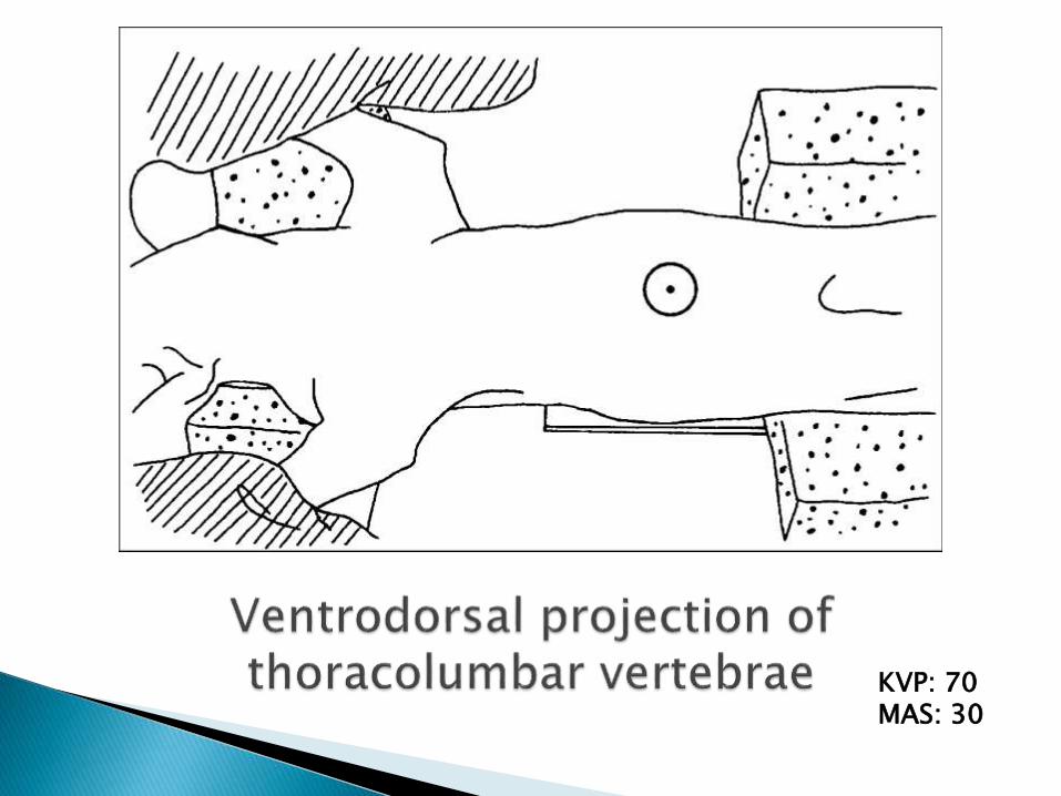
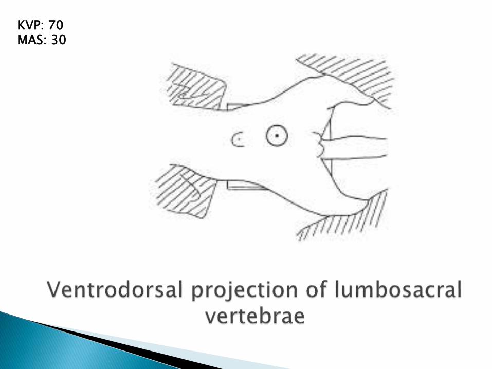
KVP: 70MAS: 30
KVP: 65MAS: 25-30
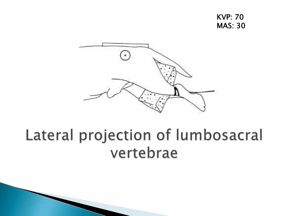
KVP: 70MAS: 30
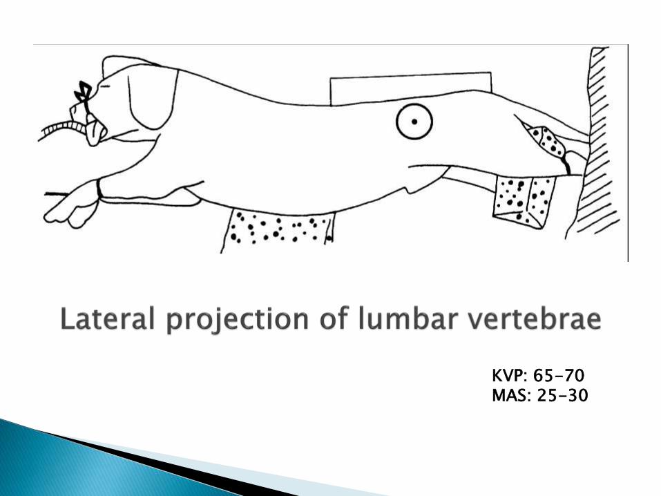
KVP: 65-70MAS: 25-30
KVP: 70MAS: 30
KVP: 70MAS: 30
KVP: 70MAS: 30

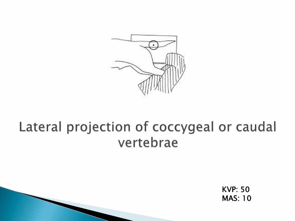
KVP: 50MAS: 10

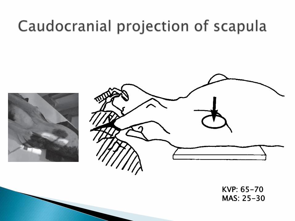
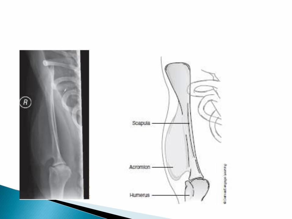
KVP: 65-70MAS: 25-30
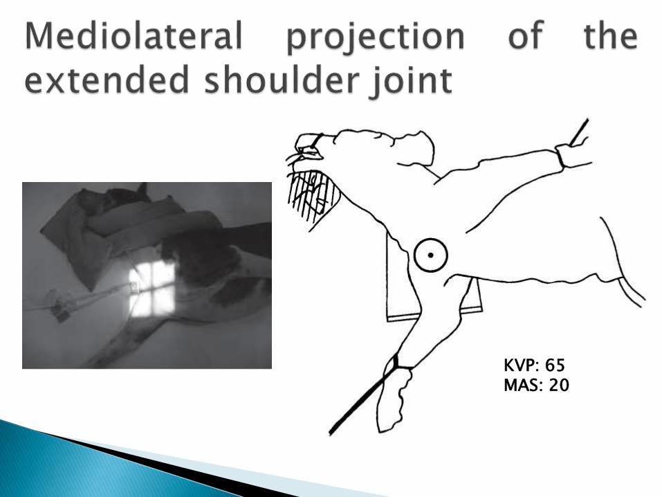
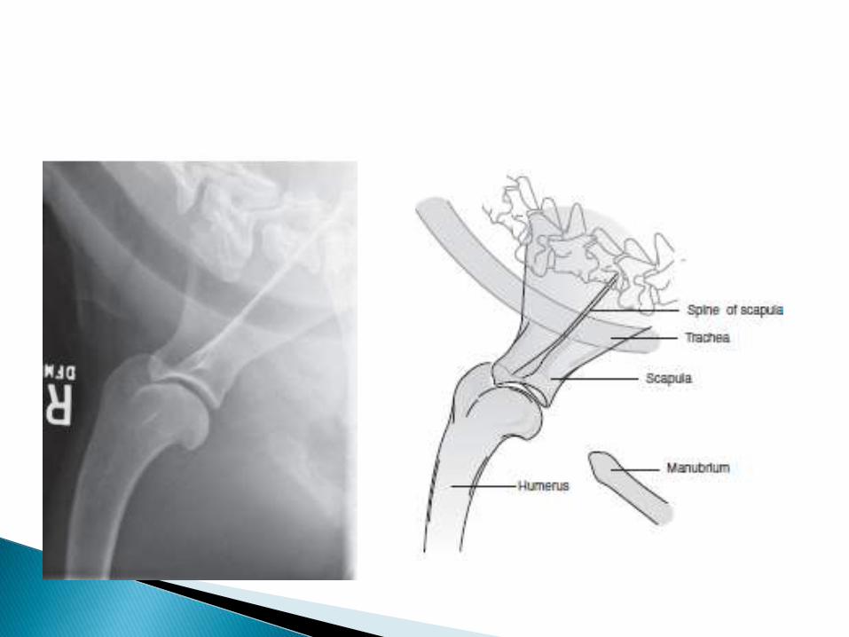
KVP: 65MAS: 20
KVP: 55MAS: 15-20
KVP: 55MAS: 15-20
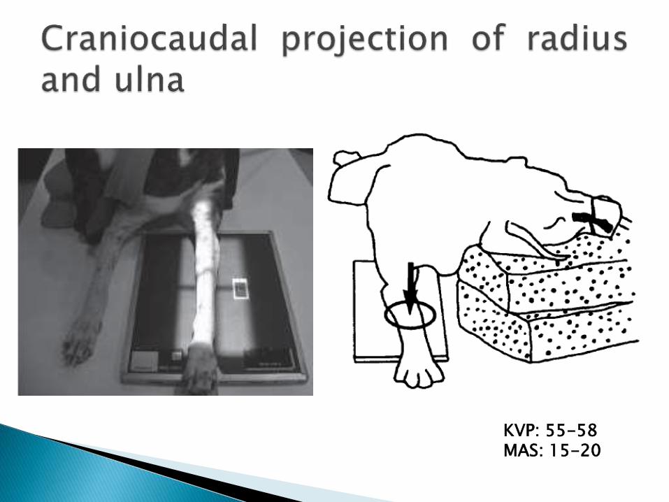
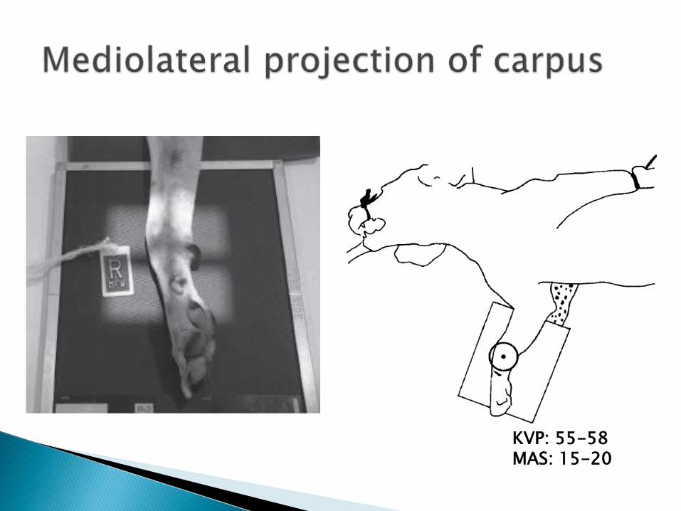
KVP: 55-58MAS: 15-20
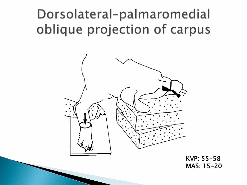
KVP: 55-58MAS: 15-20
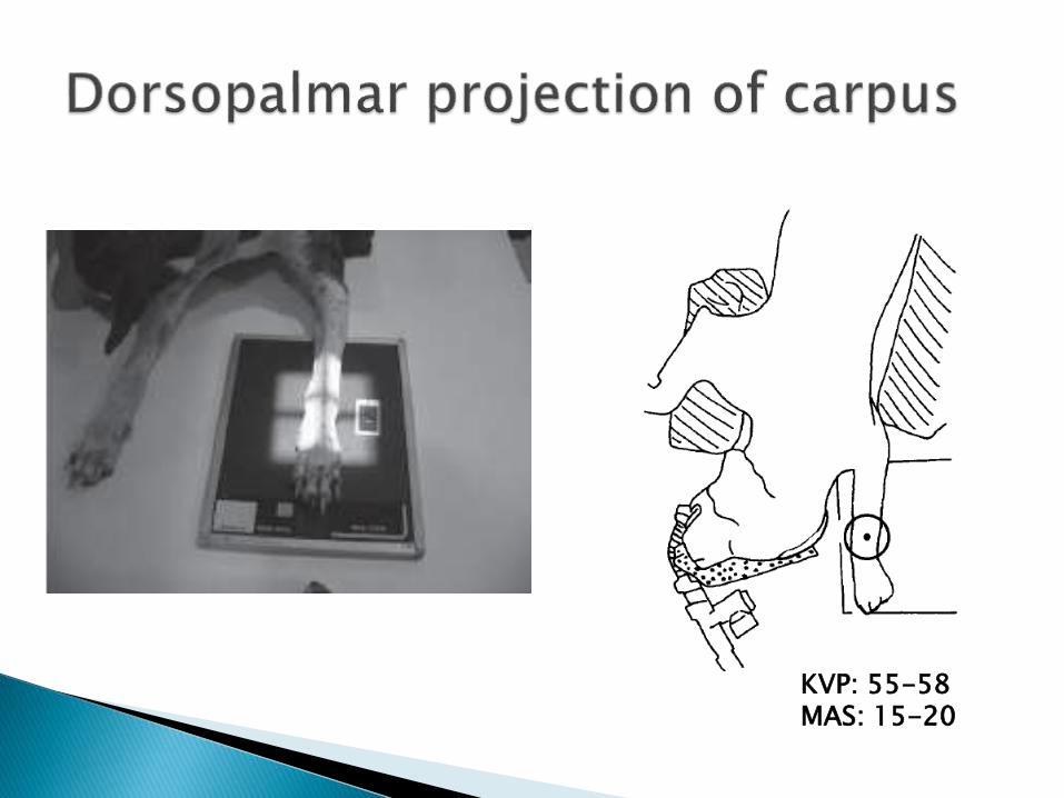
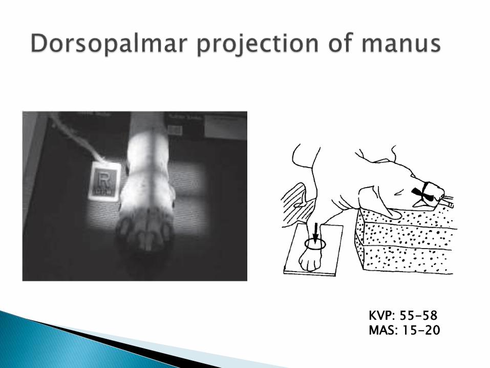
KVP: 55-58MAS: 15-20
KVP: 55-58MAS: 15-20

KVP: 55-58MAS: 15-20
KVP: 55-58MAS: 15-20

KVP: 55-58MAS: 15-20
KVP: 50MAS: 10-15
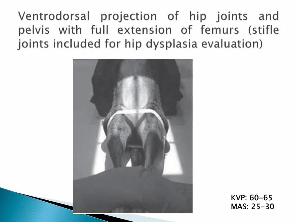
KVP: 60-65MAS: 25-30

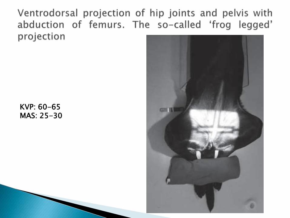
KVP: 60-65MAS: 25-30
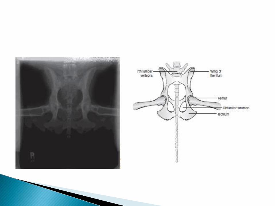
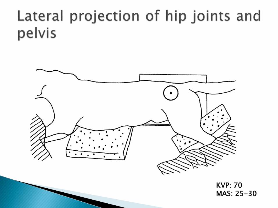
KVP: 70MAS: 25-30
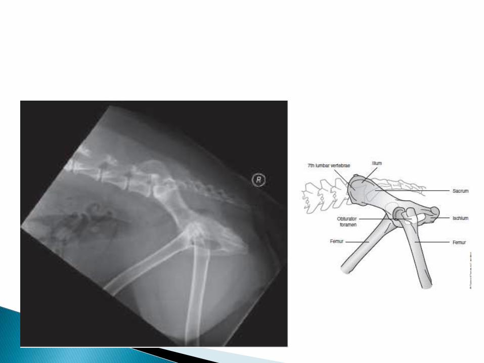
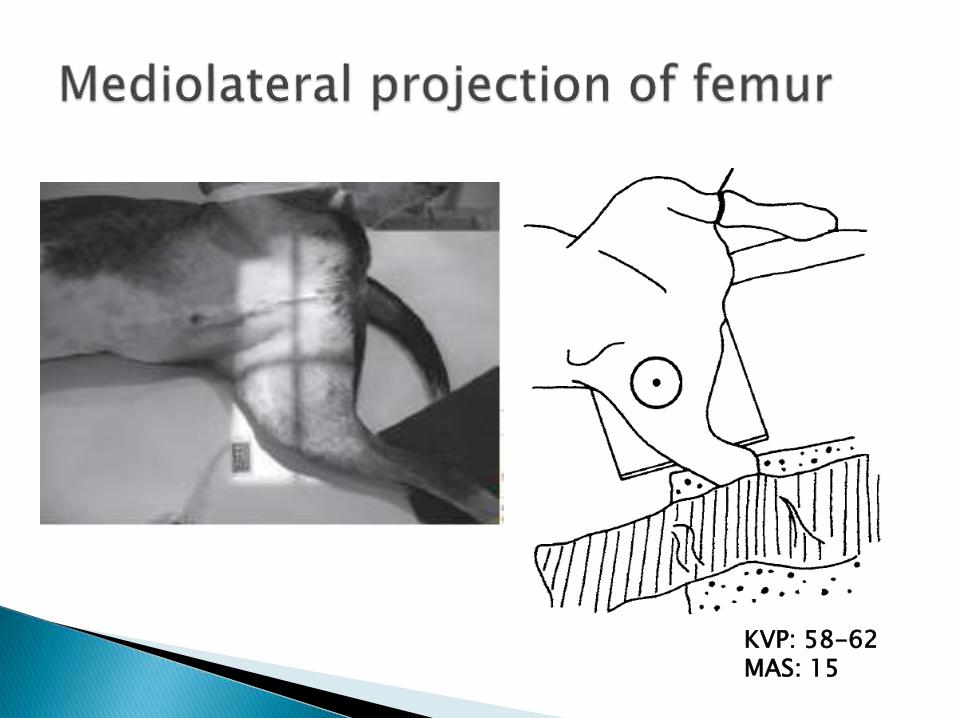
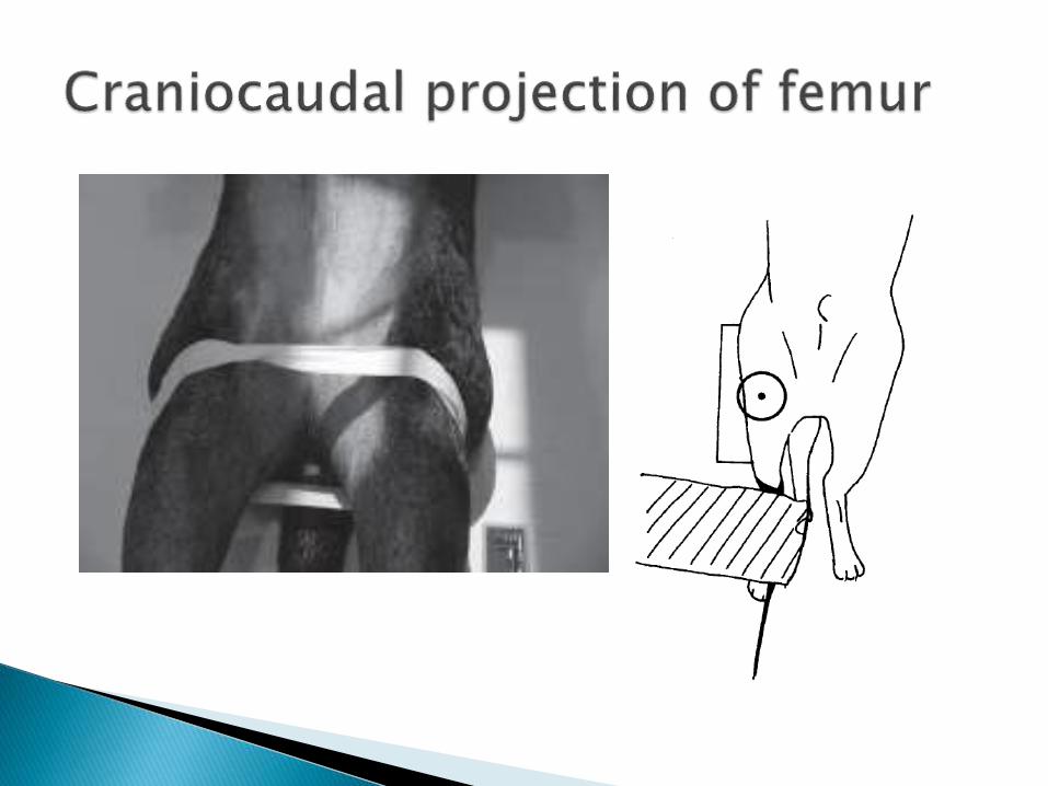
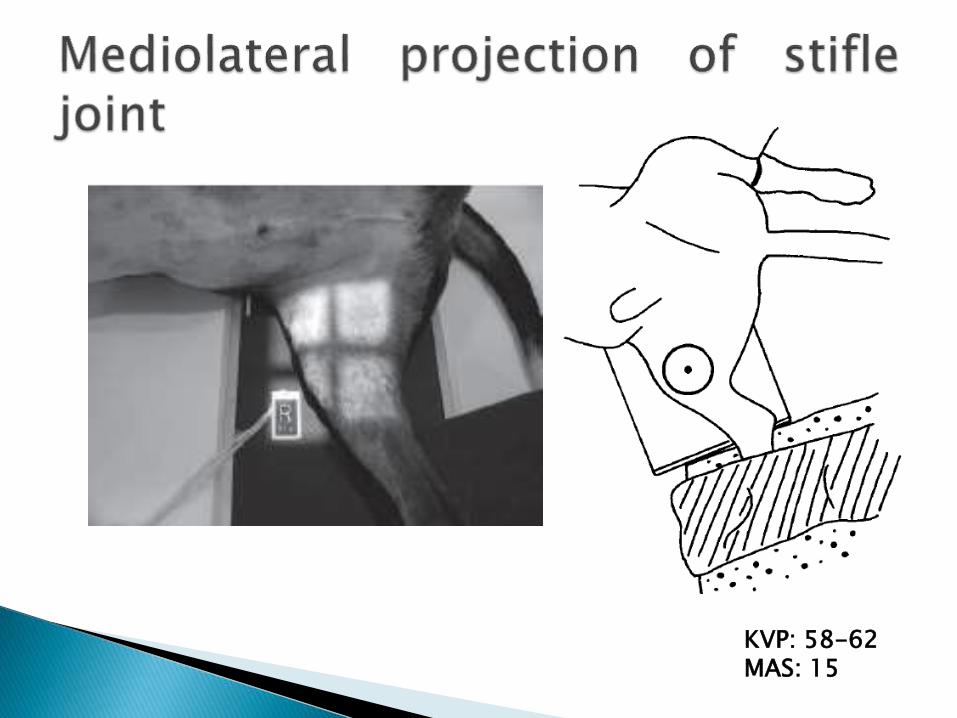
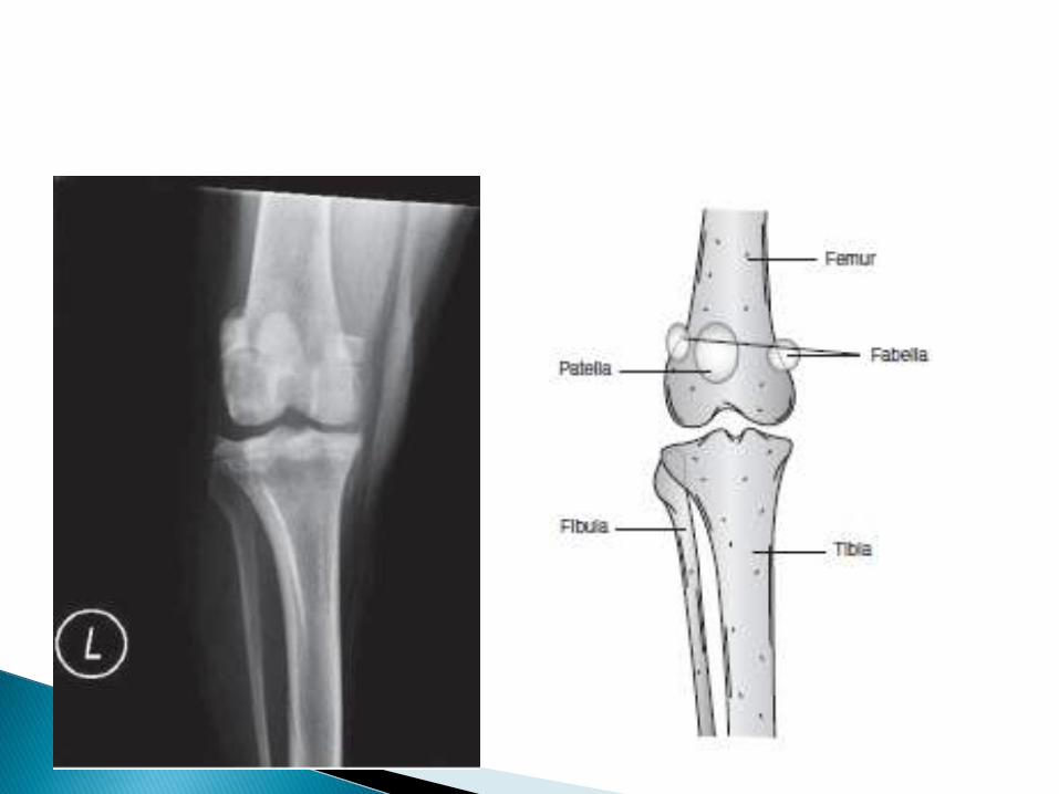
KVP: 58-62MAS: 15
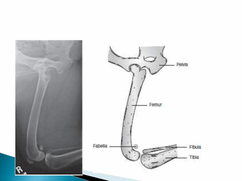
KVP: 58-62MAS: 15
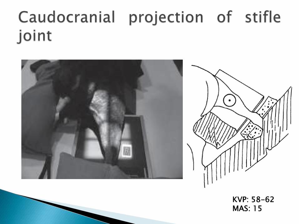
KVP: 58-62MAS: 15
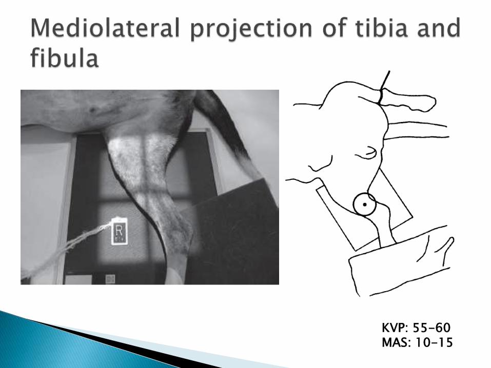
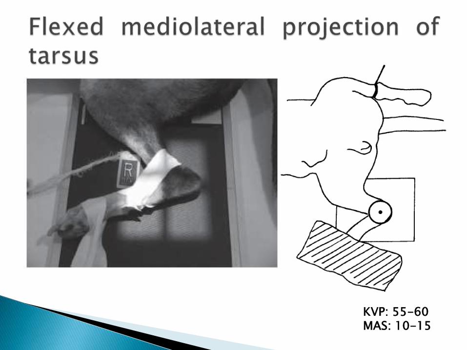
KVP: 55-60MAS: 10-15
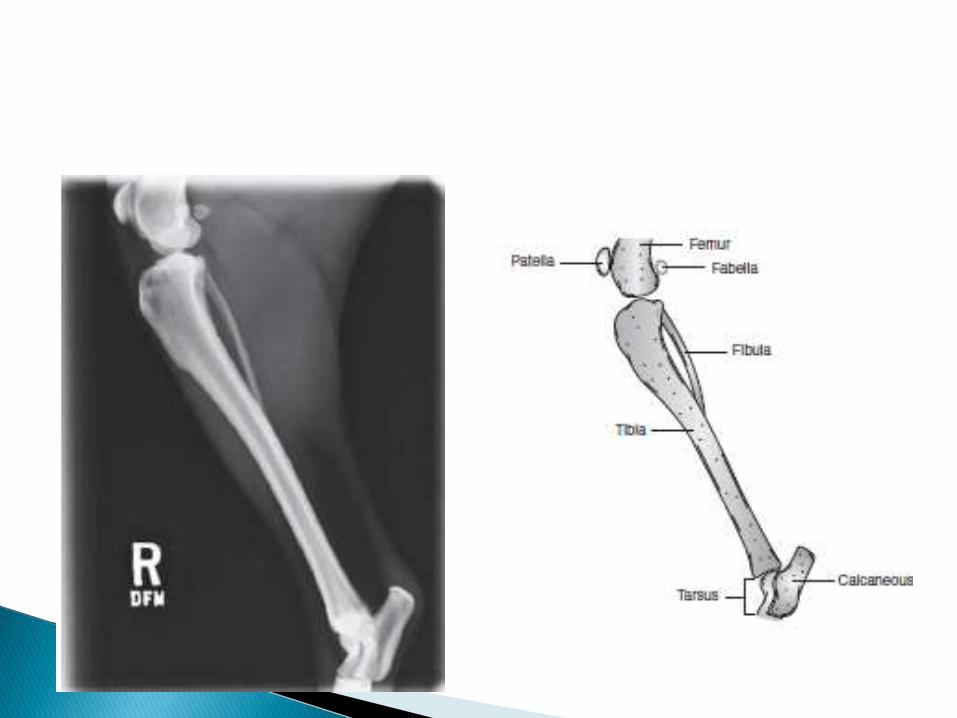

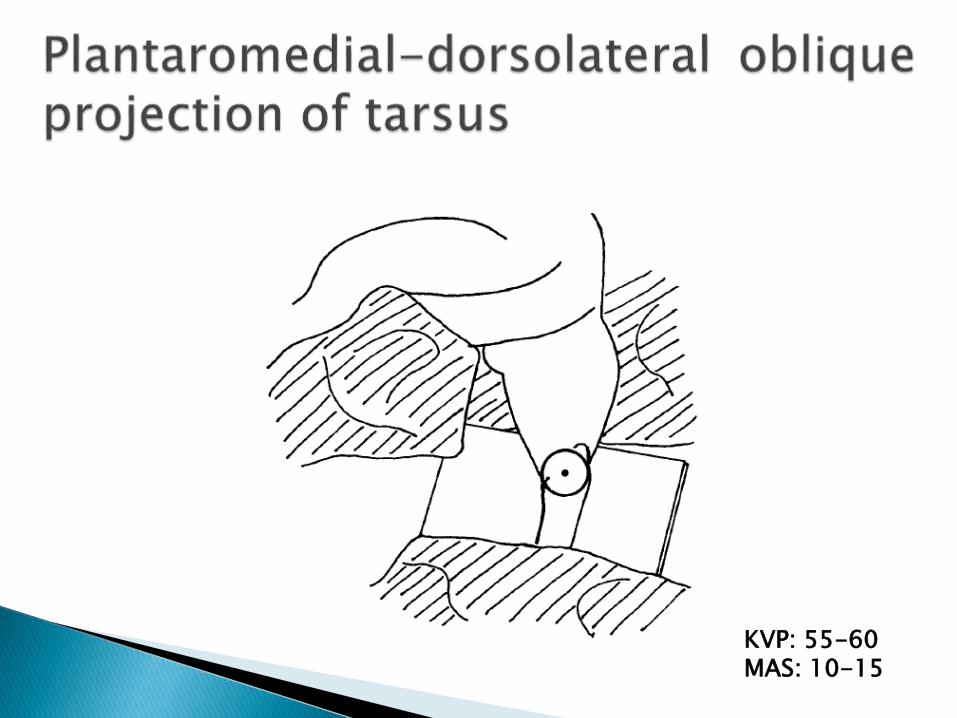
KVP: 55-60MAS: 10-15

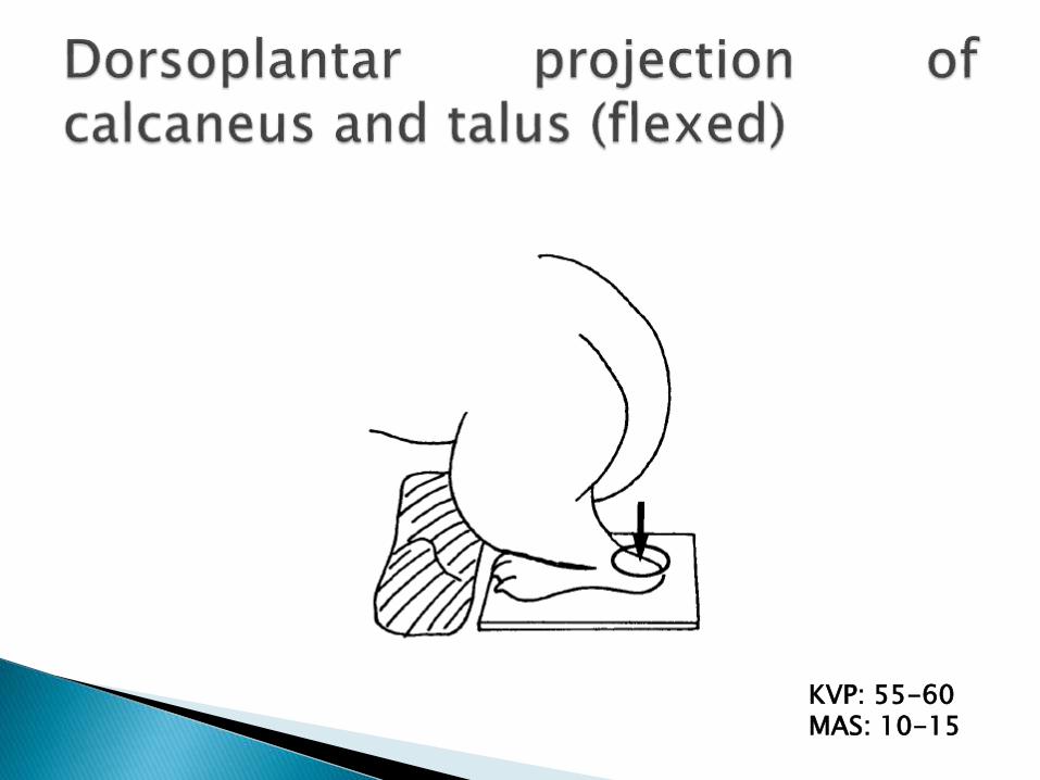
KVP: 55-60MAS: 10-15
KVP: 55-60MAS: 10-15
KVP: 55-60MAS: 10-15
KVP: 55-60MAS: 10-15
KVP: 55-60MAS: 10-15
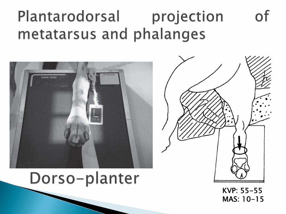
KVP: 55-55MAS: 10-15

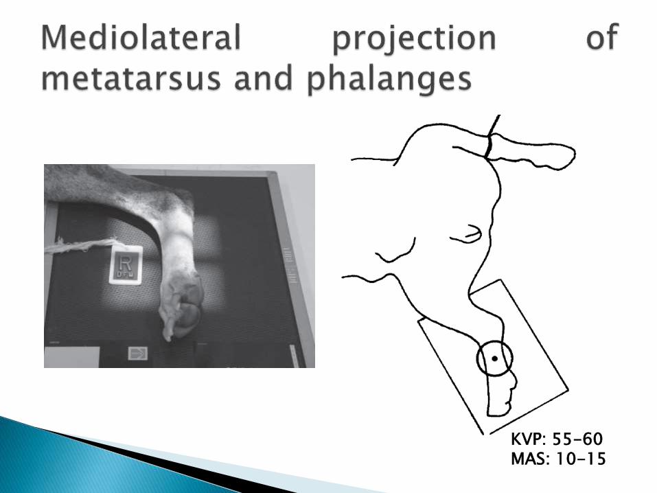
KVP: 55-60MAS: 10-15
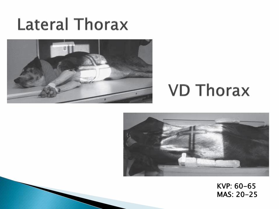
KVP: 60-65MAS: 20-25
KVP: 65-70MAS: 20-25