Embed Size (px)
Citation preview

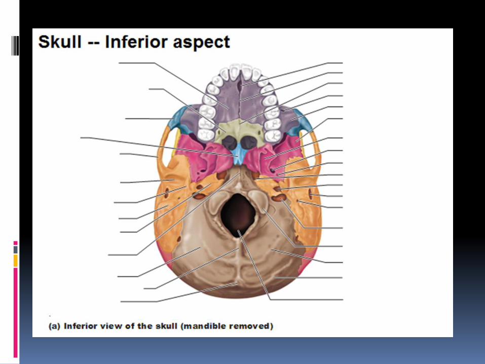
SKULL
Skull divded to
Cranium
Face

Cranium (8)
Frontal
Parietal (Paired)
Temporal (Paired)
Sphenoid
Ethmoid
Occipital

Facial (14)
Zygomatic (2)
Maxillae (2)
Nasal bone (2)
Lacrimal (2)
Vomer
Palatine (2)
Inferior Conchae (2)
Mandible


Cranial Cavity
Vault of Skull – Sutures and in the midline shallow sagittal groove that lodges superior sagittal sinus.
Base of skull
- Anterior Cranial Fossa
- Middle Cranial Fossa
- Posterior Cranial Fossa


Pterion
-Weakest point of skull- Sphenoid, Parietal, Frontal and Temporal bone meets- Anterior Division of Middle Meningeal Artery runs underneath

Base of Skull: Ant Cranial Fossa Houses frontal lobe
Anteriorly: Post Wall of Frontal bone
Posteriorly: Lesser wing of Sphenoid
Medial: Anterior Clinoid Process
Lateral : Frontal bone & Pterion
Floor: Cribriform plate, orbital plate of frontal bone, ethmoid, & crista galli.

Ant Cranial Fossa

Main Anatomic Features in ACF Frontal crest :- It’s a midline bony ridge
that projects upwards and provide attachment to the falx cerebri.
Crista galli :- (latin for cock’s comb) Provides the site for anterior most attachment of the falx cerebri.
Cribriform plate :- It is a sheet of bone which contains numerous small foramina – these transmit olfactory nerve fibres(CN I) into the nasal cavity

Middle Cranial Fossa
Anteriorly: Lesser wing of Sphenoid
Posteriorly: Petrous part of Temporal Bone
Laterally: Squamous parts of Temporal bone
Greater Wing of Sphenoid
Parietal bone
Floor: Greater wing of Sphenoid
Squamous and Petrous parts of Temporal Bone

Middle Cranial Fossa

Foramina of MCF
Features Location Relevance
Superior Orbital Fissure Between Greater and Lesser Wing of Sphenoid
Lacrimal N (V1)Frontal N (V1)Trochlear N(IV th Nerve)Superior Ophthalmic VeinOculomotor N (III)Nasociliary N (V1)Abducent Nerve (VI)
Foramen Rotundum Greater wing of Sphenoid Maxillary Nerve
Foramen Ovale Greater wing of Sphenoid MALEMandibular NAccessory Meningeal NLesser Petrosal NEmissary Vein

Foramina of MCF
Features Location Relevance
Foramen Spinosum Greater Wing of Sphenoid MENMiddle Meningeal Artery & VeinEmmisary VeinSpinosus Nerve
Foramen Lacerum Apex of Petrous part of Temporal Bone
Transmits Internal Carotid Artery from Carotid Canal to enter Cavernous SinusGreater Petrosal N.
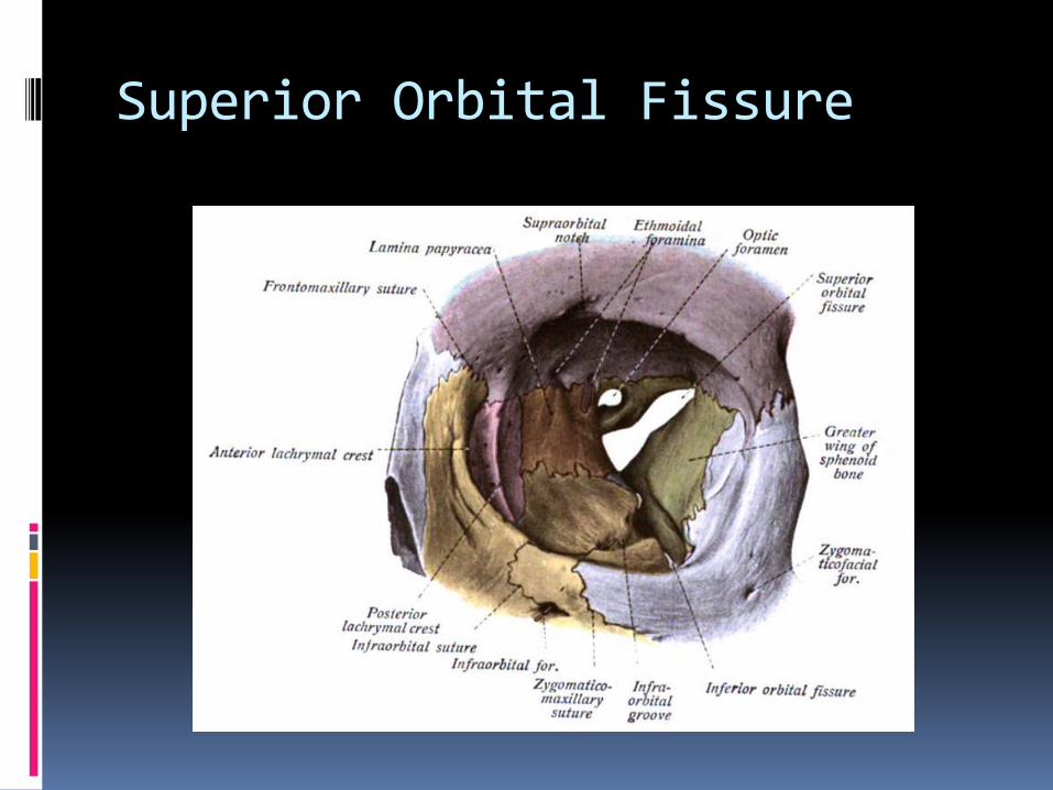
Superior Orbital Fissure
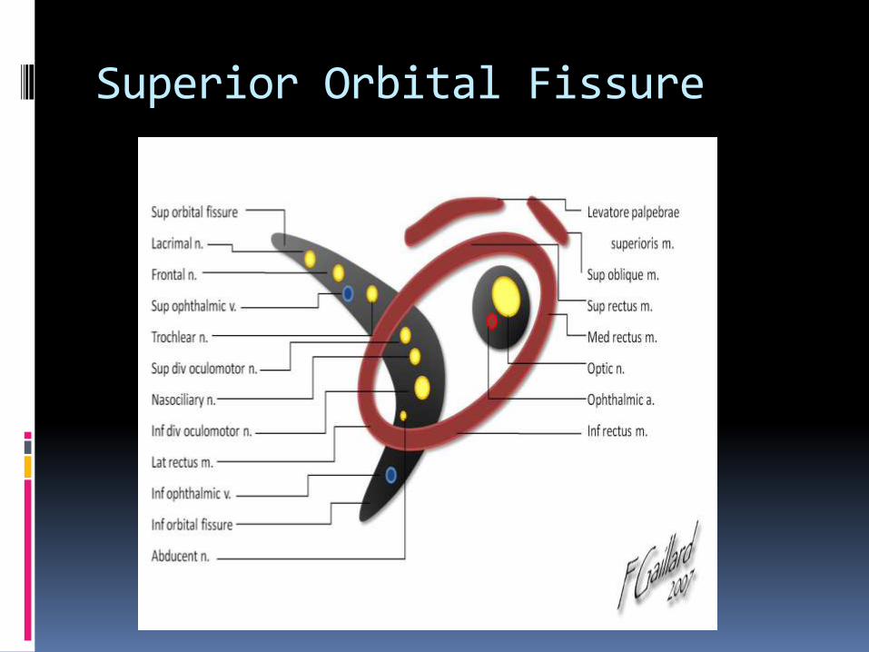
Superior Orbital Fissure

Ant Clinoid Process
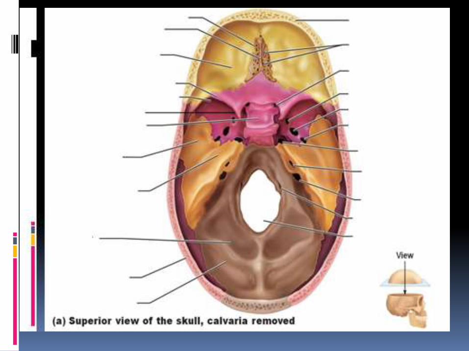

Posterior Cranial Fossa
Houses Cerebellum, pons and Medulla Oblangata
Anterior : Superior border of Petrous part of Temporal Bone
Post: Internal Surface of Squamous part of Occipital Bone
Floor: Basilar, condylar, mastoid part of temporal bone,
Squamous part of occipital bone

Foramina in PCF
Features Location Relevance
Internal Acoustic Meatus Posterior Surface of petrous part of Temporalbone
Vestibulocochlear N (VIII)Facial N (VII)
Jugular Foramen Between Petrous part of Temporal bone and occipital bone
Inferior Petrosal SinusGlossopharngeal N (IX)Vagus N (X)Acessory N (XI)Sigmoid Sinus
Hypoglossal Canal Above Anterolateralboundary of Foramen Magnum
Hypoglossal Nerve (XII)
Foramen Magnum Central part of OccipitalBone
Medulla OblangataMeningesSpinal parts of Accessory Nerves (XI)2 Vertebral Arteries