Embed Size (px)
Citation preview
NERVOUS, CIRCULATORY & RESPIRATORY SYSTEM:
•An insect's nervous system is a network of specialized cells called neurons that serve as an "information highway" within the body.
•The nervous system of insect functions to generate and transport electrical impulses, to integrate information received and to stimulate muscles for movement.
Neuron•The basic unit of nervous system that
functions in nerve impulse transmission is the nerve cell or neuron.
•A neuron is composed of:▫ A cell body where nucleus is found▫One or more receptor fibrils=Dendrites ▫An axon that branches at the tip
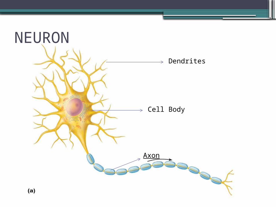
NEURONDendrites
Cell Body
Axon
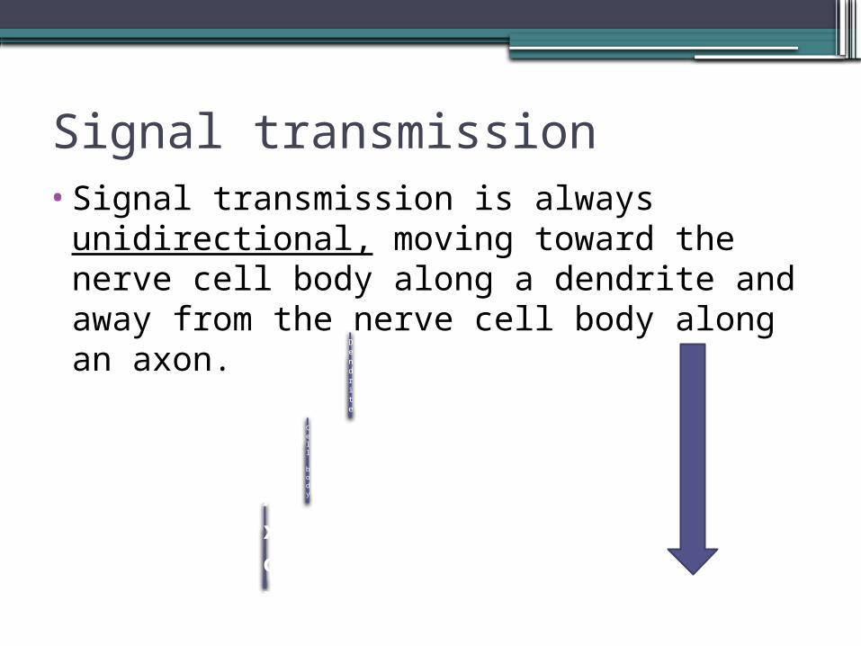
Signal transmission•Signal transmission is always
unidirectional, moving toward the nerve cell body along a dendrite and away from the nerve cell body along an axon.
Dendrite
Cell bodyA
xon
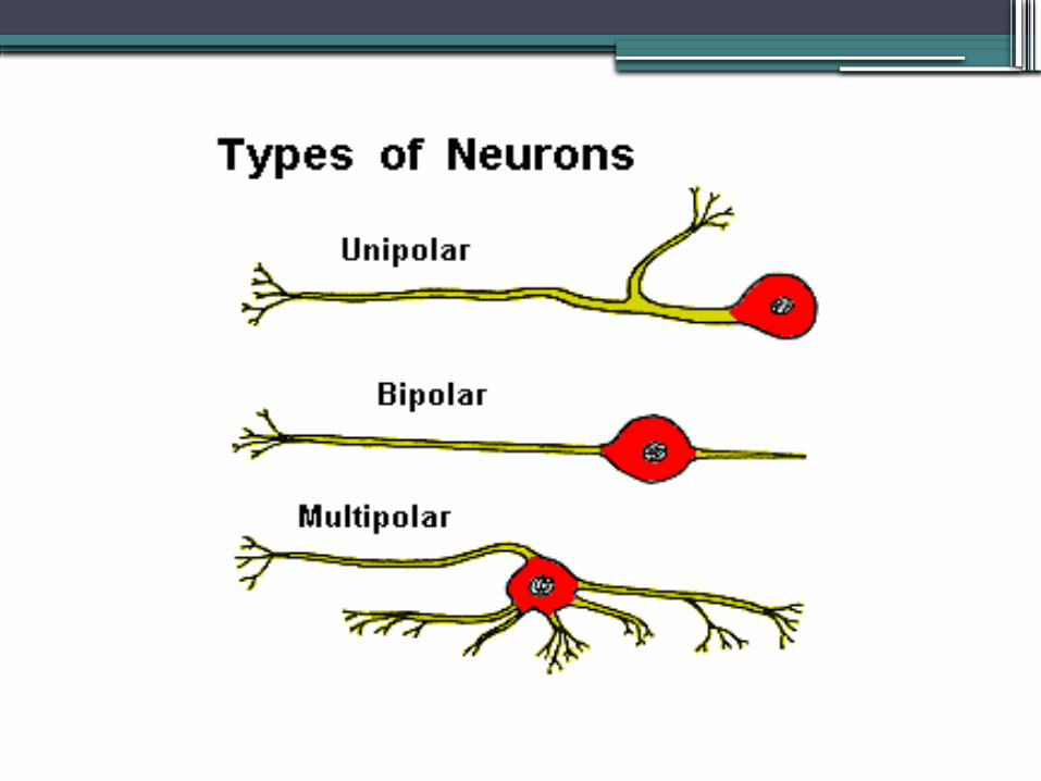
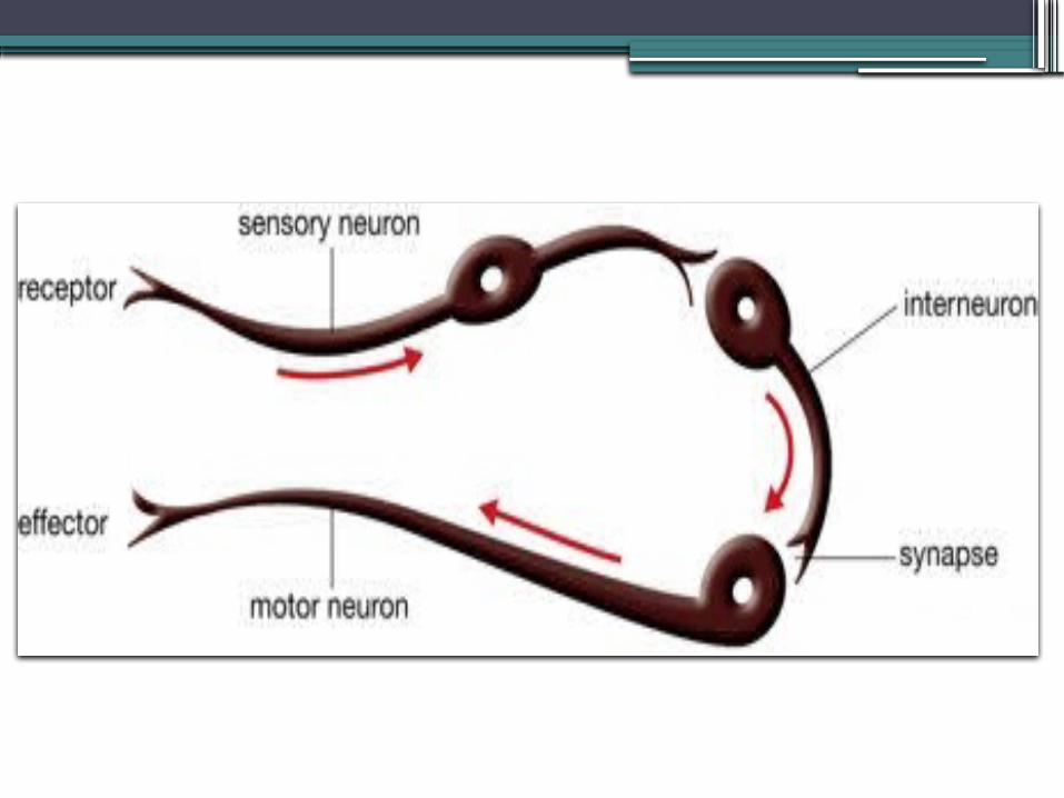
•Neurons are usually divided into three categories, depending on their function within the nervous system:
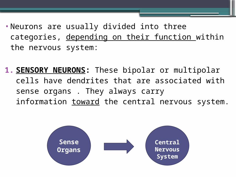
1. SENSORY NEURONS: These bipolar or multipolar cells have dendrites that are associated with sense organs . They always carry information toward the central nervous system.
Sense Organ
s
Central Nervou
s System
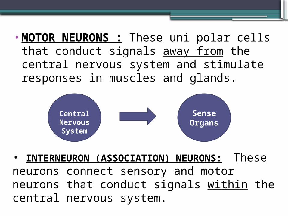
•MOTOR NEURONS : These uni polar cells that conduct signals away from the central nervous system and stimulate responses in muscles and glands.
• INTERNEURON (ASSOCIATION) NEURONS: These neurons connect sensory and motor neurons that conduct signals within the central nervous system.
Sense Organ
s
Central Nervou
s System
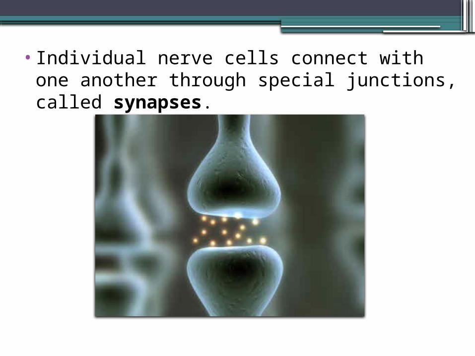
•Individual nerve cells connect with one another through special junctions, called synapses.
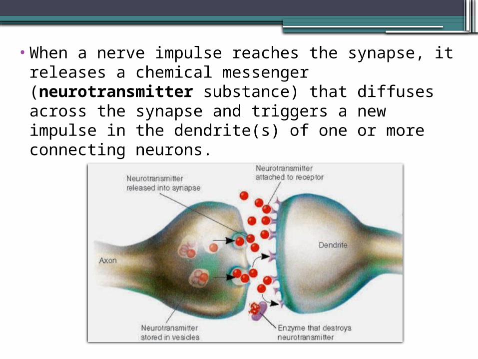
•When a nerve impulse reaches the synapse, it releases a chemical messenger (neurotransmitter substance) that diffuses across the synapse and triggers a new impulse in the dendrite(s) of one or more connecting neurons.
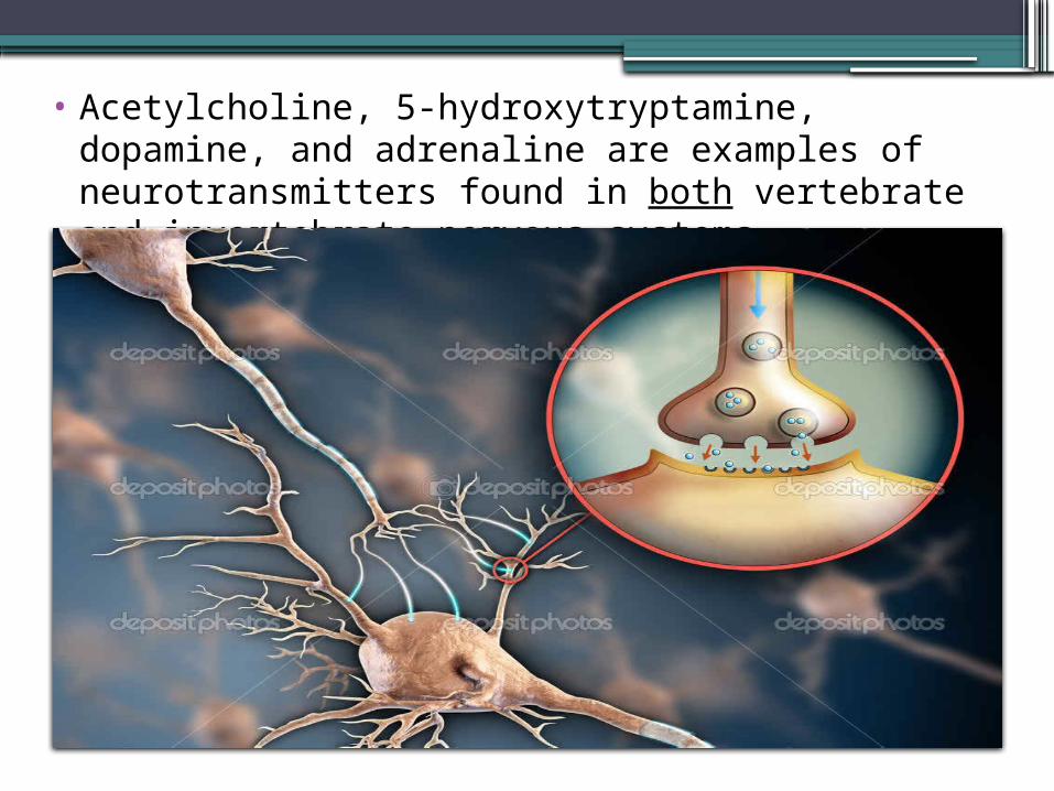
• Acetylcholine, 5-hydroxytryptamine, dopamine, and adrenaline are examples of neurotransmitters found in both vertebrate and invertebrate nervous systems.
•Nerve cells are typically found grouped in bundles. A nerve is simply a bundle of dendrites or axons that serve the same part of the body.
• A ganglion is a dense cluster of interconnected nerves that process sensory information or control motor outputs.
The Central Nervous System
•Like most other arthropods, insects have a relatively simple central nervous system with a dorsal brain linked to a ventral nerve cord that consists of paired segmental ganglia running along the ventral midline of the thorax and abdomen.
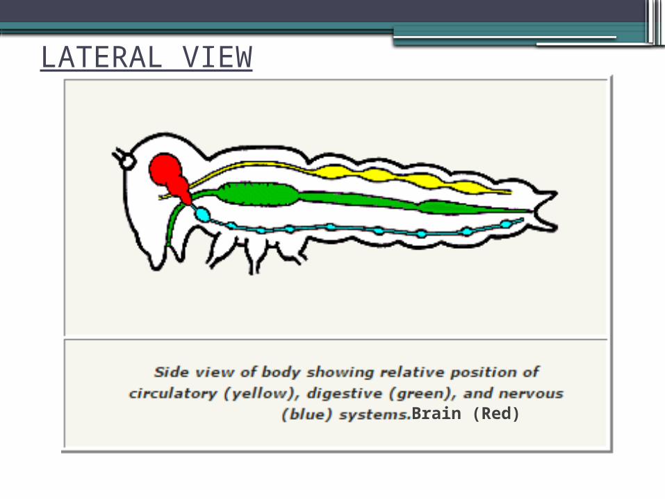
Brain (Red)
LATERAL VIEW
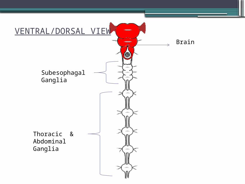
VENTRAL/DORSAL VIEWBrain
Subesophagal Ganglia
Thoracic & Abdominal Ganglia
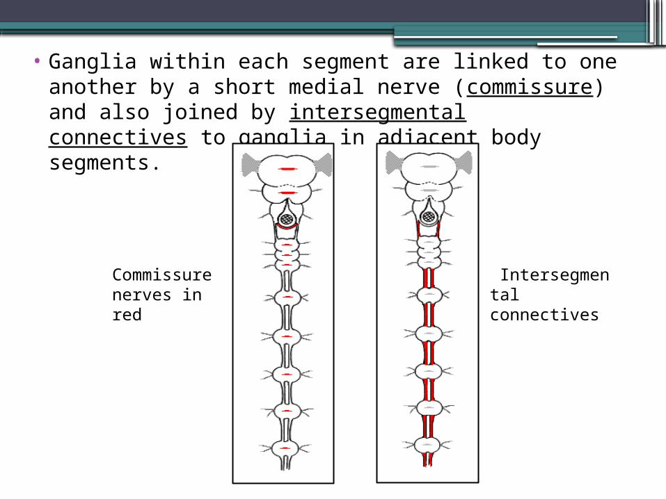
• Ganglia within each segment are linked to one another by a short medial nerve (commissure) and also joined by intersegmental connectives to ganglia in adjacent body segments.
Commissure nerves in red
Intersegmental connectives
BRAIN•An insect's brain is a complex of six fused
ganglia (three pairs) located dorsally within the head capsule.
• Each part of the brain controls (innervates:supply) a limited spectrum of activities in the insect's body:
1. Protocerebrum2. Deutocerebrum3. Tritocerebrum
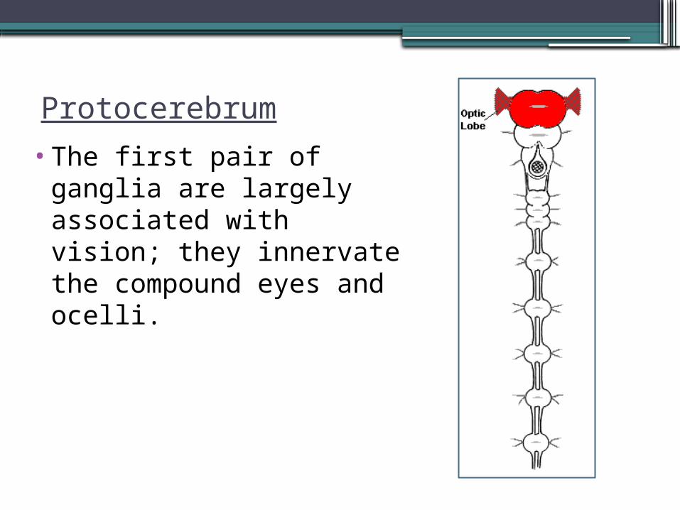
Protocerebrum•The first pair of ganglia
are largely associated with vision; they innervate the compound eyes and ocelli.
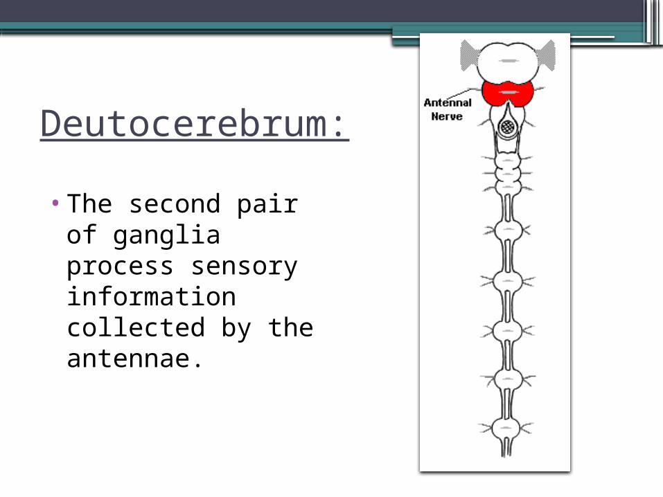
Deutocerebrum:•The second pair of
ganglia process sensory information collected by the antennae.
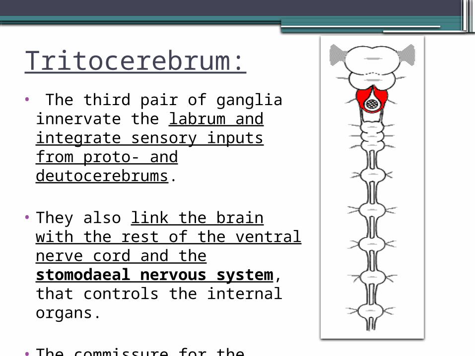
Tritocerebrum:• The third pair of ganglia
innervate the labrum and integrate sensory inputs from proto- and deutocerebrums.
• They also link the brain with the rest of the ventral nerve cord and the stomodaeal nervous system, that controls the internal organs.
• The commissure for the tritocerebrum loops around the digestive system.
SUBESOPHAGEAL GANGLION•Located ventrally in the head capsule
(just below the brain and esophagus) is another complex of fused ganglia (jointly called the subesophageal ganglion).
• The subesophageal ganglion innervates not only mandibles, maxillae, and labium, but also the hypopharynx, salivary glands, and neck muscles.
VENTRAL NERVE CORD•In the thorax, three pairs of thoracic
ganglia (sometimes fused) control locomotion by innervating the legs and wings.
• Thoracic muscles and sensory receptors are also associated with these ganglia.
•Similarly, abdominal ganglia control movements of abdominal muscles.
• Spiracles in both the thorax and abdomen are controlled by a pair of lateral nerves that arise from each segmental ganglion.
•A pair of abdominal ganglia usually fused to form a large caudal ganglion, innervates the excretory system, reproductive organs, and sensory receptors (such as cerci) located on the insect's back end.
THE STOMODAEAL NERVOUS SYSTEM
•An insect's internal organs are largely innervated by a stomodeal (or stomatogastric) nervous system.
•The stomodeal nervous system controls activities of the gut and circulatory system
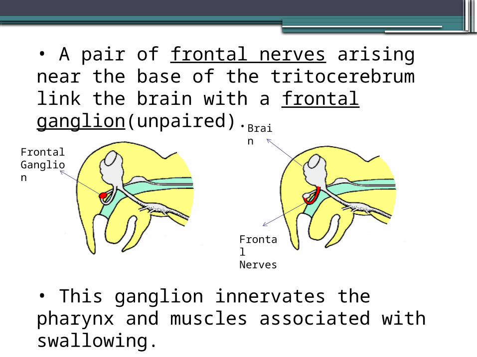
• A pair of frontal nerves arising near the base of the tritocerebrum link the brain with a frontal ganglion(unpaired).
• This ganglion innervates the pharynx and muscles associated with swallowing.
Frontal Ganglion
Brain
Frontal Nerves
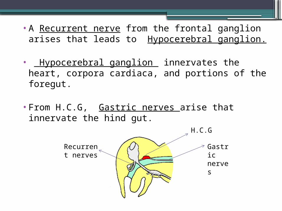
•A Recurrent nerve from the frontal ganglion arises that leads to Hypocerebral ganglion.
• Hypocerebral ganglion innervates the heart, corpora cardiaca, and portions of the foregut.
•From H.C.G, Gastric nerves arise that innervate the hind gut.
H.C.G
Gastric nerves
Recurrent nerves
•In comparison to vertebrates, an insect's nervous system is far more de-centralized.
• Most overt behavior (e.g. feeding, locomotion, mating, etc.) is integrated and controlled by segmental ganglia instead of the brain.
•In some cases, the brain may stimulate or inhibit activity in segmental ganglia but these signals are not essential for survival.
Indeed, a headless insect may survive for days or weeks (until it dies of starvation or dehydration) as long as the neck is sealed to prevent loss of blood!
Endocrine System: This system refers to the collection of glands of an organism that secrete hormones.
•A hormone is a chemical signal sent from cells in one part of an organism to cells in another part (or parts) of the same individual.
•They are often regarded as chemical messengers. Although typically produced in very small quantities, hormones may cause profound changes in their target cells.
• Their effect may be stimulatory or inhibitory.
Four categories of Hormone-Controlling cells in an insect's body:
1. Endocrine glands: Secretory structures adapted exclusively for producing hormones and releasing them into the circulatory system.
2. Internal organs: Hormone producing cells are associated with numerous organs of the body, including the reproductive system, the fat body, and parts of the digestive system.
3. Neurosecretory cells -- specialized nerve cells (neurons) that respond to stimulation by producing and secreting specific chemical messengers. Functionally, they serve as a link between the nervous system and the endocrine system
4. Neurohemal organs -- similar to glands, but they store their secretory product in a special chamber until stimulated to release it by a signal from the nervous system (or another hormone).
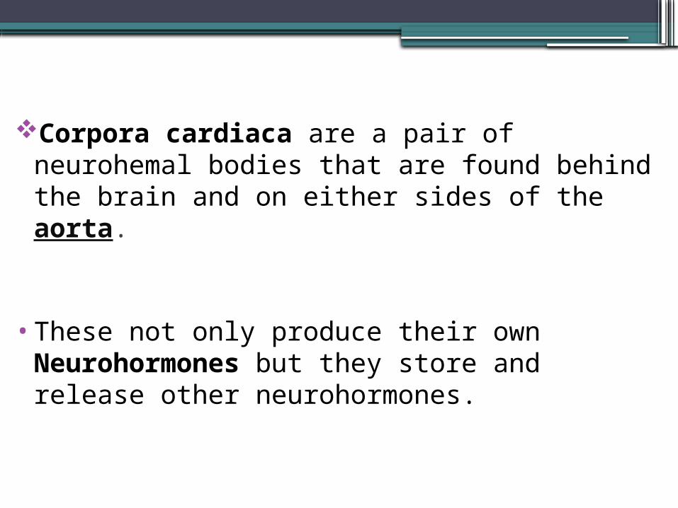
Corpora cardiaca are a pair of neurohemal bodies that are found behind the brain and on either sides of the aorta.
•These not only produce their own Neurohormones but they store and release other neurohormones.
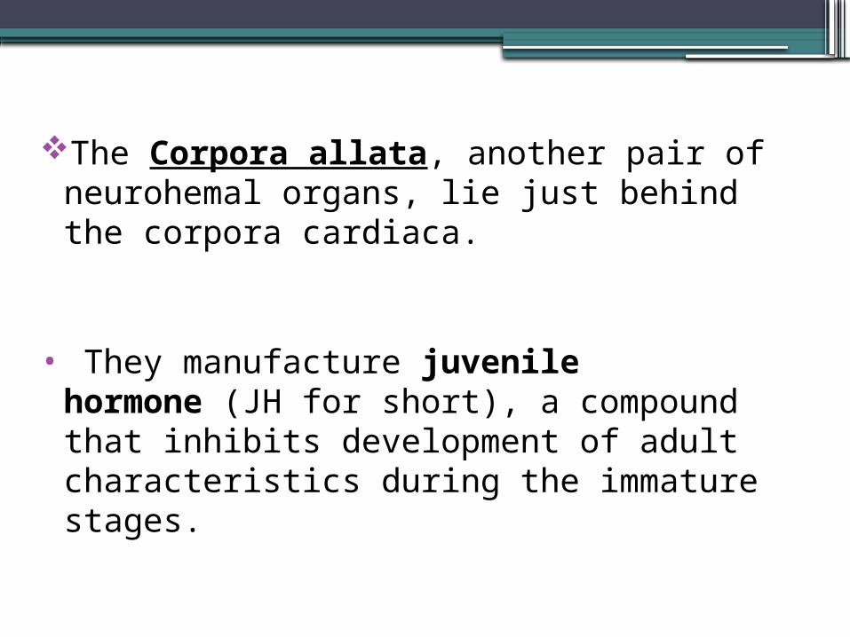
The Corpora allata, another pair of neurohemal organs, lie just behind the corpora cardiaca.
• They manufacture juvenile hormone (JH for short), a compound that inhibits development of adult characteristics during the immature stages.
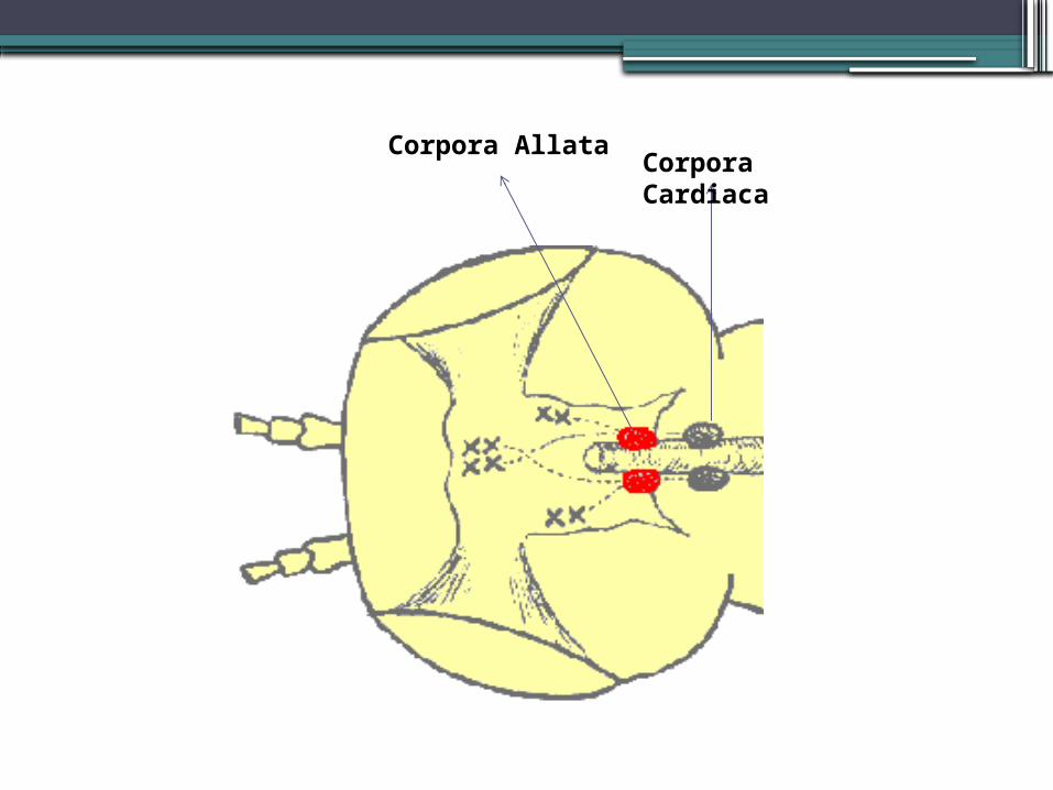
Corpora Allata Corpora Cardiaca
CIRCULATORY SYSTEM
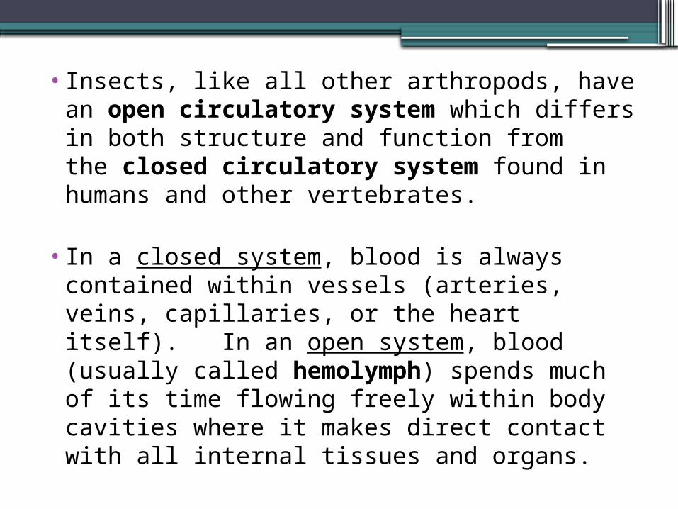
•Insects, like all other arthropods, have an open circulatory system which differs in both structure and function from the closed circulatory system found in humans and other vertebrates.
• In a closed system, blood is always contained within vessels (arteries, veins, capillaries, or the heart itself). In an open system, blood (usually called hemolymph) spends much of its time flowing freely within body cavities where it makes direct contact with all internal tissues and organs.
Functions of Insect Circulatory System•The circulatory system is responsible for
movement of nutrients, salts, hormones, and metabolic wastes throughout the insect's body.
• In addition, it plays several critical roles in defense: ▫ It seals off wounds through a clotting reaction,▫ It encapsulates and destroys internal parasites
or other invaders▫In some species, it produces (or sequesters)
distasteful compounds that provide a degree of protection against predators.
Dorsal Vessel•A dorsal vessel is the major structural
component of an insect's circulatory system.
•This tube runs longitudinally through the thorax and abdomen, along the inside of the dorsal body wall.
• It is a fragile, membranous structure that collects hemolymph in the abdomen and conducts it forward to the head.
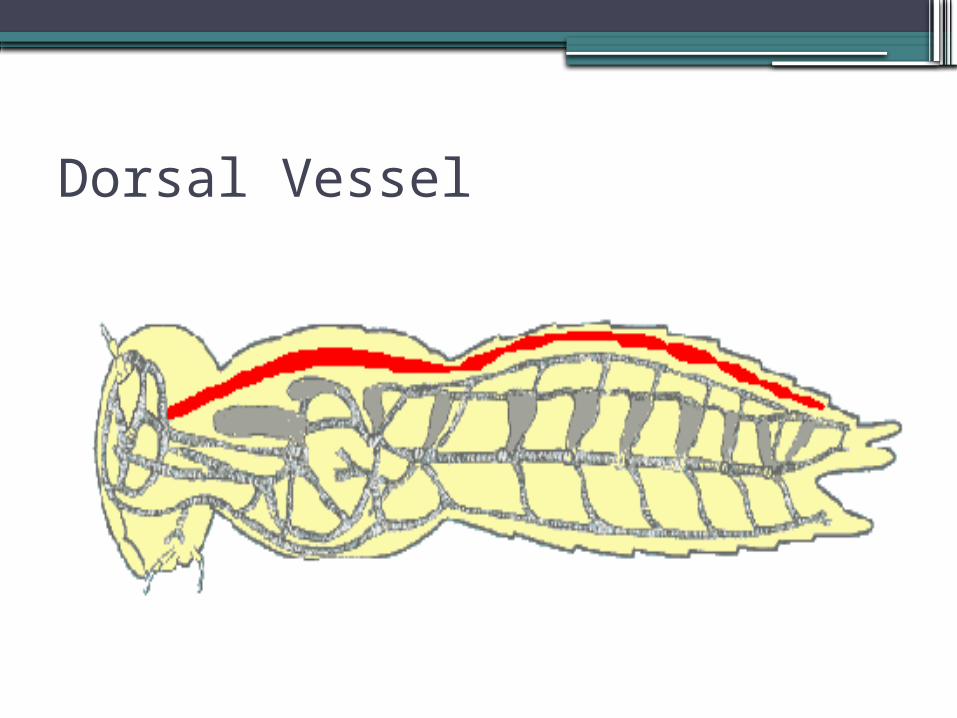
Dorsal Vessel
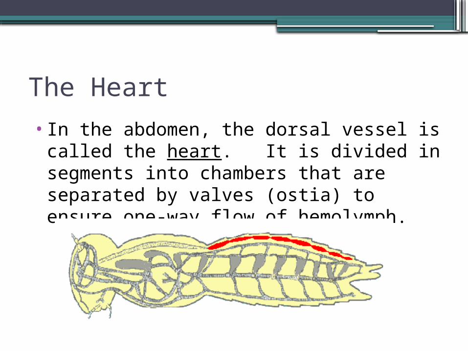
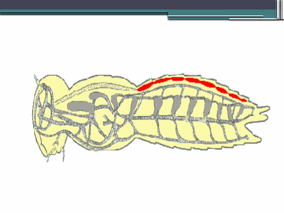
The Heart•In the abdomen, the dorsal vessel is
called the heart. It is divided in segments into chambers that are separated by valves (ostia) to ensure one-way flow of hemolymph.
• A pair of alary muscles are attached laterally to the walls of each chamber.
•Contractions of the these muscles force the hemolymph forward from chamber to chamber.
•During each diastolic phase (relaxation), the ostia open to allow inflow of hemolymph from the body cavity.
•The heart's contraction rate varies considerably from species to species -- typically in the range of 30 to 200 beats per minute.
•The rate tends to fall as ambient temperature drops and rise as temperature (or the insect's level of activity) increases.
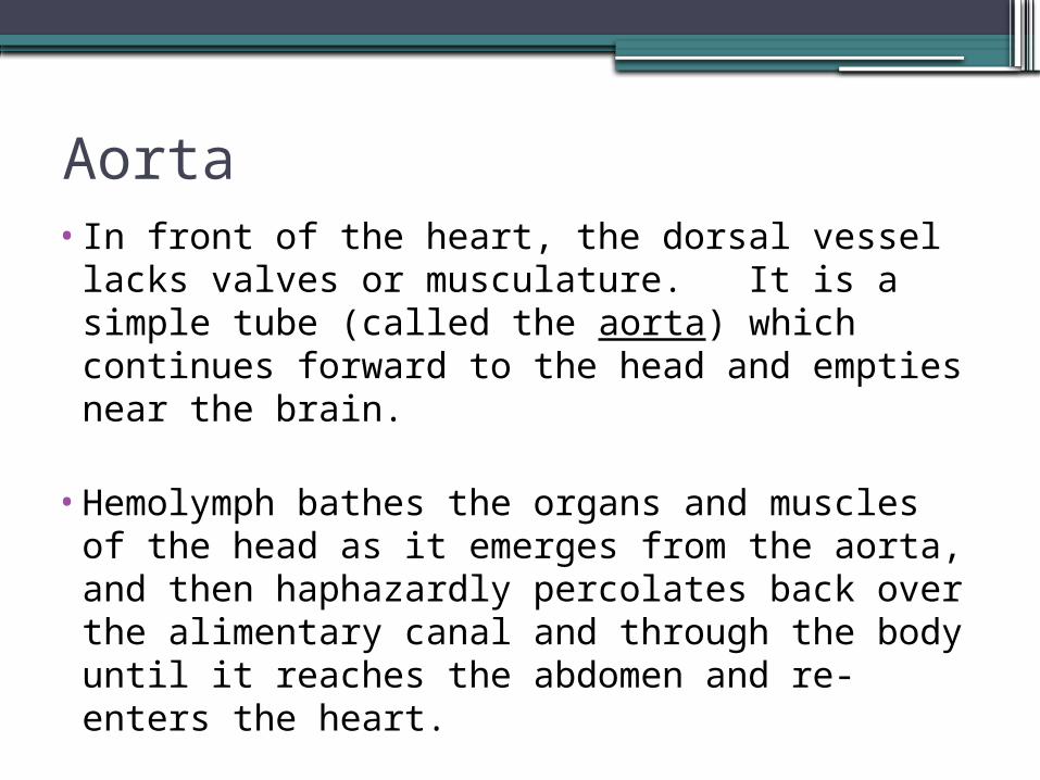
Aorta•In front of the heart, the dorsal vessel lacks
valves or musculature. It is a simple tube (called the aorta) which continues forward to the head and empties near the brain.
•Hemolymph bathes the organs and muscles of the head as it emerges from the aorta, and then haphazardly percolates back over the alimentary canal and through the body until it reaches the abdomen and re-enters the heart.
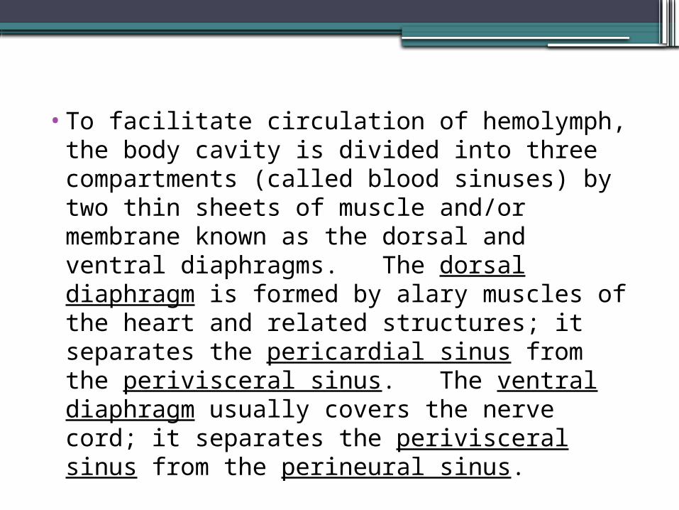
•To facilitate circulation of hemolymph, the body cavity is divided into three compartments (called blood sinuses) by two thin sheets of muscle and/or membrane known as the dorsal and ventral diaphragms. The dorsal diaphragm is formed by alary muscles of the heart and related structures; it separates the pericardial sinus from the perivisceral sinus. The ventral diaphragm usually covers the nerve cord; it separates the perivisceral sinus from the perineural sinus.
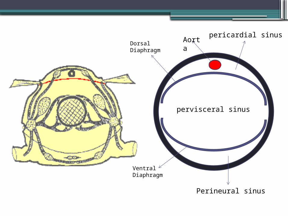
Aorta
Dorsal Diaphragm
Ventral Diaphragm
pericardial sinus
pervisceral sinus
Perineural sinus

Heamolymph Flow in Insect
Composition of blood (Haemolymph)
• About 90% of insect haemolymph is plasma: a watery fluid -- usually clear, but sometimes greenish or yellowish in color.
• Compared to vertebrate blood, it contains relatively high concentrations of amino acids, proteins, sugars, and inorganic ions.
• The remaining 10% of haemolymph volume is made up of various cell types (collectively known as haemocytes); they are involved in the clotting reaction, phagocytosis, and/or encapsulation of foreign bodies.
• Oxygen is delivered by the tracheal system, not the circulatory system.
RESPIRATION
• Physiological Respiration:
Transport of oxygen from the outside air to the cells within tissues, and the transport of carbon dioxide in the opposite direction.
• Cellular respiration:
The metabolic process by which an organism obtains energy by reacting oxygen with glucose to give water, carbon dioxide and 38ATP (energy).
Although physiologic respiration is necessary to sustain cellular respiration and thus life in animals, the processes are distinct: cellular respiration takes place in individual cells of the organism, while physiologic respiration concerns the bulk flow and transport of metabolites between the organism and the external environment.
•The respiratory system is responsible for delivering sufficient oxygen to all cells of the body and for removing carbon dioxide (CO2) that is produced as a waste product of cellular respiration.
• The respiratory system of insects (and many other arthropods) is separate from the circulatory system. It is a complex network of tubes called a tracheal system that delivers oxygen-containing air to every cell of the body.
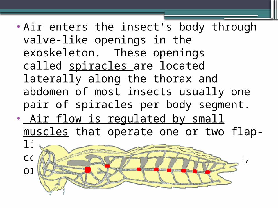
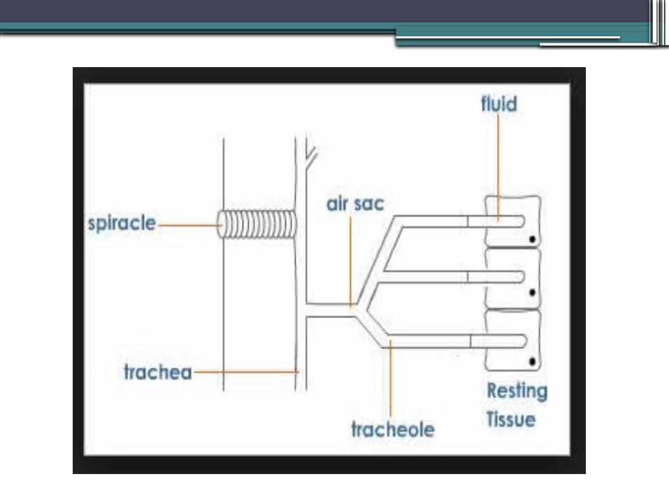
•Air enters the insect's body through valve-like openings in the exoskeleton. These openings called spiracles are located laterally along the thorax and abdomen of most insects usually one pair of spiracles per body segment.
• Air flow is regulated by small muscles that operate one or two flap-like valves within each spiracle contracting to close the spiracle, or relaxing to open it.
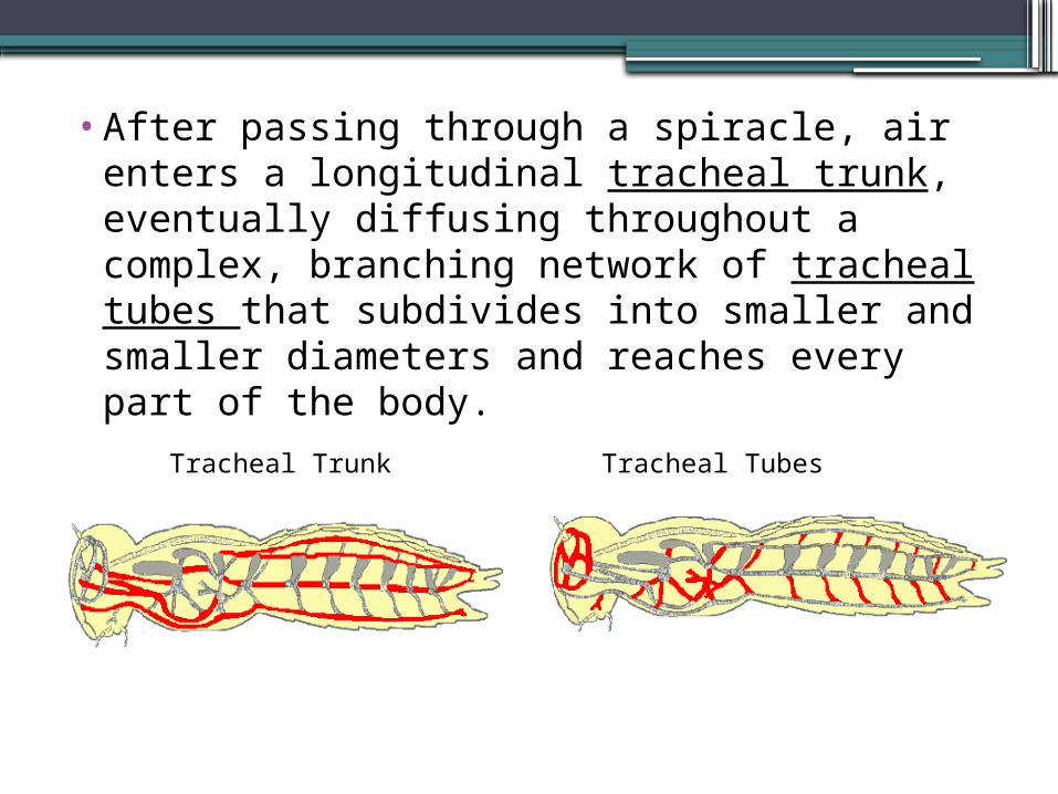
•After passing through a spiracle, air enters a longitudinal tracheal trunk, eventually diffusing throughout a complex, branching network of tracheal tubes that subdivides into smaller and smaller diameters and reaches every part of the body.
Tracheal Trunk Tracheal Tubes
•At the end of each tracheal branch, a special cell (the tracheole) provides a thin, moist interface for the exchange of gasses between atmospheric air and a living cell.
• Oxygen in the tracheal tube first dissolves in the liquid of the tracheole and then diffuses into the cytoplasm of an adjacent cell.
•At the same time, carbon dioxide, produced as a waste product of cellular respiration, diffuses out of the cell and, eventually, out of the body through the tracheal system.
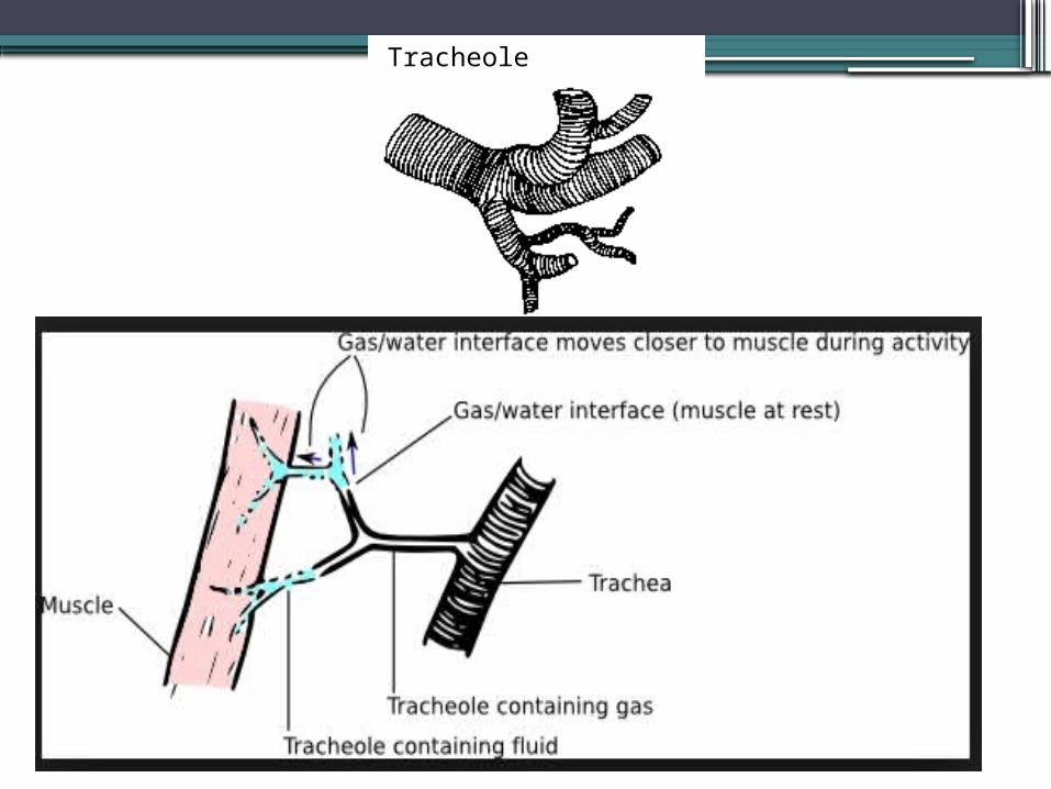
Tracheole
•In certain parts of the tracheal system air sacs, balloon-like structures that may store a reserve of air.
• In dry terrestrial environments, this temporary air supply allows an insect to survive.
• Aquatic insects consume the stored air while under water or use it to regulate buoyancy. During a molt, air sacs fill and enlarge as the insect breaks free of the old exoskeleton and expands a new one.
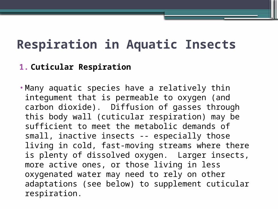
Respiration in Aquatic Insects1. Cuticular Respiration
• Many aquatic species have a relatively thin integument that is permeable to oxygen (and carbon dioxide). Diffusion of gasses through this body wall (cuticular respiration) may be sufficient to meet the metabolic demands of small, inactive insects -- especially those living in cold, fast-moving streams where there is plenty of dissolved oxygen. Larger insects, more active ones, or those living in less oxygenated water may need to rely on other adaptations (see below) to supplement cuticular respiration.
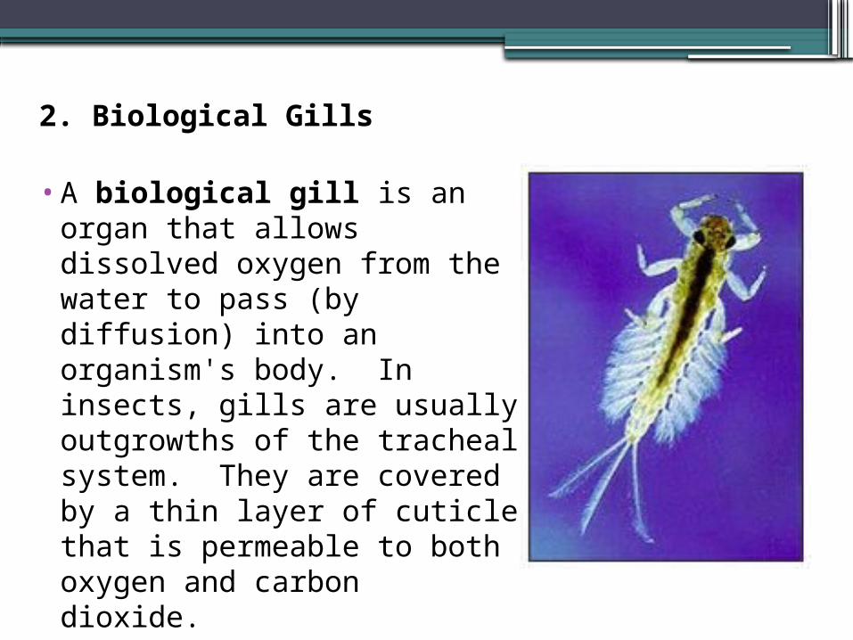
2. Biological Gills
•A biological gill is an organ that allows dissolved oxygen from the water to pass (by diffusion) into an organism's body. In insects, gills are usually outgrowths of the tracheal system. They are covered by a thin layer of cuticle that is permeable to both oxygen and carbon dioxide.
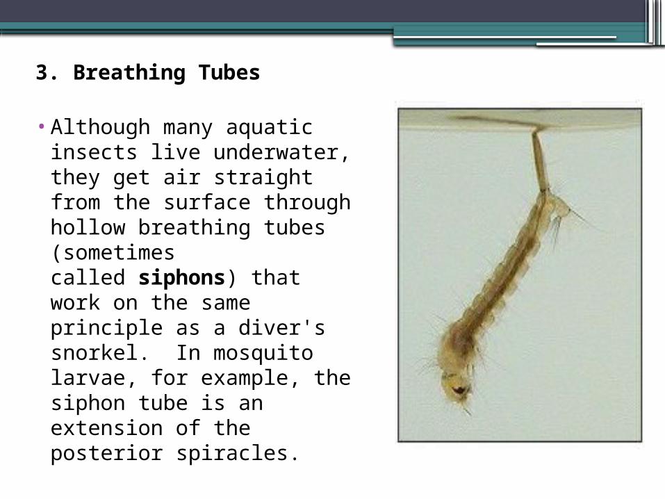
3. Breathing Tubes
• Although many aquatic insects live underwater, they get air straight from the surface through hollow breathing tubes (sometimes called siphons) that work on the same principle as a diver's snorkel. In mosquito larvae, for example, the siphon tube is an extension of the posterior spiracles.
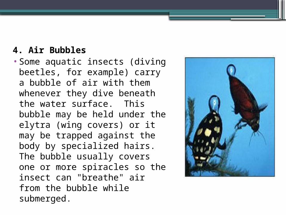
4. Air Bubbles• Some aquatic insects (diving
beetles, for example) carry a bubble of air with them whenever they dive beneath the water surface. This bubble may be held under the elytra (wing covers) or it may be trapped against the body by specialized hairs. The bubble usually covers one or more spiracles so the insect can "breathe" air from the bubble while submerged.
5. Plastron•A plastron is a special array of rigid, closely-
spaced hydrophobic hairs (setae) that create an "airspace" next to the body. Air trapped within a plastron operates as a physical gill (just like air in a bubble)
6. Hemoglobin•Hemoglobin is a respiratory pigment that
facilitates the capture of oxygen molecules. It occurs only rarely in insects -- most notably in the larvae of certain midges (family Chironomidae) known as bloodworms.
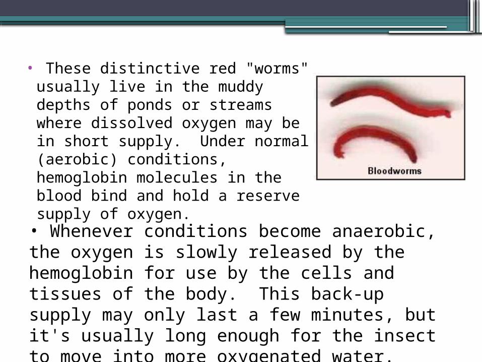
• These distinctive red "worms"
usually live in the muddy depths of ponds or streams where dissolved oxygen may be in short supply. Under normal (aerobic) conditions, hemoglobin molecules in the blood bind and hold a reserve supply of oxygen. • Whenever conditions become anaerobic, the oxygen is slowly released by the hemoglobin for use by the cells and tissues of the body. This back-up supply may only last a few minutes, but it's usually long enough for the insect to move into more oxygenated water.