
www.elsevier.com/locate/mbs
Mathematical Biosciences 208 (2007) 430–453
A geometrical model of dermal capillary clearance q
Kosmas Kretsos a,*, Gerald B. Kasting b
a University at Buffalo, State University of New York, Department of Chemical and Biological Engineering,
Furnas Hall, Buffalo, NY 14260-4200, USAb University of Cincinnati, College of Pharmacy, Health Professions Building, Cincinnati, OH 45267-0004, USA
Received 16 November 2005; received in revised form 31 July 2006; accepted 23 October 2006Available online 14 November 2006
Abstract
A new microscopic model is developed to describe the dermal capillary clearance process of skin perm-eants. The physiological structure is represented in terms of a doubly periodic array of absorbing capillar-ies. Convection-dominated transport in the blood flow within the capillaries is coupled with interstitialdiffusion, the latter process being quantified via a slender-body-theory approach. Convection across thecapillary wall and in the interstitial phase is treated as a perturbation which may be added to the diffusivetransport. The model accounts for the finite permeability of the capillary wall as well as for the geometry ofthe capillary array, based on realistic values of physiological parameters. Calculated dermal concentrationprofiles for permeants having the size and lipophilicity of salicylic acid and glucose illustrate the power andgeneral applicability of the model. Furthermore, validation of the model with published in vivo experimen-tal results pertaining to human skin permeation of hydrocortisone is presented. The model offers the pos-sibility for in-depth theoretical understanding and prediction of subsurface drug distribution in the humanskin following topical application, as well as rates of capillary clearance into the systemic circulation. Asimpler approach that treats the capillary bed as a homogeneously absorbing zone is also employed. Thelatter may be used in conjunction with the capillary exchange model to estimate measurable dermal trans-port and clearance parameters in a straightforward manner.� 2006 Elsevier Inc. All rights reserved.
0025-5564/$ - see front matter � 2006 Elsevier Inc. All rights reserved.doi:10.1016/j.mbs.2006.10.012
q Presented in part at the Controlled Release Society Annual Meeting, Glasgow, Scotland, July 2003.* Corresponding author. Present address: Entelos Inc., 110 Marsh Drive, Foster City, CA 94403, USA. Tel.: +1 650
572 5485; fax: +1 650 572 5401.E-mail address: [email protected] (K. Kretsos).

K. Kretsos, G.B. Kasting / Mathematical Biosciences 208 (2007) 430–453 431
Keywords: Capillary exchange; Mathematical model; Dermis; Slender body theory; Salicylic acid; Glucose; Hydro-cortisone
Nomenclature
Cde concentration in dermis (molm�3)Dde diffusivity in dermis (m2 s�1)Do characteristic diffusivity (m2s�1)f function used in the calculation of GG periodic Green’s function (molm�3)h length of capillary segment (m)Jr transcapillary flux (molm�2 s�1)J+1, J�1 flux above or below the capillary plexus (molm�2 s�1)k position vector for a random pointkc position vector for a cap. centerline pointko position vector for a point-sourcekper position vector for a cap. perimeter pointkde dermal clearance rate constant (s�1)Kbl/w blood/water partition coefficientKde/w dermis/water partition coefficientKwall/w cap. wall/water partition coefficientlx, ly lateral dimensions of unit cell (m)Lo characteristic length (m)N number of capillary segmentsnwall unit vector normal to cap. surfacent unit vector tangent to cap. centerlinenn unit vector normal to cap. centerlinenb unit vector binormal to cap. centerlineR source density (molm�1 s�1)Pe Peclet number (unitless)Pcap diffusive cap. permeability (ms�1)r capillary radius (m)s capillary arc length (m)s 0 cumulative capillary arc length (m)Ubl blood velocity (ms�1)Usp seepage velocity (ms�1)
Greek lettersAij and Bi matrices used in the source-densities calculationsb, c geometric capillary parameters (m)e arbitrary small parameter (unitless)h angle defining location on the capillary wall perimeter (rad)r geometric capillary parameter (unitless)

432 K. Kretsos, G.B. Kasting / Mathematical Biosciences 208 (2007) 430–453
1. Introduction
Various substances applied to the skin surface are able to permeate into deeper layers ofthe skin, and pass into the systemic circulation via microscopic blood vessels in the dermis.Thus, the quantitative prediction of the rate at which a skin penetrant is cleared throughthe microvasculature is valuable to the effective development of dermatological and transder-mally delivered drugs and to the risk assessment for chemical exposure in applications rang-ing from consumer products to chemical/biological weapons protection. Whereas theabsorption rates of most topically applied compounds are limited by the stratum corneum,some are modulated by capillary blood flow due to the nature of the compound [1,2],drug-induced vasoconstriction [3,4], and/or environmental and pathological conditions[5–7]. Furthermore, local epidermal and dermal concentration profiles, which are sensitiveto the clearance rates, are important to the understanding of diabetes [8,9], irritant andallergic contact dermatitis [10], and the pharmacodynamics of dermatological drugs [11,12]in the lower skin layers.
The archetype for capillary network modeling was set forth by Krogh [13,14]: Capillariesare assumed to be straight, parallel, identical cylinders that are evenly spaced. A no-fluxboundary condition is imposed on a polygonal tissue surface surrounding a single capillary.It thus suffices to concentrate on a single capillary and its surrounding tissue, which can beapproximated with a cylinder, and then extrapolate the results for the whole organ. Thiskey assumption was supported by physiology experiments conducted by Krogh himself [13].Many of the subsequent capillary exchange models may be regarded extensions of Krogh’scylinder [15–20]; most of them simulate the blood-tissue exchange of oxygen in various organs.Kretsos and Kasting [21] reviewed the progress of the field up to 2002. Several models haveemerged in the last three years pertaining to the microvascular exchange of mostly endogenousgases in tumor [19,22,23], peritoneum [24], isolated heart [25], peripheral nerve [18], parenchy-mal tissue in general [20], skeletal muscle [23,26], and skin [27]. The latter is an oxygen-ex-change model in which the superficial dermal capillaries are represented as pairs of counter-current, arterial/venular, straight vessels. The model that was actually solved considers stea-dy-state unidimensional transport. As illustrated by Beard and Bassingthwaighte [28] whodeveloped a three-dimensional model with straight capillaries, this reduction in dimensionsmay lead to erroneous results. Salathe [26] introduced an oxygen-exchange model in termsof a two-dimensional array of straight capillaries where axial diffusion in the tissue was ig-nored and capillary–capillary interactions between adjacent vessels were calculated. Compari-son between a capillary-loop and a straight-capillary exchange system [29] revealed that withthe use of the latter important information regarding the interstitial dispersion of a solute islost. Secomb et al. [23] followed a similar methodology to ours through the employment ofGreen’s functions in another oxygen-exchange model. Instead of imposing periodic boundaryconditions, the authors treated a specific tissue region as if it were embedded in an ‘infinitemedium’ across the boundaries of which the net flux of oxygen is zero. Their simulations per-tained to specific vascular networks whose configurations were obtained by observations ofsmall areas of rat muscle, brain, and tumor. An expansion of their model to considerably larg-er areas, e.g., in order to simulate the systemic effects of a chemical spill that occurs over sev-eral cm2 of skin area, seems intractable.

K. Kretsos, G.B. Kasting / Mathematical Biosciences 208 (2007) 430–453 433
This work introduces a skin-specific capillary exchange model that simulates the effect ofthe microvasculature on the fate of exogenous skin permeants once they have arrived at thedermis. The three-dimensional, bent-cylinder geometry of the dermal capillaries is accuratelyrepresented and mass transfer interactions among capillaries are taken into account throughthe imposition of periodic boundary conditions. The model considers both diffusive and con-vective transport mechanisms, the latter due to plasma leakage from the capillaries. Partition-ing phenomena both in the blood stream and in the interstitium, which depend on factorssuch as volume exclusion and binding to serum proteins, may be incorporated into the mod-el through the use of the appropriate values of partition coefficients. The model has thecapability of predicting steady state concentration profiles in the interstitial microenvironmentof the capillary plexus and variations in blood concentrations along the capillary loop. Thus,it is suitable to explore the topical kinetics of a skin permeant. This is illustrated for the caseof exogenously applied compounds having the same physicochemical properties as salicylicacid, glucose, and hydrocortisone. It is shown that the capillary model can work hand inhand with a simple, macroscopic, whole-skin approach to estimate the total rate of microvas-cular clearance of a given permeating substance. Furthermore, a modified version of themodel can be applied to the study and monitoring of epidermal interstitial fluid concentra-tions of endogenous substances such as glucose (for diabetics) and urea (for patients under-going dialysis treatments). A related publication describes the application of the distributed-clearance representation to the analysis of dermal concentration profiles of salicylic acid inrats [30].
2. Methods
2.1. Dermal microvasculature
The vascular physiology of the dermis has been described in detail elsewhere [21]. Briefly, thegeometry of the system involves two horizontal plexuses of vessels [31] with the superficial onestarting a few hundred microns below the skin surface. This plexus gives rise to the smaller, thin-ner parts of the microcirculation, the capillary loops. The capillaries are most important in termsof clearance into the systemic circulation since (a) they are the first permeable parts of the circu-lation encountered by a topically applied permeant and (b) they are the most permeable and mostnumerous microvessels [32]. The dermal capillary may be thought of as a bent, capital lambdashaped cylinder, most of which is located inside the papillae (ridges) near the dermal-epidermaljunction. It has an internal radius of �3 · 10�6 m and a total loop length that ranges from1.5 · 10�4 to 5 · 10�4 m [33]. The capillary ensemble may be considered to be a periodic structurein which the capillaries are perpendicular to the surface of the skin, as indicated by experimentalmeasurements [33,34].
2.2. Model formulation
The physical system is represented as an infinite array of capillaries and their surroundinginterstitium (Fig. 1(a)). The preceding and proceeding parts of the vasculature are assumed

Fig. 1. (a) The blood capillaries of the upper dermis represented as a doubly periodic array of matter-exchangingvessels perpendicular to the skin surface. (b) Definition sketch of the unit cell consisting of a single capillary loop and itssurrounding interstitium. All dimensions are in 10�6 m.
434 K. Kretsos, G.B. Kasting / Mathematical Biosciences 208 (2007) 430–453
to be non-exchanging vessels that supply (arterioles) and accept (venules) the blood flow. Thetransport processes that a topical permeant experiences in the interstitium are diffusion andconvection generated by filtration. The relative importance of diffusive and convective trans-port depends heavily on the nature of the skin permeant [21]. In the case where the inclusionof convective mechanisms is warranted, the pericapillary interstitium is thought of as a por-ous medium with the low-Reynolds number fluid flow in the pores obeying Darcy’s law[35,36]. In the absence of convection (the case studied in depth in this report) the interstit-ium is treated as a homogeneous medium having effective transport properties that expressits physiological heterogeneity. The blood flow is idealized as a plug flow of plasma. Diffu-sion in blood is considered negligible with respect to convection. More detailed models ofblood flow are available that take into account the intermittent, irregular nature of bloodflow in capillaries as well as the impact of the red blood cells [37–39]. However, they involvea level of complexity not needed for the present model, which considers only a time-averagedvalue of blood velocity.
2.2.1. Transport through the interstitiumThe dimensions parallel to the skin’s surface are denoted as x and y. The depth dimension,
z, is considered infinite (Fig. 1(b)). The validity of this assumption is addressed in the Discus-sion. There is a steady flux, J+1, of the skin permeant at z! +1. As the permeant diffusesdownwards it is, in part, systemically absorbed through the capillaries. The permeant remain-ing in the tissue continues to traverse the dermis with a different flux, J�1. The convectivepart of interstitial transport, generated by the filtration of plasma, is characterized in termsof an averaged seepage velocity, Usp. It is reasonable to assume (a) that this velocity will have

K. Kretsos, G.B. Kasting / Mathematical Biosciences 208 (2007) 430–453 435
its maximum value just outside the capillary wall and (b) that as z!�1 the convective fluxwould disappear since it is a localized phenomenon occurring in the space between the vascu-lar and lymphatic capillaries. Since the capillary array is periodic in the lateral dimensions (xand y), periodic conditions apply at the respective boundaries of the unit cell. Due to the sym-metry/periodicity of the problem, a net convective flux from one unit cell to another cannotexist, thus, the corresponding boundary conditions involve only diffusive fluxes. Eqs. (1)through (7) describe transport in the interstitium,
r � ð�DderCde þUspCdeÞ ¼ 0; ð1ÞCdejx¼0 ¼ Cdejx¼lx
; ð2ÞoCde
ox
����x¼0
¼ oCde
ox
����x¼lx
; ð3Þ
Cdejy¼0 ¼ Cdejy¼ly; ð4Þ
oCde
oy
����y¼0
¼ oCde
oy
����y¼ly
; ð5Þ
Dde
oCde
oz
����z!�1
! J�1; ð6Þ
DdeoCde
oz
����z!þ1
! Jþ1; ð7Þ
with Cde representing the permeant concentration in the dermis and Dde its diffusivity.
2.2.2. Transcapillary fluxIt is assumed that the capillary wall is homogeneous and of constant thickness. The total tran-
scapillary flux, Jr, is the sum of the diffusive and convective fluxes between the blood and tissuephases.
Jr ¼ P cap
Cbl
Kbl=w
� CdejrKde=w
� �þ U spjrKwall=w
2
Cbl
Kbl=w
þ CdejrKde=w
� �; ð8Þ
where Pcap is the (diffusive) capillary permeability coefficient. Kwall/w, Kbl/w, and Kde/w are the par-tition coefficients of the permeant in the capillary wall, blood and interstitial dermis, respectively,referred to bulk aqueous solution. For an ionizable compound it is convenient to define the bulksolution as a pH 7.4 aqueous buffer, so that the ionization state of the solute is clearly specified[30]. The terms Cdejr and Uspjr represent the tissue concentration and seepage velocity at the cap-illary wall, for a capillary of radius r. Since blood is considered to be well mixed over the capillarycross section, an analogous distinction is not necessary for Cbl. The transcapillary flux defined byEq. (8) should be equal to the flux normal to the capillary wall that enters the tissue, given by thefollowing equation:
Jr ¼ ð�DderCdejrÞ � nwall þ U spjrCdejr; ð9Þ
where nwall is the normal to the capillary surface outward unit vector. The finalconsideration regarding the transcapillary formalism is imposed by the lack of radial
436 K. Kretsos, G.B. Kasting / Mathematical Biosciences 208 (2007) 430–453
symmetry for the tissue concentration on the capillary surface, i.e., Cdejr depends onthe angle h that determines location on the perimeter of a capillary cross section. Thisis accounted for by combining Eqs. (8) and (9) and averaging them over the capillaryperimeter:
P capCbl
Kbl=w
� hCdejriKde=w
� �þ U spjrKwall=w
2
Cbl
Kbl=w
þ hCdejriKde=w
� �
¼ �DdehrCdejr � nwalli þ U spjrhCdejri; ð10Þ
with hi denoting the ‘ 12p
R 2p0
. . . dh’ averaging operation.
2.2.3. Mass and volume balances inside the capillaryWe consider a slice of the capillary thin enough that can be approximated by a straight cylinder
extending from length s to s + ds. A volume balance for this slice results in a relationship betweenthe (average) blood velocity Ubl and the (average) seepage velocity Uspjr by which plasma leaksfrom the capillary.
dU bl
ds¼ � 2U spjr
r: ð11Þ
The net flux of the permeant leaving the blood phase through the capillary wall is equal to �Jr. Amass balance for the capillary slice yields the following equation:
rU bl
2
dCbl
ds¼ �J r: ð12Þ
Eqs. (1)–(7) and (10)–(12) constitute the governing equations. In order to minimize the num-ber of parameters and the complexity of the solution the equations were made dimensionlesswith the use of suitable characteristic parameters. All length figures became dimensionlesswith a characteristic length, Lo, equal to 10�4 m (chosen as a round number having an orderof magnitude between those of the capillary length and of the capillary radius). The lateraldimensions of the unit cell were chosen to be lx = ly = 10�4m (yielding a realistic averagecapillary density of O(108)/m2 skin). The dermal tissue diffusion coefficient was made dimen-sionless with the use of a characteristic diffusivity, Do, equal to 10�11 m2 s�1, stemming fromthe results of our previous work [30]. Blood velocity was made dimensionless withUo = 10�4 ms�1 which is the order of magnitude of a typically reported velocity of capillaryblood flow [40]. Henceforth, variables and parameters that were made dimensionless aredenoted by a ‘hat’ (^). The dimensionless groups that arose from this process are two Pecletnumbers (defined by Eqs. (13) and (14); Peint for transport in the interstitium and Pebl fortransport in the blood phase). Generally, in mass transfer systems, the Peclet number is ameasure of the importance of convection relative to diffusion; as Pe! 0, diffusiondominates.
Peint ¼U spLo
Do
; ð13Þ
Pebl ¼U blrDo
: ð14Þ

K. Kretsos, G.B. Kasting / Mathematical Biosciences 208 (2007) 430–453 437
2.3. Methods of solution
2.3.1. Capillary centerline/surfaceParametric equations were used to generate the capillary centerline and subsequently the sur-
face of the capillary wall. An image of the capillary network [31] was used as a guide in orderto define the shape of the centerline:
x ¼ lx
2þ b tanhðrÞ; y ¼ ly
2; z ¼ �cr2: ð15Þ
The parameters b = c = 2 · 10�5 m were defined by the actual size and geometry of a single cap-illary loop. The interval �2.74 < r < 2.74 suffices to define a capillary loop of length �3 · 10�4 m.The corresponding representation of the capillary wall was constructed by considering the unitvectors tangential to the centerline, nt, normal to it, nn, and the corresponding binormal vector,nb. The capillary surface (for a constant capillary radius of 3 · 10�6 m) is defined through the fol-lowing equations
x ¼ lx=2þ b tanhðrÞ þ 3� 10�6C�ðrÞ cosðuÞ; ð16Þy ¼ ly=2þ 3� 10�6E�ðrÞ sinðuÞ; ð17Þz ¼ �cr2 þ 3� 10�6DðrÞ cosðuÞ; ð18Þ
with u 2 [�p, p] defining position on the circumference of the capillary. The components of theunit vectors are given in the Appendix A.
2.3.2. Perturbation analysis for the inclusion of convective transportConvective transport in the tissue was treated as a perturbation to diffusion that could be added
to the formalism, if necessary. Mathematically, this was realized through a perturbationanalysis [41]. A small-impact convection translates into a small Peclet number, i.e., of the formPeint = ewint with e� 1 and wint a to-be-defined function. The first approximation, which is accu-rate to within an error of order e, is equivalent to making the assumption that there is no filtrationor convection through the interstitium (Peint = 0). The present analysis pertaining to the lowmolecular weight compounds glucose, salicylic acid, and hydrocortisone considers purely diffusivetransport processes, i.e., convection is negligible. It has been argued that in skin there exists a low-level filtration coupled with the local lymph flow which prevents the formation of edema [21].Convective transport through the dermal interstitium, may or may not be important dependingon the properties of the permeant. It is recognized that for low- to medium-size substances, a cat-egory that includes practically all skin permeants, diffusion dominates [21]. However, if the per-meant binds extensively to serum proteins, e.g., albumin, then it will follow to a substantialextent the protein’s convective path to lymph. In this case, exclusion of interstitial and transcap-illary convection may lead to erroneous results. The possible inclusion of filtration/convection isgenerally discussed elsewhere [42].
2.3.3. Slender body theory formulationSince the capillary radius is much smaller than its length we may, to a good approximation, ap-
ply slender body theory which makes use of point-source solutions [43]. The solution for the dermal

438 K. Kretsos, G.B. Kasting / Mathematical Biosciences 208 (2007) 430–453
concentration field may thus be approximated in terms of a line distribution of point sources locat-ed on the capillary centerline. The first step of this analysis was to determine the correspondingdoubly periodic Green’s function, Gðk; koÞ, which yields the tissue concentration at an arbitrarypoint k ¼ ðx; y; zÞ resulting by taking into account the effect of a single unit source located at a pointko ¼ ðxo; yo; zoÞ inside the unit cell. The work for the derivation of Gðk; koÞ was significant, but in-volved standard methods, so details [42] are omitted here (the end result is shown in the AppendixA). Due to the slow convergence of the resulting double series as k! ko, another approach forcalculations near ko had to be employed. There exists a variety of such methods [44–46]. In many,one has to ‘extract’ the singularity, the cause of slow convergence, by breaking the Green’s functioninto two parts which are calculated separately. The same principle was employed here:
Gðk; koÞ ¼1
4pjk� kojþ f ðk; koÞ: ð19Þ
Values of Gðk; koÞ near ko were used to obtain values of the auxiliary function f near, and by inter-polation at, the singularity. These values were then fitted to a convenient polynomial function rep-resenting the behavior of f in the vicinity of ko.
The integration of Gðk; koÞ over the capillary centerline yields the disturbance to the concentra-tion field caused collectively by the point-sources. This disturbance must be added to the ‘back-ground’ linear concentration profile that would have prevailed in the absence of the capillaries.This background profile was chosen to be equal to the one that specifies the concentration asymp-totically as z! �1 (from Eq. (6)). Thus, the concentration at point k ¼ ðx; y; zÞ is given by theequation given below:
CdeðkÞ ¼ ðJ�1zþ gÞ þZ
cRðsÞGðk; koðsÞÞds; ð20Þ
where RðsÞ is the source density along the capillary centerline c on which position is quantified byarc length s.
In the absence of convective interstitial transport the change of the blood-phase concentrationalong the capillary loop is given by the combination of Eqs. (10) and (12):
dCbl
ds¼ �v1Cbl þ v2hCdejri; ð21Þ
v1 ¼2P cap
Pebl � Kbl=w
; ð22Þ
v2 ¼2P cap
Pebl � Kde=w
: ð23Þ
Integrating Eq. (21) over ½so; s� yields the blood concentration at s (Eq. (24)). The value so repre-sents the arc length at the first permeable part of the capillary, where the permeant’s blood con-centration is assumed to be zero. The newly defined variable s0 represents the cumulative arclength from so to s
CblðsÞ ¼ v2 expð�v1sÞZ s
so
expðv1s0ÞhCdejrids0: ð24Þ

K. Kretsos, G.B. Kasting / Mathematical Biosciences 208 (2007) 430–453 439
Eqs. (20) and (24) were used to substitute the tissue and blood concentrations in Eq. (10). Thisresulted to an integral equation in which the only unknown was RðsÞ. The source density, akey component of this analysis, was approximated numerically by discretizing the centerlineinto j = 1, . . . , 2N intervals of equal arc length. These arcs were short enough to be wellapproximated by their respective chords. The source density was assumed to be piecewise con-stant in each of these intervals [43]. By imposing the integral equation, i.e., by defining k onthe perimeter of discrete capillary cross sections that correspond to the centers of these inter-vals (located at i = 1, . . . , 2N elevations and defined by the use of Eq. (15)) a linear systemarises:
X2N
j¼1
AijRj ¼ Bi: ð25Þ
The matrices Aij and Bi are defined by the following equations:
Aij¼ nwall �rZ sjþ1
sj
1
4pjki� koðsÞjds
* +� v3
Z sjþ1
sj
1
4pjki� koðsÞjds
* +þZ sjþ1
sj
f ðkc;i; koðsÞÞds
" #
þ v4 expð�v1siÞZ si
so
expðv1s0ÞZ sjþ1
sj
1
4pjki� koðsÞjds
* +ds0 þ
Z si
so
expðv1s0ÞZ sjþ1
sj
f ðkc;i; koðsÞÞdsds0" #
; ð26Þ
Bi¼ v3hJ�1ziþ gi� expðv1siÞZ si
so
expðv1s0ÞhJ�1ziþ gids0; ð27Þ
with
v3 ¼P cap
Dde � Kde=w
; ð28Þ
v4 ¼ v2
P cap
Dde � Kbl=w
: ð29Þ
It is worth noting that, due to the circular symmetry of the capillary cross section, a perimeter-averaged Taylor expansion of f about the center of the cross section, kc;i, is equal to f ðkc;iÞ (towithin an error of order r2Þ. It is also worth noting that the same Taylor expansion of f, togetherwith Eq. (30), was used to simplify the general term hrCdejr � nwalli (appearing in Eq. (10)) into thefirst term of Eq. (26).
hnwall � rðJ�1zþ gÞi ffi J�1hðnn cos hþ nb sin hÞ � ezi ¼ constant � hcos hi ¼ 0: ð30Þ
In order to evaluate the integrals appearing in Eqs. (26) and (27) the coordinates of the pointsources ko were formulated in terms of the arc length s through a linear relation of the appropriateposition vectors. The terms involving the inverse of jki � koðsÞj were calculated through a generalanalytical formula derived for the respective integrals (Appendix A). The terms involving thefunction f were integrated numerically. The averaged quantities appearing in Eqs. (26) and (27)were derived by calculating the integrals for multiple perimeter points, defined by Eq. (31), andaveraging them.ki ¼ kper;i ¼ kc;i þ rðnn cos hþ nb sin hÞ: ð31Þ

440 K. Kretsos, G.B. Kasting / Mathematical Biosciences 208 (2007) 430–453
2.3.4. Calculation of the tissue- and blood-phase concentrationsOnce the source strength distribution is determined, the tissue- and blood-phase concentrations
could be calculated through Eqs. (20) and (24). The validity of the results was tested by additionalcalculations within the physiologic ranges of the parameters involved as well as for the limitingcases of a very large (a) dermal diffusivity and (b) blood flow velocity.
2.3.5. Macroscopic approach – homogeneous absorbing capillary zoneA simple, macroscopic model may serve as a ‘translating’ tool for the capillary exchange model.
The results of the elaborate capillary model are thus fitted into a model that makes use of macro-scopic effective properties. The value of this approach lies in the fact that these macro-propertiescan be directly measured, or deduced, by skin permeation experiments. The macro-approach isbased on the simplifying substitution of the capillaries with a finite-depth, homogeneously absorb-ing zone in the papillary dermis. The absorption of material in this zone is characterized by a first-order rate constant kde. It is assumed that above and below the zone the permeant is transported bysteady state diffusion. Transport in each of the three dermal zones is described by Eqs. (32)–(34),while the general solution yielding the concentration in the absorbing zone is given in Eq. (35):
Dde
d2Cde
dz2¼ 0; z1 < z < þ1; ð32Þ
Dded2Cde
dz2� kdeCde ¼ 0; z2 < z < z1; ð33Þ
Dde
d2Cde
dz2¼ 0; �1 < z < z2; ð34Þ
Cde ¼ C1 exp
ffiffiffiffiffiffiffikde
Dde
sz
!þ C2 exp �
ffiffiffiffiffiffiffikde
Dde
sz
!: ð35Þ
The fluxes that enter the absorbing zone at z = z1 and exit the absorbing zone at z = z2 are onceagain defined as J+1 and J�1, respectively. The boundary conditions at z1 and z2 are given inEqs. (36)–(39):
DdedCde
dz
����z1
¼ Jþ1; ð36Þ
DdedCde
dz
����z2
¼ J�1; ð37Þ
Cdejz1¼ Jþ1z1 þ g0; ð38Þ
Cdejz2¼ J�1z2 þ g: ð39Þ
The system was solved by (a) defining the concentration profile below the absorbing zone as inputand (b) simultaneous fitting (Newton’s method) of the absorbing zone parameters (depth of the

K. Kretsos, G.B. Kasting / Mathematical Biosciences 208 (2007) 430–453 441
zone z1 � z2 and absorption rate constant kde) to the results of the capillary model (concentrationat the beginning of the absorbing zone Cdejz1
and flux above the zone J+1).
2.4. Input parameters/model validation
Salicylic acid is an anti-inflammatory agent found in over 80 topical preparations that aim totreat a variety of conditions such as psoriasis, acne, and calluses [47]. It is a small lipophilic com-pound that is highly ionized at the dermal pH. Salicylic acid is highly bound to human serumalbumin in blood. However, a recent analysis of experimental salicylic acid data in human andrat [30] yielded a dermis/water partition coefficient close to unity, which may be considered asinconsistent with a highly bound fraction of the compound extravascularly. It is likely that thebound fraction of salicylic acid in the interstitial dermis is much less than its analogue in the plas-ma phase, possibly due to the confinement of the leaked plasma proteins in a small percentage ofthe total dermal volume [48]. The second model compound was chosen to have the same proper-ties (size, lipophilicity, ionization state at physiologic pH, and dermal diffusivity) as glucose. Glu-cose is a small (MW = 180), neutral, hydrophilic compound. It is important to note that in no waydo the results simulate in vivo dermal glucose levels. A similar analysis for physiologic glucose lev-els would have to acknowledge the considerable presence of the compound in the blood phase andits consumption by the skin’s cellular compartments; the latter may induce important changes onthe physics of the system driven by the metabolic needs of the organ [49]. Rather, we chose to‘borrow’ the properties of glucose in order to illustrate the applicability of the model to a perme-ant of some interest and to draw comparisons with the clearance process of salicylic acid, e.g.,lipophilic vs. hydrophilic, etc. The parameters for these two compounds, used as input to the cap-illary model, are shown in Table 1.
The model was validated with published results pertaining to the in vivo human skin permeationof hydrocortisone [50], a synthetic steroid hormone that is administered topically for the treat-ment of various skin conditions. Briefly, Zesch and Schaefer [50] applied 10�1g polyethylenglycolcontaining 1% hydrocortisone on intact human skin. After 16 h, they took a biopsy of the treatedarea, froze it, sliced it laterally in 40 lm-thick sections, and measured the concentration in each of
Table 1Physicochemical, transport, and permeability properties of salicylic acid, hydrocortisone, and glucose-like compoundused as input for the capillary exchange model
Property Salicylic acid Glucose Hydrocortisone
MW 138.12 180.16 362.5Dermal diffusivity · 1010 m2 s�1 0.69 ± 0.25 [30] 2.56 ± 0.13 [59] 0.29 [62]Capillary permeability · 107ms�1 100a 1b 10–100c
Blood/dermis partition coefficient 4.17 [60] 1 4.17d
Dermis/water partition coefficient �1 [30] 0.65 ± 0.09 [59] 1.8 ± 0.16 [62]Capillary blood velocity · 104 ms�1 1 [40] 1 [40] 0.1–1c
a Order of magnitude estimate based on experimental studies on the permeability of small lipophilic substances [49].b Estimate based on several glucose-permeability experimental studies [61].c Varied.d Assumed to be the same with salicylic acid.

442 K. Kretsos, G.B. Kasting / Mathematical Biosciences 208 (2007) 430–453
these sections. Thus, they were able to capture the in vivo depth-concentration profile (the authorsdid not report the error of their measurements). Their reported concentration values for depthswell below the capillary plexus were used for the determination of the background concentration,J�1z + g, through a linear regression. The hydrocortisone transport and partitioning parametersserving as input for the model are summarized in Table 1. The dermal diffusivity and dermis/waterpartition coefficient were collected from the literature. The assumption that salicylic acid andhydrocortisone share the same blood/water partition coefficient was made. This assumptionwas based on the fact that both substances are (a) lipophilic and (b) have a similar protein-boundfraction in plasma [51]. The capillary permeability of hydrocortisone was generally expected to besimilar to the one for salicylic acid due to their lipophilic character and small size. However, sincethere were no data available, a parametric analysis was performed for this parameter. Further-more, since hydrocortisone is a known vasoconstrictor [52], the blood velocity was also variedduring the simulations.
3. Results and discussion
The source strength distributions for salicylic acid and glucose are shown in Fig. 2. For bothpermeants, the minimum value (strongest absorption) is observed at the apex of the loop. The
Fig. 2. Normalized source strength distribution along the capillary centerline. The arterial limb of the capillary extendsfrom �1.57 · 10�4 to 0 m (abscissa-arc length) and its venular limb extends from 0 to +1.57 · 10�4 m, with its apexsituated at 0. The centerline was discretized in 20 segments and the source strength was calculated at their respectivecenters (closed circles). The sign of the discrete source strengths indicates whether the corresponding segment acts as asink (negative) or as a source (positive). (a) Salicylic acid, (b) glucose.

K. Kretsos, G.B. Kasting / Mathematical Biosciences 208 (2007) 430–453 443
weakest absorption (highest value) is observed at the venular end, most likely for two reasons: itslocation and the concentration gradient across the capillary wall. It is the deepest permeable partof the loop, thus, the interstitial concentration at its depth is expected to be low. Also, as bloodenters the capillary at its arterial end it absorbs the diffusing permeant and the concentration dif-ference DC across the capillary wall gradually decreases. The lowest value of DC occurs at the ven-ular end, where blood concentration is expected to be maximal. At the arterial end blood enterspermeant-free thus, DC is larger driving permeation through the capillary wall. Comparison of themagnitudes of the source strength distributions for glucose and salicylic acid reveals that salicylicacid is cleared much more efficiently than glucose, i.e., at the apex of the loop the source strengthis around a hundred times larger for salicylic acid. This was anticipated since it is unlikely that thesmall lipophilic salicylic acid will suffer much restriction in traversing the endothelial cells com-prising the capillary wall. Glucose, on the other hand, is likely to permeate the barrier via inter-endothelial clefts. Although these clefts comprise roughly 1% of the total available exchange area,they allow rapid passage of a molecule like glucose [49]. Putting matters in a topical delivery per-spective, a small hydrophilic molecule would suffer much more restriction in traversing the polarpathway of the stratum corneum than the capillary wall. The difference between the permeabilityof these two barriers was estimated to be 4–5 orders of magnitude for hydrophilic permeants [53].Another important feature that arose from the salicylic acid analysis is the prediction that most ofthe capillary acts as a sink while its venular end acts as a source, i.e., it redistributes some of theabsorbed permeant to the interstitium. Starling [54] hypothesized a similar pattern for the convec-tive absorption/reabsorption of fluids.
Fig. 3 shows dermal depth-concentration profiles for the two compounds. The ‘background’concentration that would have existed if the capillaries and the concomitant clearance processwere absent (J�1z + g; see Eqs. (6) and (20)) is shown (open circles). This was chosen to equal1 molm�3 at the depth where the apex of the loop is located (z = 0) and 0 at the dermal-hypoder-mal junction. The utility of the model predictions (closed circles) becomes apparent if we considerour input, the ‘background’ concentration profile, to be the result of an in vitro skin permeationexperiment, i.e., with the clearance process switched off. The model can predict the degree towhich one must increase this in vitro flux (J+1; see Eq. (7)) from the donor formulation in orderto have the same concentration levels/efficacy in the lower dermis when the formulation is appliedin vivo. For example, it is evident from Fig. 3(a) that for salicylic acid we would have to double theconcentration at the apex-of-the-loop depth in vivo in order to have the same lower-dermis con-centration levels observed in our in vitro experiment. Fig. 3(b) shows that for glucose the impact ofclearance is not as great. It is apparent that, depending on the objective, the model can use as in-put the concentration above the capillary zone, serving as our ‘background’ concentration. Thiswould translate to a known imposed flux, J+1, with the model predicting the in vivo dermal con-centration levels. The results in this case would look very similar to Fig. 3 rotated by 180�, i.e., theclearance would result to lower concentration levels at and below the capillary zone.
A more detailed picture of the local concentration field for the same calculation as above is giv-en in Fig. 4. The concentration profiles correspond to a lateral slice of the unit cell located at adepth level of 1.1 · 10�4 m below the capillary apex. The ‘background’ concentration at this levelwould have been 0.945 molm�3. It is evident that in the case of salicylic acid its strong absorptionperturbs significantly the concentration field around the capillary loop. The contour plot ofFig. 4(a) also reveals that the dual capacity of the loop as a source and a sink is not equally

Fig. 3. Dermal interstitial depth-concentration profile (Eq. (20)). The capillary extends depth-wise from 0 (apex) to�1.57 · 10�4 m. The open circles correspond to the concentration profile in the absence of vascular clearance(J�1z + g; chosen to be 1 at the capillary apex depth and 0 at the dermal-hypodermal junction). The flux above thecapillary (J+1) is predicted by the model. (a) Salicylic acid, (b) glucose.
444 K. Kretsos, G.B. Kasting / Mathematical Biosciences 208 (2007) 430–453
weighted since the arterial sink is much stronger than the venular source. Fig. 4(b) indicates anearly symmetric profile with a relatively diminished clearance of glucose that perturbs the con-centration field marginally.
The concentration of salicylic acid in the blood phase is shown in Fig. 5(a). The clearance isevidently blood-flow limited. The concentration in the capillary loop plateaus at some point atits venular part. As discussed earlier, this saturation leads to a partial redistribution of theblood-absorbed permeant back into the dermis. Apparently, there are no blood flow limitationsfor glucose (Fig. 5(b)). The permeant is absorbed by blood steadily without reaching saturation.The substantial difference in the magnitude of the blood concentration for glucose and salicylicacid can be attributed to the favorable blood/dermis partition coefficient for the latter(Kbl/de = 4.17) and the 100-fold difference in the capillary permeability of these two permeants.The clearance rate at organ level is estimated by the product of the blood concentration at thevenular end (assumed to be the last permeable part), the cross section of the capillary, the bloodvelocity, and the capillary density/skin area. For salicylic acid it is 1.31 · 10�6 mols�1 m�2 skinand for glucose it is 5.2 · 10�8 mols�1 m�2 skin.
Fitting the model-generated concentration profiles of Fig. 3 to the macro-approach schemedescribed earlier (Eqs. (32)–(39)) yielded for salicylic acid a clearance rate constant equalto kde = 1.2 · 10�2 s�1 applicable at a homogeneously absorbing zone of thicknessz1 � z2 = 1.15 · 10�4 m located in the superficial dermis. The value of the clearance rate constant

Fig. 4. Dermal interstitial depth-concentration profile (planar and contour representations). The concentration wascalculated at a lateral slice of the unit cell which cleaves the capillary loop at a depth of 1.1 · 10�4 m below its apex. Thedashed lines represent the major contours. (a) Salicylic acid, (b) glucose.
K. Kretsos, G.B. Kasting / Mathematical Biosciences 208 (2007) 430–453 445
is considerably higher than the one found in a previous investigation (kde = 9.1 · 10�4 s�1, [30]).However, the latter pertained to the case where vascular clearance was modeled as a homoge-neously distributed phenomenon occurring throughout the dermis (2 · 10�3 m-thick) whereasin the present analysis clearance occurs as a localized phenomenon in a 1.15 · 10�4 m-zone insidethe layer. The corresponding values for glucose are kde = 9.37 · 10�4 s�1 corresponding to az1 � z2 = 6.96 · 10�4 m-zone where the clearance occurs.
Fig. 6 shows the results of the model validation process. The model agreement with the exper-imental results is very good for the hypothesized set of parameters (Pcap = 10�5 ms�1,Ubl = 10�4 ms�1). The agreement continued to be good for a range of capillary permeability val-ues (Fig. 6(a)). A possible vasoconstrictive effect of hydrocortisone was also investigated(Fig. 6(b)) with the agreement between model and experimental data still being good for a modestreduction in blood flow. It appears that if this effect would result to a reduction of the local bloodvelocity by an order of magnitude then the clearance of hydrocortisone would be severelydiminished.

Fig. 5. Blood concentration profiles along the capillary loop. The arterial part of the capillary extends from�1.57 · 10�4 to 0 m and its venular part extends from 0 to 1.57 · 10�4 m, with its apex situated at 0. (a) Salicylic acid,(b) glucose.
446 K. Kretsos, G.B. Kasting / Mathematical Biosciences 208 (2007) 430–453
The validity of the treatment of dermis as an infinite medium longitudinally needs be examined.There is a general consensus that a concentration of a typical skin permeant as it diffuses in thedermis decays rapidly to very low levels due to vascular clearance [55]. This can be appreciated inFig. 6 where the experimentally determined concentration approaches zero at a depth of8 · 10�4 m from the skin surface (the total thickness of the dermis is typically 2 · 10�3 m). Thus,in the �1 direction the permeant ‘sees’ the dermis as an infinite medium and our assumption isfair. In the +1 direction we have effectively assumed that the epidermis (the layer above the der-mis, typically having a thickness of 10�4 m) can be treated as unperfused dermis, a commonassumption having its foundation to the similar water content of these two layers. We have alsoassumed that the disturbance in the concentration field caused by the capillary clearance is a localeffect that diminishes rapidly outside the clearance zone. The results of the model’s validation(Fig. 6) can be used to test this hypothesis. The model was able to reproduce experimental resultscorresponding to the viable epidermis and dermis suggesting that the infinite medium approach isa reasonable approximation. This was solidified further by additional simulations predicting theconcentration field above the capillary zone (data not shown). In the case of salicylic acid therange by which the concentration varies within a lateral slice of the unit cell is around 1 molm�3
(Fig. 4(a)). The corresponding range for a lateral unit-cell slice located 10�4 m above the capillaryzone (epidermis) is 10�2 molm�3 which is suggestive of a marginally perturbed concentrationfield.
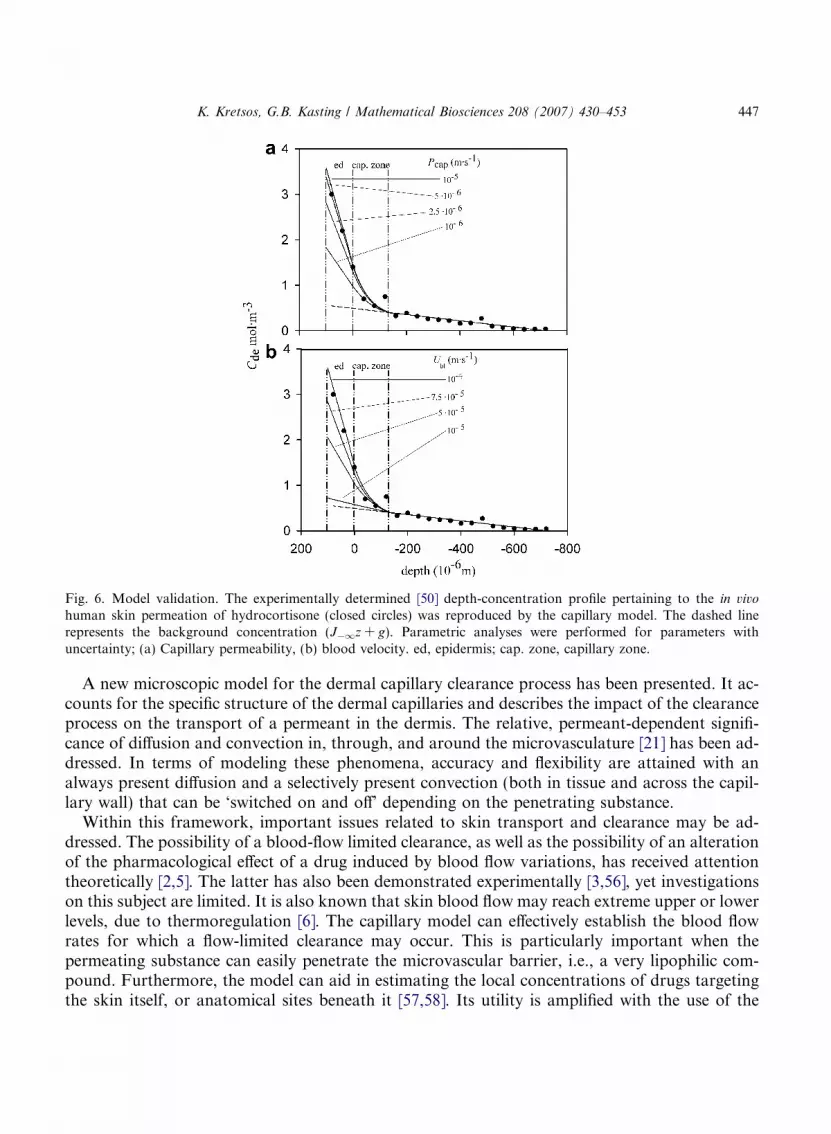
Fig. 6. Model validation. The experimentally determined [50] depth-concentration profile pertaining to the in vivo
human skin permeation of hydrocortisone (closed circles) was reproduced by the capillary model. The dashed linerepresents the background concentration (J�1z + g). Parametric analyses were performed for parameters withuncertainty; (a) Capillary permeability, (b) blood velocity. ed, epidermis; cap. zone, capillary zone.
K. Kretsos, G.B. Kasting / Mathematical Biosciences 208 (2007) 430–453 447
A new microscopic model for the dermal capillary clearance process has been presented. It ac-counts for the specific structure of the dermal capillaries and describes the impact of the clearanceprocess on the transport of a permeant in the dermis. The relative, permeant-dependent signifi-cance of diffusion and convection in, through, and around the microvasculature [21] has been ad-dressed. In terms of modeling these phenomena, accuracy and flexibility are attained with analways present diffusion and a selectively present convection (both in tissue and across the capil-lary wall) that can be ‘switched on and off’ depending on the penetrating substance.
Within this framework, important issues related to skin transport and clearance may be ad-dressed. The possibility of a blood-flow limited clearance, as well as the possibility of an alterationof the pharmacological effect of a drug induced by blood flow variations, has received attentiontheoretically [2,5]. The latter has also been demonstrated experimentally [3,56], yet investigationson this subject are limited. It is also known that skin blood flow may reach extreme upper or lowerlevels, due to thermoregulation [6]. The capillary model can effectively establish the blood flowrates for which a flow-limited clearance may occur. This is particularly important when thepermeating substance can easily penetrate the microvascular barrier, i.e., a very lipophilic com-pound. Furthermore, the model can aid in estimating the local concentrations of drugs targetingthe skin itself, or anatomical sites beneath it [57,58]. Its utility is amplified with the use of the

448 K. Kretsos, G.B. Kasting / Mathematical Biosciences 208 (2007) 430–453
accompanying macroscopic approach. As was demonstrated, the results of the admittedly com-plex microscopic model can be fitted into an easier to implement whole-skin version that considerscapillary clearance occurring throughout the dermis [30] or in a homogeneously absorbing zonelocated within the layer (present calculations). This whole-skin approach may then serve as thelink of the capillary model to experimentally measured properties.
Further steps may increase the model’s applicability and predictive power. These include (a)linking with available models that predict the distribution of the permeant in the stratum corneumand viable epidermis; (b) extending the model to the time dimension in order to simulate non-steady-state behavior, (c) including explicitly in the calculations convective transport processes,based on the framework that has already been provided, to widen the model’s applicability tolarge molecules; (d) adding to the model the lymphatic capillary network, either deterministicallyin a way similar to the treatment of blood capillaries, or stochastically by regarding them as alocalized sink at the lower boundary of the unit cell; (e) applying the model to the analysis ofthe thermoregulation process since the mathematics of transcapillary diffusion are very similarto that describing heat transfer from the blood to tissue; (f) tailoring the model to the importantproblem of the transcapillary escape of endogenous substances, e.g., glucose, urea, into the skininterstitium. The latter is directly linked to the monitoring of these substances for diabetics andfor patients with kidney failure.
Acknowledgments
As this work is, in part, part of Kosmas Kretsos doctoral dissertation we thank his academicadvisor, Dr. Johannes M. Nitsche for suggesting the problem and for his guidance and support.Financial support came from the Procter & Gamble Company’s International Program for Ani-mal Alternatives, NSF GOALI Grant BES-9818160, and NIOSH Grant R01 OH007529.
Appendix A
A.1. Unit vectors used in defining the capillary surface
The unit vectors (nt, nn, and nb) that aid in defining the capillary surface, were calculatedthrough the use of their definitive equations and their known properties, e.g., unit magnitude,direction. The change in curvature along the capillary wall is not abrupt, thus we can considerthe respective vectors of the capillary’s centerline:
nt ¼ AðrÞex þ BðrÞez ¼b=cosh2ðrÞffiffiffiffiffiffiffiffiffiffiffiffiffiffiffiffiffiffiffiffiffiffiffiffiffiffiffiffiffiffiffiffiffiffiffiffiffiffiffiffiffi
b2=cosh4ðrÞ þ 4c2r2
q ex �2crffiffiffiffiffiffiffiffiffiffiffiffiffiffiffiffiffiffiffiffiffiffiffiffiffiffiffiffiffiffiffiffiffiffiffiffiffiffiffiffiffi
b2=cosh4ðrÞ þ 4c2r2
q ez; ð40Þ
nn ¼ C�ðrÞex þ DðrÞez ¼ ffiffiffiffiffiffiffiffiffiffiffiffiffiffiffiffiffiffiffiffiffiffiffiffiffiffiffiffiffiffiffiffiffiffi
1
1þ ½AðrÞ=BðrÞ�2
sex �
ffiffiffiffiffiffiffiffiffiffiffiffiffiffiffiffiffiffiffiffiffiffiffiffiffiffiffiffiffiffiffiffiffiffiffiffiffiffiffiffiffiffi1� 1
1þ ½AðrÞ=BðrÞ�2
sez; ð41Þ
nb ¼ E�ðrÞey ¼ ½C�ðrÞBðrÞ � DðrÞAðrÞ�ey: ð42Þ

K. Kretsos, G.B. Kasting / Mathematical Biosciences 208 (2007) 430–453 449
The geometric parameters b and c are defined in the Methods. The pair E+(r), C+(r) is used toproduce one ‘limb’ of the capillary (0 < r < 2.74) and the pair E�(r), C�(r) is used for the otherlimb (�2.74 < r < 0).
A.2. Doubly periodic Green’s function
The Green’s function Gðk; koÞ is the building block for the determination of the dermal concen-tration. It is the solution of Eq. (43) [41] subjected to periodic conditions analogous to Eqs. (2)–(5).
r2Gðk; koÞ þ dðk; koÞ ¼ 0: ð43Þ
Since most skin permeants do not accumulate in the lower strata of skin it is reasonable to assumethat the concentration as z! �1 does not vary linearly with depth, but rather is constant. Thiswould translate to J�1 = 0 in Eq. (6). Since (a) we are considering steady-state conditions, (b) weare imposing periodic boundary conditions on the lateral dimensions, and (c) there is a zero-fluxcondition at z ¼ �1, whatever flux is generated by our unit source must be counterbalanced bythe flux at z ¼ þ1. This translates to Eq. (44):
oGðx; y;þ1Þoz
¼ � 1
lx � ly
: ð44Þ
The solution (Eqs. (45) and (46)) was obtained by applying the method of separation of variables.
G¼ 0þ 1
2plx
X1m¼1
1
mcos½kmðy� yoÞ�exp½kmðz� zoÞ�þ
1
2ply
X1n¼1
1
ncos½knðx� xoÞ�exp½knðz� zoÞ�
þ 4
lxly
X1n¼1
X1m¼1
fcos½knðx� xoÞ�cos½kmðy� yoÞ�g exp½kmnðz� zoÞ�
2kmn
� �; z< zo ð45Þ
G ¼ � 1
lxly
ðz� zoÞ þ1
2plx
X1m¼1
1
mcos½kmðy � yoÞ� exp½�kmðz� zoÞ�
þ 1
2ply
X1n¼1
1
ncos½knðx� xoÞ� exp½�knðz� zoÞ�
þ 4
lxly
X1n¼1
X1m¼1
fcos½knðx� xoÞ� cos½kmðy � yoÞ�g exp½�kmnðz� zoÞ�
2kmn
� �; z > zo
ð46Þ
with the eigenvalues being kn ¼ 2pn=lx and km ¼ 2pm=ly and with kmn ¼ffiffiffiffiffiffiffiffiffiffiffiffiffiffiffik2
m þ k2n
q.
A.3. Calculations involving the integral of 1/jk � koj
We define a local coordinate system, XYZ. We let the X axis coincide with a small, approxi-mately straight, capillary centerline segment (of length h) and the axes’ point of origin coincidewith the middle point of this segment (Fig. A1). A point-source situated on this segment has coor-

Fig. A1. Definition sketch of the local geometry in the calculations involving the integral 1/4pjk–koj.
450 K. Kretsos, G.B. Kasting / Mathematical Biosciences 208 (2007) 430–453
dinates (s, 0,0). The projection of k � (x, y, z) on the X axis has variable coordinates (x 0, 0,0).Since it is y2 + z2 = q2, the integral required is shown in equation given below:
I ¼Z h
2
�h2
1ffiffiffiffiffiffiffiffiffiffiffiffiffiffiffiffiffiffiffiffiffiffiffiffiffiffiffiðx0 � sÞ2 þ q2
q dx0: ð47Þ
This calculation is performed with a change of variables, by setting x 0 = s + q tanh with h 2 (�p/2,p/2). The utility of the function tanh lies in the fact that it is able to sample all possible valueswithout becoming infinite. By translating the integral’s arguments in the new variable, we haveffiffiffiffiffiffiffiffiffiffiffiffiffiffiffiffiffiffiffiffiffiffiffiffiffiffiffiðs� x0Þ2 þ q2
q¼ qj sec hj which is equal to qsech since sech > 0, " h 2 (�p/2, p/2). Moreover,
it is evident that dx 0 = qsec2hdh. Thus, the corresponding indefinite integral is:
I ind ¼Z
1ffiffiffiffiffiffiffiffiffiffiffiffiffiffiffiffiffiffiffiffiffiffiffiffiffiffiffiðx0 � sÞ2 þ q2
q dx0 ¼Z
q sec2 hdhq sec h
¼Z
sec hdh ¼ ln j sec hþ tan hj: ð48Þ
By noting that tanh = (x 0 � s)/q and that sec h ¼ffiffiffiffiffiffiffiffiffiffiffiffiffiffiffiffiffiffiffiffi1þ tan2 hp
, the indefinite integral is translatedback to the original variables in the following equation
I ind ¼ ln
ffiffiffiffiffiffiffiffiffiffiffiffiffiffiffiffiffiffiffiffiffiffiffiffiffiffiffiffiffi1þ x0 � s
q
� �2s
þ x0 � sq
������������ ¼ ln
ffiffiffiffiffiffiffiffiffiffiffiffiffiffiffiffiffiffiffiffiffiffiffiffiffiffiffiffiffi1þ x0 � s
q
� �2s
þ x0 � sq
24
35: ð49Þ
Finally, the definite integral over an arbitrary length h is
I ¼Z h
2
�h2
1
jk� kojdt
¼ ln
ffiffiffiffiffiffiffiffiffiffiffiffiffiffiffiffiffiffiffiffiffiffiffiffiffiffiffiffiffiffiffiffiffi1þ h=2� s
q
� �2s
þ h=2� sq
24
35� ln
ffiffiffiffiffiffiffiffiffiffiffiffiffiffiffiffiffiffiffiffiffiffiffiffiffiffiffiffiffiffiffiffiffiffiffiffi1þ �h=2� s
q
� �2s
þ�h=2� sq
24
35: ð50Þ

K. Kretsos, G.B. Kasting / Mathematical Biosciences 208 (2007) 430–453 451
The parameters q, s depend on the local geometry and the length of each capillary centerline seg-ment is given by h = sj+1 � sj. Thus they make Eq. (50) applicable to every segment of the capil-lary loop. However, they must be translated to the ‘global’ coordinate system whenever theformula is used. Batchelor [63] derived a similar analytical formula for the integral 1/jk � koj per-taining to a slender body with an irregular cross section and with k being very close to the body,i.e., 2q/h� 1. Eq. (50) agrees with Batchelor’s formula up to the fourth decimal when2q/h = 0.01.
Appendix B. Supplementary data
Supplementary data associated with this article can be found, in the online version, atdoi:10.1016/j.mbs.2006.10.012.
References
[1] N.A. Monteiro-Riviere, A.O. Inman, J.E. Riviere, S.C. McNeill, M.L. Francoeur, Topical penetration ofpiroxicam is dependent on the distribution of the local cutaneous vasculature, Pharm. Res. 10 (1993) 1326.
[2] G.B. Kasting, P.J. Robinson, Can we assign an upper limit to skin permeability? Pharm. Res. 10 (1993) 930.[3] N.L. Benowitz, J. Peyton, P. Olsson, C.-J. Johansson, Intravenous nicotine retards transdermal absorption of
nicotine: evidence of blood flow-limited percutaneous absorption, Clin. Pharmacol. Ther. 52 (1992) 223.[4] J.E. Riviere, B. Sage, P.L. Williams, Effects of vasoactive drugs on transdermal lidocaine iontophoresis, J. Pharm.
Sci. 80 (1991) 615.[5] G.L. Flynn, B. Stewart, Percutaneous drug penetration: choosing candidates for transdermal development, Drug
Dev. Res. 13 (1988) 169.[6] N. Charkoudian, Skin blood flow in adult human thermoregulation: How it works, when it does not, and why,
Mayo Clin. Proc. 78 (2003) 603.[7] S.M. Hammersborg, M. Farstad, O. Haugen, V. Kvalheim, H. Onarheim, P. Husby, Time course variations of
haemodynamics, plasma volume and microvascular fluid exchange following surface cooling: an experimentalapproach to accidental hypothermia, Resuscitation 65 (2005) 211.
[8] J.P. Bantle, W. Thomas, Glucose measurement in patients with diabetes mellitus with dermal interstitial fluid, J.Lab. Clin. Med. 130 (1997) 436.
[9] E.J. Service, P.C. O’Brien, S.D. Wise, S. Ness, S.M. LeBlanc, Dermal interstitial glucose as an indicator of ambientglycemia, Diabetes Care 20 (1997) 1426.
[10] M.K. Robinson, G.F. Gerberick, C.A. Ryan, P. McNamee, I.R. White, D.A. Basketter, The importance ofexposure estimation in the assessment of skin sensitization risk, Contact Dermatitis 42 (2000) 251.
[11] D.D. Tang-Liu, R.M. Matsumoto, J.I. Usansky, Clinical pharmacokinetics and drug metabolism of tazarotene: anovel topical treatment for acne and psoriasis, Clin. Pharmacokinet. 37 (1999) 273.
[12] T.-W. Wong, Y.-L. Zhao, A. Sen, S.W. Hui, Pilot study of topical delivery of methotrexate by electroporation, Br.J. Dermatol. 152 (2005) 524.
[13] A. Krogh, The number and distribution of capillaries in muscles with calculations of the oxygen pressure headnecessary for supplying the tissue, J. Physiol. (London) 52 (1919) 409.
[14] A. Krogh, The supply of oxygen to the tissues and the regulation of the capillary circulation, J. Physiol. (London)52 (1919) 457.
[15] J.B. Bassingthwaighte, T.J. Knopp, J.B. Hazelrig, A concurrent flow model for capillary-tissue exchanges, in: C.Crone, N.A. Lassen, (Eds.), Capillary Permeability, Alfred Benzon Symposium II, Academic Press, NY, 1970, pp.60–80.

452 K. Kretsos, G.B. Kasting / Mathematical Biosciences 208 (2007) 430–453
[16] C.A. Poulain, B.A. Finlayson, J.B. Bassingthwaighte, Efficient numerical methods for nonlinear-facilitated transport and exchange in a blood-tissue exchange unit, Ann. Biomed. Eng. 25 (1997)547.
[17] M.S. Titcombe, M.J. Ward, An asymptotic study of oxygen transport from multiple capillaries to skeletal muscletissue, SIAM J. Appl. Math. 60 (2000) 1767.
[18] G.C. Sharma, M. Jain, A computational solution of mathematical model for oxygen transport in peripheral nerve,Comput. Biol. Med. 34 (2004) 633.
[19] J.P. Kirkpatrick, D.M. Brizel, M.W. Dewhirst, A mathematical model of tumor oxygen and glucose mass transportand metabolism with complex reaction kinetics, Radiat. Res. 159 (2003) 336.
[20] N.M. Tsoukias, A. Popel, A model of nitric oxide capillary exchange, Microcirculation 10 (2003) 479.[21] K. Kretsos, G.B. Kasting, Dermal capillary clearance: physiology and modeling, Skin Pharmacol. Physiol. 18
(2005) 55.[22] T.S. Koh, L.H. Cheong, Z. Hou, Y.C. Soh, A physiologic model of capillary-tissue exchange for dynamic contrast-
enhanced imaging of tumor microcirculation, IEEE Trans. Biomed. Eng. 50 (2003) 159.[23] T.W. Secomb, R. Hsu, E.Y.H. Park, M.W. Dewhirst, Green’s function methods for analysis of oxygen delivery to
tissue by microvascular networks, Ann. Biomed. Eng. 32 (2004) 1519.[24] J. Waniewski, Distributed modeling of diffusive solute transport in peritoneal dialysis, Ann. Biomed. Eng. 30
(2002) 1181.[25] D.A. Beard, K.A. Schenkman, E.O. Feigl, Myocardial oxygenation in isolated hearts predicted by an anatomically
realistic microvascular transport model, Am. J. Physiol. 285 (2003) H1826.[26] E.P. Salathe, Mathematical analysis of oxygen concentration in a two dimensional array of capillaries, J. Math.
Biol. 46 (2003) 287.[27] Y. Ji, J. Liu, Vasculature based model for characterizing the oxygen transport in skin tissues – analogy to the
Weinbaum-Jiji bioheat equation, Heat Mass Transfer 40 (2004) 627.[28] D.A. Beard, J.B. Bassingthwaighte, Advection and diffusion of substances in biological tissues with complex
vascular networks, Ann. Biomed. Eng. 28 (2000) 253.[29] W. Wang, A critical parameter for transcapillary exchange of small solutes in countercurrent systems, J. Biomech.
33 (2000) 543.[30] K. Kretsos, G.B. Kasting, J.M. Nitsche, Distributed diffusion-clearance model for transient drug distribution
within the skin, J. Pharm. Sci. 93 (2004) 2820.[31] I.M. Braverman, The cutaneous microcirculation: Ultrastructure and microanatomical organization, Microcircu-
lation 4 (1997) 329.[32] W.P. Paaske, P. Sejrsen, Permeability of continuous capillaries, Dan. Med. Bull. 36 (1989) 570.[33] I.M. Braverman, A. Yen, Ultrastructure of the human dermal microcirculation. II. The capillary loops of the
dermal papillae, J. Invest. Dermatol. 68 (1977) 44.[34] T.J. Ryan, Cutaneous circulation, in: L.A. Goldsmith (Ed.), Biochemistry and Physiology of the Skin, Oxford
University Press, New York, 1983, pp. 817–877.[35] K.N. An, E.P. Salathe, A theory of interstitial fluid motion and its implications for capillary exchange, Microvasc.
Res. 12 (1976) 103.[36] G.J. Fleischman, T.W. Secomb, Effect of extravascular pressure gradients on capillary fluid exchange, Math.
Biosci. 81 (1986) 145.[37] K.L. Lin, L. Lopez, J.D. Hellums, Blood flow in capillaries, Microvasc. Res. 5 (1973) 7.[38] R. Skalak, Blood rheology, in: F.C. Hoppensteadt (Ed.), Lectures in Applied Mathematics – Mathematical
Aspects of Physiology, American Mathematical Society, Providence, Rhode Island, 1981, pp. 109–139.[39] A.A. Merrikh, J.L. Lage, Effect of blood flow on gas transport in a pulmonary capillary, J. Biomech. Eng. 127
(2005) 432.[40] J.F. Gross, J. Aroesty, Mathematical models of capillary flow: a critical review, Biorheology. 9 (1972) 225.[41] W.M. Deen, Analysis of Transport Phenomena, Oxford University Press, New York, 1998, pp. 132–164.[42] K. Kretsos, Ph. D. Thesis, Buffalo, State University of New York, NY, 2003.[43] J.M. Nitsche, On Brownian dynamics with hydrodynamic wall effects: a problem in diffusion near a fiber, and the
meaning of the no-flux boundary condition, Chem. Eng. Comm. 148–150 (1996) 623.
K. Kretsos, G.B. Kasting / Mathematical Biosciences 208 (2007) 430–453 453
[44] N. Guerin, S. Enoch, G. Tayeb, Combined method for the computation of the doubly periodic Green’s function, J.Electromagnet. Wave 15 (2001) 202.
[45] K.V. Horoshenkov, S.N. Chandler-Wilde, Efficient calculation of two-dimensional periodic and waveguideacoustic Green’s functions, J. Acoust. Soc. Am. 111 (2002) 1610.
[46] D. Shanks, Non-linear transformations of divergent and slowly convergent sequences, J. Math. Phys. 34 (1955) 1.[47] A.N. Lin, T. Nakatsui, Salicylic acid revisited, Int. J. Dermatol. 37 (1998) 335.[48] H. Wiig, M. DeCarlo, L. Sibley, E.M. Renkin, Interstitial exclusion of albumin in rat tissues measured by a
continuous infusion method, Am. J. Physiol. 263 (1992) H1222.[49] C. Crone, D.G. Levitt, Capillary permeability to small solutes, in: Handbook of Physiology. The Cardiovascular
System. Microcirculation, American Physiological Society, Bethesda, MD, 1984, pp. 411–466.[50] A. Zesch, H. Schaefer, Penetrationkinetik von radiomarkiertem hydrocortison aus verschiedenartigen salbengr-
undlagen in die menschliche haut. II. In vivo, Arch. Derm. Forsch. 252 (1975) 245.[51] A.C. Moffat, M.D. Osselton, B. Widdop (Eds.), Clarke’s analysis of drugs and poisons, third ed., Pharmaceutical
Press, London, 2003.[52] S.M. Wallace, H.M. Falkenberg, J.O. Runikis, W.D. Stewart, Skin levels and vasoconstrictor assay of topically
applied hydrocortisone, Arch. Dermatol. 115 (1979) 440.[53] G.B. Kasting, R.L. Smith, B.D. Anderson, Prodrugs for dermal delivery: solubility, molecular size, and functional
group effects, in: K.B. Sloan (Ed.), Prodrugs: Topical and Ocular Drug Delivery, Marcel Dekker, New York, 1992,pp. 117–161.
[54] E.H. Starling, On the absorption of fluids from the convective tissue spaces, J. Physiol. 19 (1896) 312.[55] R.J. Scheuplein, R.L. Bronaugh, Percutaneous absorption, in: L.A. Goldsmith (Ed.), Biochemistry and Physiology
of the Skin, Oxford University Press, New York, 1983, pp. 1255–1295.[56] P. Singh, M.S. Roberts, Effects of vasoconstriction on dermal pharmacokinetics and local tissue distribution of
compounds, J. Pharm. Sci. 83 (1994) 783.[57] S.E. Cross, M.S. Roberts, Targeting local tissues by transdermal application: understanding drug physicochemical
properties that best exploit protein binding and blood flow effects, Drug Dev. Res. 46 (1999) 309.[58] S.C. McNeill, R.O. Potts, M.L. Francoeur, Local enhanced topical delivery (LETD) of drugs: Does it truly exist?
Pharm. Res. 9 (1992) 1422.[59] E. Khalil, K. Kretsos, G.B. Kasting, Glucose partition coefficient and diffusivity in the lower skin layers, Pharm.
Res. 23 (2006) 1237.[60] P. Singh, M.S. Roberts, Dermal and underlying tissue pharmacokinetics of salicylic acid after topical application,
J. Pharmacokinet. Biopharm. 21 (1993) 337.[61] J.C. Parker, M.A. Perry, A.E. Taylor, Permeability of the microvascular barrier, in: N.C. Staub, A.E. Taylor
(Eds.), Edema, Raven Press, NY, 1984, pp. 143–187.[62] S. Borsadia, A.H. Ghanem, Y. Seta, W.I. Higuchi, G.L. Flynn, C.R. Behl, V.P. Shah, Factors to be considered in
the evaluation of bioavailability and bioequivalence of topical formulations, Skin Pharmacol. 5 (1992) 129.[63] G.K. Batchelor, Slender-body theory for particles of arbitrary cross-section in Stokes flow, J. Fluid Mech. 44
(1970) 419.
Recommended


