Nguan Soon TanTang, Jeak Ling Ding, Sander Kersten andHuang, Siu Kwan Sze, Mark Boon Yang Punugu, Chek Kun Tan, Royston-LukePengcheng Zhu, Ming Jie Tan, Lakshmi Yan Yih Goh, Mintu Pal, Han Chung Chong, Proteins to Modulate Wound HealingAngiopoietin-like 4 Interacts with MatrixSignal Transduction:
doi: 10.1074/jbc.M110.108175 originally published online August 21, 20102010, 285:32999-33009.J. Biol. Chem.
10.1074/jbc.M110.108175Access the most updated version of this article at doi:
.JBC Affinity SitesFind articles, minireviews, Reflections and Classics on similar topics on the
Alerts:
When a correction for this article is posted•
When this article is cited•
to choose from all of JBC's e-mail alertsClick here
Supplemental material:
http://www.jbc.org/content/suppl/2010/09/07/M110.108175.DC1.html
http://www.jbc.org/content/285/43/32999.full.html#ref-list-1
This article cites 45 references, 15 of which can be accessed free at
by guest on June 28, 2013http://www.jbc.org/Downloaded from

Angiopoietin-like 4 Interacts with Matrix Proteins toModulate Wound Healing*□S
Received for publication, January 26, 2010, and in revised form, July 30, 2010 Published, JBC Papers in Press, August 21, 2010, DOI 10.1074/jbc.M110.108175
Yan Yih Goh‡1, Mintu Pal‡1, Han Chung Chong‡, Pengcheng Zhu‡, Ming Jie Tan‡, Lakshmi Punugu‡, Chek Kun Tan‡,Royston-Luke Huang‡, Siu Kwan Sze‡, Mark Boon Yang Tang§, Jeak Ling Ding¶, Sander Kersten�,and Nguan Soon Tan‡2
From the ‡School of Biological Sciences, Nanyang Technological University, 60 Nanyang Drive, Singapore 637551, the §NationalSkin Centre, 1 Mandalay Road, Singapore 308205, the ¶Department of Biological Sciences, National University of Singapore, 14Science Drive, Singapore 117543, and �Wageningen University, 6700 EV Wageningen, The Netherlands
A dynamic cell-matrix interaction is crucial for a rapid cellu-lar response to changes in the environment. Appropriate cellbehavior in response to the changing wound environment isrequired for efficient wound closure. However, theway inwhichwound keratinocytesmodify the wound environment to coordi-nate with such cellular responses remains less studied.We dem-onstrated that angiopoietin-like 4 (ANGPTL4) produced bywound keratinocytes coordinates cell-matrix communication.ANGPTL4 interacts with vitronectin and fibronectin in thewound bed, delaying their proteolytic degradation by metallo-proteinases. This interaction does not interfere with integrin-matrix protein recognition and directly affects cell-matrix com-munication by altering the availability of intact matrix proteins.These interactions stimulate integrin- focal adhesion kinase,14-3-3, and PKC-mediated signaling pathways essential foreffective wound healing. The deficiency of ANGPTL4 in micedelays wound re-epithelialization. Further analysis revealedthat cell migration was impaired in the ANGPTL4-deficientkeratinocytes. Altogether, the findings provide molecular in-sight into a novel control of wound healing via ANGPTL4-de-pendent regulation of cell-matrix communication. Given theknown role of ANGPTL4 in glucose and lipid homeostasis, it is aprime therapeutic candidate for the treatment of diabeticwounds. It also underscores the importance of cell-matrix com-munication during angiogenesis and cancer metastasis.
Skin repair after an injury proceeds via a finely tuned patternof integrated biological events aimed at restoration of the epi-thelial barrier. The inflammatory stage of repair is followedby the proliferation andmigration of keratinocytes, a processcalled re-epithelialization (1). These events are regulatedspatiotemporally by several classical growth factors andcytokines, the effects of which have been well documented (2).Less studied are extracellular factors such as matricellular pro-teins and adipocytokines, both shown to have a profound local
impact duringwound repair (3, 4). Effective directed cellmigra-tion requires constant cellular interactionwith the extracellularmatrix (ECM)3 in response to the changing wound environ-ment. Although the importance of such cell-matrix communi-cation in wound healing is well recognized, themechanism thatmodifies the external wound microenvironment for coordi-nated keratinocyte behavior remains unclear.Integrins on the cell surface often function as biosensors
to constantly interrogate the wound environment and mod-ulate cell responses accordingly. Binding of integrins to theircognate matrix proteins activates intracellular signalingpathways that modulate a broad range of cellular processes,including cell migration (5). Integrin-mediated signaling re-quires that integrins bind substrate-anchored matrix proteins.This interaction provides mechanical resistance that permitstensional forces to be generated via the actomyosin system (6).In contrast, small soluble matrix protein fragments generatedby the action of proteases during re-epithelialization can com-petewith substrate-anchoredmatrix proteins for integrin bind-ing and impair cell signaling (7). Thus, productive integrin sig-naling depends on the context in which the intact matrixprotein is presented to cells. However, theway inwhichmigrat-ing wound keratinocytes coordinate the balance between sub-strate-anchored and small soluble matrix protein fragments bythe specific induction of wound integrins requires furtherinvestigation.Transcriptional regulation plays an important role in the con-
trol of keratinocytebehavior at thedifferentphasesofwoundheal-ing, but little is known about the mechanism that modifies thewoundmicroenvironment to coordinate with changes in cellu-lar behavior for cell-matrix communication. Effective cell-ma-trix communication is crucial for efficient wound healing. Sev-eral nuclear hormone receptors, one of the largest knownclasses of transcription factors, have been implicated woundrepair (8, 9). Of interest, studies have shown that nuclear hor-mone receptor peroxisome proliferator-activated receptor �/�
* This work was supported by A*STAR BMRC Grant 05/1/22/19/377, Ministryof Education Grant ARC 18/08, and Nanyang Technological UniversityGrants RGD 127/05 and 158/06.
□S The on-line version of this article (available at http://www.jbc.org) containssupplemental Figs. S1–S3 and Table S1.
1 Both authors contributed equally to this work.2 To whom correspondence should be addressed. Tel.: 65-63162941; Fax:
65-67913856; E-mail: [email protected].
3 The abbreviations used are: ECM, extracellular matrix; ANGPTL4, angiopoi-etin-like 4; nANGPTL4, N-terminal coiled-coil fragment; KANGPTL4, humankeratinocytes knockdown of ANGPTL4; cANGPTL4, C-terminal fibrinogen-like domain; KCTRL, human keratinocytes with control scrambled siRNA;MMP, matrix metalloproteinase; PLA, proximity ligation assay; PPAR, per-oxisome proliferator-activated receptor; SPR, surface plasmon resonance;WF, wound fluid/exudate; LCM, laser-capture microdissection; CM, condi-tioned medium; FACS, fluorescence-activated cell sorter.
THE JOURNAL OF BIOLOGICAL CHEMISTRY VOL. 285, NO. 43, pp. 32999 –33009, October 22, 2010© 2010 by The American Society for Biochemistry and Molecular Biology, Inc. Printed in the U.S.A.
OCTOBER 22, 2010 • VOLUME 285 • NUMBER 43 JOURNAL OF BIOLOGICAL CHEMISTRY 32999 by guest on June 28, 2013http://www.jbc.org/Downloaded from

(PPAR�/�) is an early transcription factor that modulateskeratinocyte response to inflammation during wound healing(10, 11). Most studies have focused on intracellular signaling oreventsmediated by PPAR�/� that were important for keratino-cyte survival and migration (10, 12, 13). However, the mecha-nism by which PPAR�/� alters the wound microenvironmentfor effective cell-matrix communication remains unknown.Conceivably, as an intracellular transcription factor, PPAR�/�is likely to exert such an effect via extracellular factors.Angiopoietin-like 4 (ANGPTL4) belongs to a group of se-
creted factors that play important roles in lipid and glucosemetabolism (14). Its expression is up-regulated by PPAR (15)and by hypoxia (16). ANGPTL4 is also implicated in breastcancer metastasis via the regulation of vascular integrity (17,18). The native ANGPTL4 is proteolytically cleaved, giving riseto an N-terminal coiled-coil fragment (nANGPTL4) and aC-terminal fibrinogen-like domain (cANGPTL4). Despitemultiple physiological and pathological functions, the signifi-cance of the different cleaved fragments of ANGPTL4 is onlybeginning to be understood. Importantly, the identity of thebinding partners for ANGPTL4 and the mechanism by whichANGPTL4 modulates cell migration is unknown, hamperingour understanding of its contribution to wound healing andcancer metastasis.Here, we show that ANGPTL4 is a PPAR�/� target gene in
keratinocytes and that its expression is elevated after injury.Weshow that ANGPTL4 produced by wound keratinocytes coor-dinates cell-matrix communication. Specifically, ANGPTL4interacts with vitronectin and fibronectin in the wound bed,delaying their proteolytic degradation by metalloproteinasesand thereby regulating the availability of local extracellularmatrix. This interaction does not interfere with the binding ofmatrix protein to its cognate integrin receptor or with integrin-mediated signaling. Our findings reveal a novel control of thewound environment by keratinocytes that coordinates the dy-namic interactions between integrins and components of extra-cellular matrices.
EXPERIMENTAL PROCEDURES
Reagents—Sensor CM5 chips, amine coupling kits, and Immo-biline pK buffers were from GE Healthcare. Purified vitronectin,fibronectin, and laminin were from Calbiochem. Transfectionreagent ExGen 500 was from Fermentas. Real-time PCR KAPASYBR Fast Master mix was from KAPABiosystem. DUOlinkproximity ligation assay was from Olink Bioscience. Dro-sophila Schneider 2 (S2) expression vector harboring a pro-prietary secretory signal pSSAc5.1/V5-His A was as pre-viously described (19). Double promoter pFIV-U1/H6-Purolentivirus-based siRNAvector (catalog #SI110A-1) and pPACKF1packaging plasmid kit were from System Biosciences. Purifiedmatrix proteins were purchased from Sigma. All chemicalswere from Sigma unless otherwise stated.Antibodies—p21-activated kinase (PAK), LIMK1 (LIM ki-
nase 2), PKB�, and their cognate phosphorylated forms werefrom Cell Signaling. Rac1 and cdc42 were from Cytoskeleton.�-Tubulin, His tag, laminin, fibronectin, matrix metallopro-teinases (MMPs), and HRP-conjugated secondary antibodieswere from Santa Cruz Biotechnology; vitronectin and integrin
�v�5 were from Chemicon; keratin 6 for wound keratinocytesand hair follicle, �-smooth muscle actin for myofibroblasts,F4/80 for macrophages, and CD31 for endothelial cells werefrom BioLegend. Anti-human PPAR�/� monoclonal antibod-ies were fromPerseus Proteomics Inc., Japan. Rabbit polyclonalantibodies against the C-terminal region of human (186–406amino acid) and mouse (190–410 amino acids) ANGPTL4were produced in-house. Briefly, female rabbits (New ZealandWhite, 2–2.5 kg) were injected intramuscularly with 300 �g ofrecombinant proteins homogenized with 500 �l of completeFreund’s adjuvant solution. First and second booster immuni-zation with the same immunization dose were performed 3 and6weeks after priming immunization using incomplete Freund’sadjuvant, respectively. Final harvest was done by bleeding awhole blood volume, and the rabbits were then culled withinjection of euthanasia into themarginal ear vein. The carcasseswere disposed after confirming noheart beat and corneal reflex,pedal reflex reactions. Preimmune blood sampling was col-lected as the negative control. Antibodies were purified by Pro-tein A affinity chromatography as recommended by manufac-turer (GE Healthcare).Keratinocyte Culture—Primary human keratinocytes (Cas-
cade Biologics) were cultured inQuantum153medium supple-mented with insulin, transferrin, EGF, cholera toxin, and 5%FBS (PAA Laboratories) in a 5% CO2, 37 °C humidified incuba-tor. This medium is a modification of the keratinocyte mediumpreviously described (20, 21). Medium was changed every 3days. Cells were subcultured upon reaching 70% confluence.Briefly, medium was removed, and the cells washed with PBS.Trypsin (0.25%), EDTA (1mM) in PBS was added to the culture(0.08 ml/cm2) and incubated at room temperature for 15 min.The flask was rapped gently to dislodge cells from the surface ofthe flask. PBS containing 1% dialyzed FBS was added, and thecells were collected by centrifugation. The cell pellet was resus-pended with fresh medium and subcultured in new flask at2.5 � 103 cells/cm2.Chromatin Immunoprecipitation (ChIP)—ChIP was per-
formed according to the manufacturer’s (Upstate Biotechnol-ogy) instructions with some modifications. Briefly, ChIP assaywas performed using the monoclonal PPAR�/� antibody. Cellswere treated with 1% formaldehyde at 37 °C for 15 min. Cross-linked DNA was sonicated to form fragments ranging from200 to 500 bp in length. DNA fragments were reverse cross-linked at 65 °C for 6 h. The DNA was subsequently purifiedusing Qiaquick column (Qiagen). DNA was amplified by PCRfor 20–23 cycles. The ChIP primers for the amplification of thePPAR-response element of the human ANGPTL4 gene were aspreviously described (22).SkinWounding Experiment—Wounding was performed as
previously described (23, 24). Briefly, the hair follicle cycle ofeach mouse was synchronized by shaving the back of theanimal 2 weeks before the start of the experiment. Afteranesthetizing, the mice were shaven. A full thickness mid-dorsal wound (0.5-cm2, square-shaped)was created by excisingthe skin and the underlying panniculus carnosus. Wound clo-surewasmeasured daily in a double-blinded fashion until it wascomplete. At indicated days post-wounding, the entire wound,including a 5-mmmargin, was excised.Wounds were dissected
ANGPTL4 Modulates Cell-Matrix Communication
33000 JOURNAL OF BIOLOGICAL CHEMISTRY VOLUME 285 • NUMBER 43 • OCTOBER 22, 2010 by guest on June 28, 2013http://www.jbc.org/Downloaded from

for immunohistochemistry, RNA, and protein analyses (23, 25).Six-week-old PPAR�/��/� and �/� (24) and pure bred wildtype (ANGPTL4�/�) andANGPTL4-knock-out (ANGPTL4�/�)male mice were used (26). All mice used in this study had aC57BL/6 background and were individually caged, housed in atemperature-controlled room (23 °C) on a 10-h dark/14-h lightcycle, and fed with the standard mouse chow diet. Animalexperimentswere approved by theUniversity InstitutionalAni-mal Care and Use Committee (ARF-SBS/NIE-A-0093, ARFSBS/NIE-A-0078 and ARF SBS/NIE-A-004).Western Blot and Immunofluorescence Assays—Cells or tis-
sues were lysed in ice-cold lysis buffer (20 mM NaH2PO4, 250mM NaCl, 1% Triton X-100, 0.1% SDS) supplemented withcomplete protease inhibitors (Roche Applied Science). Equalamounts of protein extracts were resolved by SDS-PAGEand electroblotted onto polyvinylidene difluoride membranesfor Western analysis. Membranes were processed accordingto standard protocol and developed using chemilumines-cence (Millipore). Equal loading/transfer was verified by Co-omassie staining of gels or by immunodetection of �-tubulin.Wound biopsies were fixed with 4% paraformaldehyde in PBSfor 2 h at 25 °C. The fixed tissues were centrally bisected,washed twice with PBS, and embedded in Tissue-Tek OCTcompound medium (Sakura) overnight at 4 °C. The tissueswere subsequently frozen at �70 °C for cryosectioning. Cryo-stat sections (8 �m) mounted on SuperFrost Plus slides wereanalyzed by immunofluorescence as previously described,except that anti-ANGPTL4 antibodies were used (23). Theslides presenting the largest wound diameter was defined asthe wound center. As a control for immunofluorescence stain-ing, 10-fold more peptide antigen was preincubated with anti-ANGPTL4 at 4 °C for 1 h before use. Images were taken using aLSM710 confocal laser scanning microscope with a Plan-Apo-chromat 40�/1.40 oil objective and ZEN software (Carl Zeiss).Flow Cytometry (FACS)—Wound tissues were subjected to
FACS analysis as previously described (27). Entire excised skinwounds were dispersed enzymatically into single cell suspen-sions. The tissue was incubated with dispase I (1 mg/ml) over-night at 4 °C, minced, and incubated in digestion buffer con-taining hyaluronidase (1 mg/ml), collagenase D (1 mg/ml),and DNase (100 unit/ml) (Sigma) in a 37 °C shaking incuba-tor for 2 h. The dispase and hyaluronidase digests werepooled and filtered through a 70-�mNylon cell strainer. Cellswere washed, pelleted, and resuspended in equal volume ofPBS containing 3% FBS. For staining of surface marker, cellswere first blocked with mouse BD Fc Block and then incu-bated with either phyoerythrin- or FITC-conjugated mono-clonal antibodies specific for F4/80 (macrophages) andCD31 (endothelial cells) or control isotype IgG on ice for 30min. After washing with PBS, the samples were subjected toflow cytometry on a FACSCalibur system (BD Biosciences).Data were analyzed using the CellQuest software (BD Bio-sciences). The analyzer threshold was adjusted on the flowcytometer channel to exclude most of the subcellular debris toreduce the background noise.Laser-capture Microdissection (LCM)—Paraffin-embedded
sections of PPAR�/��/� and -�/� wounds (10 �m) weremounted ontoMembraneSlides (Carl Zeiss). Hematoxylin- and
eosin-stained sections were then subjected to LCM usingPALM MicroBeam according to the manufacturer’s instruc-tions (Carl Zeiss). LCM tissues were collected into microcen-trifuge tubes with opaque AdhesiveCaps (Carl Zeiss). RNAwasextracted usingOptimumTMFFPERNA Isolation kit (Ambion)pooled from eight LCM tissues. RNA was reverse-transcribedusing random primers, and the resulting cDNA was used forreal-time PCR.Expression and Purification of Recombinant ANGPTL4
Proteins—The cDNA sequences encoding human full-lengthANGPTL4, nANGPTL4, and cANGPTL4 were amplified byPfu polymerase and subcloned into pSSAc5.1/V5-His-A(19). A histidine tag was introduced between the secretorysignal and the ANGPTL4 cDNA. All ligated products weretransformed into competent Escherichia coliTop 10 bacteriaand selected on Luria broth agar plates containing 80 �g/mlampicillin. Positive cloneswere confirmed byDNAsequencing.Positive constructs were co-transfected with hygromycin ex-pression vector pCoHygro (Invitrogen) into S2 cells. Recombi-nant ANGPTL4 proteins were purified from the conditionedmedium of stable ANGPTL4-expressing S2 cells by preparativeisoelectric membrane electrophoresis as described (28).Surface Plasmon Resonance (SPR) Coupled to Liquid Chro-
matography-Tandem Mass Spectrometry—Purified cANGPTL4was immobilized onto a CM5-carboxylated dextran sensorchip by amine coupling using the Surface Prep Moduleof BIACORE 3000 as recommended by the manufacturer(BIAcore). Acute wound fluid/exudate (WF) was collectedfrom two patients undergoing split-thickness skin grafting.The acute wound fluid was collected daily under sterile condi-tions from beneath a vapor-permeable membrane applied tothe donor site and changed every 24 h for 3 days postoper-atively. WF was centrifuged, aliquoted, and frozen at �70 °C.WF buffered with 50 mM Tris, pH 8.0, was introduced into thecANGPTL4-conjugated CM5 chip at a flow rate of 5�l/min for10minwith running buffer (50mMTris, pH 8.0, 100mMNaCl).After incubation for 45 s, the chamber was washed with thesame buffer, and the bound molecules were subsequentlyeluted using 10 mM glycine, pH 6.0, and collected in a recoveryvial. The CM5 chip was reused to pool more samples afterwashing with running buffer for 10 min at 20 �l/min. Therecovered cANGPTL4-binding proteins were digested withtrypsin, reduced, alkylated, and then analyzed with a FinniganSurveyorHPLC system coupled online to a LTQ-Orbitrapmassspectrometer (Thermo Electron) equipped with a nanospraysource. Proteins were identified using a Mascot search. SPRwas used to determine the dissociation constant of the inter-action between fibronectin and vitronectin with recombi-nant cANGPTL4 immobilized onto a CM5 chip. Six concen-trations (0.16, 0.32, 0.63, 1.25, 2.50, and 5.0 �M) of variousmatrix proteins were used. Each sensorgram was corrected bysubtracting a sensorgram obtained from a reference flow cellwith no immobilized protein. Anti-cANGPTL4 antibodies runagainst the immobilized cANGPTL4 determined the Rmaxvalue to be 251.8 resonance units. Global fitting of the SPR datato a Langmuir 1:1model was used to determine the dissociationconstant (KD) with Scrubber 2 software. Values are given as the
ANGPTL4 Modulates Cell-Matrix Communication
OCTOBER 22, 2010 • VOLUME 285 • NUMBER 43 JOURNAL OF BIOLOGICAL CHEMISTRY 33001 by guest on June 28, 2013http://www.jbc.org/Downloaded from

mean � S.D. of five independent preparations of recombinantproteins.Affinity Co-precipitation Assay—Purified recombinant His-
tagged ANGPTL4, nANGPTL4, or cANGPTL4 was immobi-lized onto nickel-nitrilotriacetic acid resin (GE Healthcare).The resinwaswashedwithwash buffer (50mMTris, pH7.5, 150mMNaCl, 0.1% Triton X-100) to remove excess ANGPTL4. Anequal amount of ANGPTL4-bound resin was dispensed andincubatedwith 500 ng of purifiedmatrix protein in PBS at 25 °Cfor 30 min. The resin was then thoroughly washed with washbuffer. The unbound fractions were pooled, and the boundfractions were released by SDS-PAGE loading dye. Both frac-tions were analyzed by immunoblotting with their indicatedantibodies. Resin treatedwithTris-buffered salinewas used as acontrol. In vivo co-immunoprecipitation was performed usingcorresponding antibodies as previously described (29).SucroseGradient SedimentationAssay—Proteins (1�g)were
allowed to interact at 4 °C for 2 h in 150�l of 50mMTris, pH8.0,and 100 mM NaCl. The protein mixture was size-fractionatedby ultracentrifugation for 16 h at 132,000� g at 18 °C through a5-ml sucrose density gradient (25–40%). Fractions of 300 �lwere collected, chloroform/ethanol-precipitated, and analyzedby Western blot using their respective antibodies.In Situ Proximity Ligation Assay (PLA)—Keratinocytes sub-
cultured overnight on glass chamber slides (Lab-Tek) or cryo-sections of wound biopsies were fixed with 4% paraformalde-hyde for 15 min. The slides were washed twice with PBS andblocked for 1 h at room temperature with 2% BSA in PBS con-taining 0.1%TritonX-100 followed by incubationwith the indi-cated antibody pairs overnight at 4 °C. The slides were washedas described above. DUOlinkTM in situ PLA was performed asrecommended by the manufacturer (OLink Biosciences) usinga slide incubated without primary antibody as a negative con-trol. Triple PLA was performed as previously described withminor modifications (30). Rabbit anti-cANGPTL4 (in-house),mouse anti-integrin �v�5, and goat anti-vitronectin antibodies(Chemicon) were used as proximity probes. DNAwas ligated at37 °C for 1 h. All probe sequences were as previously described(30) and were synthesized by Proligo (Sigma). As a negativecontrol, rabbit anti-cANGPTL4 and mouse anti-integrin �5proximity probes were omitted. Images were taken using aLSM710 confocal laser scanning microscope with a Plan-Apo-chromat 63�/1.40 oil objective and ZEN software (Carl Zeiss).Knockdown of ANGPTL4 and Real-time PCR—siRNA against
humanANGPTL4 and a scrambled sequence control were sub-cloned into the pFIV-H1/U6-puro siRNA lentivirus system(System Biosciences). An equimolar ratio of sense and anti-sense oligonucleotide mixture was heated to 95 °C for 5 minand allowed to anneal in 20mMTris, pH 7.8, 100mMNaCl, and0.2 mM EDTA by slow cooling to room temperature. Theannealed oligonucleotide was phosphorylated using polynucle-otide kinase before ligation with BbsI-linearized pFIV-H1/U6-puro siRNA vector. Ligated products were transformed intocompetent E. coli Top 10 bacteria and selected on Luria brothagar plates containing 80�g/ml ampicillin. Positive cloneswereconfirmed by DNA sequencing. Positive constructs were co-transfected with pPACK packing plasmids into 293TN cellsusing ExGen 500. Supernatant was collected 48 h post-trans-
fection, and pseudovirus-containing precipitate was obtainedby centrifugation at 50,000 � g for 90 min at 4 °C. Cells weretransduced using Polybrene according to the manufacturer’srecommendation. Transduced cells were enriched by 350 �g/ml puromycin selection for 2 weeks. Knockdown efficiency ofANGPTL4 and relative expression level of indicated genesweredetermined by quantitative PCR. All oligonucleotides and Taq-man probes sequences were provided in supplemental TableS1. Control and ANGPTL4-knockdown keratinocytes weredenoted as KCTRL and KANGPTL4, respectively. The interferonresponse detection kit was from System Biosciences.Matrix Protein Degradation Assay—Purified ECM proteins
(200 ng)were first allowed to interactwith various recombinantANGPTL4 proteins (200 ng) before incubation at 37 °C witheither WF or serum-free KANGPTL4-conditioned medium (CM).At the indicated times, aliquots of the reaction were stopped bythe addition of SDS-PAGE loading dye. CM was prepared asfollows; 3 � 106 KANGPTL4 cells were subcultured in a 10-cmdish the day before treatment. The next day cells were treatedwith 50�g/ml TNF-� in 3ml of serum-free basal Quantum 153medium for 12 h. CM was collected, sterile-filtered, and storedat �80 °C for use in assays. Three independent experimentsfrom twoWF samples were performed. Protease inhibition as-says were performed using the protease inhibitors pepstatin A(8 �M), EDTA (8 mM), and PMSF (1 mM) either alone or inindicated combinations in the CM. The matrix proteins wereanalyzed by Western blot using the corresponding antibodies.Statistical Analysis—Data were analyzed statistically by two-
tailed Mann-Whitney tests using SPSS software. Values wereexpressed as the mean � S.E., and p � 0.05 was consideredstatistically significant.
RESULTS
ANGPTL4 Expression Is Regulated by PPAR�/� inKeratinocytes—ANGPTL4 is a direct target gene of PPAR�/� inHaCaT cells, a non-tumorigenic human keratinocyte cellline (22). However, the role, expression, and regulation ofANGPTL4 in skin wound healing is unclear. To this end, wefirst examined the expression level of ANGPTL4 in humankeratinocytes after ligand activation of specific PPAR isotypes.Quantitative PCR revealed that ANGPTL4mRNAwas up-reg-ulated by all three PPAR isotypes (Fig. 1A), with an �8.5-foldinduction with the specific PPAR�/� ligand, GW501516. Wefound that serum, which contains undefined and complexmix-ture of lipid metabolites that can act as ligands for PPARs, alsoincreased ANGPTL4 expression by�5-fold (Fig. 1A). Next, thePPAR response element of the ANGPTL4 (22) from humankeratinocytes was analyzed by ChIP using monoclonal anti-PPAR�/�. Results showed that PPAR�/� was bound to this siteof the ANGPTL4 promoter region (Fig. 1B), indicating thatANGPTL4 is a direct target of PPAR�/� in keratinocytes.Immunoblot analysis of day 3 wound biopsies using differentpolyclonal anti-ANGPTL4 antibodies detected the nativeANGPTL4 and cANGPTL4 in the PPAR�/��/� mice, whereasthe expression of ANGPTL4 was reduced in PPAR�/��/� lit-termates (Fig. 1C). The specificity of anti-cANGTPL4 is shownin supplemental Fig. S1A and specificity of anti-nANGPTL4was as previously reported (15). Day 3 wound biopsies were
ANGPTL4 Modulates Cell-Matrix Communication
33002 JOURNAL OF BIOLOGICAL CHEMISTRY VOLUME 285 • NUMBER 43 • OCTOBER 22, 2010 by guest on June 28, 2013http://www.jbc.org/Downloaded from

used because PPAR�/� expression peaked at day 3 post-wounding (31). Immunofluorescence staining further con-firmed that ANGPTL4was highly expressed in both the woundepithelia and the wound bed in PPAR�/��/� mice, whereasreduced expression was detected in their PPAR�/��/� litter-mates (Fig. 1D). Quantitative PCR analysis of the LCM woundepithelium, dermis, and wound bed of day 3 wound biopsies
from PPAR�/��/� and PPAR�/��/� mice showed that the woundepithelium was the major producerof ANGPTL4 (supplemental Fig.S1B). These results suggested thatANGPTL4 secreted by wound kera-tinocytes may play an importantrole during wound healing.cANGPTL4 Interacts with Spe-
cific Matrix Proteins—To begin tounderstand the role of ANGPTL4during wound healing, we soughtto identify ANGPTL4-binding pro-teins using SPR-MS. Prompted byour initial observation (Fig. 1D), wehypothesized that WF may harborANGPTL4-interacting proteins. Us-ing recombinant cANGPTL4 andWF as the bait and lysate, respec-tively, we identified the ECM pro-teins, vitronectin and fibronectin,as ANGPTL4 binding partners(supplemental Fig. S1C). Recom-binant cANGPTL4 was expressedand purified from Drosophila S2culture medium (supplemental Fig.S1D). Further analyses using SPRwith purified vitronectin and fi-bronectin and ANGPTL4 revealedbinding constants (KD) of �10�7 M
(ANGPTL4 with fibronectin andvitronectin, 3.80 � 1.74 � 10�7 and3.04 � 1.33 � 10�7 M, respectively;cANGPTL4 with fibronectin andvitronectin, 3.52 � 1.41 � 10�7 and5.94 � 1.79 M, respectively. Fig. 2, Aand B). Specific anti-cANGPTL4antibodies against immobilizedcANGPTL4 determined the theo-retical Rmax value to be 251.8 reso-nance units. The experimental Rmaxvalues of fibronectin and vitro-nectin for cANGPTL4 were 238.6and 218.5 resonance units, respec-tively, suggesting a 1:1 stoichiom-etry of binding. This interactionwas specific, as the binding of anti-cANGTPL4 antibody, but not pre-immune IgG, to immobilized-ANGPTL4 blocked its interactionwith vitronectin and fibronectin (Fig.
2, C and D). Specific interactions between cANGPTL4 withvitronectin and fibronectin were confirmed by in vitro affinityco-immunoprecipitation (Fig. 2E). In addition, we also exam-ined the formation of the cANGPTL4-matrix protein complexby sedimentation using sucrose gradient ultracentrifugation,which separates proteins and protein complexes accord-ing to their native molecular weight, with larger proteins/
FIGURE 1. Reduced expression of ANGPLT4 in PPAR�/��/� mice wounds. A, relative mRNA levels ofANGPTL4 in human keratinocytes treated with different agonists selective for each PPAR isotype, ciprofibrate(30 �M, PPAR�), GW501516 (2 nM, GW, PPAR�/�), and pioglitazone (500 nM, PPAR�) are shown. Values aremean � S.E. of four independent studies. Ribosomal protein L27 used as a normalizing housekeeping gene.B, ChIP was done in keratinocytes using pre-immune IgG or monoclonal anti-PPAR�/�. Regulatory region withthe PPAR response element was immunoprecipitated with anti-PPAR�/� and specifically amplified. No ampli-fied signal was obtained with preimmune IgG. A control region upstream of PPAR response element served asthe negative control. Aliquots of chromatin were analyzed before immunoprecipitation (input). M, 100-bp DNAmarker. C, expression is shown of ANGPTL4 protein in day 3 post-wounding mice skin biopsies. Polyclonalantibodies that recognized the N-terminal (anti-nANGPTL4) and C-terminal (anti-cANGPTL4) of ANGPTL4 wereused. �-Tubulin served as loading and transfer control. n � 5. IB, immunoblot. D, shown is immunofluorescencestaining of ANGPTL4 in PPAR�/��/� and PPAR�/��/� day 3 wound biopsies using anti-cANGPTL4. Sectionswere counterstained with DAPI. Negative control is performed with anti-cANGPTL4 preincubated with antigenpeptide. Representative images from wound epithelia and wound beds were shown (n � 5). The dotted whiteline denotes epidermal-dermal junction. Scale bar, 40 �m.
ANGPTL4 Modulates Cell-Matrix Communication
OCTOBER 22, 2010 • VOLUME 285 • NUMBER 43 JOURNAL OF BIOLOGICAL CHEMISTRY 33003 by guest on June 28, 2013http://www.jbc.org/Downloaded from
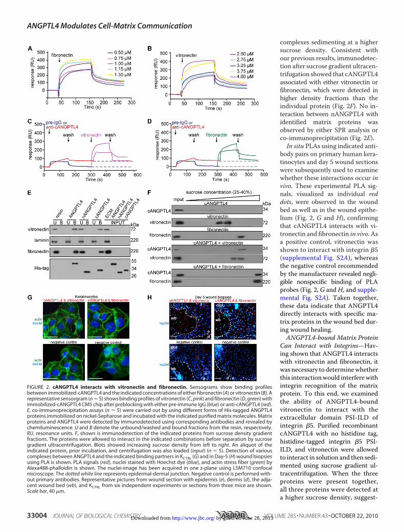
complexes sedimenting at a highersucrose density. Consistent withour previous results, immunodetec-tion after sucrose gradient ultracen-trifugation showed that cANGPTL4associated with either vitronectin orfibronectin, which were detected inhigher density fractions than theindividual protein (Fig. 2F). No in-teraction between nANGPTL4 withidentified matrix proteins wasobserved by either SPR analysis orco-immunoprecipitation (Fig. 2E).In situ PLAs using indicated anti-
body pairs on primary human kera-tinocytes and day 5 wound sectionswere subsequently used to examinewhether these interactions occur invivo. These experimental PLA sig-nals, visualized as individual reddots, were observed in the woundbed as well as in the wound epithe-lium (Fig. 2, G and H), confirmingthat cANGPTL4 interacts with vi-tronectin and fibronectin in vivo. Asa positive control, vitronectin wasshown to interact with integrin �5(supplemental Fig. S2A), whereasthe negative control recommendedby the manufacturer revealed negli-gible nonspecific binding of PLAprobes (Fig. 2,G andH, and supple-mental Fig. S2A). Taken together,these data indicate that ANGPTL4directly interacts with specific ma-trix proteins in the wound bed dur-ing wound healing.ANGPTL4-bound Matrix Protein
Can Interact with Integrins—Hav-ing shown that ANGPTL4 interactswith vitronectin and fibronectin, itwas necessary to determinewhetherthis interactionwould interferewithintegrin recognition of the matrixprotein. To this end, we examinedthe ability of ANGPTL4-boundvitronectin to interact with theextracellular domain PSI-ILD ofintegrin �5. Purified recombinantcANGPTL4 with no histidine tag,histidine-tagged integrin �5 PSI-ILD, and vitronectin were allowedto interact in solution and then sedi-mented using sucrose gradient ul-tracentrifugation. When the threeproteins were present together,all three proteins were detected ata higher sucrose density, suggest-
FIGURE 2. cANGPTL4 interacts with vitronectin and fibronectin. Sensograms show binding profilesbetween immobilized-cANGPTL4 and the indicated concentrations of either fibronectin (A) or vitronectin (B). Arepresentative sensorgram (n � 5) shows binding profiles of vitronectin (C, pink) and fibronectin (D, green) withimmobilized-cANGPTL4 CM5 chip after preblocking with either pre-immune IgG (blue) or anti-cANGPTL4 (red).E, co-immunoprecipitation assays (n � 5) were carried out by using different forms of His-tagged ANGPTL4proteins immobilized on nickel-Sepharose and incubated with the indicated purified matrix molecules. Matrixproteins and ANGPTL4 were detected by immunodetected using corresponding antibodies and revealed bychemiluminescence. U and B denote the unbound/washed and bound fractions from the resin, respectively.RU, resonance units. F, shown is immunodetection of the indicated proteins from sucrose density gradientfractions. The proteins were allowed to interact in the indicated combinations before separation by sucrosegradient ultracentrifugation. Blots showed increasing sucrose density from left to right. An aliquot of theindicated protein, prior incubation, and centrifugation was also loaded (input) (n � 5). Detection of variouscomplexes between ANGPTL4 and the indicated binding partners in KCTRL (G) and in Day-5 (H) wound biopsiesusing PLA is shown. PLA signals (red), nuclei stained with Hoechst dye (blue), and actin stress fiber (green) byAlexa488-phalloidin is shown. The nuclei-image has been acquired in one z-plane using LSM710 confocalmicroscope. The dotted white line represents epidermal-dermal junction. Negative control is performed with-out primary antibodies. Representative pictures from wound section with epidermis (e), dermis (d), the adja-cent wound bed (wb), and KCTRL from six independent experiments or sections from three mice are shown.Scale bar, 40 �m.
ANGPTL4 Modulates Cell-Matrix Communication
33004 JOURNAL OF BIOLOGICAL CHEMISTRY VOLUME 285 • NUMBER 43 • OCTOBER 22, 2010 by guest on June 28, 2013http://www.jbc.org/Downloaded from

ing that ANGPTL4-bound matrixprotein could still interact withPSI-ILD of integrin �5 (Fig. 3A). Invivo co-immunoprecipitation andtriple PLA were used to furtherconfirm this observation. Co-immu-noprecipitation from human keratin-ocyte lysate using antibodiesagainst integrin �v�5, vitronectin,or cANGPTL4 followed by immu-nodetection showed that the cor-responding two proteins were alsofound in the immunoprecipitates(Fig. 3B). Finally, triple PLA furtherrevealed the close proximity ofANGPTL4, integrin �5, and vi-tronectin at focal adhesions (Fig.3C). In retrospect, the PLA signalsfrom ANGPTL4 and matrix pro-teins detected in wound epithelium(Fig. 2H) represented ANGPTL4-bound matrix proteins that hadinteracted with their cognate in-tegrins in keratinocytes. Takentogether, our results suggest thatthe binding of ANGPTL4 to matrixproteins, such as vitronectin, doesnot prevent the matrix protein fromassociating with its cognate integrin.ANGPTL4 Interacts with Matrix
Proteins and Delays Their Degra-dation—Directed migration of woundkeratinocytes over the provisionalwound bed requires the controlledturnover of matrix proteins by pro-teases (32). We next examined theeffect of the interaction betweenANGPTL4 and matrix proteins onthe turnover rate ofmatrix proteins.We preincubated purified ECMproteins with various recombinantANGPTL4 proteins and subjectedthe mixture to WF. Our resultsrevealed that the degradation ofvitronectin and fibronectin wasslower in the presence of ANGPTL4and cANGPTL4 compared with ei-ther the vehicle control or nANGPTL4(Fig. 3D). As a control, laminin-5,which does not bind to cANGPTL4,was degraded at a similar rateregardless of the presence ofANGPTL4 (Fig. 3D).To eliminate the possible contri-
bution of endogenous ANGPTL4from WF, we performed a similarmatrix degradation experiment withserum-free CM. We initially sup-
FIGURE 3. ANGPTL4 modulates matrix protein degradation. A, shown are immunoblots of cANGPTL4,His-tagged integrin �5 PSI-ILD, and vitronectin of sucrose density gradient fractions. The proteins wereallowed to interact in the indicated combinations before separation by sucrose density gradient ultracen-trifugation. Blots showed increasing sucrose density from left to right. B, in vivo co-immunoprecipitation(IP; n � 3) was performed by using antibodies against integrin �5 (I), vitronectin (V), and cANGPTL4 (A). Ascontrol, pre-immune IgG (C) was used. Antibodies were covalently cross-linked to Protein G on agarosebeads were incubated with total keratinocyte cell lysate. Immunoprecipitates were detected by immuno-blot using corresponding antibodies and revealed by chemiluminescence. Native ANGPTL4 of �55 kDawas after longer exposure time as denoted by asterisk. Total cell lysate served as the input. (In, integrin �5;Vn, vitronectin; Ag, ANGPTL4). C, a triple PLA showed the ternary complex in keratinocytes. Triple PLAsignals (red) nuclei were stained with Hoechst dye (blue) and Alexa488-phallodin for actin fiber (green).Representative PLA images from three independent experiments are shown. Negative control is withoutanti-cANGPLT4 and anti-integrin proximity probes. Scale bar, 40 �m. Immunodetection of the matrixproteins, vitronectin, and fibronectin after incubation for indicated time with WF (D) or TNF-�-treatedKANGPTL4 (E) CM in the presence of indicated protease inhibitors is shown. Laminin, which does not interactwith cANGPTL4, serves as control. Three independent experiments from two wound fluids were per-formed. Values below denote change in mean -fold expression compared with input.
ANGPTL4 Modulates Cell-Matrix Communication
OCTOBER 22, 2010 • VOLUME 285 • NUMBER 43 JOURNAL OF BIOLOGICAL CHEMISTRY 33005 by guest on June 28, 2013http://www.jbc.org/Downloaded from

pressed endogenous ANGPTL4 expression by RNA interfer-ence in human keratinocytes. Keratinocytes were transducedwith a lentivirus-mediated ANGPTL4 or control scrambledsiRNA. The ANGPTL4 expression level in ANGPTL4-knock-down keratinocytes (KANGPTL4) was reduced by 90% comparedwith control siRNA keratinocytes (KCTRL) (supplemental Fig.S2B). The expression of �-tubulin remained unchanged, as didthe transfer and loading control. The expression of ANGPTL3,a closely related member of the family, remained unchanged,indicating the specificity of the knockdown. The induction ofinterferon responses has been reported as a challenge to thespecificity of some RNAi approaches (33). Real-time PCR anal-ysis of key interferon response genes OAS1, OAS2, MX1,
and ISGF3� revealed no signifi-cant difference between KANGPTL4and either wild type non-trans-duced cells or KCTRL (supplementalFig. S2C). These data suggest thatgene silencing is not associated withnonspecific interferon-response in-duction, namely, an off-target effect.Next, we stimulated the expressionof proteases in KANGPTL4 by TNF-�treatment and used the resultingserum-free CM for a matrix proteindegradation assay. Consistent withour previous results, the degrada-tion of vitronectin and fibronectinwas slower in the presence ofANGPTL4 and cANGPTL4 (sup-plemental Fig. S2D). Using differentprotease inhibitors, we furthershowed that ANGPTL4mainly pro-tected the degradation of vitronec-tin and fibronectin from MMPs(Fig. 3E). SPR analysis failed todetect any interaction betweenrecombinant MMP2 or MMP9 andcANGPTL4, arguing against adirect role of ANGPTL4 in the inhi-bition of MMPs (supplemental Fig.S2E). Taken together, our resultsshowed a physical interaction be-tween ANGPTL4 with specific ma-trix proteins that resulted in theselective delay of the degradation ofmatrix proteins by MMPs duringwound healing.ANGPTL4 Deficiency Delays
Wound Re-epithelialization—Weshowed that ANGPTL4 producedby keratinocytes interacts with vi-tronectin and fibronectin in thewound bed and delays their proteo-lytic degradation by MMPs. Tounderscore the in vivo relevance ofANGPTL4 in the degradation ofspecific matrix proteins by MMPs,
we examined the expression of MMPs and matrix proteins inANGPTL4�/� and ANGPTL4�/� wound biopsies. Keratino-cytes synthesize and secrete mainlyMMP-1, -2, -9 and -10, andtheir expression is required to regenerate the injured tissue(32). Immunoblot analysis of day 5 ANGPTL4�/� andANGPTL4�/� wound biopsies showed that the protein levelsof vitronectin and fibronectin, but not laminin, was reduced(Fig. 4A). Our analysis did not reveal significant differences inthe protein level of major MMPs (Fig. 4A), indicating that thedifferential matrix protein level was a consequence of increasedsusceptibility of matrix proteins to proteolytic degradation.We hypothesized that such actions would have a direct
impact on wound healing. We first examined keratinocyte
FIGURE 4. ANGPTL4 knock-out mice displayed impaired wound re-epithelialization. A, shown is an immu-noblot analysis of MMP-1, -2, -9, -10, fibronectin, laminins, and vitronectin from day 5 wound biopsies ofANGPTL4�/� and ANGPTL4�/� mice. Values below the band represent the mean -fold differences in proteinexpression levels relative to ANGPTL4�/� from eight wound biopsies for each genotype. �-Tubulin served asloading and transfer controls. B, shown are wound closure kinetics of KCTRL and KANGPTL4 treated with mitomy-cin C (2 �g/ml) on the indicated matrix protein-coated surfaces. Representative time-lapsed images ofwounded cultures are shown. Yellow dotted lines represent the scratch gap at the time of wounding. The graphshows the distance to be covered by the migrating keratinocytes as the percentage of 0 h (�100%) in vitrowound gap distance (�S.E., n � 5, using the Mann-Whitney test). C, shown are the wound closure kinetics ofANGPTL4�/� and ANGPTL4�/� mice. Wound surface areas are plotted as percentage of day 0 (�100%) woundsurface area (�S.E., n � 10, using the Mann-Whitney test). Arrows indicate the mean time for complete woundclosure. D, shown are immunoblot analyses of the indicated proteins from ANGPTL4�/� and ANGPTL4�/� miceday 5 wound biopsies (n � 5). Values below the bands represent the mean -fold differences in protein expres-sion levels when compared with ANGPTL4�/�, which was assigned the value 1. GSK-3, glycogen synthasekinase-3; KLC, kinesin-light chain; PAK, p21-activated kinase; PDK1, 3-phosphoinositide-dependent kinase-1;LIMK, LIM kinase. �-Tubulin served as the loading and transfer control.
ANGPTL4 Modulates Cell-Matrix Communication
33006 JOURNAL OF BIOLOGICAL CHEMISTRY VOLUME 285 • NUMBER 43 • OCTOBER 22, 2010 by guest on June 28, 2013http://www.jbc.org/Downloaded from

migration using in vitro scratch wound assays on surfacescoated with matrix proteins using KCTRL and KANGPTL4treated with mitomycin C to exclude any effects of prolifera-tion. Our results showed that KANGPTL4 re-populated the invitro wound significantly more slowly on fibronectin- andvitronectin-coated surfaces compared with KCTRL (Fig. 4B). Nosignificant difference was observed on laminin-coated surfaces.Next, we examined the healing of full-thickness skin wounds inANGPTL4�/� andANGPTL4�/�mice.Our analysis of the day3–10 wound biopsies showed a delayed re-epithelialization ofANGPTL4�/� wounds compared with ANGPTL4�/� (Fig.4C). The length of the wound epidermis measured from thefirst hair follicle to the tip of thewound epithelial tongue is usedas an indicator of keratinocyte migration, and this was alsoreduced in ANGPTL4�/� wounds (supplemental Fig. S3A). Nodifference in wound contraction, defined by the distancebetween the first hair follicle on either side of the wound edge,was observed. Immunohistochemical staining of wound bi-opsies for keratin 6 identifies the wound epithelia and hairfollicles, whereas �-smooth muscle actin reveals the myofi-broblasts (supplemental Fig. S3, B and C). We harvested theentire wound along with a 5-mm perimeter of the surroundingtissue. Cell counts indicated cell number per wound at day 3(ANGPTL4�/� 3.20 � 0.39 � 106; ANGPTL4�/� 3.01 �0.44 � 106, n � 4) and day 7 (ANGPTL4�/� 3.54 � 0.26 � 106;ANGPTL4�/� 3.33 � 0.54 � 106, n � 4). The cell suspensionwas analyzed on FACS after staining with antibodies againstF4/80 for macrophages showed no significant difference be-tween ANGPTL4�/� and ANGPTL4�/� biopsies (day 3,ANGPTL4�/� 15.1 � 1.2%; ANGPTL4�/� 14.1 � 2.1%; day 7,ANGPTL4�/� 18.3 � 2.6%; ANGPTL4�/� 18.7 � 3.8% of cellsin wound, n � 3) (supplemental Fig. S3D). We observed a con-sistently lower number of CD31� endothelial cells inANGPTL4�/� compared with ANGPTL4�/� wounds (day 3,ANGPTL4�/� 15.7 � 2.4%; ANGPTL4�/� 10.1 � 1.8%; day7, ANGPTL4�/� 19.7 � 3.6%; ANGPTL4�/� 14.7 � 2.8% ofcells in wound, n � 3) (supplemental Fig. S3E). Our resultsshowed that ANGPTL4 deficiency delays wound re-epithelial-ization associated, at least in part, with an increase in matrixprotein degradation.ANGPTL4 Deficiency Affects Focal Adhesion Kinase- and
14-3-3�-dependent Signaling Pathways—Given that integrinsare receptors for matrix proteins and having shown thatANGPTL4 deficiency affects matrix protein integrity andwoundhealing, it is conceivable that the underlyingmechanisminvolves integrin-mediated signaling. Indeed, the expression orphosphorylation of downstream effectors like focal adhesionkinase- and 14-3-3-dependent signaling cascades was reducedin ANGPTL4�/� compared with ANGPTL4�/� wounds (Fig.4D). 14-3-3 associates with integrins tomodulate cellmigrationvia a focal adhesion kinase-independent mechanism involvingprotein kinase C (PKC) (34). ANGPTL4�/� wounds alsoshowed decreased expression of RACK1, indicating an attenu-ated PKC-mediated signal transduction (Fig. 4D) (35). Areduced activation of focal adhesion kinase is also known toconverge with a decreased activation of the Raf-MEK-ERK sig-naling pathway (36).
The downstream mediators of the PI3K cascade such as3-phosphoinositide-dependent kinase-1 (PDK1), PKB�, andglycogen synthase kinase 3� (GSK-3�) were also altered (Fig.4D). Glycogen synthase kinase 3� is a target of PKB� known tophosphorylate kinesin light chain and, thus, to negatively regu-late kinesin-based motility and integrin recycling (37, 38). Weobserved hyperphosphorylated kinesin light chain 2 inANGPTL4�/�wounds, which suggested that integrin recyclingmay be impaired in ANGPTL4-deficient keratinocytes (Fig.4D). Small Rho GTPases are effectors of PI3K pathway. Amongthem, cdc42 andRac1 are pivotal intracellularmediators for theformation of lamellipodia and cell migration. They activatedownstream effectors such as p21-activated kinase, which inturn activate LIM kinases (39). ANGPTL4 deficiency led to areduction in the phosphorylation of p21-activated kinase 1(PAK1) and LIM kinase1 (LIMK1) (Fig. 4D). These would havea direct impact on lamellipodia formation and migration, con-sistent with our earlier observation that wound healing wasdelayed inKANGPTL4 (Fig. 4B). Taken together, our results showthat ANGPTL4 deficiency impairs the activation of numerousintegrin-initiated downstream signaling cascades, includingfocal adhesion kinase and 14-3-3, to mediate gene expressioninvolved in cell migration.
DISCUSSION
Wound healing is a complex process that involves a cascadeof overlapping events, including inflammation, re-epithelializa-tion, and remodeling, all directed at the restoration of the epi-dermal barrier. Re-epithelialization is accomplished byincreased keratinocyte proliferation and the guided migrationof the keratinocytes over the wound. Cellular interactions withECM proteins, i.e. cell-matrix communication, among others,coordinates the individual events, enabling temporal and spa-tial control as well as ordered changes in keratinocyte behaviorand phenotype. We revealed a newly discovered cell-matrixcommunication role for ANGPTL4 in influencing wound re-epithelialization. ANGPTL4 binds and delays the degradationof specific matrix proteins; they are, thus, available as intactcomponents of ECM to regulate cell-matrix communication.During wound healing, migrating cells must display appro-
priate cellular behavior in response to the changing woundenvironment to enable effective wound closure. Integrins onthe cell surface function as biosensors to constantly monitorchanges in themicroenvironment. Simultaneously, the contextin which the cognate matrix protein is presented to the cellsdictates productive integrin activation. At a low protein ratio ofsoluble to substrate-anchored matrix that is well below thatrequired for blocking adhesion, onemay observe an acceleratedturnover of the integrin-matrix protein interaction (40). Thedeficiency in ANGPTL4 has a dramatic effect on wound clo-sure. ANGPTL4 binds to specific matrix proteins via its C-ter-minal fibrinogen-like domain and delays their degradation byproteases. This association, however, does not interfere withintegrin-matrix protein recognition. Instead, it directly affectsintegrin-mediated signaling by altering the balance betweensubstrate-anchored matrix proteins and soluble matrix proteinfragments, thereby modifying the availability of the local sub-
ANGPTL4 Modulates Cell-Matrix Communication
OCTOBER 22, 2010 • VOLUME 285 • NUMBER 43 JOURNAL OF BIOLOGICAL CHEMISTRY 33007 by guest on June 28, 2013http://www.jbc.org/Downloaded from

strate-anchored ECM and consequently modulating cellularbehavior.Numerous studies have shown that inflammation-induced
PPAR�/� is crucial for wound repair. PPAR�/� confers anti-apoptotic properties to keratinocytes, in part via transcriptionalcontrol of the PKB� signaling pathway and by maintaining asufficient number of viable wound keratinocytes at the woundedge (10, 12). Recently, PPAR�/� was shown to potentiatecell polarization and directed migration during re-epitheli-alization (13). Most of these studies focused on intracellularsignaling or events mediated by PPAR�/� that were impor-tant for cell survival and migration. Clearly, cell-matrix com-munication is needed for effective directed cell migration dur-ing wound healing. However, the mechanism by whichPPAR�/� modifies the wound microenvironment to coordi-nate cell-matrix communication remains unknown. Conceiv-ably, as an intracellular transcription factor, PPAR�/� is likelyto exert such an effect via an extracellular factor. The expres-sion ofANGPTL4, onlyweakly detectable in normal intact skin,was markedly elevated during the re-epithelialization phase ofwound healing, as was similarly observed in PPAR�/�-knock-out mice (31). We provide evidence that PPAR�/� stimulatesthe expression of the adipocytokine, ANGPTL4, in keratino-cytes, which allows themigrating keratinocytes tomodulate thewound microenvironment and to coordinate with cellularresponses. Similar to its effect on integrin-matrix interactions,integrin-focal adhesion kinase-mediated signaling and keyintracellular signaling cascades involved in actin polymeriza-tion and for the establishment of a leading lamellipodium inmigrating cells are dependent onANGPTL4. Besides regulatingcell proliferation, PI3K/PKB�, PDK1, and glycogen synthasekinase-3� are involved in the coordinated assembly and disas-sembly of actin filaments and integrin recycling and contributeto the motility of rapidly migrating cells, such as wound keratin-ocytes (42). The ANGPTL4-knock-out mice did not have anyobvious skin abnormalities but displayed altered epidermal dif-ferentiation (data not shown). This suggests that a low level ofANGPTL4 may be required for normal skin homeostasis andmay play an important role during wound repair. The role ofPPAR�/� in epidermal differentiation and maintenance of alipid barrier is well recognized, although the underlying mech-anism remains unclear (43). Although not the focus of thisstudy, we observed that the expression of 14-3-3, as assessed byimmunoblot, was diminished in ANGPTL4�/� compared withANGPTL4�/� mice, and this reduction may play a role in reg-ulating epidermal differentiation. Studies have shown that14-3-3 associates with PKC, which has a well established role inepidermal differentiation (44).Emerging works have shown adipocytokines such as lep-
tin, to have a profound local impact on wound healing (45).However, the mechanism for the observed beneficial effecton wound repair is unclear, and research efforts are currentlydirected toward understanding the molecular regulation. Weidentified ANGPTL4, an adipocytokine, as having a beneficialeffect on wound healing in part due to its effect on the integrityof matrix proteins and cell-matrix communication. Given thatANGPTL4 is involved in lipid and glucose homeostasis and thatANGPTL4 decreases blood glucose and improves glucose tol-
erance in mice (41), it is a prime therapeutic candidate for dis-eases such as diabetes and for wound healing. A better under-standing of its role in wound healing, especially in diabetic andchronic wounds, would provide better wound management.Altogether, ANGPTL4modulates cell-matrix communicationsthrough its interactions with and effects on matrix proteins.Importantly, it provides a novel means by which migratingwound keratinocytes can scrutinize the changes in the woundECM and modulate their cell behavior.
Acknowledgments—We thank Dr. Samuel Ko and Anna Teo (CarlZeiss, Singapore Pte Ltd.) for expertise in image acquisition using theLSM710 confocalmicroscope,MiraxMIDI, andPALMLaser-capturemicrodissection.
REFERENCES1. Hynes, R. O. (2002) Cell 110, 673–6872. Werner, S., and Grose, R. (2003) Physiol. Rev. 83, 835–8703. Bornstein, P., and Sage, E. H. (2002) Curr. Opin Cell Biol. 14, 608–6164. Bulcao, C., Ferreira, S. R., Giuffrida, F. M., and Ribeiro-Filho, F. F. (2006)
Curr. Diabetes Rev. 2, 19–285. Caswell, P. T., and Norman, J. C. (2006) Traffic 7, 14–216. Ginsberg,M.H., Partridge, A., and Shattil, S. J. (2005)Curr. Opin Cell Biol.
17, 509–5167. Giannone, G., and Sheetz, M. P. (2006) Trends Cell Biol. 16, 213–2238. Tan, N. S., Michalik, L., Desvergne, B., and Wahli, W. (2003) Am. J. Clin.
Dermatol. 4, 523–5309. Grose, R., Werner, S., Kessler, D., Tuckermann, J., Huggel, K., Durka, S.,
Reichardt, H. M., and Werner, S. (2002) EMBO Rep. 3, 575–58210. Tan, N. S., Michalik, L., Noy, N., Yasmin, R., Pacot, C., Heim, M., Fluh-
mann, B., Desvergne, B., andWahli, W. (2001)Genes Dev. 15, 3263–327711. Tan, N. S., Michalik, L., Desvergne, B., andWahli, W. (2004) Expert Opin.
Ther. Targets. 8, 39–4812. Di-Poï, N., Tan, N. S., Michalik, L., Wahli, W., and Desvergne, B. (2002)
Mol. Cell 10, 721–73313. Tan, N. S., Icre, G., Montagner, A., Bordier-ten-Heggeler, B., Wahli, W.,
and Michalik, L. (2007)Mol. Cell. Biol. 27, 7161–717514. Oike, Y., Akao, M., Kubota, Y., and Suda, T. (2005) Trends Mol. Med. 11,
473–47915. Mandard, S., Zandbergen, F., Tan, N. S., Escher, P., Patsouris, D., Koenig,
W., Kleemann, R., Bakker, A., Veenman, F., Wahli, W., Muller, M., andKersten, S. (2004) J. Biol. Chem. 279, 34411–34420
16. Belanger, A. J., Lu, H., Date, T., Liu, L. X., Vincent, K. A., Akita, G. Y.,Cheng, S. H., Gregory, R. J., and Jiang, C. (2002) J. Mol. Cell Cardiol. 34,765–774
17. Minn, A. J., Gupta, G. P., Siegel, P.M., Bos, P. D., Shu,W., Giri, D. D., Viale,A., Olshen, A. B., Gerald, W. L., and Massague, J. (2005) Nature 436,518–524
18. Padua, D., Zhang, X. H., Wang, Q., Nadal, C., Gerald, W. L., Gomis, R. R.,and Massague, J. (2008) Cell 133, 66–77
19. Tan, N. S., Ho, B., and Ding, J. L. (2002) Protein Eng. 15, 337–34520. Rheinwald, J. G., and Green, H. (1977) Nature 265, 421–42421. Rheinwald, J. G., and Green, H. (1975) Cell 6, 331–34322. Schug, T. T., Berry, D. C., Shaw,N. S., Travis, S. N., andNoy,N. (2007)Cell
129, 723–73323. Chong, H. C., Tan, M. J., Philippe, V., Tan, S. H., Tan, C. K., Ku, C. W.,
Goh, Y. Y., Wahli, W., Michalik, L., and Tan, N. S. (2009) J. Cell Biol. 184,817–831
24. Tan, N. S., Michalik, L., Desvergne, B., andWahli,W. (2005) J. Biol. Chem.280, 18163–18170
25. Tan, S. H., Pal, M., Tan,M. J., Wong,M. H., Tam, F. U., Teo, J.W., Chong,H. C., Tan, C. K., Goh, Y. Y., Tang, M. B., Cheung, P. C., and Tan, N. S.(2009) J. Biol. Chem. 284, 18047–18058
26. Koster, A., Chao, Y. B., Mosior, M., Ford, A., Gonzalez-DeWhitt, P. A.,
ANGPTL4 Modulates Cell-Matrix Communication
33008 JOURNAL OF BIOLOGICAL CHEMISTRY VOLUME 285 • NUMBER 43 • OCTOBER 22, 2010 by guest on June 28, 2013http://www.jbc.org/Downloaded from
Hale, J. E., Li, D., Qiu, Y., Fraser, C. C., Yang, D. D., Heuer, J. G., Jaskunas,S. R., and Eacho, P. (2005) Endocrinology 146, 4943–4950
27. Chen, L., Tredget, E. E., Wu, P. Y., and Wu, Y. (2008) PloS one 3, e188628. Tan, N. S., Ho, B., and Ding, J. L. (2000) FASEB J. 14, 859–87029. Tan, N. S., Michalik, L., Di-Poï, N., Ng, C. Y., Mermod, N., Roberts, A. B.,
Desvergne, B., and Wahli, W. (2004) EMBO J. 23, 4211–422130. Soderberg, O., Gullberg, M., Jarvius, M., Ridderstråle, K., Leuchowius,
K. J., Jarvius, J., Wester, K., Hydbring, P., Bahram, F., Larsson, L. G., andLandegren, U. (2006) Nat. Methods 3, 995–1000
31. Michalik, L., Desvergne, B., Tan, N. S., Basu-Modak, S., Escher, P., Rieus-set, J., Peters, J. M., Kaya, G., Gonzalez, F. J., Zakany, J., Metzger, D.,Chambon, P., Duboule, D., and Wahli, W. (2001) J. Cell Biol. 154,799–814
32. Page-McCaw,A., Ewald, A. J., andWerb, Z. (2007)Nat. Rev.Mol. Cell Biol.8, 221–233
33. Bridge, A. J., Pebernard, S., Ducraux,A.,Nicoulaz, A. L., and Iggo, R. (2003)Nat. Genet. 34, 263–264
34. Dellambra, E., Patrone, M., Sparatore, B., Negri, A., Ceciliani, F., Bon-danza, S., Molina, F., Cancedda, F. D., and De Luca, M. (1995) J. Cell Sci.108, 3569–3579
35. Schechtman, D., and Mochly-Rosen, D. (2001) Oncogene 20, 6339–6347
36. Porter, G. W., Khuri, F. R., and Fu, H. (2006) Semin Cancer Biol. 16,193–202
37. Morfini, G., Szebenyi, G., Elluru, R., Ratner, N., and Brady, S. T. (2002)EMBO J. 21, 281–293
38. Roberts, M. S., Woods, A. J., Dale, T. C., Van Der Sluijs, P., and Norman,J. C. (2004)Mol. Cell. Biol. 24, 1505–1515
39. Knaus, U. G., and Bokoch, G. M. (1998) Int. J. Biochem. Cell Biol. 30,857–862
40. Legler, D. F.,Wiedle, G., Ross, F. P., and Imhof, B. A. (2001) J. Cell Sci. 114,1545–1553
41. Xu, A., Lam, M. C., Chan, K. W., Wang, Y., Zhang, J., Hoo, R. L., Xu, J. Y.,Chen, B., Chow,W. S., Tso, A.W., and Lam, K. S. (2005) Proc. Natl. Acad.Sci. U.S.A. 102, 6086–6091
42. Enomoto, A., Murakami, H., Asai, N., Morone, N., Watanabe, T., Kawai,K., Murakumo, Y., Usukura, J., Kaibuchi, K., and Takahashi, M. (2005)Dev. Cell 9, 389–402
43. Burdick, A. D., Kim, D. J., Peraza, M. A., Gonzalez, F. J., and Peters, J. M.(2006) Cell. Signal. 18, 9–20
44. Denning, M. F. (2004) Int. J. Biochem. Cell Biol. 36, 1141–114645. Frank, S., Stallmeyer, B., Kampfer, H., Kolb, N., and Pfeilschifter, J. (2000)
J. Clin. Invest. 106, 501–509
ANGPTL4 Modulates Cell-Matrix Communication
OCTOBER 22, 2010 • VOLUME 285 • NUMBER 43 JOURNAL OF BIOLOGICAL CHEMISTRY 33009 by guest on June 28, 2013http://www.jbc.org/Downloaded from
Recommended


