
lable at ScienceDirect
Experimental Eye Research xxx (2016) 1e16
Contents lists avai
Experimental Eye Research
journal homepage: www.elsevier .com/locate/yexer
Research article
Aqueous outflow regulation: Optical coherence tomographyimplicates pressure-dependent tissue motion
Chen Xin a, c, Ruikang K. Wang a, b, Shaozhen Song a, Tueng Shen a, b, Joanne Wen b,Elizabeth Martin d, Yi Jiang b, Steven Padilla b, Murray Johnstone b, *
a Department of Bioengineering, University of Washington, USAb Department of Ophthalmology, University of Washington, USAc Department of Ophthalmology, Beijing Anzhen Hospital, Capital Medical University, Chinad Department of Ophthalmology, Cook County Hospital System, USA
a r t i c l e i n f o
Article history:Received 28 February 2016Received in revised form21 May 2016Accepted in revised form 9 June 2016Available online xxx
Keywords:GlaucomaAqueousIntraocular pressureSchlemm’s canalTrabecular meshworkCollector channelsLymphaticsPulsatile flowOptical coherence tomography
* Corresponding author. Department of Ophthalmoton, Eye Institute, 1259 NE Pacific St., HSB T163KBox7190, USA.
E-mail addresses: [email protected] (C. Xin(R.K. Wang), [email protected] (S. Song), [email protected](J. Wen), [email protected] (E. Martin)(Y. Jiang), [email protected] (S. Padilla), [email protected] (M. Johnstone).
http://dx.doi.org/10.1016/j.exer.2016.06.0070014-4835/© 2016 The Authors. Published by Elsevie
Please cite this article in press as: Xin, C., etissue motion, Experimental Eye Research (
a b s t r a c t
Glaucoma is a leading cause of blindness worldwide and results from damage to the optic nerve.Currently, intraocular pressure is the only treatable risk factor. Changes in aqueous outflow regulatepressure; regulation becomes abnormal in glaucoma. From inside the eye aqueous flows out through thetrabecular meshwork into a venous sinus called Schlemm’s canal, next into collector channels and finallyreturns to the episcleral vessels of the venous system. The location of aqueous outflow regulation isunknown. Ex vivo and in vivo studies implicate both pressure-dependent trabecular tissue motion andtissues distal to Schlemm’s canal in regulation of aqueous outflow. Technologies have not previouslybeen available to study these issues. New ex vivo imaging in human eyes identifies hinged flaps or leafletsat collector channel entrances using a high-resolution spectral domain optical coherence tomography(SD-OCT) platform. The hinged flaps open and close in synchrony with pressure-dependent trabecularmeshwork motion. The SD-OCT platform images from the trabecular meshwork surface while experi-mentally changing transtrabecular pressure gradients. New in vivo imaging in human eyes uses a motionsensitive technology, phase-sensitive OCT to quantitate real-time pulse-dependent trabecular tissuemotion as well as absence of such motion when aqueous access to the outflow system is blocked. Therecent studies suggest that aqueous outflow regulation results from synchronous pressure-dependentmotion involving a network of interconnected tissues including those distal to Schlemm’s canal. Thenew imaging technologies may shed light on glaucoma mechanisms and provide guidance in themanagement of medical, laser and surgical decisions in glaucoma.© 2016 The Authors. Published by Elsevier Ltd. This is an open access article under the CC BY license
(http://creativecommons.org/licenses/by/4.0/).
1. Introduction
1.1. Overview
Glaucoma is a leading cause of irreversible blindness in theworld and results from damage to the optic nerve (Quigley and
logy, University of Washing-357190, Seattle, WA 98195-
), [email protected] (T. Shen), [email protected], [email protected]@gmail.com,
r Ltd. This is an open access article
t al., Aqueous outflow regula2016), http://dx.doi.org/10.10
Broman, 2006). Intraocular pressure (IOP) is the only treatablerisk factor (Heijl et al., 2009). Control of IOP regulation resideswithin the aqueous outflow system of the eye (Grant, 1958) and IOPregulation becomes abnormal in glaucoma (Grant, 1958; Gabelt &Kaufman, 2011). This article focuses on intrinsic abnormalities ofthe outflow system in glaucoma in contrast to identifiable extrinsicfactors. The intrinsic outflow system abnormality in glaucoma isunknown but is described as primary open angle glaucoma (POAG);the term is reflective of the lack of a clearly identified cause. Theterm “glaucoma” in this review article will refer only to the enig-matic disease of POAG.
1.2. Pathway of aqueous outflow
Aqueous flows from the anterior chamber through the
under the CC BY license (http://creativecommons.org/licenses/by/4.0/).
tion: Optical coherence tomography implicates pressure-dependent16/j.exer.2016.06.007

C. Xin et al. / Experimental Eye Research xxx (2016) 1e162
trabecular meshwork (TM) into Schlemm’s canal (SC) followed bypassage into collector channel entrances (CCE) along SC externalwall. From the CCE, aqueous-containing vessels course outward todischarge aqueous to visible episcleral and conjunctival veins onthe scleral surface. Because aqueous must flow through these tis-sues, TM tissue configuration determines aqueous outflow and IOPregulation.
1.3. Localized regulation of aqueous outflow
A location between the TM lamellae and the lining of SC innerwall is called the juxtacanalicular space. A prevalent view is thatextracellular matrix material (ECM) in the juxtacanalicular spaceacts like a filter to provide passive resistance to aqueous outflow.Modulation of the properties of the ECM, in conjunction with SCinner wall endothelium interactions, is thought to provide anadjustable resistance. A large body of evidence and carefullyreasoned arguments favor the juxtacanalicular space as the pri-mary site of IOP regulation (Gabelt & Kaufman, 2011; Johnstone,2009). While ECM in the juxtacanalicular space is undoubtedlyimportant and perhaps central to IOP control, imaging evidencesuggests additional factors may be involved in controlling aqueousoutflow and IOP.
1.4. Distributed regulation of aqueous outflow
A passive filter at a single location in the juxtacanalicular spacemay solely regulate aqueous outflow. If so, there is no need forembarking on the studies of tissue motion described in this review.Section 2 provides a diverse background of evidence pointing to thebenefit of exploring features of a more complex regulatory frame-work. The background evidence points to the benefit of studies ofTM and CCE entrance motion. Such studies can help to provide anintegrated regulatory model to predict and explain in vivo aqueousoutflow system behavior in glaucoma.
Evidence from imaging demonstrates pulsatile aqueousoutflow and pulse-dependent, pressure-induced physical motionof both the TM and the CCE. In vivo imaging studies reveal coor-dinated, continuously oscillating pressure-dependent pulse wavesof aqueous leaving SC (Goldmann, 1946; Ascher, 1942a) and syn-chronous changes of shape of the pathways through whichaqueous flows (Li et al., 2013). Rather than being limited to a singlesite, aqueous outflow and IOP regulation may result from tensionalintegration involving subcellular (Johnstone, 2014), cellular(Johnstone, 1979) and tissue-level (Johnstone, 1979, 2004)prestress that permits coordinated synchronous tissue-wideresponses.
1.5. Article goals
1) To review evidence for the role of distributed tissue-widepressure-dependent motion in controlling aqueous outflow. 2)To describe a new spectral domain optical coherence (SD-OCT)technology that permits ex vivo high resolution imaging of hingedcollagen flaps at CCE, a tissue geometry that allows the entrancesto open and close. 3) To use the same SD-OCT technology toidentify synchronous pressure-dependent TM and CCE motion. 4)To describe a second new imaging technology with high sensi-tivity to tissue motion, phase-based OCT (PhS-OCT) that permitsin vivo imaging of pulse-dependent motion of outflow systemstructures.
Please cite this article in press as: Xin, C., et al., Aqueous outflow regulatissue motion, Experimental Eye Research (2016), http://dx.doi.org/10.10
2. Coordinated proximal and distal tissue motion
2.1. Glaucoma surgery points to distal resistance
2.1.1. SC glaucoma surgeries bypass the TMA series of recently developed minimally invasive glaucoma
surgeries (MIGS) bypass the TM providing access to the distaloutflow pathways of the eye (Bahler et al., 2012; Johnstone et al.,2014). The procedures are relatively effective, typically loweringIOP to the mid teens (Kaplowitz et al., 2014); risks are modestmaking the procedures an attractive alternative to glaucomafiltering surgery (Pfeiffer et al., 2015; Grover et al., 2014; Neuhann,2015). However, it may be argued that the procedures are far fromeffective in many patients and at times do not lower the pressuremore than phacoemulsification alone. Also the procedures do nottypically reduce IOP to near episcleral venous pressure (EVP) levelsas might be expected if most of the outflow system resistance wasin the TM (Richter and Coleman, 2016). Together these findingssuggest that the relative ineffectiveness of MIGS needs to be betterexplained.
2.1.2. IOP levels found after SC surgery suggest distal resistanceThe typical post surgery IOP that is achieved (Brandao and
Grieshaber, 2013; Saheb and Ahmed, 2012) suggests that resis-tance distal to SC is important and is probably close to the externalwall of SC (Schuman et al., 1999). Perfusion studies also find half ormore of the resistance is distal to the TM (Ellingsen and Grant,1972;Rosenquist et al., 1989). In addition, experimental microsurgery(Ellingsen and Grant, 1972; Johnstone and Grant, 1973a; VanBuskirk, 1982) and anatomic studies demonstrate many attach-ments between the TM (Johnstone, 1974, 2004; Rohen and Rentsch,1968; Smit and Johnstone, 2002) and the CCE area, suggesting thatcontrol of outflow may not be limited to a single site, but rathermay be a result of coordinated behavior of connected tissues.
The relative efficacy of the MIGS and yet the inability to explainresidual distal outflow resistance point to the need to better un-derstand global tissue mechanics of the outflow system (Loewenand Schuman, 2013), particularly the distal pathways. Rapidlyevolving new OCT imaging technology that can image synchronousTM and CCE motion in the laboratory (Li et al., 2013) and trabeculartissue motion in humans in real time (Hariri et al., 2014; Sun et al.,2015) suggest it may be possible to gain a better understanding ofintrinsic outflow control mechanisms.
2.2. Imaging of pulsatile aqueous outflow abnormalities inglaucoma
2.2.1. Pulsatile aqueous outflow into episcleral veinsReports of the discovery of pulsatile aqueous outflow and
aqueous vein identification occurred simultaneously; the pulsatilenature of aqueous outflow was a salient feature discussed in theoriginal papers as a means of aqueous vein recognition (Goldmann,1946; Ascher, 1942a). Pulsatile outflow originates from SC and issynchronous with the ocular pulse (Fig. 1) (Ascher, 1961; Johnstoneet al., 2010). The ocular pulse in turn results from changes inchoroidal volume that occur with the cardiac cycle (Phillips et al.,1992). Video imaging demonstrates pulse-dependent patterns ofaqueous outflow from SC into CCE (Johnstone, 2006) (Fig. 2).Directly verifiable video imaging also provides quantitative mea-surements of the volume of the pulse waves of aqueous enteringthe aqueous veins (Stepanik, 1954).
2.2.2. Pulsatile aqueous outflow abnormalities identify glaucomaand its severity
Pulsatile outflow from SC into the aqueous veins is altered in
tion: Optical coherence tomography implicates pressure-dependent16/j.exer.2016.06.007

Fig. 1. (Panel A-Systole) Cardiac source of pulsatile flow. Systole-induced choroidal vasculature expansion (red arrows). Transient intraocular pressure (IOP) increase (large blackarrows). Aqueous pulse wave distends the trabecular meshwork forcing it outward into Schlemm’s canal (SC). One-way channels into SC prevent backflow (small curved arrows).Distention of the TM into SC reduces SC volume (black arrows on blue curved surface outlining SC. SC pressure increases. Small black arrow denotes aqueous discharge from SC.Aqueous pulse wave then enters the aqueous vein. (Panel A-Diastole) Blood enters the left ventricle (green circle of arrows). Double red arrows indicate absence of a choroidalpressure wave in diastole. IOP transiently decreases. TM recoils inward during diastole (green arrows). Aqueous enters SC (large blue arrow). (Panel B) During diastole episcleral veinpressure (EVP) is slightly higher than aqueous vein pressure (AVP), resulting in a relative EVP [). The EVP [ causes episcleral vein blood to move toward or into an aqueous vein. Thenext systole causes a transient AVP [. The oscillations result in pulsatile flowmanifestations in the aqueous veins. The AVP [ causes transient movement of a standing aqueous waveinto a tributary episcleral vein (B1), transient elimination of a lamina of blood (B2), a bolus of blood swept into the increased aqueous stream (B3), an oscillating increase in diameterof the aqueous component of a persistent laminar (B4) or a trilaminar (B5) aqueous flow wave. From: Johnstone M, Jamil A, Martin E. Aqueous Veins and Open Angle Glaucoma. In:Schacknow P, Samples J, editors. The Glaucoma Book. New York: Springer, 2010. p. 65e78.
Fig. 2. Pulsatile aqueous movement along a collector channel viewed in vivowith a gonioscopy lens. Parallel white lines above the area of the trabecular meshwork (TM) depict thecourse of movement of blood-tinged aqueous (white arrows) along an aqueous vein in sequential video frames encompassing one systolic pulse wave. AeC (Gonioscopic videocourtesy of R. Stegmann). From: Johnstone MA. A New Model Describes an Aqueous Outflow Pump and Explores Causes of Pump Failure in Glaucoma. In: Grehn H, Stamper R,editors. Essentials in Ophthalmology: Glaucoma II. Heidelberg: Springer, 2006.
C. Xin et al. / Experimental Eye Research xxx (2016) 1e16 3
glaucoma patients (Ascher, 1961, 1949, 1953). For example, onestudy noted pulsatile flow in 196 aqueous veins of 111 normalsubjects but in only 6% of glaucoma patients. No pulsatile flow was
Please cite this article in press as: Xin, C., et al., Aqueous outflow regulatissue motion, Experimental Eye Research (2016), http://dx.doi.org/10.10
noted in patients with an IOP higher than 28 mm Hg. Pulsatileaqueous outflow issues in the subset of patients with normalpressure glaucoma is yet to be systematically studied.
tion: Optical coherence tomography implicates pressure-dependent16/j.exer.2016.06.007

C. Xin et al. / Experimental Eye Research xxx (2016) 1e164
The compensation maximum test uses oph-thalmodynamometry to increase IOP while imaging aqueous veins(Kleinert,1951; Stambaugh et al., 1954). In normal subjects pulsatileoutflow increases as IOP increases while in glaucoma patients,pulsatile outflow slows or stops (Ascher, 1961; Johnstone, 2006). Inthe aqueous influx test, distal compression of an aqueous vein re-sults in an increase in pulsatile outflow in the proximal aqueousveins in normal subjects (Ascher, 1942b, 1944; De Vries, 1947). Inglaucoma, pressure on distal episcleral veins causes proximal veinsto fill with blood (Ascher, 1961; Goldmann, 1948; Thomassen,1949).
In glaucoma patients with reduced or absent pulsatile outflow,outflow medications that reduce IOP increase pulsatile aqueousoutflow; medications include miotics (Ascher, 1942a, 1942c; DeVries, 1947; Thomassen, 1947; Cambiaggi, 1958), adrenergics(Ascher, 1942a, 1942c; De Vries, 1947; Thomassen,1947; Cambiaggi,1958) and prostaglandins (Johnstone et al., 2011). Pulsatile aqueousoutflow improvement is made manifest by an increase in ampli-tude, speed and length of distal aqueous pulse wave progressionalong the vein. Beyond the duration of the effect of themedications,pulsatile outflow again returns to its diminished or absent status.Aqueous pulse waves induced at surgery have been found to havepotential use in predicting the success of SC microsurgery (Fellmanand Grover, 2014).
2.3. SC blood reflux reveals TM motion abnormalities in glaucoma
2.3.1. Normal physical activity results in SC pressure gradientreversal
Physiologic activities such as gymnastics and yoga commonlyinvolve body inversion. The pressure reversal in the systemicvasculature causes a rise in episcleral venous pressure (EVP). Therise in EVP causes the pressure in SC to also become higher than IOPcausing a reversal of pressure gradients across the TM (Weinrebet al., 1984). The pressure reversal in SC causes blood to enter thecanal (Friberg et al., 1987). Aqueous in the anterior chamber cannotflow against the higher pressure in SC; a highly significant corre-lation has been found between EVP and the IOP rise associated withincreased SC pressure (Friberg et al., 1987). For every mm Hg rise inEVP, there is thought to be an almost equal rise in IOP.
Two studies reported an IOP increase within seconds from themid-teens to mid-30s following inversion (Weinreb et al., 1984;Friberg and Weinreb, 1985). Following inversion, IOP and SC pres-sures rise to as high as 43 mm Hg (Friberg et al., 1987). Syndromesinvolving venous obstruction and arteriovenous anomalies cancause marked EVP and SC pressure elevation that prevents aqueousfrom leaving the anterior chamber. The resulting EVP-dependentIOP elevation can lead to intractable glaucoma (Phelps, 1978;Phelps et al., 1982).
2.3.2. SC pressure reversals moves the TM far away from SC externalwall
The canal is little more than a potential space at physiologicpressures (Johnstone and Grant, 1973b; Grierson and Lee, 1975a).Intentional SC pressure gradient reversal used clinically causesblood to reflux into and dilate SC (Kronfeld et al., 1942). In vivostudies in primates demonstrate that with as little as a 4 mmreversal of pressure gradients the TM moves far from SC externalwall and the TM collapses resulting in a widely dilated canal lumen(Johnstone et al., 1980). Gonioscopy is a clinical technique thatplaces a special lens on the surface of the eye. The lens permitsdirect imaging of the TM under high power magnification of a sli-tlamp. Use of a suction or flanged goniolens as well as aqueousaspiration intentionally causes pressure to be higher in SC than inthe anterior chamber.
Please cite this article in press as: Xin, C., et al., Aqueous outflow regulatissue motion, Experimental Eye Research (2016), http://dx.doi.org/10.10
2.3.3. SC pressure reversal identifies glaucoma and its severityThe TM is transparent, permitting direct observation and im-
aging of blood entering SC. In normal eyes direct imaging revealsthat SC pressure reversal causes blood to rapidly fill the lumen ofSC; with cessation of pressure reversal SC rapidly empties. In mildglaucoma, SC fills and empties slowly, in more advanced glaucomathere is very slow patchy SC filling. In far advanced glaucoma, thereis a complete absence of entry of blood into SC (Kronfeld, 1949;Suson and Schultz, 1969; Schirmer, 1969, 1971; Phelps et al.,1972). Poor filling of SC following SC pressure reversal in theoperating room is predictive of failure of canaloplasty, which is aform of SC glaucoma microsurgery (Grieshaber et al., 2010).
2.4. Laboratory studies characterize loss of TM motion in glaucoma
2.4.1. Perfusion studies characterize tissue level TM motionBy fixing eyes in vivo at a series of systematically controlled
perfusion pressures, steady state pressure-dependent TM re-sponses have been characterized (Johnstone, 1974; Johnstone andGrant, 1973b; Grierson and Lee, 1975a). In living eyes with normalEVP but IOP below EVP, the TM collapses into a solid sheet of tissueand the SC lumen is widely dilated (Johnstone, 1974; Johnstone andGrant, 1973b; Johnstone et al., 1980; Grierson and Lee, 1975b). Atincreasing pressures, the TM distends progressively into SC and atphysiologic pressures begins to develop progressive apposition toSC external wall (Fig. 3) (Grierson and Lee, 1975b). The tissue-levelconfiguration changes resulting in SC closure are consistent withconclusions from earlier studies of Grant and colleagues doingperfusion studies (Ellingsen and Grant, 1971, 1972) as well as laterlens depression studies of Van Buskirk (Van Buskirk, 1982; VanBuskirk, 1976).
2.4.2. Perfusion studies characterize cellular level TM motionAt the cellular level, the cells of the entire SC inner wall move
outward. Individual SC endothelial cells, where untethered by cellprocesses to underlying trabecular lamellae, move outward into SC,forming large pseudovacuoles (Johnstone, 1979; Johnstone andGrant, 1973b; Grierson and Lee, 1975a). Juxtacanalicular cells andprocesses deform at the process origins consistent with the pres-ence of cellular stress (Johnstone, 1979; Johnstone and Grant,1973b; Grierson and Lee, 1974a; Grierson et al., 1978). As pres-sures increase further, the TM becomes appositional to SC externalwall preventing aqueous access to CCE (Johnstone and Grant,1973b; Grierson and Lee, 1975b). Endothelial-lined cylindrical re-gions arising from SC inner wall also move into CCE (Grierson andLee,1974b); a similar finding occurs in the aqueous plexus of bovineeyes with elevated IOP (Battista et al., 2008).
2.4.3. Perfusions demonstrate TM motion causes resistance inglaucoma
Ellingsen and Grant compared two anterior chamber perfusionconditions; first with iridectomy so that the lens remained in aneutral position; second without iridectomy where anteriorchamber perfusion caused reverse pupillary block. The reversepupillary block caused the lens-iris diaphragm to move backward.Backward lens motion exerted tension on the zonules and ciliarybody. The increasing tension on the attached trabecular tendonsand on the scleral spur caused the TM to move away from SCexternal wall. Chamber deepening caused a profound reduction inresistance, completely eliminating the increasing resistance previ-ously found with increasing IOP (Ellingsen and Grant, 1971). Moreimportantly, the effect of tension causing SC wall separationwas farmore pronounced in glaucoma eyes. The effect of chamber deep-ening that separates SC wall and eliminates increasing resistancewith increasing IOP points to a physical relationship between TM
tion: Optical coherence tomography implicates pressure-dependent16/j.exer.2016.06.007

Fig. 3. Pressure-dependent trabecular meshwork (TM) configuration. A and B aremicrographs of two eyes of the same primatewith eyes fixed simultaneously in vivo. (A, C,& F); LowIOP; IOP ¼ 0 mm Hg, episcleral venous pressure ~ 8 mm Hg. The scleral spur (SS) is rotated inward toward the anterior chamber. The lumen of Schlemm’s canal (SC) is large; the TMtissues are collapsed with obliteration of the juxtacanalicular space (JCS). (D, G) (Neutral lOP) IOP¼ 0, EVP¼ 0 the trabecular tissues are in a neutral position, SC lumen size is modest.(B, E, H) (High lOP) lOP¼ 25 mmHg, EVP ~ 8 mmHg during fixation; IOP reduced to 0 mm Hg after fixationwith animal still alive. Blood refluxes into SC. The SS is rotated toward SC.The lumen of SC is reduced to a potential space. SC inner wall endothelium distends to reach Schlemm’s canal external or corneoscleral wall (CSW). The JCS is large. Large spaces arepresent between the trabecular lamellae. Red blood cells (RBC) are present in SC but not the JCS. (N, nucleus of Schlemm’s canal endothelial cell.) (RBC dimensions ~ 8 mm) From:Johnstone MA. Grant WM: Pressure-dependent changes in structure of the aqueous outflow system in human and monkey eyes, Am J Ophthalmol 75:380, 1973.
C. Xin et al. / Experimental Eye Research xxx (2016) 1e16 5
movement, SC lumen enlargement and resistance to aqueousoutflow.
The finding that increasing resistance with increasing IOP is farmore pronounced in glaucomatous eyes is consistent with theconcept that SC wall apposition is a factor in the resistance issue inglaucoma (Ellingsen and Grant, 1971). More importantly, the find-ings suggest that if a therapeutic approach couldbe found that placestension of the scleral spur and opens SC, the resistance issue inglaucoma might be reversible. Of interest, pilocarpine does achievethe above anatomic aim (Johnstone, 2009). For the duration of thepilocarpine action, the glaucoma abnormality also appears to befunctionally reversible. Pulsatile aqueous outflow improves (Ascher,1942c, 1961; Thomassen, 1947; Cambiaggi, 1958) indicative of animproved ability of the outflow tissues to move. The increased pul-satile flow is followed by a reduction in IOP that persists until thepharmacologic effect of pilocarpinedisappears (Ascher,1942c,1961).
2.4.4. Perfusion studies demonstrate locations of motion-dependentresistance
Experimental perfusion in ex vivo eyes by Grant and colleaguessuggest that perhaps 50% of resistance or less can be attributed tothe TM (Ellingsen and Grant, 1972; Rosenquist et al., 1989;Johnstone and Grant, 1973a; Ellingsen and Grant, 1971). Studiesby Van Buskirk used lens depression to place tension on the zonules
Please cite this article in press as: Xin, C., et al., Aqueous outflow regulatissue motion, Experimental Eye Research (2016), http://dx.doi.org/10.10
(Van Buskirk, 1976; Van Buskirk and Grant, 1973). The tensiontransmitted through the ciliary body places tension on the TMrotating it away from SC external wall. Van Buskirk histologicallydemonstrated progressive movement of trabecular lamellae awayfrom each other, movement of SC inner wall endothelium awayfrom the lamellae to enlarge the juxtacanalicular space, andmovement of SC endothelium away from SC external wall tomarkedly enlarge SC lumen (Van Buskirk, 1982).
There was a corresponding highly linear reduction in outflowresistance associated with lens depression that rotates the scleralspur inward with a resulting separation of the walls of SC (VanBuskirk, 1982). The group of studies points to TM motion as beingpressure sensitive and an important factor in outflow resistance. Italso suggests the need to maintain tightly controlled elasticity andcompliance of the TM tissues to optimize spacing between the TMlamellae, between the TM lamellae and SC inner wall endothelium(the juxtacanalicular space) and particularly the relationships be-tween SC walls.
2.5. Laboratory evidence of tissue and cell stiffening in glaucoma
2.5.1. Pulsatile aqueous outflow, cell deformation and shear stressrelationships
Regulation of TM motion is thought to become impaired in
tion: Optical coherence tomography implicates pressure-dependent16/j.exer.2016.06.007

C. Xin et al. / Experimental Eye Research xxx (2016) 1e166
glaucoma. Evidence includes a reduced ability of SC lumen tochange dimensions and diminished pulsatile aqueous outflow inaqueous veins; both phenomena are thought to have tissuestiffening as an underlying cause. The cellular components of theTM, particularly the individual SC endothelium and juxtacana-licular cells, move and deform in response to IOP changes withinthe physiologic range (Johnstone, 1979; Johnstone and Grant,1973a; Grierson and Lee, 1975a). The ability to sense environ-mental stimuli such as IOP fluctuations and cyclic IOP oscilla-tions results when pressure-dependent, tensionally integratedcells undergo deformation and experience shear stress (Ingber,2008).
Cellular deformation responses associated with pulse-inducedmotion are dependent on the properties of intracellular contrac-tile machinery. Alterations of the TM contractile state havedemonstrated that a reduction of actomysin contractility is asso-ciated with an expansion of spaces of the TM in human tissue(Gonzalez et al., 2016). Recently these mechanisms have beenprobed in vivo in a living mouse model where actomysin inhibitionalso has been shown to modulate outflow resistance (Ko et al.,2016). Cochlin has been shown to be capable of regulating IOP(Goel et al., 2012), to modulate trabecular cell elongation (Goelet al., 2011), to be associated with glaucomatous TM(Bhattacharya et al., 2005), and to also have a role in mechano-sensing of shear stress (Goel et al., 2012).
2.5.2. Cyclic stresses, gene expression, cytoskeletal networks, signaltransduction
In an ex vivo model, cyclic changes in IOP are found to alterconventional aqueous outflow (Ramos and Stamer, 2008). Becausea synchronous increase in cellularity associated with cytoskeletalcontractile machinery occurs, it is proposed that cyclic changes actto alter cellular contractile mechanisms (Ramos et al., 2009). Suchfindings have led to the proposal that a physiologic benefitimportant to IOP homeostasis is conferred by the cyclic mechanicalstresses. A need to improve our understanding of the linkage be-tween cyclic stresses, TMmotion, and IOP regulatory homeostasis issuggested by these studies.
Mechanical stresses lead to alterations in gene expression (Lunaet al., 2009a; Liton et al., 2005; Tumminia et al., 1998; Tamm et al.,1999), changes in cytoskeletal networks (Junglas et al., 2012;Mitton et al., 1997) and modulation of signal transduction (Lunaet al., 2009b). Cells of the outflow system determine extracellularmatrix (ECM) composition. Mechanical stretching modulates thecomposition of the ECM in the trabecular beams and juxtacana-licular space (Bradley et al., 2003; Vittal et al., 2005). Elasticity andcompliance of the TM is in turn controlled by ECM composition.Modulation of ECM composition becomes abnormal in glaucoma(Keller et al., 2009; Stamer and Acott, 2012).
2.5.3. Tissue stiffeningStudies implicate changes in cell and ECM stiffness as probable
factors in the glaucoma process (Filla et al., 2011; Clark et al., 2013).A relationship between increased TM tissue stiffness and openangle glaucoma is found in recent elastic modulus determinationstudies (Last et al., 2011; Raghunathan et al., 2013; Thomasy et al.,2013); the change in tissue stiffness properties is also identified inclinical research studies (Johnstone, 2006; Suson and Schultz, 1969;Schirmer, 1969). Studies involving steroids (Raghunathan et al.,2015) and the Wnt pathway (Morgan et al., 2015) further impli-cate increased stiffness of cells and ECM of the TM tissues as factorsinvolved in the outflow regulation failure in glaucoma.
Please cite this article in press as: Xin, C., et al., Aqueous outflow regulatissue motion, Experimental Eye Research (2016), http://dx.doi.org/10.10
2.6. IOP and CCE motion mediated by TM attachments
2.6.1. Connections between the TM and SC external wall may holdCCE open
Studies by Rohen and Rentsch (Rohen and Rentsch, 1968) werethe first to point out that the organization of tissues surroundingthe CCE permits them to move and that the motion may be capableof altering CCE lumen dimensions. Their studies further observethat structures arising from the TM span across SC and connect tothe regions of CCE. They conclude that the configuration of thestructures connecting the TM and SCmay help to hold the CCE openas pressure increases. Fig. 4 demonstrates attachments between theTM and CCE as originally described by Rohen and Rentsch (Rohenand Rentsch, 1968).
In support of the findings of a correlation between pressure-dependent TM motion and CCE relationships, a micro computer-ized tomography (CT) study (Hann et al., 2011) reports that the CCEsize is markedly different in an immersion fixed eye (27.5 mm)compared with a perfused eye fixed at 10 mm Hg (40.5 mm). Inanother perfusion study (Hann et al., 2014) a 3.7-fold increase inCCE occlusion was found in glaucoma compared with normal eyes.In glaucoma eyes, SC volume, CCE area and CCE diameter weredecreased compared to normal eyes at like pressures.
2.6.2. The lymphatic connectionSchlemm’s canal has recently been shown to be a lymphatic-like
vessel (Aspelund et al., 2014). The aqueous outflow system acquireslymphatic identity through upregulation of the master lymphaticregulator PROX1. As a result, SC tissues express the lymphatic valvemarker FOXC2 but also integrin alpha9, continuous vascularendothelial cadherin junctions and basement membrane featureslike the collecting lymphatics (Park et al., 2014). Recently, disrup-tion of genetic mechanisms associated with lymphatic develop-ment has been shown to be present and to cause defectivedevelopment of outflow pathways leading to glaucoma in a labo-ratory model (Thomson et al., 2014). These linkages betweenabnormal lymphatic developmental signals and glaucoma suggestthat like the lymphatics, pulsatile behavior may be an importantfactor in movement of aqueous. In addition, imaging of abnor-malities of pulsatile behavior may yield information important tounderstanding the glaucoma process.
Extracellular fluid homeostasis is tightly controlled by regula-tion of return of lymphatic fluid to the vascular. Although onceviewed as passive conduits like arteries, lymphatic vessel seg-ments between valves exhibit structural behavior in common withventricles (Quick et al., 2007, 2009). It is well established that theycan actively pump lymph against an axial pressure gradient fromlow-pressure tissue to the great veins of the neck (Quick et al.,2009). When outlet pressure falls below inlet pressure they cantransition from pump to conduit behavior (Quick et al., 2007,2009).
Lymph flow is regulated by pulsatile unidirectional flowrequiring tissuemotion and valves. Aqueous and lymph both returnto the vascular system by such unidirectional pulsatile mecha-nisms. A lymphatic-like model has been proposed to explain pul-satile aqueous outflow (Johnstone, 1974, 2004) and failure ofpulsatile outflow in glaucoma (Johnstone, 2006). A chokepointdistal to SC that minimizes aqueous backflow is useful to provide amore satisfactory model. The findings of CCE connections to the TMas noted in Section 2.6.1 above and recent evidence that the CCEundergo synchronous changes in shape (Hariri et al., 2014) as dis-cussed in Section 4.5 below suggest the presence of such functionaldistal chokepoints.
tion: Optical coherence tomography implicates pressure-dependent16/j.exer.2016.06.007

Fig. 4. Organization of aqueous outflow pathways. Scanning electron microscopy (SEM) of a macaca nemestrina monkey eye. (A, B) are paired mirror image radial sections oftrabecular meshwork (TM), Schlemm’s canal (SC) and collector channel ostia (CCO). A cylindrical attachment structure (CAS) has a lumen that communicates with the juxtaca-nalicular space (JCS) and the collector channel entrance (CCE). Red rectangle indicates TM region organized parallel to probable path of preferential aqueous flow into large openfunnel at origin of a CAS. Intrascleral collector channel (ISCC). SC external wall (EW). (C) SEM section parallel to the circumference of SC. CAS span across SC to hinged flaps at CCE. Adirect path for aqueous flow from SC into the CCE and ISCC is visible.
C. Xin et al. / Experimental Eye Research xxx (2016) 1e16 7
3. TM motion identification with commercial OCT
3.1. Motion identified with commercial OCT
SC lumen dimensions are dependent on and are reflective of TMmotion. For example, in vivo experimental studies demonstrate thatwhen IOP increases, SC lumen becomes smaller resulting from TMdistention into SC (Johnstone and Grant, 1973b; Grierson and Lee,1975a). OCT imaging permits dynamic noninvasive real-time as-sessments of living tissue (Huang et al., 1991; Fercher et al., 2003;Tomlin and Wang, 2005). The technology provides high-resolution (<10 mm) and high-speed imaging of three-dimensional (3- D) tissue structures making the OCT a potentiallyvaluable tool for studying the outflow system in normal and glau-coma eyes.
Commercially available OCT instruments have made strides inassessing TM motion associated with changes in SC dimensions. Acorrelation has been shown between cross-sectional SC area andIOP in several studies; the results are consistent with findings froma morphometric analysis system showing pressure-dependentmovement in ex vivo human and nonhuman primate (NHP) eyes(Johnstone and Grant, 1973b). The landmark study of Kagemann
Please cite this article in press as: Xin, C., et al., Aqueous outflow regulatissue motion, Experimental Eye Research (2016), http://dx.doi.org/10.10
and colleagues demonstrates that SC area decreases in response toan acute change in IOP in healthy human subjects (Kagemann et al.,2014) (Fig. 5). Furthermore, SC area is significantly smaller inglaucomatous compared to normal eyes (Kagemann et al., 2010;Wang et al., 2012).
3.2. Limitations in assessing TM motion with commercial OCTsystems
Challenges remain in using OCT imaging technology in theaqueous outflow system. It remains difficult to quantify SC di-mensions or characterize structures spanning across SC. In addi-tion, image resolution has been insufficient to delineate details ofCCE and their relationships to intrascleral collector channels in thedeep scleral plexus. Of greater concern has been the inability tocharacterize the time scale of TM motion in these tissues, particu-larly tracking of pulse-dependent motion.
Scattering from the limbal tissue and shadowing from thevasculature above SC reduces the effective power of the imagingbeam reaching the canal. These factors diminish image contrast,preventing accurate delineation of the borders of the canal whilealso precluding characterization of structures within the canal,
tion: Optical coherence tomography implicates pressure-dependent16/j.exer.2016.06.007

Fig. 5. B-scans of Schlemm’s canal at baseline (A) and during acute IOP elevation (B)matched based upon vascular landmarks (yellow arrows). Magnification reveals adecrease in Schlemm’s canal cross-sectional area from baseline (C) to high IOP (D).Scale bars: 500 mm (scans have an anisotropic aspect ratio, and for that reason thehorizontal and vertical scale bars are of different lengths). (For interpretation of thereferences to colour in this figure legend, the reader is referred to the web version ofthis article.)
C. Xin et al. / Experimental Eye Research xxx (2016) 1e168
structures surrounding CCE and relationships in the deep scleralplexus (Kagemann et al., 2010). When longer wavelengths are usedto enhance light penetration into the tissue, image contrast im-proves but axial resolution diminishes (Usui et al., 2011).
An endoscopic OCT probe to image the outflow system from theTM surface circumvents the scattering and shadowing problems,permitting identification of CCE (Ren et al., 2011). However, theimaging rate of the probe of 0.5 frames per second prevents it frombeing useful in assessing TM or CCE motion in response to cyclicpulse-induced variations of IOP.
In vivo studies have inevitable motion artifact. Comparativestudies are problematic because it is difficult to repeatedly imagethe same location. The lumen of SC is also normally little more thana potential space with the TM in close approximation to SC externalwall creating a particularly difficult additional challenge forassessing TM-CCE relationships (Johnstone and Grant, 1973b;Grierson and Lee, 1975a). An additional problem results from theinability to study relationships of structures or characterize themotion of structures connecting the TM and the scleral flaps sur-rounding CCE.
4. Ex vivo SD-OCT high-resolution platform for structure andmotion detection
4.1. The technology
SD-OCT systems are pixel based and capable of an optimizedimage resolution in the range of <4 mm if designed properly. Images
Please cite this article in press as: Xin, C., et al., Aqueous outflow regulatissue motion, Experimental Eye Research (2016), http://dx.doi.org/10.10
are of structure only. Motion can be characterized with SD-OCT byanalyzing serial time-series structural framesbutonly if resolution ishigh and intervals between successive frames is short. Patient mo-tion, scleral scattering and vessel shadowing prevent SD-OCT sys-tems fromobtainingahigh resolutionof theSCandcollector channelregion in vivo. In addition, the poor signal to noise ratio preventsstudies of rapid pulse-dependent motion. We recently reportedstudies that circumvented these issues by using a stable ex vivopreparation that images the outflow system from the trabecularmeshwork rather than the scleral surface (Hariri et al., 2014).
The absence of motion or vessel shadowing along with greatlyreduced scattering provided a much-improved signal to noise ratioand sufficiently high resolution to image details of SC and CCEstructural relationships. SD-OCT imaging while simultaneouslychanging transtrabecular pressures permitted identifying time-dependent changes in structural images of SC and the CCE. Suchtime-dependent measurements permit determining pulseamplitude-dependent tissue velocities and displacement.
4.2. Crucial guidance for ex vivo experimental design provided byin vivo studies
Because of a normal TM configuration that places the meshworkclose to SC external wall and because of the scleral thickness, it hasnot been possible to provide high resolution imaging of real time SCand CCE entrancemotion; yetmany studies point to the importanceof gaining an understanding of the motion of this region.
Knowledge gained from eyes of normal and glaucoma patientsprovides a clear rationale for the use of the laboratory-based studiesto be described below. Section 2.3.1 discusses the fact that EVPvaries markedly under physiologic conditions. A physiologic pres-sure reversal and blood reflux into SC occurs when EVP pressure ishigher than IOP. Various degrees of body inversion result frommany physiologic activities that then lead to reversal of pressuregradients with attendant blood reflux into the canal. In associationwith blood reflux EVP, SC pressure and IOP can rise into the 40s.
Clinical use of reversal of SC pressure gradients with bloodreflux into SC is reported in the classic outflow system studies thatstratify normal, glaucoma suspects and glaucoma patients dis-cussed in detail in Section 2.3.3; along with pulsatile outflowchanges these are the only type of studies that provide evidence ofdirectly verifiable outflow system abnormalities in glaucoma. Thetechnique of SC pressure reversal is also used in clinical researchstudies to predict who will have success following a SC glaucomaprocedure as noted in Section 2.3.3. Knowledge of the effects ofin vivo SC pressure reversal developed through systematic physi-ology studies provides a rational and intuitive foundation for use ofthe technique to be described in the following section.
4.3. Ex vivo SD-OCT platform components
A recently reported experimental OCT system (Hariri et al., 2014)circumvents the imaging limitations and challenges outlined inSection 3.2. Segments of the outflow system of ex vivo non-human(NH) primate eyes were imaged from the TM surface using a high-resolution OCT system with a spatial resolution approaching 4 mm(Hariri et al., 2014). The setup avoids scleral light scattering andvessel shadowing as well as eliminatingmotion artifact. In addition,the ex vivo environment eliminates the imposing limitations pre-sented by in vivo tissue motion artifacts.
A cannula attached to a perfusion reservoir is inserted into SC.Reservoir height adjustments permit maintaining steady statepressures in SC. Switching between perfusion reservoirs at differentheights provides a means to track instantaneous pressure-dependent motion. The system permits quantitative assessment
tion: Optical coherence tomography implicates pressure-dependent16/j.exer.2016.06.007

C. Xin et al. / Experimental Eye Research xxx (2016) 1e16 9
of TM, SC and CCE structural relationships under steady stateconditions using both 2D and 3D modalities. Continuous 2D OCTimaging while switching between reservoirs permits real timetracking of pressure-dependent tissue motion.
Because SC is often little more than a potential space preventingsatisfactory delineation of SC and CCE walls with OCT imaging, thecurrent SC perfusion technique makes use of SC dilation to permitfar more clear delineation of relationships. The experimental plat-formmirrors the clinical studies that have been used so successfullyin identifying outflow system abnormalities in glaucoma as out-lined in Section 2.3. With this ex vivo system, steady-state pressureconditions permit measurement of TM tissue responses undercontrolled conditions as well as reconstruction of SC volumes witha much higher resolution than that obtained with transcleralmeasurements. The lumen of SC, CCE and structures surroundingCCE can be easily identified. Both structural element motion andresultant lumen dimension changes can be quantified.
5. Ex vivo SD-OCT images pressure-dependent position ofhinged flaps at CCE
The study by Rohen and Rentsch (Rohen and Rentsch, 1968) is
Fig. 6. Representative two-dimensional (2-D) structural OCT images from the limbal region o(red arrow). (B) Red arrows identify trabecular meshwork (TM) and SC location ~150 mm beappearance of SC at the same location as in (B) following introduction of perfusate. Video av(C). (D) is a scanning electron microscopy (SEM) image of the outflow system of a nemestrinwith an intrascleral collector channel that turns circumferentially in SC. Cylindrical attachmhinged flap at the CCO. (E) is the SEM section adjacent to the image in D showing how theoriented intrascleral collector channel. (F) is the 2� enlargement the OCT image croppedStructural features of the outflow system are mirrored in both the SEM images of the eye (Dof SEM. Original SEM images: 337� magnification. CM, ciliary muscle. From: Hariri S, Johnpressure-dependent motion using high-resolution spectral domain optical coherence tomogthis figure legend, the reader is referred to the web version of this article.)
Please cite this article in press as: Xin, C., et al., Aqueous outflow regulatissue motion, Experimental Eye Research (2016), http://dx.doi.org/10.10
the first to describe hinged collagen flaps at CCE entrances. Thestudy notes the hinged flaps are attached at only one end and thusare free to move. Their study also describes connections betweenthe TM and the CCE hinged flaps. The study furthermore points outthat the connections are necessary to hold CCE open sufficiently toallow aqueous flow.
5.1. Ex vivo steady-state measurements of hinged flap pressure-dependent position
With the high-resolution SD-OCT platform described above(Hariri et al., 2014), the CCE hinged flaps at CCE entrances as well asthe connections to the TM can be imaged. Resolution is sufficientlyhigh that comparisons can be made between structural features ofCCE obtained by SD-OCT and scanning electron microscopy as seenin Fig. 6. CCE typically enter the sclera and then join intrascleralchannels in the deep scleral plexus (Fig. 6D and E). These intra-scleral channels are typically oriented parallel to SC circumferenceand have a thin scleral septum separating them from SC (Fig. 6E andF). These parallel intrascleral collector channels also open and closeFig. 6B and C. Systematic steady-state changes in IOP demonstratethat the hinged flaps at CCE and intrascleral collector channels
f a nemestrina monkey eye. (A) shows a perfusion cannula inside Schlemm’s canal (SC)yond the cannula tip before introduction of perfusate. (C) shows the maximally dilatedailable at (www.youtube.com/watch?v¼QhN4yJAzYeY) showing transition from (B) toa primate eye showing a collector channel entrance or ostia (CCO) that communicatesent structures (CAS) provide connections between the TM and a septum that creates aCCO region transitions from a channel communicating with SC to a circumferentiallyfrom (C) (red dashed box) and permits a more detailed comparison of relationships.& E) and the enlarged OCT image (F) that illustrate SD-OCT resolution approaching thatstone M, Jiang Y et al. Platform to investigate aqueous outflow system structure andraphy. J Biomed Opt. 2014; 19:106013. (For interpretation of the references to colour in
tion: Optical coherence tomography implicates pressure-dependent16/j.exer.2016.06.007
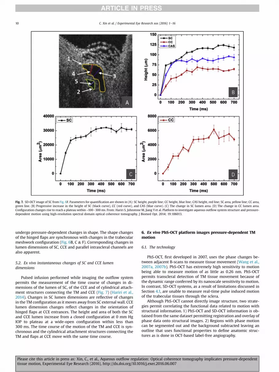
Fig. 7. SD-OCT image of SC from Fig. 6F. Parameters for quantification are shown in (A): SC height, purple line; CC height, blue line; CAS height, red line; SC area, yellow line; CC area,green line. (B) Progressive increase in the height of SC (black curve), CC (red curve), and CAS (blue curve). (C) The change in SC lumen area. (D) The change in CC lumen area.Configuration changes rise to reach a plateau within ~100e300 ms. From: Hariri S, Johnstone M, Jiang Yet al. Platform to investigate aqueous outflow system structure and pressure-dependent motion using high-resolution spectral domain optical coherence tomography. J Biomed Opt. 2014; 19:106013.
C. Xin et al. / Experimental Eye Research xxx (2016) 1e1610
undergo pressure-dependent changes in shape. The shape changesof the hinged flaps are synchronous with changes in the trabecularmeshwork configuration (Fig. 6B, C & F). Corresponding changes inlumen dimensions of SC, CCE and parallel intrascleral channels arealso apparent.
5.2. Ex vivo instantaneous changes of SC and CCE lumendimensions
Pulsed infusion performed while imaging the outflow systempermits the measurement of the time course of changes in di-mensions of the lumen of SC, of the CCE and of cylindrical attach-ment structures connecting the TM and CCE (Fig. 7) (Hariri et al.,2014). Changes in SC lumen dimensions are reflective of changesin the TM configuration as it moves away from SC external wall. CCElumen dimension changes reflect changes in the orientation ofhinged flaps at CCE entrances. The height and area of both the SCand CCE lumen increase from a closed configuration at 0 mm HgIOP to plateau at a wide-open configuration within less than300 ms. The time course of the motion of the TM and CCE is syn-chronous and the cylindrical attachment structures connecting theTM and flaps at CCE move with the same time course.
Please cite this article in press as: Xin, C., et al., Aqueous outflow regulatissue motion, Experimental Eye Research (2016), http://dx.doi.org/10.10
6. Ex vivo PhS-OCT platform images pressure-dependent TMmotion
6.1. The technology
PhS-OCT, first developed in 2007, uses the phase changes be-tween adjacent B-scans to measure tissue movement (Wang et al.,2007a, 2007b). PhS-OCT has extremely high sensitivity to motionbeing able to measure motion of as little as 0.26 nm. PhS-OCTpermits transcleral detection of TM tissue movement because ofthe dynamic range conferred by its nanoscale sensitivity to motion.In contrast, SD-OCT systems, as a result of limitations discussed inSection 4.1, are unable to measure real-time pulse induced motionof the trabecular tissues through the sclera.
Although PhS-OCT cannot directly image structure, two strate-gies permit correlating the functional data related to motion withstructural information. 1) PhS-OCT and SD-OCT information is ob-tained from the same dataset permitting registration and overlay ofphase data onto structural images. 2) Regions with phase motioncan be segmented out and the background subtracted leaving anoutline that uses functional properties to define anatomic struc-tures as is done in OCT-based label-free angiography.
tion: Optical coherence tomography implicates pressure-dependent16/j.exer.2016.06.007

C. Xin et al. / Experimental Eye Research xxx (2016) 1e16 11
6.2. Ex vivo PhS-OCT imaging of pressure-dependent TM motion
Perfusion of whole enucleated NH primate eyes from a reservoirsystem controlled mean IOP while inducing sinusoidal pulse tran-sients (Li et al., 2012) (Fig. 8). Measurements were carried out at aseries of seven different mean pressures while maintaining pulseamplitude of 3 mm Hg and frequency of 1 pulse/second (1 Hz). Thestudy demonstrated TM motion synchronous with pulse-inducedIOP transients. SC lumen size decreased as IOP increased (Fig. 8A,B& C). Pulsatile TMmotion also decreased as IOP increased andwasabsent at 40 mm Hg (Fig. 8D, E & F). Quantitative measurements inthis ex vivo model included velocity, displacement, strain rate, andTM structural motion (Fig. 9A) parameters important for assessingthe biomechanical properties of tissues.
7. In vivo PhS-OCT platform images pulse-dependent TMmotion in humans
PhS-OCT has recently succeeded in detecting pulse induced TMmotion in vivo in twenty eyes of 10 human subjects (Li et al., 2013)(Fig. 9B). The key to this success is the development of a phasecompensation algorithm that permits removal of the confoundingeffects of bulk motion. A correlation between the TM motion and
Fig. 8. IOP-dependence of SC deformation (SD-OCT images) and TMmovement (PhS-OCT meImages of the same SC cross-sections at an intraocular pressure (IOP) of 8 (A),10 (B), and 40 (C)and TMdisplacement (blue lines) versus time at each IOP. The TMmean displacement (TMD) issensitive optical coherence tomography characterization of pulse-induced trabecular meshw
Please cite this article in press as: Xin, C., et al., Aqueous outflow regulatissue motion, Experimental Eye Research (2016), http://dx.doi.org/10.10
the digital pulsewas found to be highly significant (P< 0.0001). Thedigital pulse led the TM motion by a mean time of 0.5 ± 0.48 s. Asignificant linear relationship was present between the phase lagand the pulse rate (P<. 05). Phase lag was affected by age but didnot quite reach significance (P ¼ 0.074).
Comparison of normal and abnormal outflow system regions inthe same eye of a patient (Fig. 10) was made possible because aunilateral primary iris cyst closed the angle on the temporal side ofthe eyewhile the nasal angle remainedwide open (Sun et al., 2015).Because the iris cyst completely occluded the temporal angle, thatregion of the outflow system did not communicate with the ante-rior chamber. In the normal nasal angle TM tracings of velocity anddisplacement amplitude were relatively large and synchronouswith the digital pulse. In the occluded temporal angle, TM velocityand amplitudes were barely discernable.
8. Aqueous outflow system OCT imaging in the managementof glaucoma
8.1. Current status
Kagemann et al. (2014) achieved an important milestone inoutflow imaging in humans by demonstrating a decrease in SC area
asurements) derived from the same data set in ex vivowhole eye primate experiments.mmHg, respectively. Fig. D to F images are corresponding plots of IOP (dashed red lines)markedly reducedwith the increase of themean IOP. From: Li P, Reif R, Zhi Z et al. Phase-ork displacement in ex vivo nonhuman primate eyes. J Biomed Opt. 2012; 17:076026.
tion: Optical coherence tomography implicates pressure-dependent16/j.exer.2016.06.007

Fig. 9. (A) Ex vivo PhS-OCT whole eye experiments. Temporal plots over 3.5 s of IOP measurements provided by an in-line pressure transducer synchronized with PhS-OCT system.(inset (A)) IOP; (inset (B)) trabecular meshwork velocity (TMV); (inset (C)) trabecular meshwork displacement (TMD); (Inset(D)) trabecular meshwork strain rate (TMSR) and (inset(E)) normalized SC size (SCA). The experimental conditions were mean IOP 8 mmHg, pulse amplitude 3 mmHg and 1 pulse/second. The dashed vertical lines indicate the time of theIOP pulse peaks. (B) In vivo PhS-OCT in human subjects. Arrival times of human digital pulse vs. TM motion peak. A significant correlation (R2 ¼ 0.998, P < 0.0001) of theinstantaneous time between the digital pulse peaks [digital p-time, marked by black thin arrows in Fig. 3(D)] and the TM pulse minima [TM f-time, marked by blue bold arrows inFig. 3(D)] is found. The results demonstrate the temporal synchronization between the cardiac pulse and the TM motion. (A) from: Li P, Reif R, Zhi Z et al. Phase-sensitive opticalcoherence tomography characterization of pulse-induced trabecular meshwork displacement in ex vivo nonhuman primate eyes. J Biomed Opt. 2012; 17:076026. (B) from: Li P, ShenTT, Johnstone M, Wang RK. Pulsatile motion of the trabecular meshwork in healthy human subjects quantified by phase-sensitive optical coherence tomography. Biomed OptExpress. 2013; 4:2051e2065. (For interpretation of the references to colour in this figure legend, the reader is referred to the web version of this article.)
C. Xin et al. / Experimental Eye Research xxx (2016) 1e1612
in response to an increase in IOP using a commercially available SD-OCT system. However, the resolution of current commercial sys-tems is insufficient to make them an easily used clinical tool formonitoring outflow system motion.
Laboratory-based PhS-OCT is capable of imaging pulse-dependent TM motion from the scleral surface but would requirethe involvement of industry to develop a commercial system forexternal use or a probe for direct observation through the TM. Thelaboratory-based PhS-OCT system described in this review dem-onstrates that such a system with its high degree of sensitivity tomotion can image TM pulse-dependent behavior from the scleralsurface in human subjects.
Laboratory-based OCT studies also show that the TM and CCEcan be imaged from the TM surface and their motion quantifiedwith an optimized system. The laboratory technique of pressurereversal that allows detailed study of TM and CCE motion andrelationships is already used in the operating room to predict theprobability of surgical success (see Section 4.0). The imagingcapabilities of a trans-trabecular OCT system as described above
Please cite this article in press as: Xin, C., et al., Aqueous outflow regulatissue motion, Experimental Eye Research (2016), http://dx.doi.org/10.10
suggest that development of an OCT probe would be veryuseful in a surgical environment to assess and target areas withCCE to determine the functional status of the distal outflowsystem.
Circumferential flow around SC has been demonstrated to bequite limited (Grant, 1958). For that reason placement of a MIGSdevice at a single location can be expected to provide access to onlya small area of SC circumference and few CCE. Studies also showthat aqueous outflow is not distributed equally around the 360�
circumference of SC (Johnstone, 2006). If CCE and distal outflowpathways are nonfunctional at the chosen location for deviceinsertion the procedure can be expected to fail. Use of OCT tech-nology may permit identifying regions of the trabecular meshworkand CCE that experience pulse-induced motion. Pulse-inducedmotion is indicative of a region with mobile tissues capable ofchanging shape to accommodate aqueous flow. Locations withactive motion and aqueous flow also suggest the presence of afunctional distal outflow system that may be an optimal target forMIGS placement.
tion: Optical coherence tomography implicates pressure-dependent16/j.exer.2016.06.007

Fig. 10. (A) Phase-sensitive optical coherence tomography (PhS-OCT) assessment of dynamic motion of TM of a human subject in vivo. The nasal anterior chamber (AC) angle isnormal. An iris cyst closes the temporal angle preventing access of aqueous to the temporal outflow system. OCT cross-sectional image size ¼ 12 mm � 10 mm. (B) The pulse tracingis in red. Velocity and displacement tracings (in blue) of the trabecular meshwork (TM) motion in the normal nasal angle are easily identified and are synchronous with theperipheral pulse. (C) TM velocity and amplitude tracings in the temporal closed angle are barely discernable. From: Y S, P L, Johnstone M, K W, T S. Pulsatile motion of trabecularmeshwork in a patient with iris cyst by phase-sensitive optical coherence tomography. Quantitative Imaging in Medicine and Surgery. 2015; 5:171e173.
C. Xin et al. / Experimental Eye Research xxx (2016) 1e16 13
8.2. Future imaging possibilities
PhS-OCT is non-contact, non-invasive and measures tissueproperties associated with maintaining IOP homeostasis. Researchstudies using in vivo slitlamp and gonioscopic imaging identifydefective outflow system motion in humans with glaucoma. De-fects include both reduced/absent aqueous vein pulsation (Section2.2) and lack of TM motion (Section 2.3). The clinical techniquescurrently used to identify defective TM motion are sufficientlylaborious and time consuming that they have not beenwidely usedfor detection of outflow system abnormalities in a clinical envi-ronment. However non-invasive techniques that can image outflowsystem function in the office should prove to be highly useful forboth detecting andmonitoring abnormalities that result in pressureelevation.
Please cite this article in press as: Xin, C., et al., Aqueous outflow regulatissue motion, Experimental Eye Research (2016), http://dx.doi.org/10.10
The current use of random IOP measurements to detect andmonitor glaucoma is problematic because the approach capturesonly a small part of the IOP profile. Pressures undergo diurnalchanges and also numerous transient IOP elevations from baselinein the order of 10 mm Hg associated with blinking and eye move-ment. IOP measurements are typically made randomly 3e4 timesper year representing sampling of ~12 s of the 31 million seconds inthe year. Diurnal pressures vary considerably and often rise mark-edly at night when no measurements are made. Measurements arealso made while specifically avoiding blinking or eye movementthus preventing capture of any of the constantly recurring transientIOP elevations. Furthermore, medication compliance unknowns area confounding factor further clouding the assessment of the truepressure profile.
Motion of the TM in response to the ocular pulse is indicative of
tion: Optical coherence tomography implicates pressure-dependent16/j.exer.2016.06.007

C. Xin et al. / Experimental Eye Research xxx (2016) 1e1614
elasticity and compliance of the trabecular tissues, properties thatdetermine IOP homeostasis. Knowledge of TM motion revealed byOCT imaging may permit identifying individual patients who areexperiencing progressive difficulty in maintaining homeostasis noteasily revealed by random IOP measurements. Such imaging mayprovide a sensitive predictive tool to help decide if preemptiveescalation of medical, laser or surgical therapy is appropriate beforethe patient develops the late warning of progressive structural andfunctional damage to the visual system.
The ability to assess motion with PhS-OCT may improve selec-tion of candidates for SC surgery. By eliminating inappropriatecandidates, patients would be spared unnecessary surgery. At thesame time procedure success rates as a result of appropriate se-lection might be considerably higher. Blood reflux into SC while inthe operating room has been shown to be a predictor of the like-lihood of SC surgical success. Blood reflux into SC depends on theability of the TM to move. Use of a probe inserted inside the eye toimage through the TM may provide high-resolution images toidentify CCE motion indicative of a functional region of the distaloutflow system.
The optimal placement of MIGS devices might be substantiallyimproved if functioning CCE could be clearly identified. Regionswhere CCE are permanently closed or that have inadequate motioncould be avoided. Patients would be spared from MIGS surgerywhen CCE dysfunction indicates a limited possibility for successand could instead move directly to more appropriate alternatives.
9. Summary
In vivo clinical studies are capable of using imaging to defineaqueous outflow system abnormalities in glaucoma. These studiespoint to an abnormality of trabecular tissue motion that preventsnormal pulsatile aqueous outflow and also prevents blood refluxinto SC. Such motion abnormalities have been thought to resultfrom a stiffening of trabecular tissues. The available clinical tech-niques are difficult and time consuming.
A recently developed high resolution SD-OCT platform thatimages the outflow system from the TM surface identifies hingedflaps at CCE. The hinged flaps open and close in synchrony withpulse-dependent TMmotion. Another recently developed PhS-OCTtechnology images pulse-dependent TMmotion in human subjects.Rapidly evolving OCT imaging techniques may develop into pre-dictive clinical tools that will assist in the medical and surgicalmanagement of glaucoma.
10. Conflicts of interest
The authors have no conflicts of interest to declare.
11. Individual contributions to the article
Article Preparation, Organization, Planning and Direction: Rui-kang Wang, Chen Xin, Joanne Wen, Tueng Shen, Murray Johnstone.
Research: Ruikang Wang, Chen Xin, Elizabeth Martin, Yi Jiang,Seven Padilla Shaozhen Song, Tueng Shen, Murray Johnstone.
Acknowledgements
This work was supported in part by research grants from theNational Eye Institute (R01EY024158), theW. H. Coulter FoundationTranslational Research Partnership Program, and the Office ofResearch Infrastructure Programs of the National Institutes ofHealth through Grant No. P51OD010425 through the WashingtonNational Primate Research Center. SightLife Eyebank provided tis-sues. Part of this work was conducted at the University of
Please cite this article in press as: Xin, C., et al., Aqueous outflow regulatissue motion, Experimental Eye Research (2016), http://dx.doi.org/10.10
Washington NanoTech User Facility, a member of the NSF NationalNanotechnology Infrastructure Network, and the Biology ImagingFacility at the University of Washington. The content is solely theresponsibility of the authors and does not necessarily represent theofficial views of the grant giving bodies.
References
Ascher, K.W., 1942. Aqueous veins. Am. J. Ophthamol. 25, 31e38.Ascher, K.W., 1942. Physiologic importance of the visible elimination of intraocular
fluid. Am. J. Ophthamol. 25, 1174e1209.Ascher, K.W., 1942. Aqueous Veins: II. Local pharmacologic effects on aqueous veins
III. Glaucoma and the aqueous veins. Am. J. Ophthamol. 25, 1301.Ascher, K.W., 1944. Backflow phenomena in aqueous veins. Am. J. Ophthamol. 27,
1074.Ascher, K.W., 1949. Aqueous veins and their significance for pathogenesis of glau-
coma. Arch. Ophthamol. 42, 66.Ascher, K.W., 1953. Aqueous veins: their status eleven years after their detection.
A M. A Arch. Ophthamol. 49, 438.Ascher, K.W., 1961. The Aqueous Veins: Biomicroscopic Study of Aqueous Humor
Elimination. Charles C Thomas, Springfield, Ill, p. 251.Aspelund, A., Tammela, T., Antila, S., et al., 2014 Sep 2. The Schlemm’s canal is a
VEGF-C/VEGFR-3-responsive lymphatic-like vessel. J. Clin. Invest. 124 (9),3975e3986.
Bahler, C.K., Hann, C.R., Fjield, T., Haffner, D., Heitzmann, H., Fautsch, M.P., 2012.Second-generation trabecular meshwork bypass stent (iStent inject) increasesoutflow facility in cultured human anterior segments. Am. J. Ophthalmol. 153,1206e1213.
Battista, S.A., Lu, Z., Hofmann, S., Freddo, T., Overby, D.R., Gong, H., 2008. Reductionof the available area for aqueous humor outflow and increase in meshworkherniations into collector channels following acute IOP elevation in bovine eyes.Invest. Ophthalmol. Vis. Sci. 49, 5346e5352.
Bhattacharya, S.K., Rockwood, E.J., Smith, S.D., et al., 2005. Proteomics revealCochlin deposits associated with glaucomatous trabecular meshwork. J. Biol.Chem. 280, 6080e6084.
Bradley, J.M., Kelley, M.J., Rose, A., Acott, T.S., 2003. Signaling pathways used intrabecular matrix metalloproteinase response to mechanical stretch. Invest.Ophthalmol. Vis. Sci. 44, 5174e5181.
Brandao, L.M., Grieshaber, M.C., 2013. Update on minimally invasive glaucomasurgery (MIGS) and new implants. J. Ophthalmol. 2013, 705915.
Cambiaggi, A., 1958. Effeto della jaluronidasi sulla pressone intraocular e sull’asettodella vene dell’accqueo. Boll Soc Biol. Sper. 34, 1e7.
Clark, R., Nosie, A., Walker, T., et al., 2013. Comparative genomic and proteomicanalysis of cytoskeletal changes in dexamethasone-treated trabecular mesh-work cells. Mol. Cell Proteomics 12, 194e206.
De Vries, S., 1947. De Zichtbare Afvoer Van Het Kamerwater. Drukkerij Kinsbergen,Amsterdam, p. 90.
Ellingsen, B.A., Grant, W.M., 1971. The relationship of pressure and aqueous outflowin enucleated human eyes. Invest. Ophthalmol. 10, 430e437.
Ellingsen, B.A., Grant, W.M., 1972. Trabeculotomy and sinusotomy in enucleatedhuman eyes. Invest. Ophthalmol. 11, 21e28.
Fellman, R.L., Grover, D.S., 2014. Episcleral venous fluid wave: intraoperative evi-dence for patency of the conventional outflow system. J. Glaucoma 23,347e350.
Fercher, A., Derxler, W., CK H, T K, 2003. Optical coherence tomography-principlesand applications. Rep. Prog. Phys. 66, 239.
Filla, M.S., Schwinn, M.K., Nosie, A.K., Clark, R.W., Peters, D.M., 2011. Dexametha-sone-associated cross-linked actin network formation in human trabecularmeshwork cells involves beta3 integrin signaling. Invest. Ophthalmol. Vis. Sci.52, 2952e2959.
Friberg, T.R., Weinreb, R.N., 1985. Ocular manifestations of gravity inversion. JAMA253, 1755e1757.
Friberg, T.R., Sanborn, G., Weinreb, R.N., 1987. Intraocular and episcleral venouspressure increase during inverted posture. Am. J. Ophthalmol. 103, 523e526.
Gabelt, B.A., Kaufman, P.L., 2011. Production and flow of aqueous humor. In:Kaufman, P.L., Alm, A. (Eds.), Adler’s Physiology of the Eye. Elsevier, Edinburgh,pp. 274e307.
Goel, M., Sienkiewicz, A.E., Picciani, R., Lee, R.K., Bhattacharya, S.K., 2011. Cochlininduced TREK-1 co-expression and annexin A2 secretion: role in trabecularmeshwork cell elongation and motility. PLoS One 6, e23070.
Goel, M., Sienkiewicz, A.E., Picciani, R., Wang, J., Lee, R.K., Bhattacharya, S.K., 2012.Cochlin, intraocular pressure regulation and mechanosensing. PLoS One 7,e34309.
Goldmann, H., 1946. Abfluss des Kammerwassers beimMenschen. Ophthalmologica111, 146e152.
Goldmann, H., 1948. Uber Abflussdruck und Glasstab-phanomen. Pathogenese deseinfachen Glaukoms. Ophthalmologica 116, 193.
Gonzalez, J.M., Ko, M.K., Pouw, A., Tan, J.C., 2016. Tissue-based multiphoton analysisof actomyosin and structural responses in human trabecular meshwork. Sci.Rep. 6, 21315.
Grant, W.M., 1958. Further studies on facility of flow through the trabecularmeshwork. Arch. Ophthal. 60, 523e533.
tion: Optical coherence tomography implicates pressure-dependent16/j.exer.2016.06.007

C. Xin et al. / Experimental Eye Research xxx (2016) 1e16 15
Grierson, I., Lee, W.R., 1974. Junctions between the cells of the trabecular meshwork.Albr. Von. Graefes Arch. Klin. Exp. Ophthalmol. 192, 89e104.
Grierson, I., Lee, W.R., 1974. Changes in the monkey outflow apparatus at gradedlevels of intraocular pressure: a qualitative analysis by light microscopy andscanning electron microscopy. Exp. Eye Res. 19, 21e33.
Grierson, I., Lee, W.R., 1975. The fine structure of the trabecular meshwork at gradedlevels of intraocular pressure. (1) Pressure effects within the near-physiologicalrange (8e30 mmHg). Exp. Eye Res. 20, 505e521.
Grierson, I., Lee, W.R., 1975. The fine structure of the trabecular meshwork at gradedlevels of intraocular pressure. (2) Pressures outside the physiological range (0and 50 mmHg). Exp. Eye Res. 20, 523e530.
Grierson, I., Lee, W.R., Abraham, S., Howes, R.C., 1978. Associations between the cellsof the walls of Schlemm’s canal. Albr. Von. Graefes Arch. Klin. Exp. Ophthalmol.208, 33e47.
Grieshaber, M.C., Pienaar, A., Olivier, J., Stegmann, R., 2010. Clinical evaluation of theaqueous outflow system in primary open-angle glaucoma for canaloplasty.Invest. Ophthalmol. Vis. Sci. 51, 1498e1504.
Grover, D.S., Godfrey, D.G., Smith, O., Feuer, W.J., Montes de Oca, I., Fellman, R.L.,2014. Gonioscopy-assisted transluminal trabeculotomy, ab interno trabeculot-omy: technique report and preliminary results. Ophthalmology 121, 855e861.
Hann, C.R., Bentley, M.D., Vercnocke, A., Ritman, E.L., Fautsch, M.P., 2011. Imagingthe aqueous humor outflow pathway in human eyes by three-dimensionalmicro-computed tomography (3D micro-CT). Exp. Eye Res. 92, 104e111.
Hann, C.R., Vercnocke, A.J., Bentley, M.D., Jorgensen, S.M., Fautsch, M.P., 2014.Anatomic changes in Schlemm’s canal and collector channels in normal andprimary open-angle glaucoma eyes using low and high perfusion pressures.Invest. Ophthalmol. Vis. Sci. 55, 5834e5841.
Hariri, S., Johnstone, M., Jiang, Y., et al., 2014. Platform to investigate aqueousoutflow system structure and pressure-dependent motion using high-resolution spectral domain optical coherence tomography. J. Biomed. Opt. 19,106013.
Heijl, A., Bengtsson, B., Hyman, L., Leske, M.C., 2009. Natural history of open-angleglaucoma. Ophthalmology 116, 2271e2276.
Huang, D., Swanson, E.A., Lin, C.P., et al., 1991. Optical coherence tomography. Sci-ence 254, 1178e1181.
Ingber, D.E., 2008. Tensegrity-based mechanosensing from macro to micro. Prog.Biophys. Mol. Biol. 97, 163e179.
Johnstone, M.A., 1974. Pressure-dependent changes in configuration of the endo-thelial tubules of Schlemm’s canal. Am. J. Ophthalmol. 78, 630e638.
Johnstone, M.A., 1979. Pressure-dependent changes in nuclei and the process ori-gins of the endothelial cells lining Schlemm’s canal. Invest. Ophthalmol. Vis. Sci.18, 44e51.
Johnstone, M.A., 2004. The aqueous outflow system as a mechanical pump: evi-dence from examination of tissue and aqueous movement in human and non-human primates. J. Glaucoma 13, 421e438.
Johnstone, M.A., 2014. Intraocular pressure regulation: findings of pulse-dependenttrabecular meshwork motion lead to unifying concepts of intraocular pressurehomeostasis. J. Ocul. Pharmacol. Ther. 30, 88e93.
Johnstone, M.A., Grant, W.M., 1973. Microsurgery of Schlemm’s canal and the hu-man aqueous outflow system. Am. J. Ophthalmol. 76, 906e917.
Johnstone, M.A., Grant, W.G., 1973. Pressure-dependent changes in structures of theaqueous outflow system of human and monkey eyes. Am. J. Ophthalmol. 75,365e383.
Johnstone, M.A., Tanner, D., Chau, B., Kopecky, K., 1980. Concentration-dependentmorphologic effects of cytochalasin B in the aqueous outflow system. Invest.Ophthalmol. Vis Sci. 19, 835e841.
Johnstone, M.A., 2006. A new model describes an aqueous outflow pump and ex-plores causes of pump failure in glaucoma. In: Grehn, H., Stamper, R. (Eds.),Essentials in Ophthalmology: Glaucoma II. Springer, Heidelberg.
Johnstone, M., 2009. Aqueous humor outflow. In: Stamper, R., Lieberman, M.F.,Drake, M.V. (Eds.), Diagnosis and Therapy of the Gaucomas. Mosby, St. Louis,pp. 22e46.
Johnstone, M., Jamil, A., Martin, E., 2010. Aqueous veins and open angle glaucoma.In: Schacknow, P., Samples, J. (Eds.), The Glaucoma Book. Springer, New York,pp. 65e78.
Johnstone, M., Martin, E., Jamil, A., 2011. Pulsatile flow into the aqueous veins:manifestations in normal and glaucomatous eyes. Exp. Eye Res. 92, 318e327.
Johnstone, M.A., Saheb, H., Ahmed II, Samuelson, T.W., Schieber, A.T., Toris, C.B.,2014. Effects of a Schlemm canal scaffold on collector channel ostia in humananterior segments. Exp. Eye Res. 119, 70e76.
Junglas, B., Kuespert, S., Seleem, A.A., et al., 2012. Connective tissue growth factorcauses glaucoma by modifying the actin cytoskeleton of the trabecular mesh-work. Am. J. Pathol. 180, 2386e2403.
Kagemann, L., Wollstein, G., Ishikawa, H., et al., 2010. Identification and assessmentof Schlemm’s canal by spectral-domain optical coherence tomography. Invest.Ophthalmol. Vis. Sci. 51, 4054e4059.
Kagemann, L., Wang, B., Wollstein, G., et al., 2014. IOP elevation reduces Schlemm’scanal cross-sectional area. Invest. Ophthalmol. Vis. Sci. 55, 1805e1809.
Kaplowitz, K., Schuman, J.S., Loewen, N.A., 2014. Techniques and outcomes ofminimally invasive trabecular ablation and bypass surgery. Br. J. Ophthalmol.98, 579e585.
Keller, K.E., Aga, M., Bradley, J.M., Kelley, M.J., Acott, T.S., 2009. Extracellular matrixturnover and outflow resistance. Exp. Eye Res. 88, 676e682.
Kleinert, H., 1951. The compensation maximum: a new glaucoma sign in aqueousveins. Arch. Ophthamol. 46, 618e624.
Please cite this article in press as: Xin, C., et al., Aqueous outflow regulatissue motion, Experimental Eye Research (2016), http://dx.doi.org/10.10
Ko, M.K., Kim, E.K., Gonzales, J.M., Tan, J.C., 2016. Dose- and time-dependent effectsof actomyosin inhibition on live mouse outflow resistance and aqueousdrainage tissues. Sci. Rep. 6, 1e12.
Kronfeld, P.C., 1949. Further gonioscopic studies on the canal of Schlemm. AMAArch. Ophthal. 41, 393.
Kronfeld, P.C., McGarry, H.T., Smith, H.E., 1942. Gonioscopic study on the canal ofSchlemm. Am. J. Ophthal. 25, 1163.
Last, J.A., Pan, T., Ding, Y., et al., 2011. Elastic modulus determination of normal andglaucomatous human trabecular meshwork. Invest. Ophthalmol. Vis. Sci. 52,2147e2152.
Li, P., Reif, R., Zhi, Z., et al., 2012. Phase-sensitive optical coherence tomographycharacterization of pulse-induced trabecular meshwork displacement in ex vivononhuman primate eyes. J. Biomed. Opt. 17, 076026.
Li, P., Shen, T.T., Johnstone, M., Wang, R.K., 2013. Pulsatile motion of the trabecularmeshwork in healthy human subjects quantified by phase-sensitive opticalcoherence tomography. Biomed. Opt. Express 4, 2051e2065.
Liton, P.B., Luna, C., Bodman, M., Hong, A., Epstein, D.L., Gonzalez, P., 2005. Inductionof IL-6 expression by mechanical stress in the trabecular meshwork. Biochem.Biophys. Res. Commun. 337, 1229e1236.
Loewen, N.A., Schuman, J.S., 2013. There has to be a better way: evolution of in-ternal filtration glaucoma surgeries. Br. J. Ophthalmol. 97, 1228e1229.
Luna, C., Li, G., Liton, P.B., Epstein, D.L., Gonzalez, P., 2009. Alterations in geneexpression induced by cyclic mechanical stress in trabecular meshwork cells.Mol. Vis. 15, 534e544.
Luna, C., Li, G., Qiu, J., Challa, P., Epstein, D.L., Gonzalez, P., 2009. Extracellular releaseof ATP mediated by cyclic mechanical stress leads to mobilization of AA intrabecular meshwork cells. Invest. Ophthalmol. Vis. Sci. 50, 5805e5810.
Mitton, K.P., Tumminia, S.J., Arora, J., Zelenka, P., Epstein, D.L., Russell, P., 1997.Transient loss of alphaB-crystallin: an early cellular response to mechanicalstretch. Biochem. Biophys. Res. Commun. 235, 69e73.
Morgan, J.T., Raghunathan, V.K., Chang, Y.R., Murphy, C.J., Russell, P., 2015. Wntinhibition induces persistent increases in intrinsic stiffness of human trabecularmeshwork cells. Exp. Eye Res. 132, 174e178.
Neuhann, T.H., 2015. Trabecular micro-bypass stent implantation during small-incision cataract surgery for open-angle glaucoma or ocular hypertension:long-term results. J. Cataract. Refract Surg. 41, 2664e2671.
Park, D.Y., Lee, J., Park, I., et al., July 2014. Lymphatic regulator PROX1 determinesSchlemm canal integrity and identity. J. Clin. Invest. 124 (9), 3960e3974.
Pfeiffer, N., Garcia-Feijoo, J., Martinez-de-la-Casa, J.M., et al., 2015. A randomizedtrial of a Schlemm’s canal microstent with phacoemulsification for reducingintraocular pressure in open-angle glaucoma. Ophthalmology 122, 1283e1293.
Phelps, C.D., 1978. The pathogenesis of glaucoma in Sturge-Weber syndrome.Ophthalmology 85, 276e286.
Phelps, C.D., Asseff, C.F., Weisman, R.L., Podos, S.M., Becker, B., 1972. Blood refluxinto Schlemm’s canal. Arch. Ophthalmol. 88, 625e631.
Phelps, C.D., Thompson, H.S., Ossoinig, K.C., 1982. The diagnosis and prognosis ofatypical carotid-cavernous fistula (red-eyed shunt syndrome). Am. J. Oph-thalmol. 93, 423e436.
Phillips, C.I., Tsukahara, S., Hosaka, O., Adams, W., 1992. Ocular pulsation correlateswith ocular tension: the choroid as piston for an aqueous pump? OphthalmicRes. 24, 338e343.
Quick, C.M., Venugopal, A.M., Gashev, A.A., Zawieja, D.C., Stewart, R.H., 2007.Intrinsic pump-conduit behavior of lymphangions. Am. J. Physiol. Regul. Integr.Comp. Physiol. 292, R1510eR1518.
Quick, C.M., Ngo, B.L., Venugopal, A.M., Stewart, R.H., 2009. Lymphatic pump-conduit duality: contraction of postnodal lymphatic vessels inhibits passiveflow. Am. J. Physiol. Heart Circ. Physiol. 296, H662eH668.
Quigley, H.A., Broman, A.T., 2006. The number of people with glaucoma worldwidein 2010 and 2020. Br. J. Ophthalmol. 90, 262e267.
Raghunathan, V.K., Morgan, J.T., Dreier, B., et al., 2013. Role of substratum stiffnessin modulating genes associated with extracellular matrix and mechano-transducers YAP and TAZ. Invest. Ophthalmol. Vis. Sci. 54, 378e386.
Raghunathan, V.K., Morgan, J.T., Park, S.A., et al., 2015. Dexamethasone stiffenstrabecular meshwork, trabecular meshwork cells, and matrix. Invest. Oph-thalmol. Vis. Sci. 56, 4447e4459.
Ramos, R.F., Stamer, W.D., 2008. Effects of cyclic intraocular pressure on conven-tional outflow facility. Invest. Ophthalmol. Vis. Sci. 49, 275e281.
Ramos, R.F., Sumida, G.M., Stamer, W.D., 2009. Cyclic mechanical stress andtrabecular meshwork cell contractility. Invest. Ophthalmol. Vis. Sci. 50,3826e3832.
Ren, J., Gille, H.K., Wu, J., Yang, C., 2011. Ex vivo optical coherence tomographyimaging of collector channels with a scanning endoscopic probe. Invest. Oph-thalmol. Vis. Sci. 52, 3921e3925.
Richter, G.M., Coleman, A.L., 2016. Minimally invasive glaucoma surgery: currentstatus and future prospects. Clin. Ophthalmol. 10, 189e206.
Rohen, J.W., Rentsch, F.J., 1968. Morphology of Schlemm’s canal and related vesselsin the human eye. Albr. Von. Graefes Arch. Klin. Exp. Ophthalmol. 176, 309e329.
Rosenquist, R., Epstein, D., Melamed, S., Johnson, M., Grant, W.M., 1989. Outflowresistance of enucleated human eyes at two different perfusion pressures anddifferent extents of trabeculotomy. Curr. Eye Res. 8, 1233e1240.
Saheb, H., Ahmed II, 2012. Micro-invasive glaucoma surgery: current perspectivesand future directions. Curr. Opin. Ophthalmol. 23, 96e104.
Schirmer, K.E., 1969. Reflux of blood in the canal of Schlemm quantitated. Canad. J.Ophthalmol. 4, 40e44.
Schirmer, K.E., 1971. Gonioscopic assessment of blood in Schlemm’s canal.
tion: Optical coherence tomography implicates pressure-dependent16/j.exer.2016.06.007

C. Xin et al. / Experimental Eye Research xxx (2016) 1e1616
Correlation with glaucoma tests. Arch. Ophthalmol. 85, 263e267.Schuman, J.S., Chang, W., Wang, N., de Kater, A.W., Allingham, R.R., 1999. Excimer
laser effects on outflow facility and outflow pathway morphology. Invest.Ophthalmol. Vis. Sci. 40, 1676e1680.
Smit, B.A., Johnstone, M.A., 2002. Effects of viscoelastic injection into Schlemm’scanal in primate and human eyes: potential relevance to viscocanalostomy.Ophthalmology 109, 786e792.
Stambaugh, J., Fuhs, J., Ascher, K.W., 1954. Study of the compensation-maximumtest on aqueous veins. A M. A Arch. Ophthamol. 51, 24.
Stamer, W.D., Acott, T.S., 2012. Current understanding of conventional outflowdysfunction in glaucoma. Curr. Opin. Ophthalmol. 23, 135e143.
Stepanik, J., 1954. Measuring velocity of flow in aqueous veins. Am. J. Ophthal. 37,918.
Sun, Y., Li, P., Johnstone, M., Wang, K., Shen, T., 2015. Pulsatile motion of trabecularmeshwork in a patient with iris cyst by phase-sensitive optical coherence to-mography. Quantitative Imaging Med. Surg. 5, 171e173.
Suson, E.B., Schultz, R.O., 1969. Blood in schlemm’s canal in glaucoma suspects. Astudy of the relationship between blood-filling pattern and outflow facility inocular hypertension. Arch. Ophthalmol. 81, 808e812.
Tamm, E.R., Russell, P., Epstein, D.L., Johnson, D.H., Piatigorsky, J., 1999. Modulationof myocilin/TIGR expression in human trabecular meshwork. Invest. Oph-thalmol. Vis. Sci. 40, 2577e2582.
Thomassen, T.L., 1947. The venous tension of eyes suffering from simple glaucoma.Acta Ophthamol. 25, 221.
Thomassen, T.L., 1949. The glass-rod test in glaucomatous eyes. Br. J. Ophthamol. 35,773.
Thomasy, S.M., Morgan, J.T., Wood, J.A., Murphy, C.J., Russell, P., 2013. Substratumstiffness and latrunculin B modulate the gene expression of the mechano-transducers YAP and TAZ in human trabecular meshwork cells. Exp. Eye Res.113, 66e73.
Thomson, B.R., Heinen, S., Jeansson, M., et al., 2014. A lymphatic defect causes ocular
Please cite this article in press as: Xin, C., et al., Aqueous outflow regulatissue motion, Experimental Eye Research (2016), http://dx.doi.org/10.10
hypertension and glaucoma in mice. J. Clin. Invest. 124, 4320e4324.Tomlin, P.H., Wang, R.K., 2005. Theory, development and applications of optical
coherence tomography. J. Phys. D. Appl. Phys. 38, 2519.Tumminia, S.J., Mitton, K.P., Arora, J., Zelenka, P., Epstein, D.L., Russell, P., 1998.
Mechanical stretch alters the actin cytoskeletal network and signal trans-duction in human trabecular meshwork cells [see comments]. Invest. Oph-thalmol. Vis. Sci. 39, 1361e1371.
Usui, T., Tomidokoro, A., Mishima, K., et al., 2011. Identification of Schlemm’s canaland its surrounding tissues by anterior segment fourier domain optical coher-ence tomography. Invest. Ophthalmol. Vis. Sci. 52, 6934e6939.
Van Buskirk, E.M., 1976. Changes in facility of aqueous outflow induced by lensdepression and intraocular pressure in excised human eyes. Am. J. Ophthalmol.82, 736e730.
Van Buskirk, E.M., 1982. Anatomic correlates of changing aqueous outflow facility inexcised human eyes. Invest. Ophthalmol. Vis. Sci. 22, 625e632.
Van Buskirk, E.M., Grant, W.M., 1973. Lens depression and aqueous outflow inenucleated primate eyes. Am. J. Ophthalmol. 76, 632e640.
Vittal, V., Rose, A., Gregory, K.E., Kelley, M.J., Acott, T.S., 2005. Changes in geneexpression by trabecular meshwork cells in response to mechanical stretching.Invest. Ophthalmol. Vis. Sci. 46, 2857e2868.
Wang, R.K., Kirkpatrick, S., Hinds, N., 2007. Phase-sensitive optical coherenceelastography for mapping tissue microstrains in real time. Appl. Phys. Lett. 90,164105.
Wang, R.K., Jacques, S.L., Ma, Z., Hurst, S., Hanson, S.R., Gruber, A., 2007. Threedimensional optical angiography. Opt. Express 15, 4083e4097.
Wang, F., Shi, G., Li, X., et al., 2012. Comparison of Schlemm’s canal’s biologicalparameters in primary open-angle glaucoma and normal human eyes withswept source optical. J. Biomed. Opt. 17, 116008.
Weinreb, R.N., Cook, J., Friberg, T.R., 1984. Effect of inverted body position onintraocular pressure. Am. J. Ophthalmol. 98, 784e787.
tion: Optical coherence tomography implicates pressure-dependent16/j.exer.2016.06.007
Recommended


