ARTICLE IN PRESS
Journal of Biomechanics 42 (2009) 405–417
Contents lists available at ScienceDirect
journal homepage: www.elsevier.com/locate/jbiomech
Journal of Biomechanics
0021-92
doi:10.1
� Corr
E-m
www.JBiomech.com
Review
Biomechanical properties of the mandibular condylar cartilage and theirrelevance to the TMJ disc
M. Singh, M.S. Detamore �
Department of Chemical and Petroleum Engineering, University of Kansas, 1530 W. 15th Street, Room 4132, Lawrence, KS 66045-7609, USA
a r t i c l e i n f o
Article history:
Accepted 17 December 2008Mandibular condylar cartilage plays a crucial role in temporomandibular joint (TMJ) function, which
includes facilitating articulation with the TMJ disc, reducing loads on the underlying bone, and
Keywords:
Condyle
TMJ
Structure
Biomechanics
Finite element modeling
90/$ - see front matter & 2009 Elsevier Ltd. A
016/j.jbiomech.2008.12.012
esponding author. Tel.: +1785 864 4943; fax:
ail address: [email protected] (M.S. Detamore
a b s t r a c t
contributing to bone remodeling. To improve our understanding of the TMJ function in normal and
pathological situations, accurate and validated three-dimensional (3-D) finite element models (FEMs) of
the human TMJ may serve as valuable diagnostic tools as well as predictors of thresholds for tissue
damage resulting from parafunctional activities and trauma. In this context, development of reliable
biomechanical standards for condylar cartilage is crucial. Moreover, biomechanical characteristics of the
native tissue are important design parameters for creating functional tissue-engineered replacements.
Towards these goals, biomechanical characteristics of the condylar cartilage have been reviewed here,
highlighting the structure–function correlations. Structurally, condylar cartilage, like the TMJ disc,
exhibits zonal and topographical heterogeneity. Early structural investigations of the condylar cartilage
have suggested that the tissue possesses a somewhat transversely isotropic orientation of collagen
fibers in the fibrous zone. However, recent tensile and shear evaluations have reported a higher stiffness
of the tissue in the anteroposterior direction than in the mediolateral direction, corresponding to an
anisotropic fiber orientation comparable to the TMJ disc. In a few investigations, condylar cartilage
under compression was found to be stiffer anteriorly than posteriorly. As with the TMJ disc, further
compressive characterization is warranted. To draw inferences for human tissue using animal models,
establishing stiffness–thickness correlations and regional evaluation of proteoglycan/glycosaminogly-
can content may be essential. Efforts directed from the biomechanics community for the characteriza-
tion of TMJ tissues will facilitate the development of reliable and accurate 3-D FEMs of the human TMJ.
& 2009 Elsevier Ltd. All rights reserved.
Contents
1. Introduction . . . . . . . . . . . . . . . . . . . . . . . . . . . . . . . . . . . . . . . . . . . . . . . . . . . . . . . . . . . . . . . . . . . . . . . . . . . . . . . . . . . . . . . . . . . . . . . . . . . . . . 406
2. Biomechanical properties of the condylar cartilage as a reflection of ECM organization . . . . . . . . . . . . . . . . . . . . . . . . . . . . . . . . . . . . . . . . . . 406
2.1. Tension and shear . . . . . . . . . . . . . . . . . . . . . . . . . . . . . . . . . . . . . . . . . . . . . . . . . . . . . . . . . . . . . . . . . . . . . . . . . . . . . . . . . . . . . . . . . . . . 407
2.1.1. Tensile and shear evaluations . . . . . . . . . . . . . . . . . . . . . . . . . . . . . . . . . . . . . . . . . . . . . . . . . . . . . . . . . . . . . . . . . . . . . . . . . . . 407
2.1.2. Organization and distribution of collagen. . . . . . . . . . . . . . . . . . . . . . . . . . . . . . . . . . . . . . . . . . . . . . . . . . . . . . . . . . . . . . . . . . 407
2.1.3. Distribution of dermatan sulfate proteoglycans . . . . . . . . . . . . . . . . . . . . . . . . . . . . . . . . . . . . . . . . . . . . . . . . . . . . . . . . . . . . . 411
2.1.4. Summary: structure–property–function correlation under tension. . . . . . . . . . . . . . . . . . . . . . . . . . . . . . . . . . . . . . . . . . . . . . 411
2.2. Compression. . . . . . . . . . . . . . . . . . . . . . . . . . . . . . . . . . . . . . . . . . . . . . . . . . . . . . . . . . . . . . . . . . . . . . . . . . . . . . . . . . . . . . . . . . . . . . . . . 411
2.2.1. Compressive evaluations . . . . . . . . . . . . . . . . . . . . . . . . . . . . . . . . . . . . . . . . . . . . . . . . . . . . . . . . . . . . . . . . . . . . . . . . . . . . . . . 411
2.2.2. Spatial variation of proteoglycans/GAGs . . . . . . . . . . . . . . . . . . . . . . . . . . . . . . . . . . . . . . . . . . . . . . . . . . . . . . . . . . . . . . . . . . . 412
2.2.3. Summary: structure–property–function correlation under compression . . . . . . . . . . . . . . . . . . . . . . . . . . . . . . . . . . . . . . . . . 412
3. A perspective on 3-D computational reconstruction of the human TMJ . . . . . . . . . . . . . . . . . . . . . . . . . . . . . . . . . . . . . . . . . . . . . . . . . . . . . . . 412
4. Discussion . . . . . . . . . . . . . . . . . . . . . . . . . . . . . . . . . . . . . . . . . . . . . . . . . . . . . . . . . . . . . . . . . . . . . . . . . . . . . . . . . . . . . . . . . . . . . . . . . . . . . . . 414
5. Summary . . . . . . . . . . . . . . . . . . . . . . . . . . . . . . . . . . . . . . . . . . . . . . . . . . . . . . . . . . . . . . . . . . . . . . . . . . . . . . . . . . . . . . . . . . . . . . . . . . . . . . . . 415
Conflicts of interest statement . . . . . . . . . . . . . . . . . . . . . . . . . . . . . . . . . . . . . . . . . . . . . . . . . . . . . . . . . . . . . . . . . . . . . . . . . . . . . . . . . . . . . . . 415
Acknowledgment . . . . . . . . . . . . . . . . . . . . . . . . . . . . . . . . . . . . . . . . . . . . . . . . . . . . . . . . . . . . . . . . . . . . . . . . . . . . . . . . . . . . . . . . . . . . . . . . . . 415
References . . . . . . . . . . . . . . . . . . . . . . . . . . . . . . . . . . . . . . . . . . . . . . . . . . . . . . . . . . . . . . . . . . . . . . . . . . . . . . . . . . . . . . . . . . . . . . . . . . . . . . . 415
ll rights reserved.
+1785 864 4967.
).
ARTICLE IN PRESS
M. Singh, M.S. Detamore / Journal of Biomechanics 42 (2009) 405–417406
1. Introduction
The mandibular condyle, the temporomandibular joint (TMJ)disc and the fossa–eminence complex together form the two-compartment articulation of the TMJ (Piette, 1993). Temporo-mandibular disorders (TMDs) have considerable prevalencewith 16–59% of the population having symptoms and 33–86%having clinical signs (Carlsson and LeResche, 1995). According tothe diagnostic classification system of the American Academyof Orofacial Pain (AAOP), TMDs are subcategorized into twoprimary classes: articular and muscle-related disorders, based ontheir anatomic origin (Buescher, 2007). Biomechanics as a field isimportant for the TMJ for two primary reasons. First, biomecha-nical dysfunctions such as clicking, locking, or lateral deviation inmouth opening, are symptoms that generally accompany manyTMDs of articular origin. The other reason, of special interest tothe clinical community, is that biomechanics is a likely con-tributor in the development of such TMDs, e.g., biomechanicalalterations resulting from parafunctional behavior or trauma thatmay lead to the development of or exacerbate an existingpathological condition. In other words, we are interested in TMJbiomechanics from both the cause and effect perspectives inrelation to TMDs.
What kinds of forces are necessary to irreparably damage thetissues in the TMJ, and what are the associated magnitudes anddurations of these forces? Fortunately, a growing number ofresearchers are building three-dimensional (3-D) finite-elementmodels (FEMs) of the human TMJ to answer this question. A majorlimitation with such models pertains to the inclusion of materialproperties that do not represent the constituent tissues, whichmay render them inaccurate. In this context, developmentof reliable biomechanical standards for TMJ cartilaginous tissues,including the mandibular condylar cartilage, will be a useful inputto further the development of 3-D FEMs of the human TMJ. Amongbiomechanical studies of the cartilaginous tissues of TMJ, theTMJ disc has by far received the most attention (see reviewsby Detamore and Athanasiou, 2003a, 2003b). This reviewprimarily focuses on the mandibular condylar cartilage, anarticular cartilage that is distinct from the articular hyalinecartilages of appendicular skeleton (Milam, 2003), specificallyinspecting independent evaluations of biomechanical properties
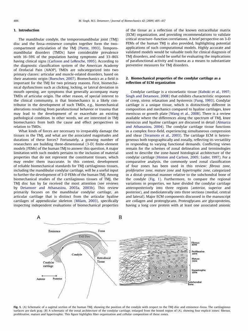
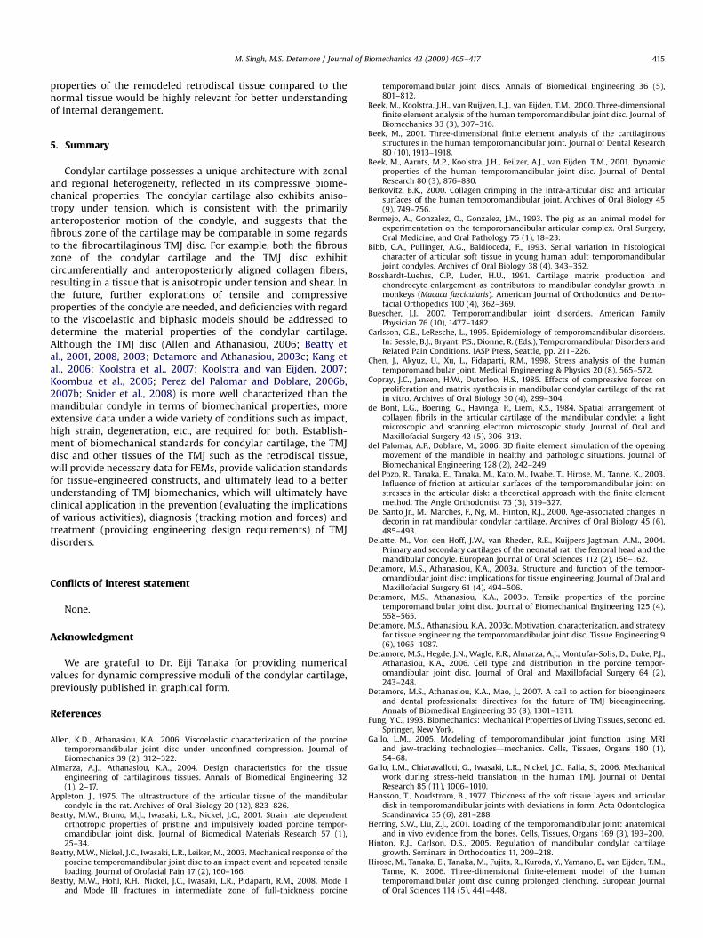
Fig. 1. (A) Schematic of a sagittal section of the human TMJ, showing the position of
surfaces are dark gray. (B) A schematic of the zonal architecture of the condylar carti
proliferative, mature and hypertrophic. This figure highlights fiber organization and ce
of the tissue as a reflection of the known extracellular matrix(ECM) organization, and providing recommendations to validatecritical structure–function correlations. A brief perspective on 3-DFEMs of the human TMJ is also provided, highlighting potentialapplications of such computational models. Highly accurate andvalidated models would be valuable tools for clinical diagnosis ofTMJ disorders, and could be useful for evaluating the implicationsof parafunctional activity and trauma as a means to substantiatepreventive measures for TMJ disorders.
2. Biomechanical properties of the condylar cartilage as areflection of ECM organization
Condylar cartilage is a viscoelastic tissue (Kuboki et al., 1997;Singh and Detamore, 2008) that exhibits characteristic responsesof creep, stress relaxation and hysteresis (Fung, 1993). Condylarcartilage is a unique tissue, which is distinctively different incomposition and mechanics compared to hyaline cartilages, kneemeniscus or growth plate (Wang et al., 2008). There is a reviewavailable where the differences along the spectrum of TMJ, kneemeniscus and hyaline cartilages are discussed in detail (Almarzaand Athanasiou, 2004). The condylar cartilage tissue functionsin a complex force-field, experiencing simultaneous compressionand shear (Teramoto et al., 2003). The cartilage ECM is hetero-geneous both topographically and zonally, reflecting its versatilityin responding to varying functional demands. Conflicting viewsremain for the schemes of zonal delineation and terminologiesused to describe the zone-based histological architecture of thecondylar cartilage (Hinton and Carlson, 2005; Luder, 1997). For acomparative analysis, the commonly used zonal classificationof four zones has been used in this review: fibrous zone,proliferative zone, mature zone and hypertrophic zone, categorizedin a distal–proximal manner relative to the subchondral bone ofthe condyle (Fig. 1). Furthermore, to compare the regionalvariations in properties, we have divided the condylar cartilageanteroposteriorly into three regions (anterior, superior andposterior), and mediolaterally into three sections (medial, centraland lateral). Major ECM components discussed in the manuscriptare collagen and proteoglycans. Proteoglycans are glycoproteins,having a long core protein with at least one associated anionic
the condyle with respect to the TMJ disc and eminence–fossa. The cartilaginous
lage, enlarged from the boxed region of (A), showing four explicit zones: fibrous,
llular composition of these zones.
ARTICLE IN PRESS
M. Singh, M.S. Detamore / Journal of Biomechanics 42 (2009) 405–417 407
glycosaminoglycan (GAG) side-chain (Detamore and Athanasiou,2003a; Mow et al., 1980). Among primary GAG molecules inthis context are chondroitin sulfate (CS), keratin sulfate (KS) and
dermatan sulfate (DS). Large chondroitin sulfate proteoglycans(CSPGs), such as aggrecan (with CS and KS side-chains) andversican (with CS side-chains exclusively), contain several GAGmoieties. In contrast, dermatan sulfate proteoglycans (DSPGs)are small proteoglycans with one (decorin) or two (biglycan)GAG side-chains. Biomechanical characterization studies of thecondylar cartilage are discussed under the following twosubsections—(a) tension/shear and (b) compression, where ECMorganization with respect to aforementioned major ECM compo-nents is discussed, followed by a synopsis of important futureconsiderations.
2.1. Tension and shear
Condylar cartilage experiences tensile loads primarily due toshear and friction produced by mandibular motion especially inthe regions closer to the joint synovium (Singh and Detamore,2008). Unlike under compression, viscoelasticity of the articularcartilage under tension is primarily governed by the solidconstituents in a relatively flow-independent manner (Huanget al., 2001). Collagen, which forms more than 60% of the dryweight of condylar cartilage in rabbits (Pietila et al., 1999), is theprimary ECM component responsible for the tensile resistance ofthe soft tissues (Huang et al., 2001). In the following subsections,tensile and shear properties of the condylar cartilage and collagenorganization in the tissue are reviewed. In addition, distribution ofDSPGs, which also play a role in the enhancement of tensileproperties (Detamore and Athanasiou, 2003a), is briefly discussed.
2.1.1. Tensile and shear evaluations
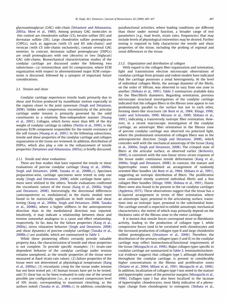
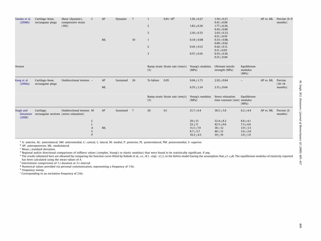
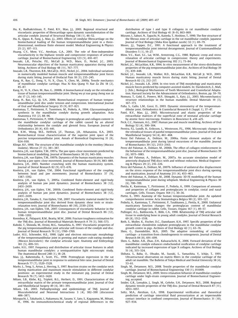
There are four studies that have reported the tensile or shearevaluations of porcine condylar cartilage (Kang et al., 2000a;Singh and Detamore, 2008; Tanaka et al., 2008b, c). Specimenpreparation-wise, cartilage specimens were tested in only onestudy (Singh and Detamore, 2008), while cartilage–bone speci-mens were utilized in the others. Tensile evaluations confirmedthe viscoelastic nature of the tissue (Kang et al., 2000a; Singhand Detamore, 2008). Interestingly, the directional differences(anteroposterior vs. mediolateral) in the elastic moduli werefound to be statistically significant in both tensile and sheartesting (Kang et al., 2000a; Singh and Detamore, 2008; Tanakaet al., 2008b), where a higher stiffness in the anteroposteriordirection than in the mediolateral direction was reported.Intuitively, it may indicate a relationship between shear andtension somewhat analogous to a cause and effect relationship,respectively. So far, data for the failure properties (Kang et al.,2000a), stress relaxation behavior (Singh and Detamore, 2008)and shear dynamics of porcine condylar cartilage (Tanaka et al.,2008b, c) are available, which are summarized in Table 1.
While these pioneering efforts certainly provide valuableproperty data, the characterization of tensile and shear propertiesis not complete. To provide specific examples: (1) strain-ratedependent behavior of the condylar cartilage under tensionremains unexplored, as the tensile properties of the tissue weremeasured at fixed strain-rate values; (2) failure properties of thetissue were not determined at physiological temperature (Kanget al., 2000a); (3) viscoelastic creep for the tissue under tensionhas not been tested yet; (4) human tissues have yet to be tested;and (5) shear has so far been evaluated in only one of the severalpossible jaw configurations (i.e., at a constant compression levelof 10% strain; corresponding to maximum clenching, as theauthors noted) (Tanaka et al., 2008b, c). In addition, considering
parafunctional activities, where loading conditions are differentthan those under normal function, a broader range of testparameters (e.g., load levels, strain rates, frequencies) that mayinclude levels of physiological extremities may be desired. Furthertesting is required to fully characterize the tensile and shearproperties of the tissue, including the probing of regional andzonal differences in the tissue.
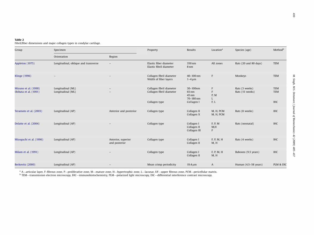
2.1.2. Organization and distribution of collagen
With regard to the collagen fiber organization and orientation,light and transmission electron microscopic observations ofcondylar cartilage from primate and rodent models have indicatedthat the cartilage possesses a zonal heterogeneity. At the levelof individual collagen fibrils, the average diameter of the fibrils,on the order of 100 nm, was observed to vary from one zone toanother (Shibata et al., 1991). Table 2 summarizes available datafor the fiber/fibrils diameters. Regarding orientation, previousmicroscopic structural investigations of the condylar cartilageindicated that the collagen fibers in the fibrous zone appear to runpredominately parallel to the surface but not to each other,forming sheet-like structures (de Bont et al., 1984; Klinge, 1996;Luder and Schroeder, 1990; Mizuno et al., 1990; Shibata et al.,1991), indicating a transversely isotropic fiber orientation. How-ever, in a recent macroscopic investigation of the condylarcartilage, an anisotropic fiber orientation in the fibrous zoneof porcine condylar cartilage was observed via polarized light,where the predominant orientation of collagen fibers was in theanteroposterior direction (Singh and Detamore, 2008), whichcoincides well with the mechanical anisotropy of the tissue (Kanget al., 2000a; Singh and Detamore, 2008). The crimped state offibers at the articular surface, as observed earlier (Berkovitz,2000), is consistent with the non-linear stress–strain response ofthe tissue under continuous tensile deformation (Kang et al.,2000a; Singh and Detamore, 2008). In contrast, the mature andhypertrophic zones exhibited an arrangement of randomlyoriented fiber bundles (de Bont et al., 1984; Shibata et al., 1991),suggesting an isotropic distribution of fibers. The proliferativezone contained mostly scattered individual fibrils and a fewcollagen fiber bundles (Klinge, 1996; Shibata et al., 1991). Elasticfibers were also found to be present in the rat condylar cartilages(Appleton, 1975). These observations suggest that the tissue has abi-layered arrangement in terms of fiber orientation, wherean anisotropic layer, proximal to the articulating surface, transi-tions into an isotropic layer, proximal to the subchondral bone.The cartilage overall is expected to exhibit anisotropic mechanicalcharacteristics, the extent of which may primarily depend on thethickness ratio of the fibrous zone to the entire cartilage.
It is known that tensile forces correspond more to fibroblasticactivity, leading to the production of collagen type I, whilecompressive forces tend to be correlated with chondrocytes andthe increased production of collagen type II and large chondroitinsulfate proteoglycans (Detamore et al., 2006). Therefore, thedistribution of the primary collagen types (I and II) in the condylarcartilage may reflect biomechanical/functional requirements ofthe tissue (Mizoguchi et al., 1996). Major collagen types specific tocondylar cartilage are summarized in Table 2. Immunohistochem-ical evidence suggests that collagen type I, although distributedthroughout the condylar cartilage, is present in considerablyhigher concentrations in the fibrous and proliferative zones(Delatte et al., 2004; Milam et al., 1991; Mizoguchi et al., 1996).In addition, localization of collagen type I was noted in the matureand hypertrophic zones of the posterior margins (Mizoguchi et al.,1996). Collagen type I has also been localized in the lacunaeof hypertrophic chondrocytes, most likely indicative of a pheno-typic change from chondrogenic to osteogenic (Shibata et al.,
ARTIC
LEIN
PRESS
Table 1Mechanical properties of condylar cartilage under different testing modalities.
Group Sample Method Regiona Directionb Loading
profile
Specimens
tested
Testing parameters Mechanical propertiesc Regional
differencesd
Species (age)
Compression Ramp stress
(MPa)
Strain rate (mm/
min)/frequency
(Hz)
Modulus of
elasticity (MPa)
Creep time constant
(s)
Equilibrium
modulus
(MPa)
Kuboki et al.
(1997)e
Cartilage–bone,
entire cartilage
intact
Indentation (creep) A Normal Sustained 10 0.51 1.04/� – 125.5723.3 2.684 – Porcine (7
months)
1.02 112.5733.0 3.591
1.53 104.0728.8 4.751
Intermittent 10 0.51 1.04/0.33f 146.5735.2 3.355
1.02 134.4715.0 5
1.53 139.8725.0 6.623
Ramp stress
(MPa)
Frequency (Hz) Modulus of
elasticity (MPa)
Poisson’s ratio Equilibrium
modulus
(MPa)
Hu et al.
(2001)
Cartilage–bone,
rectangular plugs
Atomic force
microscopy
AM Normal Dynamic 18 – 14 2.3470.26 0.4670.05 – AM vs.
others
Rabbit (6
weeks)
AL 1.51 0.41 AL vs.
others
PM 1.1170.07 0.38 PM vs.
others
PL 0.9570.06 0.3170.05 PL vs.
others
Patel and Mao
(2003)
Cartilage–bone,
rectangular plugs
Atomic force
microscopy
AM Normal Dynamic 18 – 14 0.9570.15 – – – Rabbit (7
days)
PL 1.0270.22 –
Applied %
strain range
Frequency (Hz) Dynamic complex
modulusg (MPa)
Storage and loss
modulig (MPa)
Equilibrium
modulus
(MPa)
Tanaka et al.
(2006)
Cartilage–bone,
rectangular plugs
Indentation (dynamic) AM Normal Dynamic 10 170.2 0.01–10h 1.4070.39 1.3670.38,
0.3470.07
– AM vs. PM Porcine (6–9
months)
AL 1.1570.33 1.1270.32,
0.2470.07
AM vs. PL
PM 0.8170.21 0.7970.21,
0.1670.04
AL vs. PM
PL 0.7370.26 0.7270.25,
0.1670.05
AL vs. PL
Shear % strain
amplitude
Frequency (Hz) Complex shear
modulusi (MPa)
Storage and loss
modulii (MPa)
Equilibrium
modulus
(MPa)
Tanaka et al.
(2008c)
Cartilage–bone,
rectangular plugs
Shear (dynamic),
compressive strain
(10%)
C AP Dynamic 10 1 0.01–10h 1.5670.27 1.5070.27,
0.4170.06
– – Porcine (6–9
months)
2 1.8270.26 1.7770.26,
0.4570.06
3 2.1070.33 2.0370.33,
0.5170.10
M.
Sing
h,
M.S.
Deta
mo
re/
Jou
rna
lo
fB
iom
echa
nics
42
(20
09
)4
05
–417
40
8
ARTIC
LEIN
PRESS
Tanaka et al.
(2008b)
Cartilage–bone,
rectangular plugs
Shear (dynamic),
compressive strain
(10%)
C AP Dynamic 7 1 0.01–10h 1.5670.27 1.5070.27,
0.4170.06
– AP vs. ML Porcine (6–9
months)
2 1.8270.26 1.7770.26,
0.4570.06
3 2.1070.33 2.0370.33,
0.5170.10
ML 10 1 0.3470.08 0.3370.08,
0.0970.02
2 0.4370.12 0.4270.11,
0.1170.03
3 0.5770.10 0.5570.10,
0.1570.04
Tension Ramp strain
(%)
Strain rate (mm/s) Young’s modulus
(MPa)
Ultimate tensile
strength (MPa)
Equilibrium
modulus
(MPa)
Kang et al.
(2000a)
Cartilage–bone,
rectangular plugs
Unidirectional tension – AP Sustained 26 To failure 0.05 9.0471.73 2.9270.84 – AP vs. ML Porcine
(10–14
months)ML 6.5571.24 2.1570.64
Ramp strain
(%)
Strain rate (mm/s) Young’s modulus
(MPa)
Stress relaxation
time constant (min)
Equilibrium
modulus
(MPa)
Singh and
Detamore
(2008)
Cartilage,
rectangular sections
Unidirectional tension
(stress relaxation)
M AP Sustained 7 20 0.1 21.774.4 38.575.8 6.274.4 AP vs. ML Porcine (6
months)
C 29713 32.478.2 8.874.1
L 22711 42.579.6 7.374.6
A ML 11.377.9 38712 3.973.5
S 8.773.7 40713 3.672.0
P 10.274.5 39719 3.971.9
a A: anterior, AL: anterolateral, AM: anteromedial, C: central, L: lateral, M: medial, P: posterior, PL: posterolateral, PM: posteromedial, S: superior.b AP: anteroposterior, ML: mediolateral.c Mean7standard deviation.d Regional and/or directional comparisons of stiffness values (complex, Young’s or elastic modulus) that were found to be statistically significant, if any.e The results tabulated here are obtained by comparing the function curve-fitted by Kuboki et al., i.e., A(1�exp(�t/z)), to the Kelvin model having the assumption that m15m0. The equilibrium modulus of elasticity reported
has been calculated using the mean values of A.f Intermittent compression of 1 s duration at 2 s interval.g Numerical values provided via personal communication, representing a frequency of 1 Hz.h Frequency sweep.i Corresponding to an excitation frequency of 2 Hz.
M.
Sing
h,
M.S.
Deta
mo
re/
Jou
rna
lo
fB
iom
echa
nics
42
(20
09
)4
05
–417
40
9
ARTIC
LEIN
PRESS
Table 2Fibril/fiber dimensions and major collagen types in condylar cartilage.
Group Specimen Property Results Locationa Species (age) Methodb
Orientation Region
Appleton (1975) Longitudinal, oblique and transverse – Elastic fiber diameter 350 nm All zones Rats (20 and 80 days) TEM
Elastic fibril diameter 8 nm
Klinge (1996) – – Collagen fibril diameter 40–100 nm F Monkeys TEM
Width of fiber layers 1–4mm
Mizuno et al. (1990) Longitudinal (ML) – Collagen fibril diameter 30–100nm F Rats (3 weeks) TEM
Shibata et al. (1991) Longitudinal (ML) – Collagen fibril diameter 65 nm F Rats (15 weeks) TEM
45 nm P, M
70–180 nm L
Collagen type Col lagen I F, L IHC
Teramoto et al. (2003) Longitudinal (AP) Anterior and posterior Collagen type Collagen II M, H, PCM Rats (8 weeks) IHC
Collagen X M, H, PCM
Delatte et al. (2004) Longitudinal (AP) – Collagen type Collagen I F, P, M Rats (neonatal) IHC
Collagen II M,H
Collagen III F
Mizoguchi et al. (1996) Longitudinal (AP) Anterior, superior Collagen type Collagen I F, P, M, H Rats (4 weeks) IHC
and posterior Collagen II M, H
Milam et al. (1991) Longitudinal (AP) – Collagen type Collagen I F, P, M, H Baboons (9.5 years) IHC
Collagen II M, H
Berkovitz (2000) Longitudinal (AP) – Mean crimp periodicity 19.4mm A Human (4.5–58 years) PLM & DIC
a A—articular layer, F–fibrous zone, P—proliferative zone, M—mature zone, H—hypertrophic zone, L—lacunar, UF—upper fibrous zone, PCM—pericellular matrix.b TEM—transmission electron microscopy, IHC—immunohistochemistry, PLM—polarized light microscopy, DIC—differential interference contrast microscopy.
M.
Sing
h,
M.S.
Deta
mo
re/
Jou
rna
lo
fB
iom
echa
nics
42
(20
09
)4
05
–417
41
0
ARTICLE IN PRESS
M. Singh, M.S. Detamore / Journal of Biomechanics 42 (2009) 405–417 411
1991). In contrast, collagen type II has been detected almostexclusively in the mature and hypertrophic zones (Milam et al.,1991; Mizoguchi et al., 1996; Teramoto et al., 2003; Wang et al.,2008), and to a lesser degree in the proliferative zone (Visnapuuet al., 2000). The distribution of collagen types I and II maysuggest tension to be the primary mode of loading in the fibrouszone, and compression to be the primary mode in the mature andhypertrophic zones, except for the posterior margins that mayexperience considerable tensile loads.
In addition to zonal heterogeneity, regional differences in thetensile and shear properties, which were found to be significantfor the TMJ disc (Beek et al., 2001; Detamore and Athanasiou,2003b; Kim et al., 2003; Nickel and McLachlan, 1994; Tanakaet al., 2001b; Teng et al., 1991), can be expected as the condylarcartilage functions in a multi-directional force field. Althoughregional differences in tensile properties of the condylar cartilagein a given direction were found not to be significantly different(Singh and Detamore, 2008), regional data for collagen contentand degree of anisotropy may provide valuable validation.
2.1.3. Distribution of dermatan sulfate proteoglycans
Decorin is an ECM component considered to enhance thetensile properties by increasing the collagen fiber diameterand modulating fibril synthesis, as reviewed by Detamore andAthanasiou (2003a). A higher expression of decorin in the fibrouszone of the cartilage compared to other zones, as observed in ratmodels (Del Santo et al., 2000), may reflect the fulfillment of thefunctional demand, i.e., enhancement of the tensile resistance dueto the presence of considerably higher tensile loads. Condylarcartilage showed no presence of biglycan in rat condylar cartilages(Mao et al., 1998).
2.1.4. Summary: structure–property–function correlation under
tension
In summary, these studies suggest that shear forces havea pronounced effect on the condylar cartilage in the fibrouszone, especially near the articular surface, as evidenced by theanisotropic distribution of collagen fibers, almost exclusivepresence of collagen type I, and considerable localization of
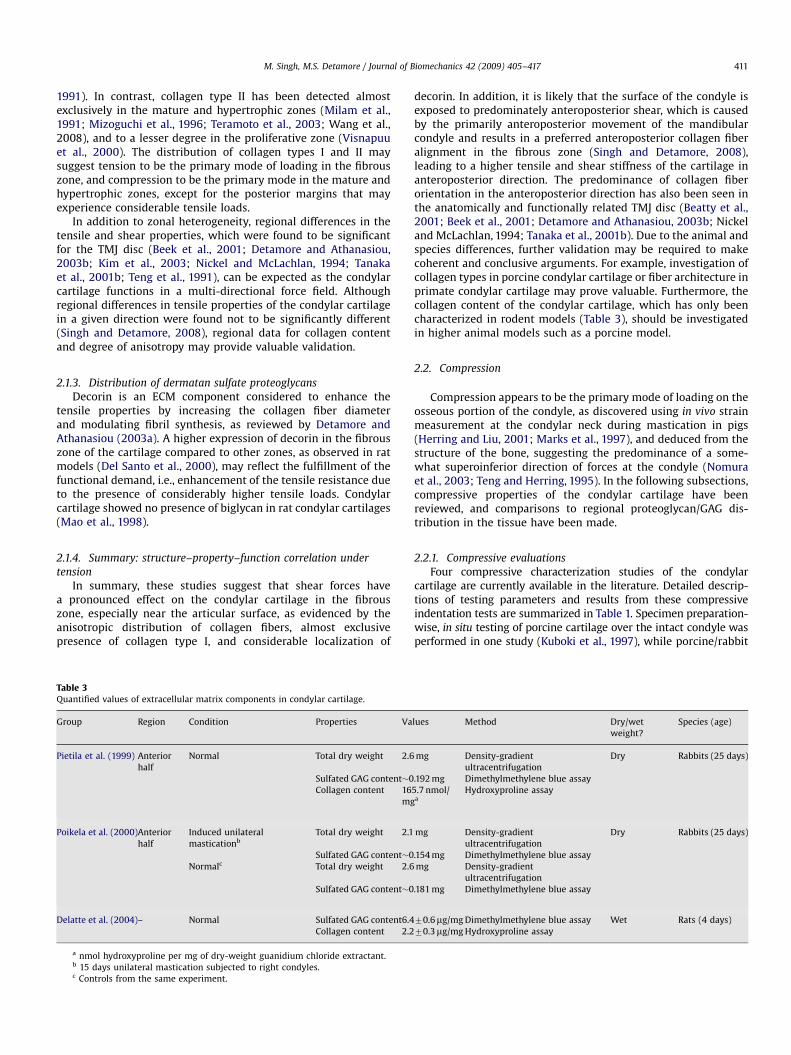
Table 3Quantified values of extracellular matrix components in condylar cartilage.
Group Region Condition Properties Va
Pietila et al. (1999) Anterior
half
Normal Total dry weight 2.6
Sulfated GAG content�0
Collagen content 16
mg
Poikela et al. (2000)Anterior
half
Induced unilateral
masticationb
Total dry weight 2.1
Sulfated GAG content�0
Normalc Total dry weight 2.6
Sulfated GAG content�0
Delatte et al. (2004)– Normal Sulfated GAG content6.4
Collagen content 2.2
a nmol hydroxyproline per mg of dry-weight guanidium chloride extractant.b 15 days unilateral mastication subjected to right condyles.c Controls from the same experiment.
decorin. In addition, it is likely that the surface of the condyle isexposed to predominately anteroposterior shear, which is causedby the primarily anteroposterior movement of the mandibularcondyle and results in a preferred anteroposterior collagen fiberalignment in the fibrous zone (Singh and Detamore, 2008),leading to a higher tensile and shear stiffness of the cartilage inanteroposterior direction. The predominance of collagen fiberorientation in the anteroposterior direction has also been seen inthe anatomically and functionally related TMJ disc (Beatty et al.,2001; Beek et al., 2001; Detamore and Athanasiou, 2003b; Nickeland McLachlan, 1994; Tanaka et al., 2001b). Due to the animal andspecies differences, further validation may be required to makecoherent and conclusive arguments. For example, investigation ofcollagen types in porcine condylar cartilage or fiber architecture inprimate condylar cartilage may prove valuable. Furthermore, thecollagen content of the condylar cartilage, which has only beencharacterized in rodent models (Table 3), should be investigatedin higher animal models such as a porcine model.
2.2. Compression
Compression appears to be the primary mode of loading on theosseous portion of the condyle, as discovered using in vivo strainmeasurement at the condylar neck during mastication in pigs(Herring and Liu, 2001; Marks et al., 1997), and deduced from thestructure of the bone, suggesting the predominance of a some-what superoinferior direction of forces at the condyle (Nomuraet al., 2003; Teng and Herring, 1995). In the following subsections,compressive properties of the condylar cartilage have beenreviewed, and comparisons to regional proteoglycan/GAG dis-tribution in the tissue have been made.
2.2.1. Compressive evaluations
Four compressive characterization studies of the condylarcartilage are currently available in the literature. Detailed descrip-tions of testing parameters and results from these compressiveindentation tests are summarized in Table 1. Specimen preparation-wise, in situ testing of porcine cartilage over the intact condyle wasperformed in one study (Kuboki et al., 1997), while porcine/rabbit
lues Method Dry/wet
weight?
Species (age)
mg Density-gradient
ultracentrifugation
Dry Rabbits (25 days)
.192 mg Dimethylmethylene blue assay
5.7 nmol/a
Hydroxyproline assay
mg Density-gradient
ultracentrifugation
Dry Rabbits (25 days)
.154 mg Dimethylmethylene blue assay
mg Density-gradient
ultracentrifugation
.181 mg Dimethylmethylene blue assay
70.6mg/mg Dimethylmethylene blue assay Wet Rats (4 days)
70.3mg/mg Hydroxyproline assay
ARTICLE IN PRESS
M. Singh, M.S. Detamore / Journal of Biomechanics 42 (2009) 405–417412
cartilage–bone sections were utilized in the other studies (Huet al., 2001; Patel and Mao, 2003; Tanaka et al., 2006). So far, datafor the creep (Kuboki et al., 1997) and dynamic properties (Huet al., 2001; Patel and Mao, 2003; Tanaka et al., 2006) areavailable. The cartilage deforms more with sustained compressionthan with intermittent compression, and a higher stiffness wasobserved at greater loads (Kuboki et al., 1997). This phenomenonof strain stiffening under compression has also been observedwith the TMJ disc (Allen and Athanasiou, 2006). While exploringregional differences using dynamic nanoindentation experiments,a decrease in the stiffness (elastic moduli), average Poisson’s ratioand surface roughness from the anterior to the posterior side anda relatively smaller decrease from the medial to the lateral sidewere reported, demonstrating the heterogeneity of the condylarcartilage (Hu et al., 2001; Patel and Mao, 2003). The differences inthe regional properties were statistically significant for 6-week-old rabbits, but not for 7-day-old rabbits, suggesting that theregional heterogeneity of the articular surface evolves with time.In another study, regional dynamic indentation tests of porcinecondylar cartilages were performed by applying sinusoidal strainswith a wide range of loading frequencies, and complex, storageand loss moduli were reported (Tanaka et al., 2006). The complex,storage and loss moduli all increased with an increase infrequency. In agreement with the aforementioned nanoindenta-tion experiments, the anterior region of the condyle in general hadlarger complex, storage and loss moduli than the posterior regionat a given frequency, with the stiffness being highest in theanteromedial region of the four regions explored (see Table 1).Interestingly, in our observations (Singh and Detamore, 2009), arelationship between the stiffness and thickness of the regionswas observed, where the average elastic modulus was foundto increase with the thickness. A thickness–stiffness correlationcan also be gleaned from the study by Tanaka et al. (2006), wherespecimens from the stiffer regions were generally thicker.Validation of the thickness–stiffness correlation for the condylarcartilage is certainly required. Moreover, exploration of zonalproperty differences may provide valuable information regardingzonal structure–material property relationship.
2.2.2. Spatial variation of proteoglycans/GAGs
Among different zones, aggrecan, the primary proteoglycan incondylar cartilage, was found to be localized mainly in the matureand hypertrophic zones using immunohistochemical methods inporcine (Roth et al., 1997) and rat (Teramoto et al., 2003) models.Using immunohistochemistry, the presence of CS-rich versican-like proteoglycans were detected in the fibrous and proliferativezones in porcine and rat models (Mao et al., 1998; Roth et al.,1997), possibly owing to the fibroblastic (Mao et al., 1998) orprogenitor cell activity (Roth et al., 1997). Interestingly, immuno-histochemical analysis of primate condylar cartilages locatedkeratan sulfate and chondroitin sulfate only in the matureand hypertrophic zones, while their presence in the fibrous zonewas not observed (Milam et al., 1991). This observation is not inaccord with other observations (Mao et al., 1998; Roth et al., 1997)unless it is due to the differences in animal models. Amongregions, the maximum synthesis of CS-rich proteoglycans wasreported to be in the posterosuperior region of the rabbit condylarcartilage (Kantomaa et al., 1994). Histochemical observation of thecondylar cartilages of rabbits showed the amount of CS-richproteoglycans in the anterior and posterosuperior regions to behigher than in the superior region, with the highest content inthe anterior region (Kantomaa and Pirttiniemi, 1998). The studieswhere the sulfated GAG content of condylar cartilage wasquantified, in rodent models, are summarized in Table 3 as areference.
Although some information about occurrence of proteoglycansis available, the literature lacks a comprehensive regionalquantification data of proteoglycans in the condylar cartilage.One limitation of the previous efforts to quantify proteoglycancontent was the lack of thickness characterization, i.e., specimenpreparation protocols did not clearly indicate the methodsemployed to affirm the nature of the specimen, namely whetherit was a cartilage section, the entire cartilage thickness, or acartilage–bone specimen. Another important issue pertains to theanimal models used in many of these studies. Due to the smallsize and anatomical differences of the rodent TMJ relative to thehuman TMJ (Bermejo et al., 1993), further quantification of thesecomponents, both regional and overall, in more suitable animalmodels (e.g., porcine or primate) may be desired. It is worthnoting that results of biochemical assays between rats and higheranimal models have also exhibited general inconsistencies withTMJ disc studies as well (Detamore and Athanasiou, 2003c).
2.2.3. Summary: structure–property–function correlation under
compression
Condylar cartilage has a heterogeneous nature in terms of bothstructure and mechanics. There are indications that the tissuemay have a positive correlation between regional thickness andcorresponding stiffness, which may imply that regional variationsin the stiffness and thickness are related phenomena, developedin response to regional loading patterns. It may also indicate thatthe role of the cartilage is to sustain a heterogeneous in vivo
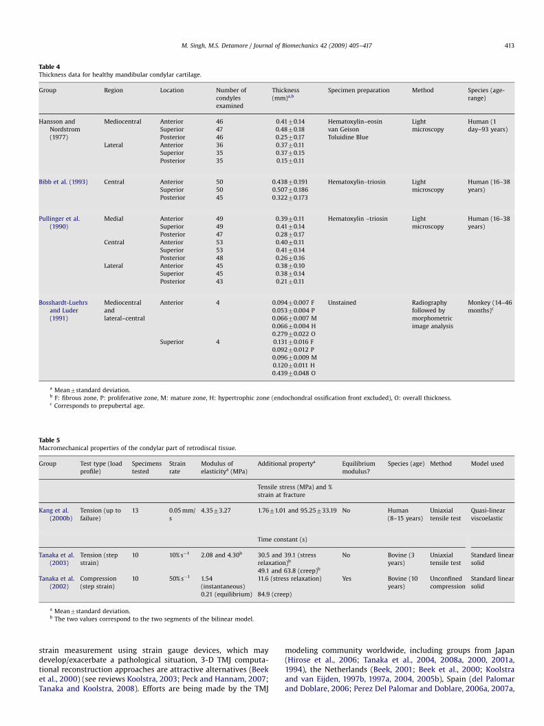
biomechanical environment, where cartilage possibly experienceshigher loads in the thicker regions. Valuable data regardingthe thickness of human condylar cartilage are available in theliterature (Bibb et al., 1993; Hansson and Nordstrom, 1977;Pullinger et al., 1990), and summarized in Table 4. Validationof the thickness–stiffness correlation in humans may improveour understanding of the nature and extent of condylar loads.If indeed such a correlation exists, it may have a direct influenceon the FEMs of the human TMJ in the future, where in vivo
imaging techniques will facilitate the determining of materialproperties of condylar cartilage in a subject-specific manner.Regarding the structure, localization of aggrecan mainly in themature and hypertrophic zones and absence from the fibrous zonesupports the notion that the zones proximal to the subchondralbone are more hyaline-like in nature and primarily serves to resistthe compression. However, comprehensive regional and zonalquantification of proteoglycans and GAGs may be warranted toclearly establish the structure–property–function correlation.A possible variation of compressive properties as a function ofdepth, also observed in knee cartilage (Verteramo and Seedhom,2004), can be anticipated, where hyaline-like mature andhypertrophic zones would be stiffer under compression than thefibrous zone. In addition, the extent of collagen fiber reinforce-ment, known to affect viscoelasticity via impeding the fluid flowthat results in fluid pressurization (Soltz and Ateshian, 1998),against compression in the condylar cartilage is required to beunderstood yet, which leads to two important questions: (a) isthere any significant difference in the collagen content of thecondylar cartilage compared to the other articular cartilages? and(b) how does the difference in the collagen architecture affect thefiber reinforcement and interstitial fluid flow in the condylarcartilage?
3. A perspective on 3-D computational reconstruction of thehuman TMJ
Due to the difficulties associated with in vivo estimation of thestress distribution using invasive experimental techniques, e.g.,
ARTICLE IN PRESS
Table 4Thickness data for healthy mandibular condylar cartilage.
Group Region Location Number of
condyles
examined
Thickness
(mm)a,b
Specimen preparation Method Species (age-
range)
Hansson and
Nordstrom
(1977)
Mediocentral Anterior 46 0.4170.14 Hematoxylin–eosin Light
microscopy
Human (1
day–93 years)Superior 47 0.4870.18 van Geison
Posterior 46 0.2570.17 Toluidine Blue
Lateral Anterior 36 0.3770.11
Superior 35 0.3770.15
Posterior 35 0.1570.11
Bibb et al. (1993) Central Anterior 50 0.43870.191 Hematoxylin–triosin Light
microscopy
Human (16–38
years)Superior 50 0.50770.186
Posterior 45 0.32270.173
Pullinger et al.
(1990)
Medial Anterior 49 0.3970.11 Hematoxylin –triosin Light
microscopy
Human (16–38
years)Superior 49 0.4170.14
Posterior 47 0.2870.17
Central Anterior 53 0.4070.11
Superior 53 0.4170.14
Posterior 48 0.2670.16
Lateral Anterior 45 0.3870.10
Superior 45 0.3870.14
Posterior 43 0.2170.11
Bosshardt-Luehrs
and Luder
(1991)
Mediocentral
and
lateral–central
Anterior 4 0.09470.007 F Unstained Radiography
followed by
morphometric
image analysis
Monkey (14–46
months)c0.05370.004 P
0.06670.007 M
0.06670.004 H
0.27970.022 O
Superior 4 0.13170.016 F
0.09270.012 P
0.09670.009 M
0.12070.011 H
0.43970.048 O
a Mean7standard deviation.b F: fibrous zone, P: proliferative zone, M: mature zone, H: hypertrophic zone (endochondral ossification front excluded), O: overall thickness.c Corresponds to prepubertal age.
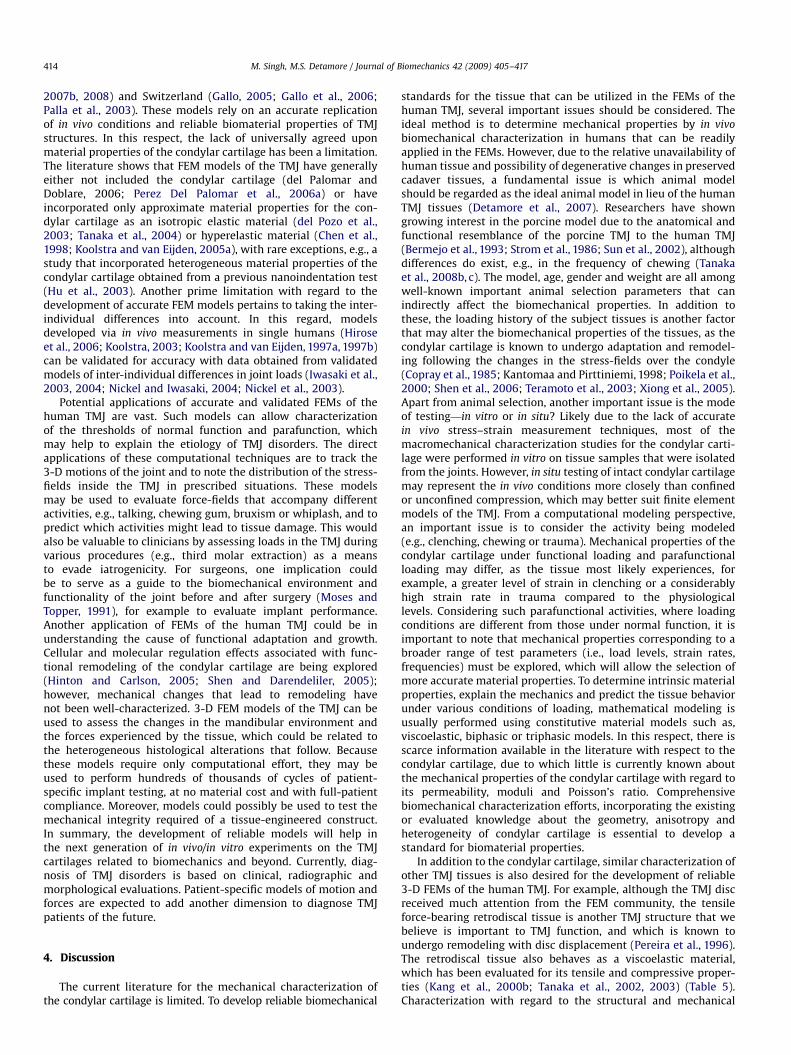
Table 5Macromechanical properties of the condylar part of retrodiscal tissue.
Group Test type (load
profile)
Specimens
tested
Strain
rate
Modulus of
elasticitya (MPa)
Additional propertya Equilibrium
modulus?
Species (age) Method Model used
Tensile stress (MPa) and %
strain at fracture
Kang et al.
(2000b)
Tension (up to
failure)
13 0.05 mm/
s
4.3573.27 1.7671.01 and 95.25733.19 No Human
(8–15 years)
Uniaxial
tensile test
Quasi-linear
viscoelastic
Time constant (s)
Tanaka et al.
(2003)
Tension (step
strain)
10 10% s�1 2.08 and 4.30b 30.5 and 39.1 (stress
relaxation)b
No Bovine (3
years)
Uniaxial
tensile test
Standard linear
solid
49.1 and 63.8 (creep)b
Tanaka et al.
(2002)
Compression
(step strain)
10 50% s�1 1.54
(instantaneous)
11.6 (stress relaxation) Yes Bovine (10
years)
Unconfined
compression
Standard linear
solid
0.21 (equilibrium) 84.9 (creep)
a Mean7standard deviation.b The two values correspond to the two segments of the bilinear model.
M. Singh, M.S. Detamore / Journal of Biomechanics 42 (2009) 405–417 413
strain measurement using strain gauge devices, which maydevelop/exacerbate a pathological situation, 3-D TMJ computa-tional reconstruction approaches are attractive alternatives (Beeket al., 2000) (see reviews Koolstra, 2003; Peck and Hannam, 2007;Tanaka and Koolstra, 2008). Efforts are being made by the TMJ
modeling community worldwide, including groups from Japan(Hirose et al., 2006; Tanaka et al., 2004, 2008a, 2000, 2001a,1994), the Netherlands (Beek, 2001; Beek et al., 2000; Koolstraand van Eijden, 1997b, 1997a, 2004, 2005b), Spain (del Palomarand Doblare, 2006; Perez Del Palomar and Doblare, 2006a, 2007a,
ARTICLE IN PRESS
M. Singh, M.S. Detamore / Journal of Biomechanics 42 (2009) 405–417414
2007b, 2008) and Switzerland (Gallo, 2005; Gallo et al., 2006;Palla et al., 2003). These models rely on an accurate replicationof in vivo conditions and reliable biomaterial properties of TMJstructures. In this respect, the lack of universally agreed uponmaterial properties of the condylar cartilage has been a limitation.The literature shows that FEM models of the TMJ have generallyeither not included the condylar cartilage (del Palomar andDoblare, 2006; Perez Del Palomar et al., 2006a) or haveincorporated only approximate material properties for the con-dylar cartilage as an isotropic elastic material (del Pozo et al.,2003; Tanaka et al., 2004) or hyperelastic material (Chen et al.,1998; Koolstra and van Eijden, 2005a), with rare exceptions, e.g., astudy that incorporated heterogeneous material properties of thecondylar cartilage obtained from a previous nanoindentation test(Hu et al., 2003). Another prime limitation with regard to thedevelopment of accurate FEM models pertains to taking the inter-individual differences into account. In this regard, modelsdeveloped via in vivo measurements in single humans (Hiroseet al., 2006; Koolstra, 2003; Koolstra and van Eijden, 1997a, 1997b)can be validated for accuracy with data obtained from validatedmodels of inter-individual differences in joint loads (Iwasaki et al.,2003, 2004; Nickel and Iwasaki, 2004; Nickel et al., 2003).
Potential applications of accurate and validated FEMs of thehuman TMJ are vast. Such models can allow characterizationof the thresholds of normal function and parafunction, whichmay help to explain the etiology of TMJ disorders. The directapplications of these computational techniques are to track the3-D motions of the joint and to note the distribution of the stress-fields inside the TMJ in prescribed situations. These modelsmay be used to evaluate force-fields that accompany differentactivities, e.g., talking, chewing gum, bruxism or whiplash, and topredict which activities might lead to tissue damage. This wouldalso be valuable to clinicians by assessing loads in the TMJ duringvarious procedures (e.g., third molar extraction) as a meansto evade iatrogenicity. For surgeons, one implication couldbe to serve as a guide to the biomechanical environment andfunctionality of the joint before and after surgery (Moses andTopper, 1991), for example to evaluate implant performance.Another application of FEMs of the human TMJ could be inunderstanding the cause of functional adaptation and growth.Cellular and molecular regulation effects associated with func-tional remodeling of the condylar cartilage are being explored(Hinton and Carlson, 2005; Shen and Darendeliler, 2005);however, mechanical changes that lead to remodeling havenot been well-characterized. 3-D FEM models of the TMJ can beused to assess the changes in the mandibular environment andthe forces experienced by the tissue, which could be related tothe heterogeneous histological alterations that follow. Becausethese models require only computational effort, they may beused to perform hundreds of thousands of cycles of patient-specific implant testing, at no material cost and with full-patientcompliance. Moreover, models could possibly be used to test themechanical integrity required of a tissue-engineered construct.In summary, the development of reliable models will help inthe next generation of in vivo/in vitro experiments on the TMJcartilages related to biomechanics and beyond. Currently, diag-nosis of TMJ disorders is based on clinical, radiographic andmorphological evaluations. Patient-specific models of motion andforces are expected to add another dimension to diagnose TMJpatients of the future.
4. Discussion
The current literature for the mechanical characterization ofthe condylar cartilage is limited. To develop reliable biomechanical
standards for the tissue that can be utilized in the FEMs of thehuman TMJ, several important issues should be considered. Theideal method is to determine mechanical properties by in vivo
biomechanical characterization in humans that can be readilyapplied in the FEMs. However, due to the relative unavailability ofhuman tissue and possibility of degenerative changes in preservedcadaver tissues, a fundamental issue is which animal modelshould be regarded as the ideal animal model in lieu of the humanTMJ tissues (Detamore et al., 2007). Researchers have showngrowing interest in the porcine model due to the anatomical andfunctional resemblance of the porcine TMJ to the human TMJ(Bermejo et al., 1993; Strom et al., 1986; Sun et al., 2002), althoughdifferences do exist, e.g., in the frequency of chewing (Tanakaet al., 2008b, c). The model, age, gender and weight are all amongwell-known important animal selection parameters that canindirectly affect the biomechanical properties. In addition tothese, the loading history of the subject tissues is another factorthat may alter the biomechanical properties of the tissues, as thecondylar cartilage is known to undergo adaptation and remodel-ing following the changes in the stress-fields over the condyle(Copray et al., 1985; Kantomaa and Pirttiniemi, 1998; Poikela et al.,2000; Shen et al., 2006; Teramoto et al., 2003; Xiong et al., 2005).Apart from animal selection, another important issue is the modeof testing—in vitro or in situ? Likely due to the lack of accuratein vivo stress–strain measurement techniques, most of themacromechanical characterization studies for the condylar carti-lage were performed in vitro on tissue samples that were isolatedfrom the joints. However, in situ testing of intact condylar cartilagemay represent the in vivo conditions more closely than confinedor unconfined compression, which may better suit finite elementmodels of the TMJ. From a computational modeling perspective,an important issue is to consider the activity being modeled(e.g., clenching, chewing or trauma). Mechanical properties of thecondylar cartilage under functional loading and parafunctionalloading may differ, as the tissue most likely experiences, forexample, a greater level of strain in clenching or a considerablyhigh strain rate in trauma compared to the physiologicallevels. Considering such parafunctional activities, where loadingconditions are different from those under normal function, it isimportant to note that mechanical properties corresponding to abroader range of test parameters (i.e., load levels, strain rates,frequencies) must be explored, which will allow the selection ofmore accurate material properties. To determine intrinsic materialproperties, explain the mechanics and predict the tissue behaviorunder various conditions of loading, mathematical modeling isusually performed using constitutive material models such as,viscoelastic, biphasic or triphasic models. In this respect, there isscarce information available in the literature with respect to thecondylar cartilage, due to which little is currently known aboutthe mechanical properties of the condylar cartilage with regard toits permeability, moduli and Poisson’s ratio. Comprehensivebiomechanical characterization efforts, incorporating the existingor evaluated knowledge about the geometry, anisotropy andheterogeneity of condylar cartilage is essential to develop astandard for biomaterial properties.
In addition to the condylar cartilage, similar characterization ofother TMJ tissues is also desired for the development of reliable3-D FEMs of the human TMJ. For example, although the TMJ discreceived much attention from the FEM community, the tensileforce-bearing retrodiscal tissue is another TMJ structure that webelieve is important to TMJ function, and which is known toundergo remodeling with disc displacement (Pereira et al., 1996).The retrodiscal tissue also behaves as a viscoelastic material,which has been evaluated for its tensile and compressive proper-ties (Kang et al., 2000b; Tanaka et al., 2002, 2003) (Table 5).Characterization with regard to the structural and mechanical
ARTICLE IN PRESS
M. Singh, M.S. Detamore / Journal of Biomechanics 42 (2009) 405–417 415
properties of the remodeled retrodiscal tissue compared to thenormal tissue would be highly relevant for better understandingof internal derangement.
5. Summary
Condylar cartilage possesses a unique architecture with zonaland regional heterogeneity, reflected in its compressive biome-chanical properties. The condylar cartilage also exhibits aniso-tropy under tension, which is consistent with the primarilyanteroposterior motion of the condyle, and suggests that thefibrous zone of the cartilage may be comparable in some regardsto the fibrocartilaginous TMJ disc. For example, both the fibrouszone of the condylar cartilage and the TMJ disc exhibitcircumferentially and anteroposteriorly aligned collagen fibers,resulting in a tissue that is anisotropic under tension and shear. Inthe future, further explorations of tensile and compressiveproperties of the condyle are needed, and deficiencies with regardto the viscoelastic and biphasic models should be addressed todetermine the material properties of the condylar cartilage.Although the TMJ disc (Allen and Athanasiou, 2006; Beatty etal., 2001, 2008, 2003; Detamore and Athanasiou, 2003c; Kang etal., 2006; Koolstra et al., 2007; Koolstra and van Eijden, 2007;Koombua et al., 2006; Perez del Palomar and Doblare, 2006b,2007b; Snider et al., 2008) is more well characterized than themandibular condyle in terms of biomechanical properties, moreextensive data under a wide variety of conditions such as impact,high strain, degeneration, etc., are required for both. Establish-ment of biomechanical standards for condylar cartilage, the TMJdisc and other tissues of the TMJ such as the retrodiscal tissue,will provide necessary data for FEMs, provide validation standardsfor tissue-engineered constructs, and ultimately lead to a betterunderstanding of TMJ biomechanics, which will ultimately haveclinical application in the prevention (evaluating the implicationsof various activities), diagnosis (tracking motion and forces) andtreatment (providing engineering design requirements) of TMJdisorders.
Conflicts of interest statement
None.
Acknowledgment
We are grateful to Dr. Eiji Tanaka for providing numericalvalues for dynamic compressive moduli of the condylar cartilage,previously published in graphical form.
References
Allen, K.D., Athanasiou, K.A., 2006. Viscoelastic characterization of the porcinetemporomandibular joint disc under unconfined compression. Journal ofBiomechanics 39 (2), 312–322.
Almarza, A.J., Athanasiou, K.A., 2004. Design characteristics for the tissueengineering of cartilaginous tissues. Annals of Biomedical Engineering 32(1), 2–17.
Appleton, J., 1975. The ultrastructure of the articular tissue of the mandibularcondyle in the rat. Archives of Oral Biology 20 (12), 823–826.
Beatty, M.W., Bruno, M.J., Iwasaki, L.R., Nickel, J.C., 2001. Strain rate dependentorthotropic properties of pristine and impulsively loaded porcine tempor-omandibular joint disk. Journal of Biomedical Materials Research 57 (1),25–34.
Beatty, M.W., Nickel, J.C., Iwasaki, L.R., Leiker, M., 2003. Mechanical response of theporcine temporomandibular joint disc to an impact event and repeated tensileloading. Journal of Orofacial Pain 17 (2), 160–166.
Beatty, M.W., Hohl, R.H., Nickel, J.C., Iwasaki, L.R., Pidaparti, R.M., 2008. Mode Iand Mode III fractures in intermediate zone of full-thickness porcine
temporomandibular joint discs. Annals of Biomedical Engineering 36 (5),801–812.
Beek, M., Koolstra, J.H., van Ruijven, L.J., van Eijden, T.M., 2000. Three-dimensionalfinite element analysis of the human temporomandibular joint disc. Journal ofBiomechanics 33 (3), 307–316.
Beek, M., 2001. Three-dimensional finite element analysis of the cartilaginousstructures in the human temporomandibular joint. Journal of Dental Research80 (10), 1913–1918.
Beek, M., Aarnts, M.P., Koolstra, J.H., Feilzer, A.J., van Eijden, T.M., 2001. Dynamicproperties of the human temporomandibular joint disc. Journal of DentalResearch 80 (3), 876–880.
Berkovitz, B.K., 2000. Collagen crimping in the intra-articular disc and articularsurfaces of the human temporomandibular joint. Archives of Oral Biology 45(9), 749–756.
Bermejo, A., Gonzalez, O., Gonzalez, J.M., 1993. The pig as an animal model forexperimentation on the temporomandibular articular complex. Oral Surgery,Oral Medicine, and Oral Pathology 75 (1), 18–23.
Bibb, C.A., Pullinger, A.G., Baldioceda, F., 1993. Serial variation in histologicalcharacter of articular soft tissue in young human adult temporomandibularjoint condyles. Archives of Oral Biology 38 (4), 343–352.
Bosshardt-Luehrs, C.P., Luder, H.U., 1991. Cartilage matrix production andchondrocyte enlargement as contributors to mandibular condylar growth inmonkeys (Macaca fascicularis). American Journal of Orthodontics and Dento-facial Orthopedics 100 (4), 362–369.
Buescher, J.J., 2007. Temporomandibular joint disorders. American FamilyPhysician 76 (10), 1477–1482.
Carlsson, G.E., LeResche, L., 1995. Epidemiology of temporomandibular disorders.In: Sessle, B.J., Bryant, P.S., Dionne, R. (Eds.), Temporomandibular Disorders andRelated Pain Conditions. IASP Press, Seattle, pp. 211–226.
Chen, J., Akyuz, U., Xu, L., Pidaparti, R.M., 1998. Stress analysis of the humantemporomandibular joint. Medical Engineering & Physics 20 (8), 565–572.
Copray, J.C., Jansen, H.W., Duterloo, H.S., 1985. Effects of compressive forces onproliferation and matrix synthesis in mandibular condylar cartilage of the ratin vitro. Archives of Oral Biology 30 (4), 299–304.
de Bont, L.G., Boering, G., Havinga, P., Liem, R.S., 1984. Spatial arrangement ofcollagen fibrils in the articular cartilage of the mandibular condyle: a lightmicroscopic and scanning electron microscopic study. Journal of Oral andMaxillofacial Surgery 42 (5), 306–313.
del Palomar, A.P., Doblare, M., 2006. 3D finite element simulation of the openingmovement of the mandible in healthy and pathologic situations. Journal ofBiomechanical Engineering 128 (2), 242–249.
del Pozo, R., Tanaka, E., Tanaka, M., Kato, M., Iwabe, T., Hirose, M., Tanne, K., 2003.Influence of friction at articular surfaces of the temporomandibular joint onstresses in the articular disk: a theoretical approach with the finite elementmethod. The Angle Orthodontist 73 (3), 319–327.
Del Santo Jr., M., Marches, F., Ng, M., Hinton, R.J., 2000. Age-associated changes indecorin in rat mandibular condylar cartilage. Archives of Oral Biology 45 (6),485–493.
Delatte, M., Von den Hoff, J.W., van Rheden, R.E., Kuijpers-Jagtman, A.M., 2004.Primary and secondary cartilages of the neonatal rat: the femoral head and themandibular condyle. European Journal of Oral Sciences 112 (2), 156–162.
Detamore, M.S., Athanasiou, K.A., 2003a. Structure and function of the tempor-omandibular joint disc: implications for tissue engineering. Journal of Oral andMaxillofacial Surgery 61 (4), 494–506.
Detamore, M.S., Athanasiou, K.A., 2003b. Tensile properties of the porcinetemporomandibular joint disc. Journal of Biomechanical Engineering 125 (4),558–565.
Detamore, M.S., Athanasiou, K.A., 2003c. Motivation, characterization, and strategyfor tissue engineering the temporomandibular joint disc. Tissue Engineering 9(6), 1065–1087.
Detamore, M.S., Hegde, J.N., Wagle, R.R., Almarza, A.J., Montufar-Solis, D., Duke, P.J.,Athanasiou, K.A., 2006. Cell type and distribution in the porcine tempor-omandibular joint disc. Journal of Oral and Maxillofacial Surgery 64 (2),243–248.
Detamore, M.S., Athanasiou, K.A., Mao, J., 2007. A call to action for bioengineersand dental professionals: directives for the future of TMJ bioengineering.Annals of Biomedical Engineering 35 (8), 1301–1311.
Fung, Y.C., 1993. Biomechanics: Mechanical Properties of Living Tissues, second ed.Springer, New York.
Gallo, L.M., 2005. Modeling of temporomandibular joint function using MRIand jaw-tracking technologies—mechanics. Cells, Tissues, Organs 180 (1),54–68.
Gallo, L.M., Chiaravalloti, G., Iwasaki, L.R., Nickel, J.C., Palla, S., 2006. Mechanicalwork during stress-field translation in the human TMJ. Journal of DentalResearch 85 (11), 1006–1010.
Hansson, T., Nordstrom, B., 1977. Thickness of the soft tissue layers and articulardisk in temporomandibular joints with deviations in form. Acta OdontologicaScandinavica 35 (6), 281–288.
Herring, S.W., Liu, Z.J., 2001. Loading of the temporomandibular joint: anatomicaland in vivo evidence from the bones. Cells, Tissues, Organs 169 (3), 193–200.
Hinton, R.J., Carlson, D.S., 2005. Regulation of mandibular condylar cartilagegrowth. Seminars in Orthodontics 11, 209–218.
Hirose, M., Tanaka, E., Tanaka, M., Fujita, R., Kuroda, Y., Yamano, E., van Eijden, T.M.,Tanne, K., 2006. Three-dimensional finite-element model of the humantemporomandibular joint disc during prolonged clenching. European Journalof Oral Sciences 114 (5), 441–448.
ARTICLE IN PRESS
M. Singh, M.S. Detamore / Journal of Biomechanics 42 (2009) 405–417416
Hu, K., Radhakrishnan, P., Patel, R.V., Mao, J.J., 2001. Regional structural andviscoelastic properties of fibrocartilage upon dynamic nanoindentation of thearticular condyle. Journal of Structural Biology 136 (1), 46–52.
Hu, K., Qiguo, R., Fang, J., Mao, J.J., 2003. Effects of condylar fibrocartilage on thebiomechanical loading of the human temporomandibular joint in a three-dimensional, nonlinear finite element model. Medical Engineering & Physics25 (2), 107–113.
Huang, C.Y., Mow, V.C., Ateshian, G.A., 2001. The role of flow-independentviscoelasticity in the biphasic tensile and compressive responses of articularcartilage. Journal of Biomechanical Engineering 123 (5), 410–417.
Iwasaki, L.R., Petsche, P.E., McCall Jr, W.D., Marx, D., Nickel, J.C., 2003.Neuromuscular objectives of the human masticatory apparatus during staticbiting. Archives of Oral Biology 48 (11), 767–777.
Iwasaki, L.R., Thornton, B.R., McCall Jr., W.D., Nickel, J.C., 2004. Individual variationsin numerically modeled human muscle and temporomandibular joint forcesduring static biting. Journal of Orofacial Pain 18 (3), 235–245.
Kang, H., Bao, G., Dong, Y., Yi, X., Chao, Y., Chen, M., 2000a. Tensile mechanicsof mandibular condylar cartilage. Hua Xi Kou Qiang Yi Xue Za Zhi 18 (2),85–87.
Kang, H., Yi, X., Chen, M., Bao, G., 2000b. A biomechanical study on the retrodiscaltissue of human temporomandibular joint. Sheng wu yi xue gong cheng xue zazhi 17 (2), 143–145.
Kang, H., Bao, G.J., Qi, S.N., 2006. Biomechanical responses of human tempor-omandibular joint disc under tension and compression. International Journalof Oral and Maxillofacial Surgery 35 (9), 817–821.
Kantomaa, T., Pirttiniemi, P., Tuominen, M., Poikela, A., 1994. Glycosaminoglycansynthesis in the mandibular condyle during growth adaptation. ActaAnatomica 151 (2), 88–96.
Kantomaa, T., Pirttiniemi, P., 1998. Changes in proteoglycan and collagen content inthe mandibular condylar cartilage of the rabbit caused by an alteredrelationship between the condyle and glenoid fossa. European Journal ofOrthodontics 20 (4), 435–441.
Kim, K.W., Wong, M.E., Helfrick, J.F., Thomas, J.B., Athanasiou, K.A., 2003.Biomechanical tissue characterization of the superior joint space of theporcine temporomandibular joint. Annals of Biomedical Engineering 31 (8),924–930.
Klinge, R.F., 1996. The structure of the mandibular condyle in the monkey (Macacamulatta). Micron 27 (5), 381–387.
Koolstra, J.H., van Eijden, T.M., 1997a. The jaw open–close movements predicted bybiomechanical modelling. Journal of Biomechanics 30 (9), 943–950.
Koolstra, J.H., van Eijden, T.M., 1997b. Dynamics of the human masticatory musclesduring a jaw open–close movement. Journal of Biomechanics 30 (9), 883–889.
Koolstra, J.H., 2003. Number crunching with the human masticatory system.Journal of Dental Research 82 (9), 672–676.
Koolstra, J.H., van Eijden, T.M., 2004. Functional significance of the couplingbetween head and jaw movements. Journal of Biomechanics 37 (9),1387–1392.
Koolstra, J.H., van Eijden, T., 2005a. Combined finite-element and rigid-bodyanalysis of human jaw joint dynamics. Journal of Biomechanics 38 (12),2431–2439.
Koolstra, J.H., van Eijden, T.M., 2005b. Combined finite-element and rigid-bodyanalysis of human jaw joint dynamics. Journal of Biomechanics 38 (12),2431–2439.
Koolstra, J.H., Tanaka, E., Van Eijden, T.M., 2007. Viscoelastic material model for thetemporomandibular joint disc derived from dynamic shear tests or strain-relaxation tests. Journal of Biomechanics 40 (10), 2330–2334.
Koolstra, J.H., van Eijden, T.M., 2007. Consequences of viscoelastic behavior in thehuman temporomandibular joint disc. Journal of Dental Research 86 (12),1198–1202.
Koombua, K., Pidaparti, R.M., Beatty, M.W., 2006. Fracture toughness estimation forthe TMJ disc. Journal of Biomedical Materials Research A 79 (3), 566–573.
Kuboki, T., Shinoda, M., Orsini, M.G., Yamashita, A., 1997. Viscoelastic properties ofthe pig temporomandibular joint articular soft tissues of the condyle and disc.Journal of Dental Research 76 (11), 1760–1769.
Luder, H.U., Schroeder, H.E., 1990. Light and electron microscopic morphologyof the temporomandibular joint in growing and mature crab-eating monkeys(Macaca fascicularis): the condylar articular layer. Anatomy and Embryology181 (5), 499–511.
Luder, H.U., 1997. Frequency and distribution of articular tissue features in adulthuman mandibular condyles: a semiquantitative light microscopic study.The Anatomical Record 248 (1), 18–28.
Mao, J.J., Rahemtulla, F., Scott, P.G., 1998. Proteoglycan expression in the rattemporomandibular joint in response to unilateral bite raise. Journal of DentalResearch 77 (7), 1520–1528.
Marks, L., Teng, S., Artun, J., Herring, S., 1997. Reaction strains on the condylar neckduring mastication and maximum muscle stimulation in different condylarpositions: an experimental study in the miniature pig. Journal of DentalResearch 76 (7), 1412–1420.
Milam, S.B., Klebe, R.J., Triplett, R.G., Herbert, D., 1991. Characterization of theextracellular matrix of the primate temporomandibular joint. Journal of Oraland Maxillofacial Surgery 49 (4), 381–391.
Milam, S.B., 2003. Pathophysiology and epidemiology of TMJ. Journal ofMusculoskeletal and Neuronal Interactions 3 (4), 382–390 (discussion406–387).
Mizoguchi, I., Takahashi, I., Nakamura, M., Sasano, Y., Sato, S., Kagayama, M., Mitani,H., 1996. An immunohistochemical study of regional differences in the
distribution of type I and type II collagens in rat mandibular condylarcartilage. Archives of Oral Biology 41 (8–9), 863–869.
Mizuno, I., Saburi, N., Taguchi, N., Kaneda, T., Hoshino, T., 1990. The fine structure ofthe fibrous zone of articular cartilage in the rat mandibular condyle. JapaneseJournal of Oral Biology (Shika Kiso Igakkai zasshi) 32 (1), 69–79.
Moses, J.J., Topper, D.C., 1991. A functional approach to the treatment oftemporomandibular joint internal derangement. Journal of CraniomandibularDisorders 5 (1), 19–27.
Mow, V.C., Kuei, S.C., Lai, W.M., Armstrong, C.G., 1980. Biphasic creep and stressrelaxation of articular cartilage in compression: theory and experiments.Journal of Biomechanical Engineering 102 (1), 73–84.
Nickel, J.C., McLachlan, K.R., 1994. In vitro measurement of the stress-distributionproperties of the pig temporomandibular joint disc. Archives of Oral Biology 39(5), 439–448.
Nickel, J.C., Iwasaki, L.R., Walker, R.D., McLachlan, K.R., McCall Jr, W.D., 2003.Human masticatory muscle forces during static biting. Journal of DentalResearch 82 (3), 212–217.
Nickel, J.C., Iwasaki, L.R., 2004. In vivo tests of TMJ morphology and masticatorymuscle forces predicted by computer-assisted models. In: Davidovitch, Z., Mah,J. (Eds.), Biological Mechanisms of Tooth Movement and Craniofacial Adapta-tion. Harvard Society for the Advancement of Orthodontics, Boston, pp. 59–70.
Nomura, T., Gold, E., Powers, M.P., Shingaki, S., Katz, J.L., 2003. Micromechanics/structure relationships in the human mandible. Dental Materials 19 (3),167–173.
Palla, S., Gallo, L.M., Gossi, D., 2003. Dynamic stereometry of the temporoman-dibular joint. Orthodontics & Craniofacial Research 6 (Suppl. 1), 37–47.
Patel, R.V., Mao, J.J., 2003. Microstructural and elastic properties of theextracellular matrices of the superficial zone of neonatal articular cartilageby atomic force microscopy. Frontiers in Bioscience 8, a18–a25.
Peck, C.C., Hannam, A.G., 2007. Human jaw and muscle modelling. Archives of OralBiology 52 (4), 300–304.
Pereira, F.J., Lundh, H., Eriksson, L., Westesson, P.L., 1996. Microscopic changes inthe retrodiscal tissues of painful temporomandibular joints. Journal of Oral andMaxillofacial Surgery 54 (4), 461–468.
Perez Del Palomar, A., Doblare, M., 2006a. Finite element analysis of thetemporomandibular joint during lateral excursions of the mandible. Journalof Biomechanics 39 (12), 2153–2163.
Perez del Palomar, A., Doblare, M., 2006b. The effect of collagen reinforcement inthe behaviour of the temporomandibular joint disc. Journal of Biomechanics 39(6), 1075–1085.
Perez del Palomar, A., Doblare, M., 2007a. An accurate simulation model ofanteriorly displaced TMJ discs with and without reduction. Medical Engineer-ing & Physics 29 (2), 216–226.
Perez del Palomar, A., Doblare, M., 2007b. Influence of unilateral disc displacementon the stress response of the temporomandibular joint discs during openingand mastication. Journal of Anatomy 211 (4), 453–463.
Perez del Palomar, A., Doblare, M., 2008. Dynamic 3D FE modelling of the humantemporomandibular joint during whiplash. Medical Engineering & Physics 30(6), 700–709.
Pietila, K., Kantomaa, T., Pirttiniemi, P., Poikela, A., 1999. Comparison of amountsand properties of collagen and proteoglycans in condylar, costal and nasalcartilages. Cells, Tissues, Organs 164 (1), 30–36.
Piette, E., 1993. Anatomy of the human temporomandibular joint. An updatedcomprehensive review. Acta Stomatologica Belgica 90 (2), 103–127.
Poikela, A., Kantomaa, T., Pirttiniemi, P., Tuukkanen, J., Pietila, K., 2000. Unilateralmasticatory function changes the proteoglycan content of mandibularcondylar cartilage in rabbit. Cells, Tissues, Organs 167 (1), 49–57.
Pullinger, A.G., Baldioceda, F., Bibb, C.A., 1990. Relationship of TMJ articular softtissue to underlying bone in young adult condyles. Journal of Dental Research69 (8), 1512–1518.
Roth, S., Muller, K., Fischer, D.C., Dannhauer, K.H., 1997. Specific properties of theextracellular chondroitin sulphate proteoglycans in the mandibular condylargrowth centre in pigs. Archives of Oral Biology 42 (1), 63–76.
Shen, G., Darendeliler, M.A., 2005. The adaptive remodeling of condylarcartilage—a transition from chondrogenesis to osteogenesis. Journal of DentalResearch 84 (8), 691–699.
Shen, G., Rabie, A.B., Zhao, Z.H., Kaluarachchi, K., 2006. Forward deviation of themandibular condyle enhances endochondral ossification of condylar cartilageindicated by increased expression of type X collagen. Archives of Oral Biology51 (4), 315–324.
Shibata, S., Baba, O., Ohsako, M., Suzuki, S., Yamashita, Y., Ichijo, T., 1991.Ultrastructural observation on matrix fibers in the condylar cartilage of theadult rat mandible. The Bulletin of Tokyo Medical and Dental University 38 (4),53–61.
Singh, M., Detamore, M.S., 2008. Tensile properties of the mandibular condylarcartilage. Journal of Biomechanical Engineering 130 (1), 011009.
Singh, M., Detamore, M.S., 2009. Stress relaxation behavior of mandibular condylarcartilage under high-strain compression. Journal of Biomechanical Engineer-ing, accepted.
Snider, G.R., Lomakin, J., Singh, M., Gehrke, S.H., Detamore, M.S., 2008. Regionaldynamic tensile properties of the TMJ disc. Journal of Dental Research 87 (11),1053–1057.
Soltz, M.A., Ateshian, G.A., 1998. Experimental verification and theoreticalprediction of cartilage interstitial fluid pressurization at an impermeablecontact interface in confined compression. Journal of Biomechanics 31 (10),927–934.
ARTICLE IN PRESS
M. Singh, M.S. Detamore / Journal of Biomechanics 42 (2009) 405–417 417
Strom, D., Holm, S., Clemensson, E., Haraldson, T., Carlsson, G.E., 1986. Grossanatomy of the mandibular joint and masticatory muscles in the domestic pig(Sus scrofa). Archives of Oral Biology 31 (11), 763–768.
Sun, Z., Liu, Z.J., Herring, S.W., 2002. Movement of temporomandibular joint tissuesduring mastication and passive manipulation in miniature pigs. Archives ofOral Biology 47 (4), 293–305.
Tanaka, E., Tanne, K., Sakuda, M., 1994. A three-dimensional finite element modelof the mandible including the TMJ and its application to stress analysis in theTMJ during clenching. Medical Engineering & Physics 16 (4), 316–322.
Tanaka, E., Rodrigo, D.P., Miyawaki, Y., Lee, K., Yamaguchi, K., Tanne, K., 2000. Stressdistribution in the temporomandibular joint affected by anterior discdisplacement: a three-dimensional analytic approach with the finite-elementmethod. Journal of Oral Rehabilitation 27 (9), 754–759.
Tanaka, E., Rodrigo, D.P., Tanaka, M., Kawaguchi, A., Shibazaki, T., Tanne, K., 2001a.Stress analysis in the TMJ during jaw opening by use of a three-dimensionalfinite element model based on magnetic resonance images. InternationalJournal of Oral and Maxillofacial Surgery 30 (5), 421–430.
Tanaka, E., Tanaka, M., Hattori, Y., Aoyama, J., Watanabe, M., Sasaki, A., Sugiyama,M., Tanne, K., 2001b. Biomechanical behaviour of bovine temporomandibulararticular discs with age. Archives of Oral Biology 46 (11), 997–1003.
Tanaka, E., Del Pozo, R., Sugiyama, M., Tanne, K., 2002. Biomechanical response ofretrodiscal tissue in the temporomandibular joint under compression. Journalof Oral and Maxillofacial Surgery 60 (5), 546–551.
Tanaka, E., Hanaoka, K., Tanaka, M., Van Eijden, T., Iwabe, T., Ishino, Y., Sasaki, A.,Tanne, K., 2003. Viscoelastic properties of bovine retrodiscal tissue undertensile stress-relaxation. European Journal of Oral Sciences 111 (6), 518–522.
Tanaka, E., del Pozo, R., Tanaka, M., Asai, D., Hirose, M., Iwabe, T., Tanne, K., 2004.Three-dimensional finite element analysis of human temporomandibular jointwith and without disc displacement during jaw opening. Medical Engineering& Physics 26 (6), 503–511.
Tanaka, E., Yamano, E., Dalla-Bona, D.A., Watanabe, M., Inubushi, T., Shirakura, M.,Sano, R., Takahashi, K., van Eijden, T., Tanne, K., 2006. Dynamic compressiveproperties of the mandibular condylar cartilage. Journal of Dental Research 85(6), 571–575.
Tanaka, E., Hirose, M., Koolstra, J.H., van Eijden, T.M., Iwabuchi, Y., Fujita, R., Tanaka,M., Tanne, K., 2008a. Modeling of the effect of friction in the temporoman-dibular joint on displacement of its disc during prolonged clenching. Journal ofOral and Maxillofacial Surgery 66 (3), 462–468.
Tanaka, E., Iwabuchi, Y., Rego, E.B., Koolstra, J.H., Yamano, E., Hasegawa, T.,Kawazoe, A., Kawai, N., Tanne, K., 2008b. Dynamic shear behavior ofmandibular condylar cartilage is dependent on testing direction. Journal ofBiomechanics 41 (5), 1119–1123.
Tanaka, E., Koolstra, J.H., 2008. Biomechanics of the temporomandibular joint.Journal of Dental Research 87 (11), 989–991.
Tanaka, E., Rego, E.B., Iwabuchi, Y., Inubushi, T., Koolstra, J.H., van Eijden, T.M.,Kawai, N., Kudo, Y., Takata, T., Tanne, K., 2008. Biomechanical response ofcondylar cartilage-on-bone to dynamic shear. Journal of Biomedical MaterialsResearch A 85 (1), 127–132.
Teng, S., Xu, Y., Cheng, M., Li, Y., 1991. Biomechanical properties and collagen fiberorientation of temporomandibular joint discs in dogs: 2. Tensile mechanicalproperties of the discs. Journal of Craniomandibular Disorders 5 (2), 107–114.
Teng, S., Herring, S.W., 1995. A stereological study of trabecular architecture in themandibular condyle of the pig. Archives of Oral Biology 40 (4), 299–310.
Teramoto, M., Kaneko, S., Shibata, S., Yanagishita, M., Soma, K., 2003. Effect ofcompressive forces on extracellular matrix in rat mandibular condylarcartilage. Journal of Bone and Mineral Metabolism 21 (5), 276–286.
Verteramo, A., Seedhom, B.B., 2004. Zonal and directional variations in tensileproperties of bovine articular cartilage with special reference to strain ratevariation. Biorheology 41 (3–4), 203–213.
Visnapuu, V., Peltomaki, T., Isotupa, K., Kantomaa, T., Helenius, H., 2000.Distribution and characterization of proliferative cells in the rat mandibularcondyle during growth. European Journal of Orthodontics 22 (6), 631–638.
Wang, L., Lazebnik, M., Detamore, M.S., 2008. Hyaline cartilage cells outperformmandibular condylar cartilage cells in a TMJ fibrocartilage tissue engineeringapplication. Osteoarthritis and Cartilage.
Xiong, H., Rabie, A.B., Hagg, U., 2005. Neovascularization and mandibular condylarbone remodeling in adult rats under mechanical strain. Frontiers in Bioscience10, 74–82.
Recommended


