
Chalcogenide optical fibers for mid-infrared sensing
Bruno BureauCatherine BoussardShuo CuiRadwan ChahalMarie Laure AnneVirginie NazabalOlivier SireOlivier LoréalPierre LucasValérie MonbetJean-Louis DoualanPatrice CamyHugues TarielFrédéric CharpentierLionel QuetelJean-Luc AdamJacques Lucas

Chalcogenide optical fibers for mid-infrared sensing
Bruno Bureau,a,,* Catherine Boussard,a Shuo Cui,a Radwan Chahal,a Marie Laure Anne,a Virginie Nazabal,a
Olivier Sire,b Olivier Loréal,c Pierre Lucas,d Valérie Monbet,e Jean-Louis Doualan,f Patrice Camy,f Hugues Tariel,g
Frédéric Charpentier,h Lionel Quetel,h Jean-Luc Adam,a and Jacques Lucasa
aISCR UMR-CNRS 6226, Verres et Céramiques, Université de Rennes 1, 35042 Rennes, FrancebUniversité Européenne de Bretagne, Université de Bretagne-Sud, LIMAT-B, 56017 Vannes, FrancecINSERM UMR991, Université de Rennes 1, 35033 Rennes, FrancedUniversity of Arizona, Department of Material Science and Engineering, Tucson, Arizona 85721eUniversité Européenne de Bretagne, IRMAR, UMR 6625, 35042 Rennes, FrancefUniversité de Caen, CIMAP, UMR 6252 CEA-CNRS-ENSICaen, 14050 Caen, FrancegDIAFIR, Le Gallium, 80 Avenue des buttes de coesmes, 35700 Rennes, FrancehIDIL, rue Claude Chappe, 22300 Lannion, France
Abstract. Chalcogenide glasses are a matchless material as far as mid-infrared (IR) applications are concerned.They transmit light typically from 2 to 12 μm and even as far as 20 μm depending on their composition, andnumerous glass compositions can be designed for optical fibers. One of the most promising applications ofthese fibers consists in implementing fiber evanescent wave spectroscopy, which enables detection of themid-IR signature of most biomolecules. The principles of fiber evanescent wave spectroscopy are recalledtogether with the benefit of using selenide glass to carry out this spectroscopy. Then, two large-scale studiesin recent years in medicine and food safety are exposed. To conclude, the future strategy is presented. It focuseson the development of rare earth-doped fibers used as mid-IR sources on one hand and tellurium-based glassesto shift the limit of detection toward longer wavelength on the other hand. © The Authors. Published by SPIE under a CreativeCommons Attribution 3.0 Unported License. Distribution or reproduction of this work in whole or in part requires full attribution of the original publication,including its DOI. [DOI: 10.1117/1.OE.53.2.027101]
Keywords: chalcogenide glasses; infrared fibers; evanescent wave spectroscopy; early diagnosis; food safety.
Paper 131641 received Oct. 30, 2013; revised manuscript received Jan. 8, 2014; accepted for publication Jan. 9, 2014; publishedonline Feb. 6, 2014.
1 IntroductionThe glass-forming ability of systems rich in chalcogen ele-ments has been known for several decades but comparedwith oxide glasses, especially silicates, this class of vitreousmaterials is just emerging from its infancy. Emerging tech-nologies related to thermal imaging, as well as infrared (IR)sensors, have nucleated new projects involving IR transmit-ting materials including chalcogenide glasses.
The main attention paid to these materials relies on theirlarge optical window extending in the mid-IR and coveringusually the two atmospheric windows ranging from 3 to 5and 8 to 12 μm.1–4 This situation leads to fundamental vibra-tional modes shifted far in the IR, and rendering these glassesinteresting for the fabrication of thermal-imaging systems.This exceptional transparency, associated to suitable viscos-ity/temperature dependence, creates a good opportunity forthe development of optical fibers. The most exciting appli-cation for this fiber consists in implementing fiber evanes-cent wave spectroscopy (FEWS).5,6 Indeed, the opticalsensors operating in the mid-IR region, where the mainIR signatures of molecules and biomolecules are located,play an important role in the development ofanalytical techniques giving in situ information on metabolicpatterns.7–12
Chemical detection using chalcogenide glass fibers wasinitially reported in the late 1980s with the characterizationof butanone.13 Chemical analyses were then performed onacetone, ethanol, and sulfuric acid using Ge-Te-Se fibers.14,15
Awider range of organic species, including carcinogens suchas benzene, toluene, and trichloroethylene, were laterdetected.16–19 In parallel, AgCl/AgBr polycrystalline fibershave also been developed as sensors.20–23 They possessthe required optical quality and transmit light up to20 μm in the IR spectral domain. However, polycrystallinefibers are very sensitive to air contamination, losing theirproperties of transparency. Moreover, they are obtained byextrusion methods, which are costly and difficult to imple-ment. Last, their sensitivity is lowered due to their largediameter of about 1 mm.
During the past decade, new chalcogenide glasses trans-parent from the visible to the far IR domains have beendeveloped in order to fabricate some optical fibers for IRsensing. Thus, numerous works have been carried out indifferent domains of application such as detection of pollu-tants in waste water,24,25 monitoring of chemical proc-esses,26,27 detection of bacterial contamination in food,28
monitoring of bacterial biofilm spreading,29,30 and metabolicimaging of tumorous tissues31,32 and human biological fluidssuch as serum, plasma,33 or human cells.34–39 The aim of thepresent article is to give an overview of the works that havebeen carried out, demonstrating the potential of chalcogenideglass fibers for implementing mid-IR FEWS experiments.
2 Mid-IR FEWSThe advantage of the FEWS is to perform remote, real-timeanalyses in situ. The principle of this IR spectroscopy isbased on the fact that the light propagating in the opticalfiber provides an evanescent wave at the interface betweenthe fiber and the surrounding area. If a chemical or biological
*Address all correspondence to: Bruno Bureau, E-mail: [email protected]
Optical Engineering 027101-1 February 2014 • Vol. 53(2)
Optical Engineering 53(2), 027101 (February 2014)

species is in direct physical contact with the fiber and hasabsorption bands in the IR spectral region, then the evanes-cent waves will be partially absorbed at each reflection,leading to a reduction of the fiber transmission which canthen be measured.
The FEWSmethod is quite simple to implement, since themeasurement necessitates only a standard spectrometerequipped with special kits to focus the light and an MCTdetector cooled by liquid nitrogen. The beam, producedby a blackbody source, is focused at the input of the fiberby two off-axis parabolic mirrors coated with gold. At theoutput of the fiber, the signal is again focused by twoparabolic mirrors on the sensitive part of the MCT detector.The absorbance spectrum A is obtained by using Eq. (1):
A ¼ log
�IrefIs
�; (1)
where Iref corresponds to the intensity when the fiber is in theair, and Is when the fiber is in contact with the sample toanalyze. The critical point is to fabricate the optical fiberstransmitting light in the mid-IR, which contains the signatureof most chemical and biological molecules through thefundamental vibration modes of their functional groups.
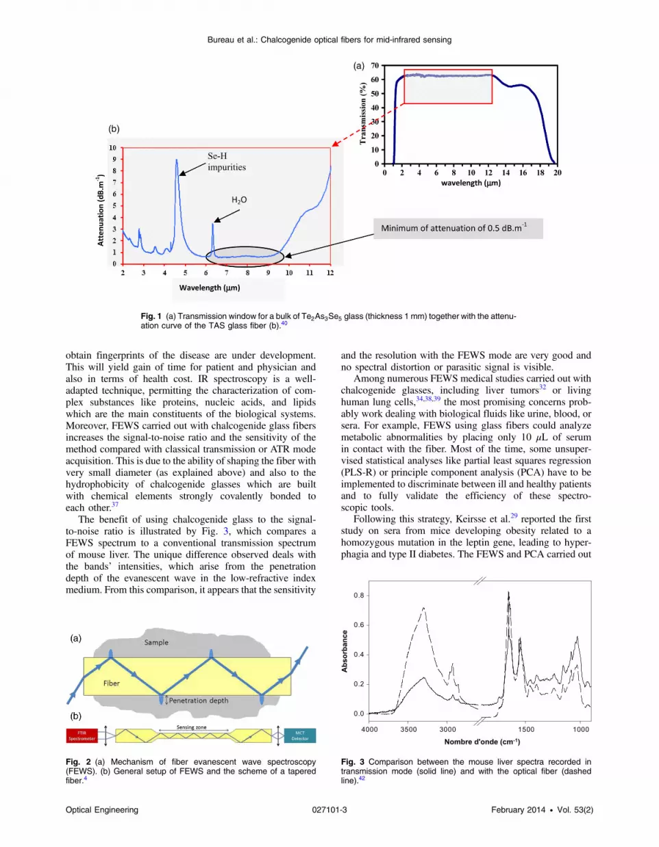
A large range of glass formulations are available to obtainsuitable optical fibers with large IR transparency ranges andlow energy losses. Among chalcogens, selenium is a goodglass former, providing very stable glasses quite easy toshape. In particular, the Te2As3Se5 glass composition(TAS glass) is an interesting compromise with aTg ¼ 137°C, which enables implementing experiments atroom temperature. This glass offers a large spectral window,typically ranging from 2 to 16 μm for a bulk with a thicknessof 1 mm. Moreover, this glass composition exhibits an excel-lent resistance to devitrification, thus permitting to shape itinto an optical fiber. The attenuation curve of the fiber isgiven in Fig. 1. The minimum of attenuation is less than1 dBm−1 and is located between 6.5 and 9 μm.Obviously, this value is far from the one obtained with silicaglass fiber, but the light transmission is sufficient for shortdistance applications such as remote spectroscopy.Overall, the fiber spectral window encompasses the mid-infrared domain, since transparency is observed from 800to 4000 cm−1 on FEWS spectra.
On the other hand, the optical index of a TAS glass is high(n1 ¼ 2.8). Thus, the optical conditions for total internalreflection are fulfilled for all optical rays entering theTAS glass fiber. The number of bound modes M for acircular fiber, depending on the wavelength λ, is estimatedby the following equation:
MðλÞ ¼ 2π2r2ðn21 − n22Þλ2
; (2)
where r is the fiber radius, n1 is the index of the fiber core,and n2 is the index of the cladding. With a diameter of400 μm, a fiber index of 2.8 and the index of the air of1, and the number of modes can be estimated approximatelybetween 37,500 at 12 μm (833 cm−1) and 135,000 at 2 μm(5000 cm−1). Thus, the light propagation in a multimodeTAS glass fiber is complex. To cope with this complexity,a background spectrum (called Iref above) is collected beforeeach experiment. So, many effects can be neglected: the
entrance and exit conditions of the IR beam, the interactionand attenuation along the optical signal transportationsection, the transition of the modes during the taper to thesensing zone, the absorption due to the fiber, and any effectrelated to fiber bending or surface roughness.
The number of reflections over a length L of a fiber with adiameter d depends on Eq. (3):
Nðθ; d; LÞ ¼ L ·tanð90 − θÞ
d; (3)
with θ being the angle of incidence from normal. In thepresent situation, it is known that the propagation withinwaveguides can be efficiently described by classical geomet-ric optics. With these considerations, a model of the fiberoptic probe’s response was presented to help in predictionsand to simulate data.41 It was shown that to improve the sen-sitivity of the sensor, the diameter of the fiber should belocally reduced to create a tapered sensing zone, whichwill be brought into contact with the sample to be analyzed.This could also be easily understood by considering that thenumber of reflections into the fiber is much higher when thefiber diameter decreases, as depicted in Fig. 2. For thefollowing application, the diameter of the fiber has beenlocally reduced from 400 μm to about 100 μm in the sensingzone, where the targeted samples are brought into contactwith the fiber. This design is absolutely essential to benefitof an enhanced signal-to-noise ratio.
Note that the evanescent wave intensity decays exponen-tially with distance from the surface of the fiber. So, thesensitive area is mostly localized within 1 μm from thefiber surface. Also, the penetration depth is a function ofthe glass index as well as of the wavelength of the propagat-ing light, according to Eq. (4):5,6
dp ¼ λ
2πffiffiffiffiffiffiffiffiffiffiffiffiffiffiffiffiffiffiffiffiffiffiffiffiffiffiffiffin22 sin
2 θi − n21p ; (4)
where λ is the wavelength, n2 and n1 are the refractive indi-ces of the glass and surrounding area, respectively, and θi isthe angle of incidence of the wave in the fiber. The penetra-tion of the evanescent wave increases linearly with thewavelength. So, the spectra collected in evanescent modeshow typically lower intensities at shorter wavelengths incomparison with those of transmission spectra. This isclearly visible in spectra collected in attenuated total reflec-tion (ATR) mode using a flat ATR plate, where θi is strictlyequal to 45 deg. For FEWS, one has also to consider thecomplex geometry of the optical fiber, in which the distribu-tion of angle of incidence into the fiber makes the dpinfluences more difficult to analyze and anticipate.
In order to illustrate the efficiency of the methods, variousapplications have been selected in the frame of two healthstrategic fields of application.
3 Application in Early DiagnosticsThe biomedical domain is always searching for new diagnos-tic methods using noninvasive approaches and, whenpossible, in real time. The diagnosis of a disease requiresaggregation of positive clinical, biological, and imagingcriteria, and other negative criteria excluding other diagnosis.This is the reason new methods that enable physicians to
Optical Engineering 027101-2 February 2014 • Vol. 53(2)
Bureau et al.: Chalcogenide optical fibers for mid-infrared sensing
obtain fingerprints of the disease are under development.This will yield gain of time for patient and physician andalso in terms of health cost. IR spectroscopy is a well-adapted technique, permitting the characterization of com-plex substances like proteins, nucleic acids, and lipidswhich are the main constituents of the biological systems.Moreover, FEWS carried out with chalcogenide glass fibersincreases the signal-to-noise ratio and the sensitivity of themethod compared with classical transmission or ATR modeacquisition. This is due to the ability of shaping the fiber withvery small diameter (as explained above) and also to thehydrophobicity of chalcogenide glasses which are builtwith chemical elements strongly covalently bonded toeach other.37
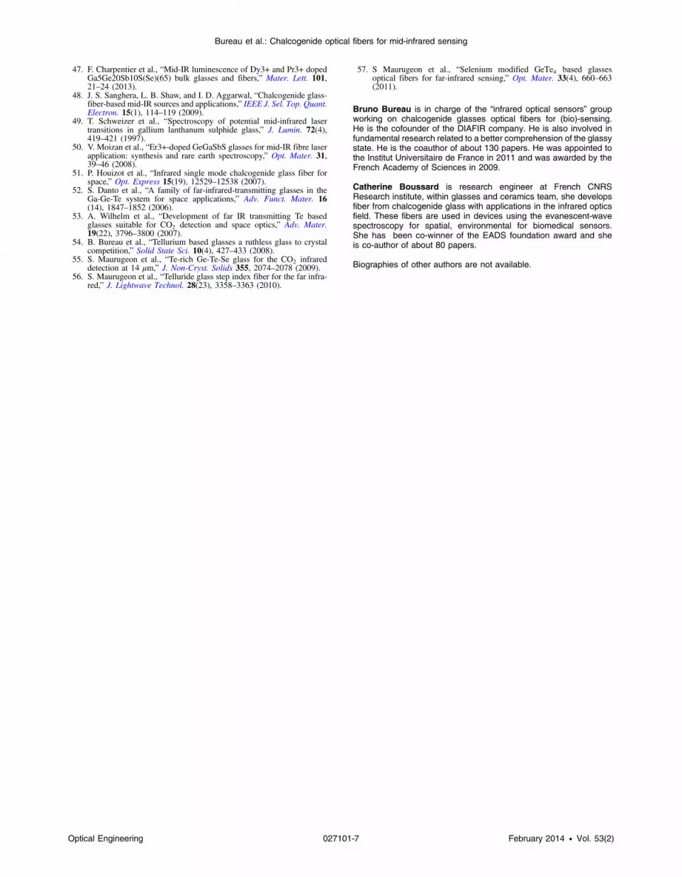
The benefit of using chalcogenide glass to the signal-to-noise ratio is illustrated by Fig. 3, which compares aFEWS spectrum to a conventional transmission spectrumof mouse liver. The unique difference observed deals withthe bands’ intensities, which arise from the penetrationdepth of the evanescent wave in the low-refractive indexmedium. From this comparison, it appears that the sensitivity
and the resolution with the FEWS mode are very good andno spectral distortion or parasitic signal is visible.
Among numerous FEWS medical studies carried out withchalcogenide glasses, including liver tumors32 or livinghuman lung cells,34,38,39 the most promising concerns prob-ably work dealing with biological fluids like urine, blood, orsera. For example, FEWS using glass fibers could analyzemetabolic abnormalities by placing only 10 μL of serumin contact with the fiber. Most of the time, some unsuper-vised statistical analyses like partial least squares regression(PLS-R) or principle component analysis (PCA) have to beimplemented to discriminate between ill and healthy patientsand to fully validate the efficiency of these spectro-scopic tools.
Following this strategy, Keirsse et al.29 reported the firststudy on sera from mice developing obesity related to ahomozygous mutation in the leptin gene, leading to hyper-phagia and type II diabetes. The FEWS and PCA carried out
Fig. 1 (a) Transmission window for a bulk of Te2As3Se5 glass (thickness 1 mm) together with the attenu-ation curve of the TAS glass fiber (b).40
Fig. 2 (a) Mechanism of fiber evanescent wave spectroscopy(FEWS). (b) General setup of FEWS and the scheme of a taperedfiber.4
Nombre d'onde (cm-1)10001500300035004000
Abs
orba
nce
0.0
0.2
0.4
0.6
0.8
Fig. 3 Comparison between the mouse liver spectra recorded intransmission mode (solid line) and with the optical fiber (dashedline).42
Optical Engineering 027101-3 February 2014 • Vol. 53(2)
Bureau et al.: Chalcogenide optical fibers for mid-infrared sensing

in 1100 to 1000 cm−1 range, which corresponds to the sugarring vibration bands, have permitted discrimination ofpathological sera from control.
Then, a study was conducted to evaluate whether mid-IRFEWS was able to discriminate metabolic diseases inpatients. The serum of one control group and three groupsof patients exhibiting chronic liver diseases [genetic hemo-chromatosis, alcoholic cirrhosis, and dysmetabolic hepatosi-derosis (DYSH)] was studied. These metabolic disturbancespotentially impact serum quality, thus giving rise to mid-IRsignature in the related patient’s serum. PLS-R, applied tothe recorded spectra, has allowed discrimination of patientswith cirrhosis and DYSH from a control patient group. Theseresults strongly suggest that the concept of metabolic profil-ing using mid-IR FEWS could be a way to investigatediseases having metabolic consequences in patients.Figure 4 better illustrates the discriminant ability of theprotocol. Figure 4(a) shows the FEWS spectra collectedfrom human sera with and without cirrhosis. Classical analy-sis methods are not able to differentiate between the twogroups of spectra. On the other hand, Fig. 4(b) shows thePCA map which enables one to fairly distinguish betweenthe metabolic states, i.e., control or cirrhotic.
Thus, chalcogenide glass FEWS possesses aptitudes forearly characterization of metabolic anomalies in differentpathologic environments. Hopefully, in the future, thanksto the inertia of chalcogenide glass fibers toward biologicalsubstances,29 the fibers may be implemented directly onpatients by guiding the probe light onto the area of interest,in situ and in vivo, rather than performing biopsies.32,33,43,44
4 Application in Food SafetyFor more than two decades, “food scandals” have beenbrought up in the international press and arouse a legitimateand durable fear in populations. Assessing food microbio-logical safety, traceability, health allegations, or adulterationis becoming a major challenge and must be considered as apublic health matter. A preliminary work had been carriedout in 2006 on the spreading of bacterial biofilm in aPetri plate, namely Proteus mirabilis.30 It had been shownthat mid-IR FEWS without any statistical study, permittedone to distinguish between the swarming and the vegetativephenotypes of the contaminant biofilm. More recently, acomplete work showed that the protocol exposed inSec. 3, including PCA, enabled the identification of somecontaminants in food matrices: milk, minced meat, and
Fig. 4 (a) FEWS spectrum of human serum with and without cirrhosis. (b) The corresponding principalcomponent analysis (PCA) map.4,33
Fig. 5 Mid-infrared (IR) FEWS PCA map for milk and cheese contaminated by various pathogens.28
Optical Engineering 027101-4 February 2014 • Vol. 53(2)
Bureau et al.: Chalcogenide optical fibers for mid-infrared sensing

cheese.28 For each, two experimental conditions weretested. First, enrichment by endogenous flora, which are nat-urally present in the samples and, second, enrichment bythree pathogenic germs: Listeria, Staphylococcus, andSalmonella. Although these pathogens have the same bio-chemical constituents, namely proteins, polysaccharides,phospholipids, and nucleic acids, the biochemical diversitywithin these biochemical classes from one strain to anotherare sufficient to provide distinct FT-IR spectra for eachpathogen. The most useful FT-IR features for bacterial iden-tification appear at wavenumbers around 1000 to 3000 cm−1
and correspond to the deformation, bending, stretching, andring vibrations of various functional groups. Also, thestatistical analyses were performed on the regions 1000 to1800 cm−1 and 2800 to 3000 cm−1, providing the greatestcontribution to the total variance in the FT-IR spectraldata. As examples, Fig. 5 depicts the PCA map for contami-nated milk and cheese by bacterial germs.
Concerning the differentiation between the pathogens, thebest results were obtained for milk and cheese, likely due to abetter physical contact between the fiber and the samples,than when studying meat.
So, from the PCA maps, some trends can be pulled out. Inaddition, logistic-PLS go farther with the discrimination ofthe pathogens strains with a classification error lower than3.5%. These results permit optimism in the potential ofFEWS for early detection of pathogens in food matrices,which could be extended to various applications in the healthfield.
5 Conclusion and PerspectivesIn the future, there are plans to develop alternative IR opticalfibers to be tested for medical diagnosis and food safety. Tworoutes will be explored: first, tellurium glasses and second,
rare earth (RE)-doped chalcogenide glasses. These perspec-tives are clearly upstream and will need strong progress inmaterial sciences before developing any prototype.Nevertheless, the work has already been initiated in thetwo following frameworks.
5.1 RE-Doped Chalcogenide Fibers
Thus, recently, some innovative optical fibers have beendeveloped for CO2 detection.45–47 The ability to detectand quantify CO2 has become increasingly critical for themonitoring of global warming. Since the emission of thisgreenhouse gas increases every year, some solutions mustbe found to reduce or control these CO2 emissions. Oneof them is the capture and the storage of CO2 in naturalunderground geological formations, but this requires specificmonitoring of the storage wells. In that frame, some RE-doped chalcogenide glass fibers have been developed to per-form a mid-IR source, pumped by a commercial laser diodeand used as a remote mid-IR optical sensor. Indeed, the REtrivalent ions incorporated in chalcogenides host matrix withlow-phonon energy can generate light from visible to mid-IR.48–50 Dy3þ ions were selected because, after being opti-cally pumped at 920 nm, the glassy fiber exhibits a mid-IR broad emission corresponding to the transition between6H11∕2 and 6H13∕2 levels, encompassing the CO2 absorptionbands centered at 4.3 μm (Fig. 6). A mid-IR sensor prototypehas been, hence, designed, which enables detecting CO2 in awide concentration range from 100% to lower than500 ppm.47 It will be very interesting to test the efficiencyof this spectroscopic “active” tool for medical applicationsby selecting the appropriate RE to the targeted metabolicderegulation. The main benefit of this technology, as com-pared with “passive” selenide fibers, lies in the compactness
Fig. 6 Fluorescence spectra of Dy3þ-doped chalcogenide glass showing the CO2 absorption bandsaround 4.3 μm pumped by commercial diode laser at 920 nm.
Fig. 7 Optical scheme of the detection of the mid-IR signal coming from an exo-planet through space.The beam profile on the right corresponds to a single-mode TAS glass at 10 μm.
Optical Engineering 027101-5 February 2014 • Vol. 53(2)
Bureau et al.: Chalcogenide optical fibers for mid-infrared sensing

of the final devices and the brightness of the mid-IR fluores-cent sources.
5.2 Germanium Telluride Fibers
Alternatively, much effort has been paid in the developmentof IR glasses transparent far in the IR in order to detect signsof life on earth-like planets. The presence of life is materi-alized by the presence of water, ozone, and CO2 in the planetatmosphere. The three molecules absorb in the IR regionaround 6, 9, and 15 μm, respectively, and one needs todevelop fiber transmitting light from 6 to 20 μm to detectthem following the scheme displayed in Fig. 7.51 Themost efficient strategy to expand the spectral window ofchalcogen glasses is to use the heavy atoms such as telluriumin order to lower the phonon energy and to shift the multi-phonon cut-off to longer wavelengths. Thanks to EuropeanSpace Agency supports in the frame of the DARWINprogram, new chalcogen glasses, exclusively based ontellurium, have been developed for the making of thesesingle-mode fibers.52–57
Among telluride glasses, the Te-Ge-X, with X ¼ As, Ga,Se, or I, systems have shown good glass-forming stability, andoptical fibers have recently been successfully produced whichtransmit light up to 15 μm compared with a 11-μm limit forselenide glass fibers. In the future, this spectral wideningcould be crucial to detect relevant mid-IR signatures thatare not reachable with the TAS glass in biology and medicine.
AcknowledgmentsThe authors thank the French ANR (Emergence, TECSAN,and OPTIC CO2), the ADEME, the European Space Agency,National Science Foundation under Grant Number ECCS-1201865, the CNRS International Associated Laboratoryfor Materials & Optics, and the Partner University Fundfor financial grants and supports.
References
1. B. Bureau et al., “Recent advances in chalcogenide glasses,” J. Non-Cryst. Solids 345, 276–283 (2004).
2. X. H. Zhang et al., “Glass to see beyond the visible,” Chemistry 14(2),432–442 (2008).
3. B. Bureau et al., “Forming glasses from Se and Te,”Molecules 14(11),4337–4350 (2009).
4. S. Cui et al., “From selenium to tellurium based glass optical fibers forinfrared spectroscopies,” Molecules 18(5), 5373–5388 (2013).
5. N. J. Harrick, “Principles of internal reflection spectroscopy,” inInternal Reflection Spectroscopy, W. Sons, Ed., Harrick ScientificCorporation, New York (1967).
6. N. J. Harrick, “Principles of internal reflection spectroscopy,” InternalReflection Spectroscopy, Harrick Scientific Corporation, New York(1979).
7. L. J. Bellamy, The Infrared Spectra of Complex Molecules, Meuthen,London (1975).
8. A. D. Cross and R. A. Jones, An Introduction to Practical Infra-RedSpectroscopy, Butterworths, London (1969).
9. U. P. Fringeli and S. H. Gunthard, in Membrane Spectroscopy, E.Grell, Ed., Springer-Verlag, New York (1981).
10. R. Mendelsohn and H. H. Mantsch, in Progress in Lipid ProteinInteractions, A. Watts and J. J. H. H. M. De Pont, Eds., Elsevier,New York (1986).
11. M. Jackson and H. H. Mantsch, in Infrared Spectroscopy ofBiomolecules, H. H. Mantsch and D. Chapman, Eds., Wiley-Liss,Chichester (1996).
12. D. Naumann et al., in Modern Techniques for Rapid MicrobiologicalAnalysis, W. H. Nelson, Ed., VCH, New York (1991).
13. D. A. C. Compton et al., “In situ FTIR analysis of a composite curingreaction using a mid-IR transmitting fiber,” Appl. Spectrosc. 42, 972–979 (1988).
14. J. Heo et al., “Remote fiber optic chemical sensing using EW inter-actions in chalcogenide glass fibers,” Appl. Opt. 30(27), 3944–3951(1991).
15. M. Rodrigues, “Chalcogenide glass fibers for remote spectroscopicchemical sensing,” Proc. SPIE 1591, 225–235 (1992).
16. J. S. Sanghera et al., “IR evanescent absorption spectroscopy of toxicchemicals using chalcogenide glass fibers,” J. Am. Ceram. Soc. 78,2198–2202 (1995).
17. J. S. Sanghera et al., “IR evanescent absorption spectroscopy withchalcogenide glass fibers,” Appl. Opt. 33(27), 6315–6322 (1994).
18. J. S. Sanghera et al., “Infrared transmitting fiber optics for biomedicalapplications,” Proc. SPIE 3596, 178–187 (1999).
19. M. Katz, I. Schnitzer, and A. Bornstein “Quantitative evaluation ofchalcogenide glass fiber evanescent wave spectroscopy,” Appl. Opt.33(25), 5888–5894 (1994).
20. E. M. Kosower et al., “Protein and cell Fourier transform infrared-attenuated total internal reflectance on silver halide fibers underreagent-accessible conditions,” Proc. SPIE 2131, 71–82 (1994).
21. J. Spielvogel et al., “Cancer diagnostics using Fourier transform fiber-optic infrared evanescent wave spectroscopy (FTIR-FEWS),” Proc.SPIE 3262, 185–191 (1998).
22. J. Spielvogel, R. Hibst, and A. Katzir, “Monitoring the diffusion oftopically applied drugs through human and pig skin using fiberevanescent wave spectrosocpy (FEWS),” Proc. SPIE 3596, 99–107(1999).
23. O. Eytan et al., “Fiber optic evanescent wave spectroscopy (FEWS) forblood diagnosis: the use of polymer coated AgClBr fibers and neuralnetwork analysis,” Proc. SPIE 3596, 74–81 (1999).
24. K. Michel et al., “Monitoring of pollutant in waste water by infraredspectroscopy using chalcogenide glass optical fibers,” Sens. Actuat. BChem. 101, 252–259 (2004).
25. K. Michel et al., “Development of a chalcogenide glass fiber devicefor in situ pollutant detection,” J. Non-Cryst. Solids 326, 434–438(2003).
26. D. Le Coq et al., “Infrared glass fibers for in-situ sensing, chemical andbiochemical reactions,” C. R. Chim. 5(12), 907–913 (2002).
27. M. L. Anne et al., “Polymeristaion of an industrial resin monitored byIR-FEWS,” Sens. Actuat. B 137(2), 687–691 (2009).
28. M. L. Anne et al., “Identification of foodborne pathogens within foodmatrices by IR spectroscopy,” Sens. Actuat. B 160(1), 202–206 (2011).
29. J. Keirsse et al., “IR optical fiber sensor for biomedical applications,”Vib. Spectrosc. 32(1), 23–32 (2003).
30. J. Keirsse et al., “Mapping bacterial surface population physiology inreal-time: IR spectroscopy of Proteus mirabilis swarm colonies,” Appl.Spectrosc. 60(6), 584–591 (2006).
31. S. Hocdé et al., “Biological tissues infrared analysis by chalcogenideglass optical fiber spectroscopy,” Proc. SPIE 4158, 49–56 (2001).
32. S. Hocdé et al., “Metabolic imaging of tissues by infrared fiber-opticspectroscopy: an efficient tool for medical diagnosis,” J. Biomed. Opt.9(2), 404–407 (2004).
33. M. L. Anne et al., “FEWS using mid infrared provides useful finger-prints for metabolic profiling in humans,” J. Biomed. Opt. 14(5),054033 (2009).
34. P. Lucas et al., “Evaluation of toxic agent effects on lung cells byfiber evanescent wave spectroscopy,” Appl. Spectrosc. 59(1), 1–9(2005).
35. P. Lucas et al., “Advances in chalcogenide fiber evanescent wave bio-chemical sensing,” Anal. Biochem. 351(1), 1–10 (2006).
36. P. Lucas et al., “Spectroscopic properties of chalcogenide fibres forbiosensor applications,” Phys. Chem. Glasses Eur. J. Glass Sci.Technol. B 47(2), 88–91 (2006).
37. P. Lucas et al., “Infrared biosensors using hydrophobic chalcogenidefibers sensitized with live cells,” Sens. Actuat. B Chem. 119(2), 355–362 (2006).
38. M. Riley et al., “Biologically inspired sensing: infrared spectroscopicanalysis of cell responses to an inhalation health hazard,” Biotechnol.Prog. 22(1), 24–31 (2006).
39. M. Riley et al., “Lung cell fiber evanescent wave spectroscopicbiosensing of inhalation health hazards,” Biotechnol. Bioeng. 95(4),599–612 (2006).
40. B. Bureau et al., “Infrared optical fiber as evanescent wave bio-sen-sors,” Proc. SPIE 5691, 1–8 (2005).
41. S. MacDonald et al., “Optical analysis of infrared spectra recordedwith tapered chalcogenide glass fibers,” Opt. Mater. 25(2), 171–178 (2004).
42. J. Keirsse et al., “Chalcogenide glass fibers for in-situ infraredspectroscopy in biology and medicine,” Proc. SPIE 5459, 61–68(2004).
43. A. Seddon, “Mid-infrared (IR)—a hot topic: the potential for usingmid-IR light for non-invasive early detection of skin cancer invivo,” Phys. Stat. Solidi B 250(5), 1020–1027 (2013).
44. A. Seddon, “Potential for using mid-infrared light for non-invasive,early-detection of skin cancers in vivo,” Proc. SPIE 8576, 85760V(2013).
45. F. Charpentier et al., “Infrared monitoring of underground CO2storage using chalcogenide glass fibers,” Opt. Mater. 31(3), 496–500 (2009).
46. F. Charpentier et al., “CO2 detection using microstructured chalcoge-nide fibers,” Sens. Lett. 7(5), 745–749 (2009).
Optical Engineering 027101-6 February 2014 • Vol. 53(2)
Bureau et al.: Chalcogenide optical fibers for mid-infrared sensing
47. F. Charpentier et al., “Mid-IR luminescence of Dy3+ and Pr3+ dopedGa5Ge20Sb10S(Se)(65) bulk glasses and fibers,” Mater. Lett. 101,21–24 (2013).
48. J. S. Sanghera, L. B. Shaw, and I. D. Aggarwal, “Chalcogenide glass-fiber-based mid-IR sources and applications,” IEEE J. Sel. Top. Quant.Electron. 15(1), 114–119 (2009).
49. T. Schweizer et al., “Spectroscopy of potential mid-infrared lasertransitions in gallium lanthanum sulphide glass,” J. Lumin. 72(4),419–421 (1997).
50. V. Moizan et al., “Er3+-doped GeGaSbS glasses for mid-IR fibre laserapplication: synthesis and rare earth spectroscopy,” Opt. Mater. 31,39–46 (2008).
51. P. Houizot et al., “Infrared single mode chalcogenide glass fiber forspace,” Opt. Express 15(19), 12529–12538 (2007).
52. S. Danto et al., “A family of far-infrared-transmitting glasses in theGa-Ge-Te system for space applications,” Adv. Funct. Mater. 16(14), 1847–1852 (2006).
53. A. Wilhelm et al., “Development of far IR transmitting Te basedglasses suitable for CO2 detection and space optics,” Adv. Mater.19(22), 3796–3800 (2007).
54. B. Bureau et al., “Tellurium based glasses a ruthless glass to crystalcompetition,” Solid State Sci. 10(4), 427–433 (2008).
55. S. Maurugeon et al., “Te-rich Ge-Te-Se glass for the CO2 infrareddetection at 14 μm,” J. Non-Cryst. Solids 355, 2074–2078 (2009).
56. S. Maurugeon et al., “Telluride glass step index fiber for the far infra-red,” J. Lightwave Technol. 28(23), 3358–3363 (2010).
57. S Maurugeon et al., “Selenium modified GeTe4 based glassesoptical fibers for far-infrared sensing,” Opt. Mater. 33(4), 660–663(2011).
Bruno Bureau is in charge of the “infrared optical sensors” groupworking on chalcogenide glasses optical fibers for (bio)-sensing.He is the cofounder of the DIAFIR company. He is also involved infundamental research related to a better comprehension of the glassystate. He is the coauthor of about 130 papers. He was appointed tothe Institut Universitaire de France in 2011 and was awarded by theFrench Academy of Sciences in 2009.
Catherine Boussard is research engineer at French CNRSResearch institute, within glasses and ceramics team, she developsfiber from chalcogenide glass with applications in the infrared opticsfield. These fibers are used in devices using the evanescent-wavespectroscopy for spatial, environmental for biomedical sensors.She has been co-winner of the EADS foundation award and sheis co-author of about 80 papers.
Biographies of other authors are not available.
Optical Engineering 027101-7 February 2014 • Vol. 53(2)
Bureau et al.: Chalcogenide optical fibers for mid-infrared sensing
Recommended


