BioMed CentralBMC Cancer
ss
Open AcceResearch articleCharacterization of SV-40 Tag rats as a model to study prostate cancerCurt E Harper1, Brijesh B Patel, Leah M Cook1, Jun Wang1, Tomoyuki Shirai2, Isam A Eltoum3,4 and Coral A Lamartiniere*1,4Address: 1Department of Pharmacology and Toxicology, University of Alabama at Birmingham, Birmingham, Alabama, USA, 2Department of Experimental Pathology and Tumor Biology, Nagoya City University Graduate School of Medical Sciences, Nagoya, Japan, 3Department of Pathology, University of Alabama at Birmingham, Birmingham, Alabama, USA and 4UAB Comprehensive Cancer Center, University of Alabama at Birmingham, Birmingham, Alabama, USA
Email: Curt E Harper - [email protected]; Brijesh B Patel - [email protected]; Leah M Cook - [email protected]; Jun Wang - [email protected]; Tomoyuki Shirai - [email protected]; Isam A Eltoum - [email protected]; Coral A Lamartiniere* - [email protected]
* Corresponding author
AbstractBackground: Prostate cancer is the second most frequently diagnosed cancer in men. Animalmodels that closely mimic clinical disease in humans are invaluable tools in the fight against prostatecancer. Recently, a Simian Virus-40 T-antigen (SV-40 Tag) targeted probasin promoter rat modelwas developed. This model, however, has not been extensively characterized; hence we haveinvestigated the ontogeny of prostate cancer and determined the role of sex steroid receptor andinsulin-like growth factor-1 (IGF-1) signaling proteins in the novel SV-40 Tag rat.
Methods: The SV-40 Tag rat was histopathologically characterized for time to tumordevelopment, incidence and multiplicity and in the ventral, dorsal, lateral and anterior lobes of theprostate. Immunoassay techniques were employed to measure cell proliferation, apoptosis, and sexsteroid receptor and growth factor signaling-related proteins. Steroid hormone concentrationswere measured via coated well enzyme linked immunosorbent assay (ELISA) kits.
Results: Prostatic intraepithelial neoplasia (PIN) and well-differentiated prostate cancer developedas early as 2 and 10 weeks of age, respectively in the ventral prostate (VP) followed by in thedorsolateral (DLP). At 8 weeks of age, testosterone and dihydrotestosterone (DHT)concentrations in SV-40 Tag rats were increased when compared to non-transgenic rats. High cellproliferation and apoptotic indices were found in VP and DLP of transgenic rats. Furthermore, weobserved increased protein expression of androgen receptor, IGF-1, IGF-1 receptor, andextracellular signal-regulated kinases in the prostates of SV-40 Tag rats.
Conclusion: The rapid development of PIN and prostate cancer in conjunction with the largeprostate size makes the SV-40 Tag rat a useful model for studying prostate cancer. This studyprovides evidence of the role of sex steroid and growth factor proteins in prostate cancerdevelopment and defines appropriate windows of opportunity for preclinical trials and aids in therational design of chemoprevention, intervention, regression, and therapeutic studies usingprostate cancer rodent models.
Published: 26 January 2009
BMC Cancer 2009, 9:30 doi:10.1186/1471-2407-9-30
Received: 22 May 2008Accepted: 26 January 2009
This article is available from: http://www.biomedcentral.com/1471-2407/9/30
© 2009 Harper et al; licensee BioMed Central Ltd. This is an Open Access article distributed under the terms of the Creative Commons Attribution License (http://creativecommons.org/licenses/by/2.0), which permits unrestricted use, distribution, and reproduction in any medium, provided the original work is properly cited.
Page 1 of 11(page number not for citation purposes)
BMC Cancer 2009, 9:30 http://www.biomedcentral.com/1471-2407/9/30
BackgroundProstate cancer is the second most frequently diagnosedcancer in men, with 782,600 new cases projected to occurin 2007 [1]. It was estimated that 254,000 deaths wouldoccur from prostate cancer in the past year. One of the firstmeans of prostate cancer treatment was androgen depriva-tion and estrogen administration [2]. Hence, it is knownthat sex steroids and their receptors play a major role inprostate cancer etiology. The androgen receptor (AR) isbelieved to participate in prostate cancer progression,including its activation and up-regulation, point muta-tions, and ligand-independent activation. Testosterone, aligand for the AR, is secreted primarily by the testes and issubsequently converted to DHT via the enzyme, 5-α-reductase. DHT has a 50-fold higher binding affinity forAR and is 10 times more potent than testosterone [3].Whether or not elevated blood levels of androgens are asignificant risk factor for prostate cancer is open fordebate. Some have reported that increased testosteronelevels in the blood are associated with an increased risk ofprostate cancer [4]. Others have failed to support the"androgen hypothesis" that circulating testosterone andDHT are positively associated with prostate cancer risk [5].
Estrogens and their receptors, including estrogen receptor-alpha (ER-alpha) and estrogen receptor-beta (ER-beta),play an integral role in normal growth, differentiation,and development of the prostate. In a study of over 600men, low circulating concentrations of estradiol wereassociated with a decreased risk of prostate cancer [4]. Inthe past, estrogens have been used to treat prostate cancerbecause it resulted in prostate growth inhibitory effects.However, toxicity and poor response rate associated withestrogen therapy have hindered its progress in treatingprostate cancer. The timing of administration and doseoften dictates the type of response produced by estrogensor estrogen-like chemicals [6].
Growth factor signaling also plays a critical role in thegrowth and development of the prostate, including theproteins of the insulin-like growth factor axis. The twogrowth factors, IGF-1 and IGF-2, interact with 6 knownIGF-binding proteins (IGF-BPs), which regulate bindingto the 2 IGF receptors (IGF-1R and IGF-2R). Elevated lev-els of IGF-1 and decreased levels of IGF-BP3 in the bloodserum have been associated with an increased risk ofadvanced stage prostate cancer [7]. Additional support tothe role of IGF-1 and prostate growth can be found in IGF-1 deficient mice that exhibit decreased prostate size [8]and increased rat prostate growth with systemic adminis-tration of IGF-1 [9]. The expression of IGF-1R in prostatecancer remains controversial. It has been proposed thatreduced IGF-1R action is necessary for prostate cancer pro-gression. Tennant et al. reported that IGF-1R expression isabundant in normal and early-stage tumors, but reduced
in advanced and metastatic prostate cancer [10]. The IGF-1 signal transduction cascade ultimately leads to the phos-phorylation of intracellular substrates and activation ofthe mitogen-activated protein kinase/extracellular signal-regulated kinase (MAPK/ERK) pathway. The MAPK/ERKpathway is involved in cell differentiation, cell survival,and cell migration [11]. ERKs, downstream effectors ofgrowth factor and sex steroid receptor signaling, also reg-ulate cell proliferation and apoptosis and participate inprostate carcinogenesis [11]. Furthermore, the MAPK/ERKsignaling has the ability to re-activate the AR pathway bya hormone-independent mechanism that may lead toandrogen-independent prostate cancer [12].
Animal models that closely mimic clinical disease inhumans are invaluable tools in the fight against prostatecancer. The TRAMP model developed in 1995 has beenused extensively as a prostate cancer model over the pastdecade [13]. One limitation of the TRAMP model, how-ever, is that it is mouse-based; therefore, the prostates arevery small and necessitate several dissected prostate lobesto allow extensive mechanism of action research at theprotein level. Recently, Shirai and colleagues developedanother SV-40 Tag targeted probasin promoter rodentmodel, this one in the Sprague Dawley rat [14]. However,this model has not been extensively characterized. Accord-ingly, we set out to thoroughly characterize the histopa-thology of autochthonous prostate cancer in the SV-40Tag rat.
The goal of this study was to define appropriate windowsof opportunity for pre-clinical trails using the SV-40 Tagrat. We report the incidence and pathological changeswithin the VP and DLP as a function of time throughoutthe natural history of prostate cancer in SV-40 Tag rats.Furthermore, we evaluated the role of sex steroid receptorand IGF-1 signaling proteins, cell proliferation, and apop-tosis in the development of prostate cancer in this model.This study should aid in the rational design of chemopre-vention, intervention, regression, and therapeutic studiesusing prostate cancer rodent models.
MethodsAnimalsAnimal care and use were conducted according to estab-lished guidelines approved by the National Institutes ofHealth and the Institutional Animal Care and Use Com-mittee at the University of Alabama at Birmingham. Ani-mals were housed in rooms maintained at 24 ± 1°C witha 12 hr light-dark cycle. All animals received powderedphytoestrogen-free AIN-76A diet (Harlan Teklad GlobalDiets, Wilmington, DE) and tap water.
SV-40 Tag rat breeders were provided for us by Dr. Tomoy-uki Shirai of Nagoya City University Medical School via
Page 2 of 11(page number not for citation purposes)
BMC Cancer 2009, 9:30 http://www.biomedcentral.com/1471-2407/9/30
Drs. Gail Prins and Steve Swanson of the University of Illi-nois at Chicago. Heterozygous SV-40 Tag females werecrossed with non-transgenic males to generate hetero-zygous SV-40 Tag male offspring (SV-40 SD females × SDmale breeders). Day 21 post-conception females wereanesthetized and the offspring were caesarian-derived toyield mycoplasma-free offspring that were used to estab-lish our colony. At 3 weeks of age, the offspring wereweaned and tails were clipped. DNA was extracted using aDNeasy Tissue Kit (Qiagen, Valencia, CA), and a PCR-based screening assay was performed to evaluate trans-gene incorporation [13-15].
Rats were necropsied starting at day 1 post-partum, onceper week from 1 to 6 weeks, and every 2 weeks starting at6 weeks of age and concluding at 40 weeks. At time of dis-section, prostates were excised, weighed, and flash frozenin liquid nitrogen. Because it is unclear which lobe in therodent resembles the human peripheral zone, the pros-tatic location in humans where prostate cancer normallyoccurs [16], we chose to analyze both the DLP, which hashistorically been referred to as the homologue of thehuman prostate [17] and the VP. Blood was collected atsacrifice, centrifuged at 3,000 × g and serum was stored at-80°C until time of analysis.
HistopathologyAt necropsy, organs were examined for gross abnormali-ties. Macro-metastasis to the bone, abdominal wall,lymph nodes, liver, kidney, and lung was investigated.Prostate, testes, seminal vesicles and tumor weights werealso recorded. The prostate and organs of suspectedmetastasis were placed in cassettes, immersed in 10% for-malin, dehydrated in a series of alcohol dilutions, fixed inxylene, embedded in paraffin wax, sliced into 5 μm sec-tions, and placed on microscope slides as described byFolkvord et al. [18]. Sections were stained with hematoxy-lin and eosin prior to histopathological examination. Dr.Isam Eltoum, a Board Certified Pathologist, blindlyscored each coded sample using the following gradingscale developed specifically for rodents [16,19-21]: Grade1 (non-cancerous), Grade 2 (low-grade PIN), Grade 3(HG-PIN), Grade 4 (well-differentiated lesion), Grade 5(moderately differentiated lesion), or Grade 6 (poorly dif-ferentiated lesion).
Sex Steroid Hormone Concentrations in Blood SerumSerum total testosterone, DHT, and estradiol concentra-tions were measured in the blood serum using coated-wellenzyme- and radio-immunoassays (Diagnostic SystemsLaboratories, Inc., Webster, TX) as described by the man-ufacturer. The following kits were used: DSL 10–4000(total testosterone), DSL 9600 (DHT), and DSL 4800(estradiol). All samples were run in duplicate with eightsamples per group by Dr. Richard Parker (Obstetrics and
Gynecology Department, UAB, Birmingham, AL). Stand-ards provided by the manufacturer were used: 0.1–25 ng/mL for total testosterone, 25–2500 pg/mL for DHT, and20–6000 pg/mL for estradiol. Sensitivity for total testo-sterone, DHT, and estradiol were 0.04 ng/mL, 4 pg/mL,and 7 pg/mL, respectively.
IGF-1 Concentrations in Blood SerumIGF-1 concentrations were measured in blood serumusing a coated well ELISA kit as described by DiagnosticSystems Laboratories, Inc. All samples were run in dupli-cate with eight samples per group. Concentration (ng/mL)of samples and controls were determined by plotting themean absorbance readings of the controls and unknownsagainst the mean absorbance readings of all standards ina four-parameter curve fit.
Cell ProliferationProstate tissues (VP and DLP) were harvested and proc-essed for detecting Ki-67, a marker of cell proliferation aspreviously described [21]. Slides were viewed using aNikon Labophot-2 microscope (Nikon Corporation,Tokyo, Japan) and digitally recorded using a Nikon 8.0Mega Pixels CoolPix 8700 Digital Camera (Nikon). ForKi67 quantitation, epithelial cells were counted usingImage J software (Image J, NIH). The VP and DLP wereanalyzed separately (a minimum of 1,000 cells countedper lobe per slide. The epithelial cells staining positive(brown) for Ki67 were counted as well as the non-prolif-erative epithelial cells (stained blue). The proliferativeindex was defined as the number of positively stained epi-thelial cells divided by the total number of epithelial cellscounted × 100. Twenty-eight week old TRAMP prostatetumor with and without Ki67 primary antibody was usedas positive and negative controls, respectively.
ApoptosisThe ApopTag® Plus Peroxidase In Situ Apoptosis DetectionKit (Chemicon International, Temecula, CA) was used tomeasure apoptosis following the manufacturer's instruc-tions. The apoptotic index was defined as the number ofepithelial cells stained positive (brown) for apoptosisdivided by the total number of epithelial cells counted ×100. Visualization was performed using a Nikon lightmicroscope, Nikon digital camera, and analyzed usingImage J software (NIH).
Immunoblot AnalysesWhen possible, protein expression levels of sex steroidand growth factor receptors and their ligands were meas-ured by western blot analysis as described previously [22].Tissues (8 biological samples/treatment group) werehomogenized in lysis buffer (1% Triton X-100, 10 mMTris (pH 7.4), 1 mM EDTA, 1 mM EGTA, 1 mM Hepes (pH7.6), 2 mM Na vanadate, 0.2 mM PMSF, 2 μg/ml leupep-
Page 3 of 11(page number not for citation purposes)
BMC Cancer 2009, 9:30 http://www.biomedcentral.com/1471-2407/9/30
tin, 2 μg/ml aprotinin). Protein concentration of eachsample was determined using the Pierce BCA ProteinAssay (Pierce, Rockford, IL). The same quantity of proteinfrom each sample was separated by SDS-PAGE and trans-ferred to a nitrocellulose membrane (BioRad, Hercules,CA). The membranes were blocked and immunoblottedwith appropriate antibodies purchased from Cell Signal-ing Technology (Danvers, MA): total Extracellular Regu-lating Kinases 1 and 2 (total-ERKs 1 and 2) (#9102) andPhosphatase and Tensin Homolog (PTEN) (#9554), andfrom Santa Cruz Biotechnology (Santa Cruz, CA): AR (SC-816), ER-alpha (SC-7207), insulin-like growth factor-1receptor alpha (IGF-1R-α) (SC-712), phospho-Akt 1/2/3(SC-7985), and β-actin (SC-47778) was used as a loadingcontrol. Positive protein controls purchased from the sup-pliers of the corresponding antibodies and the use ofKaleidoscope Precision Plus Protein and Pre-stained SDS-PAGE Broad Range standards (BioRad Hercules, CA) wereemployed to identify the protein of interest. After incuba-tion with HRP-conjugated secondary antibody (Cell Sign-aling Technology), bands were detected usingchemiluminescence (Pierce) and exposed to X-ray radiog-raphy film. Band intensity was quantified using scanningdensitometry.
Enzyme Linked Immunosorbent AssaysIGF-1 protein levels were quantified in the prostate byELISA as described by Crowther et al. [23] with modifica-tions [22]. Prior to analysis, kinetic curves were set up toestablish zero order kinetics. Samples were run in tripli-cate and the absorbance at 450 nm was read in an OPTImax Microplate reader (Molecular Devices, Sunnyvale,CA). Rat liver with and without IGF-1 primary antibodywas used as a positive and negative control, respectively.
StatisticsStatistical comparisons were performed using two-tailedStudents t-test assuming unequal variances for immunob-lot analysis and ELISA. Trend analysis was implementedto compare cell proliferation and apoptosis between non-transgenic and SV-40 Tag rats. P < 0.05 was considered tobe significant. GraphPad™Prism 5.0 and GraphPad™InStat 3.0 (San Diego, CA) were used to assist in statisticalanalysis.
ResultsHistopathologyVia the ontogeny study, we found that precursor lesionand prostate tumor progression was more rapid in the VPthan the DLP of SV-40 Tag rats (Table 1). Prostates weregraded normal (Grade 1) from day 1 through 1 week post-partum. However, low-grade PIN (Grade 2) quickly devel-oped as early as 2 weeks of age in the VP and DLP. High-grade PIN (Grade 3), the precursor to prostate adenocarci-
Table 1: Histopathological analysis of the ventral and dorsolateral prostate lobes of SV-40 Tag rats fed control AIN-76A diet
HistopathologicalGrades
HistopathologicalGrades
Age n (Ventral Prostate) (Dorsolateral prostate)
1 day 2 1, 1 1, 1
1 week 4 1, 1, 1, 1 1, 1, 1, 1
2 weeks 6 2, 2, 2, 2, 2, 2 1, 1, 2, 2, 2, 2
3 weeks 2 2, 2 2, 2
4 weeks 2 3, 3 2, 2
5 weeks 4 2, 2, 3, 3 1, 2, 2, 3
6 weeks 3 3, 3, 3 3, 3, 3
8 weeks 5 3, 3, 3, 3, 3 3, 3, 3, 3, 3
10 weeks 5 3, 3, 3, 4, 4 3, 3, 3, 3, 3
12 weeks 5 4, 4, 4, 4, 5 3, 3, 3, 3, 3
14 weeks 5 4, 4, 4, 4, 4 4, 4, 4, 4, 4
16 weeks 5 4, 5, 5, 5, 5 3, 3, 4, 4, 4
18 weeks 5 4, 5, 5, 5, 5 3, 4, 4, 4, 4
20 weeks 4 5, 5, 5, 5 4, 4, 4, 4
22 weeks 4 5, 5, 5, 5 5, 5, 5, 5
24 weeks 4 5, 5, 5, 5 5, 5, 5, 5
26 weeks 4 5, 5, 5, 6 3, 5, 6, 6
28 weeks 3 5, 6, 6 5, 5, 6
30 weeks 3 4, 6, 6 4, 6, 6
32 weeks 3 5, 5, 5 5, 5, 5
36 weeks 3 5, 5, 5 5, 5, 5
40 weeks 4 5, 5, 5, 5 5, 5, 5, 5
Tissue sections were evaluated for histopathological scores of 1 (no tumor), 2 (low grade PIN), 3 (high-grade PIN), 4 (well-differentiated tumor), 5 (moderately differentiated tumor), and 6 (poorly differentiated tumor) depending on the presence and progression of lesions. Individual grades are presented for each age group and lobe.
Page 4 of 11(page number not for citation purposes)
BMC Cancer 2009, 9:30 http://www.biomedcentral.com/1471-2407/9/30
noma, developed as early as 4 and 5 to 6 weeks of age inthe VP and DLP, respectively. None of the animals aged 8weeks or younger developed cancerous lesions (Grade 4or greater).
The first incidence of prostate cancer (well-differentiatedlesions/Grade 4) developed at 10 and 14 weeks of age inthe VP and DLP, respectively. By 10 weeks of age, 40% ofanimals developed prostate cancer in the VP, whereas by14 weeks of age 100% of animals developed prostatecancer in the DLP. In addition, 100% of the rats hadprostate cancer in each lobe by 14 weeks of age. Moder-ately-differentiated lesions (Grade 5) developed as earlyas 16 and 22 weeks of age in the VP and DLP, respec-tively. Poorly-differentiated lesions (Grade 6) developedat 26–30 weeks of age in the VP and DLP. When one lobedemonstrated Grade 6 lesions, the other lobe usuallyalso exhibited Grade 6 lesions. Interestingly, from 32–40weeks only moderately-differentiated lesions (Grade 5)and not poorly differentiated lesions (Grade 6) werefound. Only 5% of SV-40 Tag rats developed macro-scopic prostate tumors in which prostate lobes wereindistinguishable at dissection (3.18 ± 2.16 grams). In30 week old SV-40 Tag rats which developed Grade 4–6lesions without macroscopic tumors, the prostate tissueweighed 1.27 ± 0.19 grams. This is to be compared to agematched non-transgenic rat prostates weighing 1.04 ±0.20 grams. Metastasis to the pelvic lymph nodes, ure-thra, or kidneys was observed in less than 5% of SV-40Tag rats at 30 weeks of age or older. Macro- or micro-metastasis was not observed in any animals youngerthan 30 weeks of age.
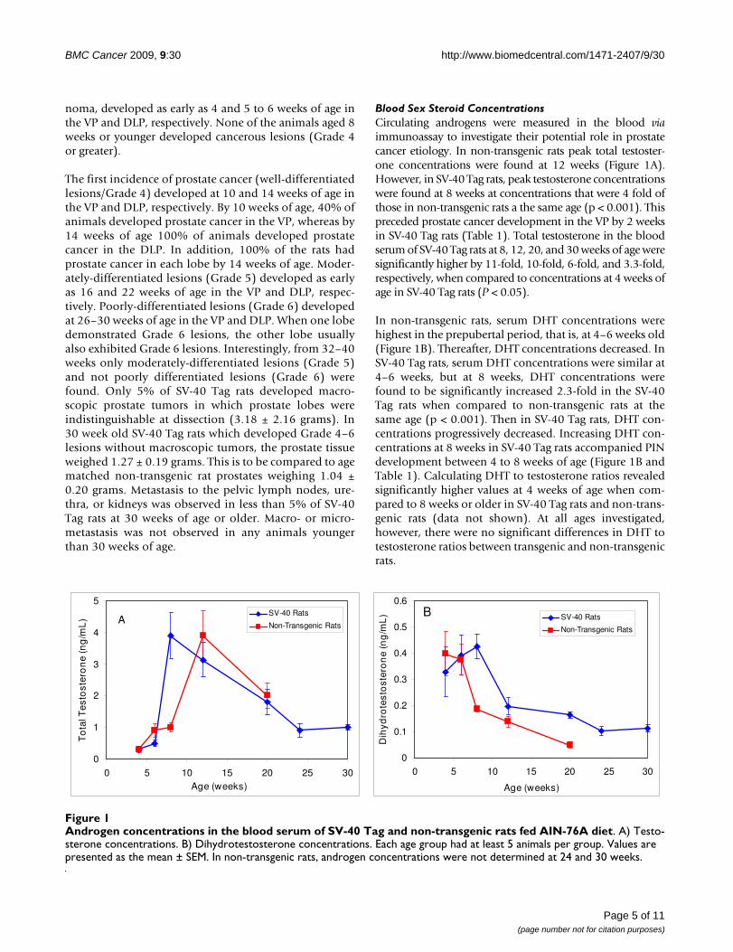
Blood Sex Steroid ConcentrationsCirculating androgens were measured in the blood viaimmunoassay to investigate their potential role in prostatecancer etiology. In non-transgenic rats peak total testoster-one concentrations were found at 12 weeks (Figure 1A).However, in SV-40 Tag rats, peak testosterone concentrationswere found at 8 weeks at concentrations that were 4 fold ofthose in non-transgenic rats a the same age (p < 0.001). Thispreceded prostate cancer development in the VP by 2 weeksin SV-40 Tag rats (Table 1). Total testosterone in the bloodserum of SV-40 Tag rats at 8, 12, 20, and 30 weeks of age weresignificantly higher by 11-fold, 10-fold, 6-fold, and 3.3-fold,respectively, when compared to concentrations at 4 weeks ofage in SV-40 Tag rats (P < 0.05).
In non-transgenic rats, serum DHT concentrations werehighest in the prepubertal period, that is, at 4–6 weeks old(Figure 1B). Thereafter, DHT concentrations decreased. InSV-40 Tag rats, serum DHT concentrations were similar at4–6 weeks, but at 8 weeks, DHT concentrations werefound to be significantly increased 2.3-fold in the SV-40Tag rats when compared to non-transgenic rats at thesame age (p < 0.001). Then in SV-40 Tag rats, DHT con-centrations progressively decreased. Increasing DHT con-centrations at 8 weeks in SV-40 Tag rats accompanied PINdevelopment between 4 to 8 weeks of age (Figure 1B andTable 1). Calculating DHT to testosterone ratios revealedsignificantly higher values at 4 weeks of age when com-pared to 8 weeks or older in SV-40 Tag rats and non-trans-genic rats (data not shown). At all ages investigated,however, there were no significant differences in DHT totestosterone ratios between transgenic and non-transgenicrats.
Androgen concentrations in the blood serum of SV-40 Tag and non-transgenic rats fed AIN-76A dietFigure 1Androgen concentrations in the blood serum of SV-40 Tag and non-transgenic rats fed AIN-76A diet. A) Testo-sterone concentrations. B) Dihydrotestosterone concentrations. Each age group had at least 5 animals per group. Values are presented as the mean ± SEM. In non-transgenic rats, androgen concentrations were not determined at 24 and 30 weeks.
0
0.1
0.2
0.3
0.4
0.5
0.6
0 5 10 15 20 25 30
Age (weeks)
Dih
ydro
test
ost
ero
ne
(ng
/mL
) SV-40 Rats
Non-Transgenic Rats
B
0
1
2
3
4
5
0 5 10 15 20 25 30Age (weeks)
To
tal T
esto
ster
on
e (n
g/m
L)
SV-40 Rats
Non-Transgenic RatsA
Page 5 of 11(page number not for citation purposes)
BMC Cancer 2009, 9:30 http://www.biomedcentral.com/1471-2407/9/30
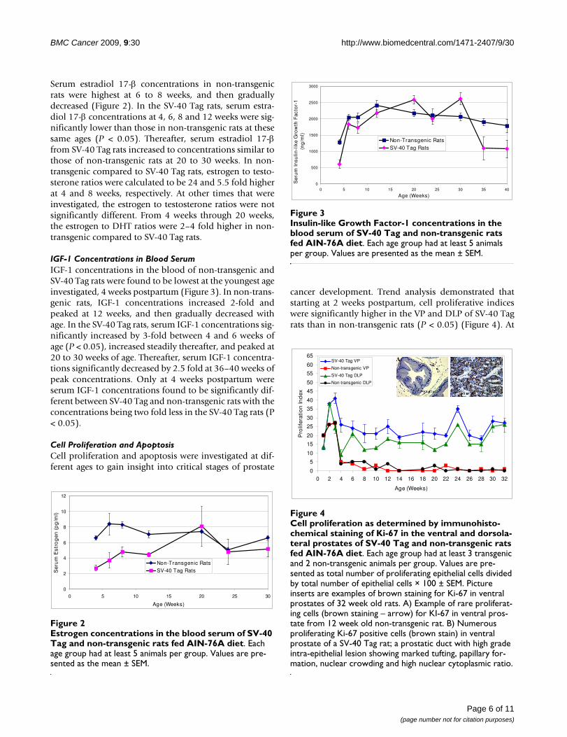
Serum estradiol 17-β concentrations in non-transgenicrats were highest at 6 to 8 weeks, and then graduallydecreased (Figure 2). In the SV-40 Tag rats, serum estra-diol 17-β concentrations at 4, 6, 8 and 12 weeks were sig-nificantly lower than those in non-transgenic rats at thesesame ages (P < 0.05). Thereafter, serum estradiol 17-βfrom SV-40 Tag rats increased to concentrations similar tothose of non-transgenic rats at 20 to 30 weeks. In non-transgenic compared to SV-40 Tag rats, estrogen to testo-sterone ratios were calculated to be 24 and 5.5 fold higherat 4 and 8 weeks, respectively. At other times that wereinvestigated, the estrogen to testosterone ratios were notsignificantly different. From 4 weeks through 20 weeks,the estrogen to DHT ratios were 2–4 fold higher in non-transgenic compared to SV-40 Tag rats.
IGF-1 Concentrations in Blood SerumIGF-1 concentrations in the blood of non-transgenic andSV-40 Tag rats were found to be lowest at the youngest ageinvestigated, 4 weeks postpartum (Figure 3). In non-trans-genic rats, IGF-1 concentrations increased 2-fold andpeaked at 12 weeks, and then gradually decreased withage. In the SV-40 Tag rats, serum IGF-1 concentrations sig-nificantly increased by 3-fold between 4 and 6 weeks ofage (P < 0.05), increased steadily thereafter, and peaked at20 to 30 weeks of age. Thereafter, serum IGF-1 concentra-tions significantly decreased by 2.5 fold at 36–40 weeks ofpeak concentrations. Only at 4 weeks postpartum wereserum IGF-1 concentrations found to be significantly dif-ferent between SV-40 Tag and non-transgenic rats with theconcentrations being two fold less in the SV-40 Tag rats (P< 0.05).
Cell Proliferation and ApoptosisCell proliferation and apoptosis were investigated at dif-ferent ages to gain insight into critical stages of prostate
cancer development. Trend analysis demonstrated thatstarting at 2 weeks postpartum, cell proliferative indiceswere significantly higher in the VP and DLP of SV-40 Tagrats than in non-transgenic rats (P < 0.05) (Figure 4). At
Estrogen concentrations in the blood serum of SV-40 Tag and non-transgenic rats fed AIN-76A dietFigure 2Estrogen concentrations in the blood serum of SV-40 Tag and non-transgenic rats fed AIN-76A diet. Each age group had at least 5 animals per group. Values are pre-sented as the mean ± SEM.
0
2
4
6
8
10
12
0 5 10 15 20 25 30
Age (Weeks)
Ser
um
Est
rog
en (
pg
/ml)
Non-Transgenic RatsSV-40 Tag Rats
Insulin-like Growth Factor-1 concentrations in the blood serum of SV-40 Tag and non-transgenic rats fed AIN-76A dietFigure 3Insulin-like Growth Factor-1 concentrations in the blood serum of SV-40 Tag and non-transgenic rats fed AIN-76A diet. Each age group had at least 5 animals per group. Values are presented as the mean ± SEM.
0
500
1000
1500
2000
2500
3000
0 5 10 15 20 25 30 35 40
Age (Weeks)
Ser
um
Insu
lin-l
ike
Gro
wth
Fac
tor-
1 (n
g/m
l)
Non-Transgenic RatsSV-40 Tag Rats
Cell proliferation as determined by immunohistochemical staining of Ki-67 in the ventral and dorsolateral prostates of SV-40 Tag and non-transgenic rats fed AIN-76A dietFigure 4Cell proliferation as determined by immunohisto-chemical staining of Ki-67 in the ventral and dorsola-teral prostates of SV-40 Tag and non-transgenic rats fed AIN-76A diet. Each age group had at least 3 transgenic and 2 non-transgenic animals per group. Values are pre-sented as total number of proliferating epithelial cells divided by total number of epithelial cells × 100 ± SEM. Picture inserts are examples of brown staining for Ki-67 in ventral prostates of 32 week old rats. A) Example of rare proliferat-ing cells (brown staining – arrow) for KI-67 in ventral pros-tate from 12 week old non-transgenic rat. B) Numerous proliferating Ki-67 positive cells (brown stain) in ventral prostate of a SV-40 Tag rat; a prostatic duct with high grade intra-epithelial lesion showing marked tufting, papillary for-mation, nuclear crowding and high nuclear cytoplasmic ratio.
0
5
10
15
20
25
30
35
40
45
50
55
60
65
0 2 4 6 8 10 12 14 16 18 20 22 24 26 28 30 32
Age (Weeks)
Pro
lifer
atio
n In
dex
SV-40 Tag VP
Non-transgenic VP
SV-40 Tag DLP
Non-transgenic DLP
Page 6 of 11(page number not for citation purposes)
BMC Cancer 2009, 9:30 http://www.biomedcentral.com/1471-2407/9/30
10 weeks of age, when 100% of the rats had developedhigh grade-PIN, cell proliferation was 7-fold and 13 foldhigher in the VP and DLP of SV-40 Tag rats than in thoseof non-transgenic rats. Furthermore, at 12 weeks of age,the cell proliferative indices in the VP and DLP of SV-40Tag rats were 50 fold and 4.5 fold higher than those innon-transgenic rats. The increase in cell proliferation inSV-40 Tag versus non-transgenic rat prostate continuedthrough the time of necropsy at 30–32 weeks where theproliferation indices were 54 and 51 fold higher in the VPand DLP, respectively. The picture inserts in Figure 4 illus-trate typical staining for the Ki-67 protein in the VP of 12week old SV-40 Tag and non-transgenic rats.
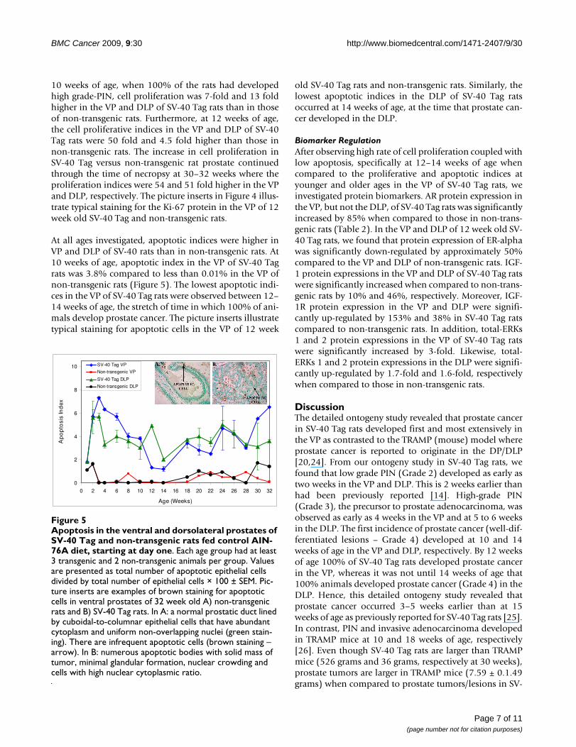
At all ages investigated, apoptotic indices were higher inVP and DLP of SV-40 rats than in non-transgenic rats. At10 weeks of age, apoptotic index in the VP of SV-40 Tagrats was 3.8% compared to less than 0.01% in the VP ofnon-transgenic rats (Figure 5). The lowest apoptotic indi-ces in the VP of SV-40 Tag rats were observed between 12–14 weeks of age, the stretch of time in which 100% of ani-mals develop prostate cancer. The picture inserts illustratetypical staining for apoptotic cells in the VP of 12 week
old SV-40 Tag rats and non-transgenic rats. Similarly, thelowest apoptotic indices in the DLP of SV-40 Tag ratsoccurred at 14 weeks of age, at the time that prostate can-cer developed in the DLP.
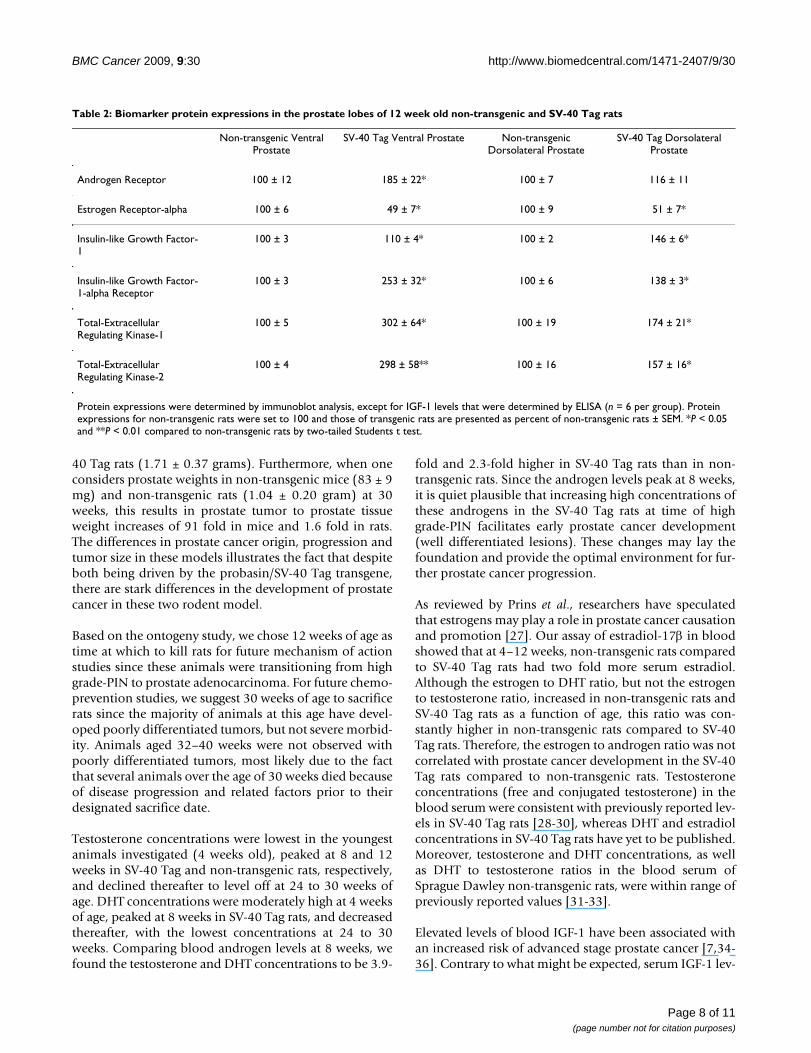
Biomarker RegulationAfter observing high rate of cell proliferation coupled withlow apoptosis, specifically at 12–14 weeks of age whencompared to the proliferative and apoptotic indices atyounger and older ages in the VP of SV-40 Tag rats, weinvestigated protein biomarkers. AR protein expression inthe VP, but not the DLP, of SV-40 Tag rats was significantlyincreased by 85% when compared to those in non-trans-genic rats (Table 2). In the VP and DLP of 12 week old SV-40 Tag rats, we found that protein expression of ER-alphawas significantly down-regulated by approximately 50%compared to the VP and DLP of non-transgenic rats. IGF-1 protein expressions in the VP and DLP of SV-40 Tag ratswere significantly increased when compared to non-trans-genic rats by 10% and 46%, respectively. Moreover, IGF-1R protein expression in the VP and DLP were signifi-cantly up-regulated by 153% and 38% in SV-40 Tag ratscompared to non-transgenic rats. In addition, total-ERKs1 and 2 protein expressions in the VP of SV-40 Tag ratswere significantly increased by 3-fold. Likewise, total-ERKs 1 and 2 protein expressions in the DLP were signifi-cantly up-regulated by 1.7-fold and 1.6-fold, respectivelywhen compared to those in non-transgenic rats.
DiscussionThe detailed ontogeny study revealed that prostate cancerin SV-40 Tag rats developed first and most extensively inthe VP as contrasted to the TRAMP (mouse) model whereprostate cancer is reported to originate in the DP/DLP[20,24]. From our ontogeny study in SV-40 Tag rats, wefound that low grade PIN (Grade 2) developed as early astwo weeks in the VP and DLP. This is 2 weeks earlier thanhad been previously reported [14]. High-grade PIN(Grade 3), the precursor to prostate adenocarcinoma, wasobserved as early as 4 weeks in the VP and at 5 to 6 weeksin the DLP. The first incidence of prostate cancer (well-dif-ferentiated lesions – Grade 4) developed at 10 and 14weeks of age in the VP and DLP, respectively. By 12 weeksof age 100% of SV-40 Tag rats developed prostate cancerin the VP, whereas it was not until 14 weeks of age that100% animals developed prostate cancer (Grade 4) in theDLP. Hence, this detailed ontogeny study revealed thatprostate cancer occurred 3–5 weeks earlier than at 15weeks of age as previously reported for SV-40 Tag rats [25].In contrast, PIN and invasive adenocarcinoma developedin TRAMP mice at 10 and 18 weeks of age, respectively[26]. Even though SV-40 Tag rats are larger than TRAMPmice (526 grams and 36 grams, respectively at 30 weeks),prostate tumors are larger in TRAMP mice (7.59 ± 0.1.49grams) when compared to prostate tumors/lesions in SV-
Apoptosis in the ventral and dorsolateral prostates of SV-40 Tag and non-transgenic rats fed control AIN-76A diet, start-ing at day oneFigure 5Apoptosis in the ventral and dorsolateral prostates of SV-40 Tag and non-transgenic rats fed control AIN-76A diet, starting at day one. Each age group had at least 3 transgenic and 2 non-transgenic animals per group. Values are presented as total number of apoptotic epithelial cells divided by total number of epithelial cells × 100 ± SEM. Pic-ture inserts are examples of brown staining for apoptotic cells in ventral prostates of 32 week old A) non-transgenic rats and B) SV-40 Tag rats. In A: a normal prostatic duct lined by cuboidal-to-columnar epithelial cells that have abundant cytoplasm and uniform non-overlapping nuclei (green stain-ing). There are infrequent apoptotic cells (brown staining – arrow). In B: numerous apoptotic bodies with solid mass of tumor, minimal glandular formation, nuclear crowding and cells with high nuclear cytoplasmic ratio.
0
2
4
6
8
10
0 2 4 6 8 10 12 14 16 18 20 22 24 26 28 30 32
Age (Weeks)
Ap
op
tosi
s In
dex
SV-40 Tag VP
Non-transgenic VP
SV-40 Tag DLP
Non-transgenic DLP
Page 7 of 11(page number not for citation purposes)
BMC Cancer 2009, 9:30 http://www.biomedcentral.com/1471-2407/9/30
40 Tag rats (1.71 ± 0.37 grams). Furthermore, when oneconsiders prostate weights in non-transgenic mice (83 ± 9mg) and non-transgenic rats (1.04 ± 0.20 gram) at 30weeks, this results in prostate tumor to prostate tissueweight increases of 91 fold in mice and 1.6 fold in rats.The differences in prostate cancer origin, progression andtumor size in these models illustrates the fact that despiteboth being driven by the probasin/SV-40 Tag transgene,there are stark differences in the development of prostatecancer in these two rodent model.
Based on the ontogeny study, we chose 12 weeks of age astime at which to kill rats for future mechanism of actionstudies since these animals were transitioning from highgrade-PIN to prostate adenocarcinoma. For future chemo-prevention studies, we suggest 30 weeks of age to sacrificerats since the majority of animals at this age have devel-oped poorly differentiated tumors, but not severe morbid-ity. Animals aged 32–40 weeks were not observed withpoorly differentiated tumors, most likely due to the factthat several animals over the age of 30 weeks died becauseof disease progression and related factors prior to theirdesignated sacrifice date.
Testosterone concentrations were lowest in the youngestanimals investigated (4 weeks old), peaked at 8 and 12weeks in SV-40 Tag and non-transgenic rats, respectively,and declined thereafter to level off at 24 to 30 weeks ofage. DHT concentrations were moderately high at 4 weeksof age, peaked at 8 weeks in SV-40 Tag rats, and decreasedthereafter, with the lowest concentrations at 24 to 30weeks. Comparing blood androgen levels at 8 weeks, wefound the testosterone and DHT concentrations to be 3.9-
fold and 2.3-fold higher in SV-40 Tag rats than in non-transgenic rats. Since the androgen levels peak at 8 weeks,it is quiet plausible that increasing high concentrations ofthese androgens in the SV-40 Tag rats at time of highgrade-PIN facilitates early prostate cancer development(well differentiated lesions). These changes may lay thefoundation and provide the optimal environment for fur-ther prostate cancer progression.
As reviewed by Prins et al., researchers have speculatedthat estrogens may play a role in prostate cancer causationand promotion [27]. Our assay of estradiol-17β in bloodshowed that at 4–12 weeks, non-transgenic rats comparedto SV-40 Tag rats had two fold more serum estradiol.Although the estrogen to DHT ratio, but not the estrogento testosterone ratio, increased in non-transgenic rats andSV-40 Tag rats as a function of age, this ratio was con-stantly higher in non-transgenic rats compared to SV-40Tag rats. Therefore, the estrogen to androgen ratio was notcorrelated with prostate cancer development in the SV-40Tag rats compared to non-transgenic rats. Testosteroneconcentrations (free and conjugated testosterone) in theblood serum were consistent with previously reported lev-els in SV-40 Tag rats [28-30], whereas DHT and estradiolconcentrations in SV-40 Tag rats have yet to be published.Moreover, testosterone and DHT concentrations, as wellas DHT to testosterone ratios in the blood serum ofSprague Dawley non-transgenic rats, were within range ofpreviously reported values [31-33].
Elevated levels of blood IGF-1 have been associated withan increased risk of advanced stage prostate cancer [7,34-36]. Contrary to what might be expected, serum IGF-1 lev-
Table 2: Biomarker protein expressions in the prostate lobes of 12 week old non-transgenic and SV-40 Tag rats
Non-transgenic Ventral Prostate
SV-40 Tag Ventral Prostate Non-transgenic Dorsolateral Prostate
SV-40 Tag Dorsolateral Prostate
Androgen Receptor 100 ± 12 185 ± 22* 100 ± 7 116 ± 11
Estrogen Receptor-alpha 100 ± 6 49 ± 7* 100 ± 9 51 ± 7*
Insulin-like Growth Factor-1
100 ± 3 110 ± 4* 100 ± 2 146 ± 6*
Insulin-like Growth Factor-1-alpha Receptor
100 ± 3 253 ± 32* 100 ± 6 138 ± 3*
Total-Extracellular Regulating Kinase-1
100 ± 5 302 ± 64* 100 ± 19 174 ± 21*
Total-Extracellular Regulating Kinase-2
100 ± 4 298 ± 58** 100 ± 16 157 ± 16*
Protein expressions were determined by immunoblot analysis, except for IGF-1 levels that were determined by ELISA (n = 6 per group). Protein expressions for non-transgenic rats were set to 100 and those of transgenic rats are presented as percent of non-transgenic rats ± SEM. *P < 0.05 and **P < 0.01 compared to non-transgenic rats by two-tailed Students t test.
Page 8 of 11(page number not for citation purposes)
BMC Cancer 2009, 9:30 http://www.biomedcentral.com/1471-2407/9/30
els were not the highest later in life during the progressionto poorly differentiated (advanced) prostate cancer in theSV-40 Tag rats. Despite a poorly differentiated tumor fre-quency of 66% at 30 weeks of age, serum IGF-1 concentra-tions gradually declined after 30 weeks of age. The lowestpost-pubertal levels were noted in the oldest animals (40weeks of age). IGF-1 levels in the blood serum of SV-40Tag rats as they aged mimicked the pattern observed innon-transgenic rats and humans but at higher concentra-tions. This demonstrates that IGF-1 levels in the bloodserum may not be the optimal marker for cancer progres-sion in this model, but simply reflects IGF-1 productionfrom the liver [37].
Because high levels of sex steroids and growth factors canoften lead to changes in cell turnover, we measured cellproliferation and apoptosis. Cell proliferation in non-transgenic rats was similar in the VP and DLP. Likewise, inSV-40 Tag rats, cell proliferation was similar in the VP andDLP, but cell proliferation was significantly higher inprostates of SV-40 Tag rats (1.5- to 54-fold higher, the lat-ter in adult animals). Specifically, proliferation was signif-icantly higher in the VP at an earlier age (2 weekspostpartum) than in the DLP (6 weeks). Increased cellproliferation is consistent with high grade-PIN and even-tually cancer lesions developing earlier in the VP than inthe DLP.
Apoptotic indices were similar in the VP and DLP of non-transgenic rats and in the VP and DLP of SV-40 Tag rats.However, there was a significantly higher rate of apoptosisin prostates of SV-40 Tag rats compared to non-transgenicrats. We speculate that the increase in apoptosis occurs asa consequence of increased cell proliferation and DNAdamaged cells being less stable and more prone to celldeath. The onset of apoptosis followed the initial surge incell proliferation and concomitant with low grade PINdevelopment in both the VP and DLP. Most striking is thedramatic decreases in apoptosis from 10–14 weeks in theVP and from 12 to 18 weeks in the DLP. This mirrors thetransition from high grade-PIN to well differentiatedlesions (cancer). Hence, the increase in cell proliferationand decrease in apoptosis correlate very well with cancerformation in SV-40 Tag rats.
After noting changes in cell proliferation and apoptosisbetween SV-40 Tag and non-transgenic rats, we investi-gated proteins that could serve as biomarkers of action. ARprotein expression in the VP was higher in 12 week oldSV-40 Tag rats when compared to non-transgenic rats.Coupling up-regulated AR with the fact that testosteroneand DHT concentrations were higher in SV-40 Tag ratsmakes a strong case for increased cell proliferation in theprostate and of prostate tumor development in this ani-mal model. Also, we found ER-alpha protein expression
was decreased in both the VP and DLP of SV-40 Tag rats.Concomitantly, we found significantly lower serum estro-gen concentrations in SV-40 Tag rats at 4–12 weeks. Theselower serum estrogen concentrations can be interruptedas reduced potential for estrogen action in SV-40 Tag pros-tate and perhaps reduced anti-androgenic action.
IGF-1 levels in the VP and DLP were elevated in SV-40 Tagrats compared to those in non-transgenic rats. Therefore,IGF-1 in the prostate tissue appears to be an importantprotein involved in prostate cancer development in thismodel. In addition to the growth factor ligand IGF-1being altered, IGF-1R and total-ERKs 1 and 2 proteinexpressions were up-regulated in the VP when SV-40 Tagrats were compared to non-transgenic rats. Takentogether, the up-regulation of the AR, IGF-1 signaling pro-teins, and their downstream effectors (ERKs) suggest apotential mechanism for prostate cancer development inthe SV-40 Tag rat model. Therefore, chemopreventive andtherapeutic agents with the ability to modulate these path-ways make attractive candidates for prevention and ther-apy in this model.
The SV-40 Tag rat and TRAMP models are similar in somerespects. As observed in humans, prostate cancer in SV-40rats is initiated early in life [14,38]. In both models, theSV-40 large T antigen is under control of the probasin pro-moter allowing androgen-regulated protein expressionspecific to the epithelium of the prostate. Transformationin the prostate occurs when the SV-40 large T antigen actsas an oncoprotein via interactions with retinoblastoma[39] and p53 tumor suppressor gene products [40,41].The small t antigen may also play a role in promotion byinteracting with protein phosphatases [42].
ConclusionThe SV-40 Tag rat provides an alternative transgenicrodent model of prostate cancer. Our ontogeny studydemonstrates that transition from PIN to well differenti-ated tumor formation occurs from 10 to 12 weeks in theVP and at 14 weeks in the DLP of SV-40 Tag rats. Thedevelopment of prostate cancer first in the VP of SV-40rats is to be contrasted to that in TRAMP mice where it firstdevelops in the DLP. An advantage of the SV-40 Tag ratover the TRAMP model is that prostates at 12 weeks in theformer are 25 times as large (781 ± 50 mg) as comparedin the latter (31 ± 2 mg), allowing for a greater number ofmechanism of action experiments to be conducted usingfewer animals. Although the cost of housing rats isroughly twice as expensive as mice, the ability to use feweranimals reduces the overall cost in the long run.
The SV-40 Tag rat displays other unique characteristicsincluding reported complete androgen dependence, lowmetastasis, and androgen-independent metastasizing
Page 9 of 11(page number not for citation purposes)
BMC Cancer 2009, 9:30 http://www.biomedcentral.com/1471-2407/9/30
taste bud neuroblastomas [43]. In fact, Asamoto et al.reported that castration at 20 weeks of age (after prostatetumors had developed) caused complete regression andinvolution of these adenocarcinomas and earlier castra-tion at 5 weeks of age completely inhibited prostate aden-ocarcinomas [14]. More detailed studies are merited toconfirm this important characteristic. In contrast, cas-trated 12 week old TRAMP mice still developed poorly dif-ferentiated tumors with metastatic potential [15,44].When castrated at 4 weeks of age (prior to high grade-PINdevelopment), some TRAMP mice developed androgen-independent prostate cancer. Although the SV-40 Tag ratmay never replace the TRAMP model, it is an additionalmodel that can be tailored to specific studies. In theTRAMP model, the ability to progress to androgen inde-pendence [15,44,45], allows it to be used to study latestage prostate cancer. On the other hand, because of itsreported androgen dependence [14] and the fact thatmost human prostate cancer is androgen dependentbefore androgen ablation treatment, the SV-40 Tag ratmodel could be useful in studying early stage prostate can-cer. The models compliment each other. It is currentlyunknown which lobe in the rat prostate is homologous tothe peripheral zone, where prostate cancer occurs in thehumans. Therefore, both the DLP and VP should be inves-tigated. Since prostate cancer develops first and to thegreatest degree in the DP/DLP of the TRAMP model, it isan ideal model to study prostate cancer in that portion ofthe prostate. On the other hand, prostate cancer developsfirst and most extensively in the VP prostate of SV-40 Tagrats, thus providing a basis to study prostate cancer in theVP with this model.
AbbreviationsAR: androgen receptor; DHT: dihydrotestosterone; DLP:dorsolateral prostate; ELISA: enyme-linked immunosorb-ent assay; ERK: extracellular regulating kinase; ER-alpha:Estrogen receptor-alpha; IGF: insulin growth factor; IGF-1R: IGF-1 receptor; MAPK: mitogen-activated proteinkinase; PIN: prostatic intraepithelial neoplasia; SV-40 Tag:Simian Virus large T antigen; TRAMP: Transgenic Adeno-carcinoma Mouse Prostate; VP: ventral prostate.
Competing interestsThe authors declare that they have no competing interests.
Authors' contributionsCEH, LMC, BJP and JW carried out the ontogeny study,cell proliferation and apoptosis assays, and analysis ofprotein expression for mechanism of action. IAE analyzedand interpreted the pathology data. CH drafted the man-uscript and LMC, BJP and JW assisted in writing the man-uscript. TS provided permission to use the SV-40 Tagmodel and CAL proposed the study design and assisted in
writing the manuscript. All authors read and approved thefinal manuscript.
AcknowledgementsWe thank Drs. Gail Prins and Steve Swanson of the University of Illinois at Chicago for providing us with SV-40 Tag rats. Furthermore, we thank Dr. Rene Desmond (UAB) for statistical assistance and Dr. Richard Parker (UAB) for steroid hormone analysis. This research was supported by NIH-NCI-P20-CA93753-03. CEH was supported by a National Cancer Institute Cancer Prevention and Control Training Program (NCI Grant CA 47888).
References1. American Cancer Society Global Cancer Facts and Figures.
American Cancer Society. Atlanta, GA 2007.2. Huggins C, Hodges CV: Studies on prostatic cancer: I. The
effect of castration, of estrogen and of androgen injection onserum phosphatases in metastatic carcinoma of the pros-tate. 1941. The Journal of Urology 2002, 168:9-12.
3. Imperato-McGinley J, Guerrero L, Gautier T, Peterson RE: Steroid5alpha-reductase deficiency in man: an inherited form ofmale pseudohermaphroditism. Science 1974,186(1470):1213-1215.
4. Gann PH, Hennekens CH, Ma J, Longcope C, Stampfer MJ: Prospec-tive study of sex hormone levels and risk of prostate cancer.Journal of the National Cancer Institute 1996, 88:118-1126.
5. Eaton NE, Reeves GK, Appleby PN, Key TJ: Endogenous sex hor-mones and prostate cancer: a quantitative review of pro-spective studies. British Journal of Cancer 1999, 80:930-934.
6. Prins GS, Birch L, Habermann H, Chang WY, Tebeau C, Putz O, Bie-berich C: Influence of neonatal estrogens on rat prostatedevelopment. Reproduction, Fertility, and Development 2001,13:241-252.
7. Chan JM, Stampfer MJ, Ma J, Gann P, Gaziano JM, Pollak M, Giovan-nucci E: Insulin-like growth factor-I (IGF-I) and IGF bindingprotein-3 as predictors of advanced-stage prostate cancer.Journal of the National Cancer Institute 2002, 94:1099-1106.
8. Torring N, Vinter-Jensen L, Pedersen SB, Sorensen FB, Flyvbjerg A,Nexo E: Systemic administration of insulin-like growth factorI (IGF-I) causes growth of the rat prostate. The Journal of Urol-ogy 1997, 158:222-227.
9. Ruan W, Powell-Braxton L, Kopchick JJ, Kleinberg DL: Evidencethat insulin-like growth factor I and growth hormone arerequired for prostate gland development. Endocrinology 1999,140:1984-1989.
10. Tennant MK, Thrasher JB, Twomey PA, Drivdahl RH, Birnbaum RS,Plymate SR: Protein and messenger ribonucleic acid (mRNA)for the type 1 insulin-like growth factor (IGF) receptor isdecreased and IGF-II mRNA is increased in human prostatecarcinoma compared to benign prostate epithelium. The Jour-nal of Clinical Endocrinology and Metabolism 1996, 81:3774-3782.
11. Robinson MJ, Cobb MH: Mitogen-activated protein kinase path-ways. Current Opinion in Cell Biology 1997, 9:180-186.
12. Yeh S, Lin HK, Kang HY, Thin TH, Lin MF, Chang C: From HER2/Neu signal cascade to androgen receptor and its coactiva-tors: a novel pathway by induction of androgen target genesthrough MAP kinase in prostate cancer cells. Proceedings of theNational Academy of Sciences of the United States of America 1999,96:5458-5463.
13. Greenberg NM, DeMayo F, Finegold MJ, Medina D, Tilley WD, Aspi-nall JO, Cunha GR, Donjacour AA, Matusik RJ, Rosen JM: Prostatecancer in a transgenic mouse. Proceedings of the National Academyof Sciences of the United States of America 1995, 92:3439-3443.
14. Asamoto M, Hokaiwado N, Cho YM, Takahashi S, Ikeda Y, Imaida K,Shirai T: Prostate carcinomas developing in transgenic ratswith SV40 T antigen expression under probasin promotercontrol are strictly androgen dependent. Cancer Research 2001,61:4693-4700.
15. Wang J, Eltoum IE, Lamartiniere CA: Genistein chemopreventionof prostate cancer in TRAMP mice. Journal of Carcinogenesis2007, 6:3.
16. Shappell SB, Thomas GV, Roberts RL, Herbert R, Ittmann MM, RubinMA, Humphrey PA, Sundberg JP, Rozengurt N, Barrios R, Ward JM,Cardiff RD: Prostate pathology of genetically engineered
Page 10 of 11(page number not for citation purposes)
BMC Cancer 2009, 9:30 http://www.biomedcentral.com/1471-2407/9/30
Publish with BioMed Central and every scientist can read your work free of charge
"BioMed Central will be the most significant development for disseminating the results of biomedical research in our lifetime."
Sir Paul Nurse, Cancer Research UK
Your research papers will be:
available free of charge to the entire biomedical community
peer reviewed and published immediately upon acceptance
cited in PubMed and archived on PubMed Central
yours — you keep the copyright
Submit your manuscript here:http://www.biomedcentral.com/info/publishing_adv.asp
BioMedcentral
mice: definitions and classification. Cancer Research 2004,64:2270-2305.
17. Price D: Comparative Aspects of Development and Structurein the Prostate. National Cancer Institute Monograph 1963, 12:1-27.
18. Folkvord JM, Viders D, Coleman-Smith A, Clark RA: Optimizationof immunohistochemical techniques to detect extracellularmatrix proteins in fixed skin specimens. J Histochem Cytochem1989, 37(1):105-113.
19. Wechter WJ, Leipold DD, Murray ED Jr, Quiggle D, McCracken JD,Barrios RS, Greenberg NM: E-7869 (R-flurbiprofen) inhibits pro-gression of prostate cancer in the TRAMP mouse. CancerResearch 2000, 60:2203-2208.
20. Kaplan-Lefko PJ, Chen TM, Ittmann MM, Barrios RJ, Ayala GE, HussWJ, Maddison LA, Foster BA, Greenberg NM: Pathobiology ofautochthonous prostate cancer in a pre-clinical transgenicmouse model. The Prostate 2003, 55:219-237.
21. Harper C, Patel BB, Wang J, Eltoum IE, Lamartiniere CA: Epigallo-catechin-3-gallate suppresses early stage, but not late stageprostate cancer in TRAMP mice: mechanisms of action. TheProstate 2007, 67:1576-1589.
22. Wang J, Eltoum IE, Lamartiniere CA: Genistein alters growth fac-tor signaling in transgenic prostate model (TRAMP). Mol CellEndocrinol 2004, 219:171-180.
23. Crowther JR: The ELISA guidebook. Methods in Molecular Biology.Clifton, NJ 2000, 149:1-413.
24. Wikstrom P, Lindahl C, Bergh A: Characterization of the autoch-thonous transgenic adenocarcinoma of the mouse prostate(TRAMP) as a model to study effects of castration therapy.The Prostate 2005, 62:148-164.
25. Cho YM, Takahashi S, Asamoto M, Suzuki S, Inaguma S, HokaiwadoN, Shirai T: Age-dependent histopathological findings in theprostate of probasin/SV40 T antigen transgenic rats: lack ofinfluence of carcinogen or testosterone treatment. Cancer Sci-ence 2003, 94:153-157.
26. Gingrich JR, Barrios RJ, Morton RA, Boyce BF, DeMayo FJ, FinegoldMJ, Angelopoulou R, Rosen JM, Greenberg NM: Metastatic pros-tate cancer in a transgenic mouse. Cancer Research 1996,56:4096-4102.
27. Prins GS, Huang L, Birch L, Pu Y: The role of estrogens in normaland abnormal development of the prostate gland. Annals ofthe New York Academy of Sciences 2006, 1089:1-13.
28. Kandori H, Suzuki S, Asamoto M, Murasaki T, Mingxi T, Ogawa K,Shirai T: Influence of atrazine administration and reduction ofcalorie intake on prostate carcinogenesis in probasin/SV40 Tantigen transgenic rats. Cancer Science 2005, 96:221-226.
29. Tang M, Ogawa K, Asamoto M, Hokaiwado N, Seeni A, Suzuki S,Takahashi S, Tanaka T, Ichikawa K, Shirai T: Protective effects ofcitrus nobiletin and auraptene in transgenic rats developingadenocarcinoma of the prostate (TRAP) and human pros-tate carcinoma cells. Cancer Science 2007, 98:471-477.
30. Zeng Y, Yokohira M, Saoo K, Takeuchi H, Chen Y, Yamakawa K, Mat-suda Y, Kakehi Y, Imaida K: Inhibition of prostate carcinogenesisin probasin/SV40 T antigen transgenic rats by raloxifene, anantiestrogen with anti-androgen action, but not nimesulide,a selective cyclooxygenase-2 inhibitor. Carcinogenesis 2005,26:1109-1116.
31. Resko JA, Feder HH, Goy RW: Androgen concentrations inplasma and testis of developing rats. The Journal of Endocrinology1968, 40:485-491.
32. Corpechot C, Baulieu EE, Robel P: Testosterone, dihydrotesto-sterone and androstanediols in plasma, testes and prostatesof rats during development. Acta Endocrinologica 1981,96:127-135.
33. Berger M, Jean-Faucher C, de Turckheim M, Veyssiere G, Jean C:Testicular and plasma androgens in newborn, immature,adult and aging mice. Archives Internationales de Physiologie et de Bio-chimie 1975, 83:239-253.
34. Chan JM, Stampfer MJ, Giovannucci E, Gann PH, Ma J, Wilkinson P,Hennekens CH, Pollak M: Plasma insulin-like growth factor-Iand prostate cancer risk: a prospective study. Science 1998,279(5350):563-566.
35. Wolk A, Mantzoros CS, Andersson SO, Bergstrom R, Signorello LB,Lagiou P, Adami HO, Trichopoulos D: Insulin-like growth factor1 and prostate cancer risk: a population-based, case-controlstudy. Journal of the National Cancer Institute 1998, 90:911-915.
36. Mantzoros CS, Tzonou A, Signorello LB, Stampfer M, TrichopoulosD, Adami HO: Insulin-like growth factor 1 in relation to pros-tate cancer and benign prostatic hyperplasia. British Journal ofCancer 1997, 76:1115-1118.
37. Sjogren K, Liu JL, Blad K, Skrtic S, Vidal O, Wallenius V, LeRoith D,Tornell J, Isaksson OG, Jansson JO, Ohlsson C: Liver-derived insu-lin-like growth factor I (IGF-I) is the principal source of IGF-I in blood but is not required for postnatal body growth inmice. Proceedings of the National Academy of Sciences of the UnitedStates of America 1999, 96:7088-7092.
38. Sakr WA, Haas G, Cassin BF, Pontes JE, Crissman JD: The fre-quency of carcinoma and intraepithelial neoplasia of theprostate in young male patients. The Journal of Urology 1993,150:379-385.
39. DeCaprio JA, Ludlow JW, Figge J, Shew JY, Huang CM, Lee WH, Mar-silio E, Paucha E, Livingston DM: SV40 large tumor antigen formsa specific complex with the product of the retinoblastomasusceptibility gene. Cell 1998, 54:275-283.
40. Lane DP, Crawford LV: T antigen is bound to a host protein inSV40-transformed cells. Nature 1979, 278:261-263.
41. Linzer DI, Levine AJ: Characterization of a 54K dalton cellularSV40 tumor antigen present in SV40-transformed cells anduninfected embryonal carcinoma cells. Cell 1979, 17:43-52.
42. Pallas DC, Shahrik LK, Martin BL, Jaspers S, Miller TB, Brautigan DL,Roberts TM: Polyoma small and middle T antigens and SV40small t antigen form stable complexes with protein phos-phatase 2A. Cell 1990, 60:167-176.
43. Asamoto M, Hokaiwado N, Cho YM, Shirai T: Effects of geneticbackground on prostate and taste bud carcinogenesis due toSV40 T antigen expression under probasin gene promotercontrol. Carcinogenesis 2002, 23:463-467.
44. Gingrich JR, Barrios RJ, Kattan MW, Nahm HS, Finegold MJ, Green-berg NM: Androgen-independent prostate cancer progres-sion in the TRAMP model. Cancer research 1997, 57:4687-4691.
45. Eng MH, Charles LG, Ross BD, Chrisp CE, Pienta KJ, Greenberg NM,Hsu CX, Sanda MG: Early castration reduces prostatic carcino-genesis in transgenic mice. Urology 1999, 54:1112-1119.
Pre-publication historyThe pre-publication history for this paper can be accessedhere:
http://www.biomedcentral.com/1471-2407/9/30/prepub
Page 11 of 11(page number not for citation purposes)
Recommended


