
JOURNAL OF NEUROINFLAMMATION
de Pablos et al. Journal of Neuroinflammation 2014, 11:34http://www.jneuroinflammation.com/content/11/1/34
RESEARCH Open Access
Chronic stress enhances microglia activation andexacerbates death of nigral dopaminergicneurons under conditions of inflammationRocío M de Pablos1,2*, Antonio J Herrera1,2, Ana M Espinosa-Oliva1,2, Manuel Sarmiento1,2,3, Mario F Muñoz1,2,Alberto Machado1,2 and José L Venero1,2
Abstract
Background: Parkinson’s disease is an irreversible neurodegenerative disease linked to progressive movementdisorders and is accompanied by an inflammatory reaction that is believed to contribute to its pathogenesis. Sincesensitivity to inflammation is not the same in all brain structures, the aim of this work was to test whether physiologicalconditions as stress could enhance susceptibility to inflammation in the substantia nigra, where death of dopaminergicneurons takes place in Parkinson’s disease.
Methods: To achieve our aim, we induced an inflammatory process in nonstressed and stressed rats (subject to achronic variate stress) by a single intranigral injection of lipopolysaccharide, a potent proinflammogen. The effect ofthis treatment was evaluated on inflammatory markers as well as on neuronal and glial populations.
Results: Data showed a synergistic effect between inflammation and stress, thus resulting in higher microglial activationand expression of proinflammatory markers. More important, the higher inflammatory response seen in stressed animalswas associated with a higher rate of death of dopaminergic neurons in the substantia nigra, the most characteristicfeature seen in Parkinson’s disease. This effect was dependent on glucocorticoids.
Conclusions: Our data demonstrate that stress sensitises midbrain microglia to further inflammatory stimulus. Thissuggests that stress may be an important risk factor in the degenerative processes and symptoms of Parkinson’s disease.
Keywords: Glucocorticoids, Lipopolysaccharide, Microglia, Parkinson’s disease, Stress, Substantia nigra
BackgroundParkinson’s disease (PD) is an age-related neurodegener-ative disorder characterised by progressive degenerationof the nigrostriatal dopaminergic (DAergic) neurons ofthe substantia nigra pars compacta (SNpc) [1]. Thisprocess results in extrapyramidal motor dysfunctionaccompanied by progressive impairment of autonomy,mood and cognitive function [1-3]. Although some geneshave been identified as being responsible for rare familialearly-onset PD [4], the aetiology of PD remains elusive.Oxidative stress, reduced expression of trophic factors,mitochondrial dysfunction, alterations of the ubiquitin
* Correspondence: [email protected] of Biochemistry and Molecular Biology, Faculty of Pharmacy,University of Seville, E-41012 Seville, Spain2Institute of Biomedicine of Seville, Virgen del Rocio University Hospital/CSIC/University of Seville, E-41013 Seville, SpainFull list of author information is available at the end of the article
© 2014 de Pablos et al.; licensee BioMed CentCommons Attribution License (http://creativecreproduction in any medium, provided the orDedication waiver (http://creativecommons.orunless otherwise stated.
proteasome system and neuroinflammatory mechanismsare thought to collaborate in the progressive demise ofSNpc neurons [1,2,5-11].Evidence suggesting that inflammation may play a
central role in the cell loss in PD has been accumulatingsince the presence of activated microglia in the substan-tia nigra (SN) of PD patients was first reported ([12]; forreview, see [13,14]). The increased number of activatedmicroglial cells is accompanied by increased expression ofproinflammatory cytokines [6,15]. Inflammation has beenshown to exist in different animal models of PD, includingthose using 1-methyl-4-phenyl-1,2,3,6-tetrahydropyridine(MPTP), 6-hydroxydopamine (6-OHDA) or rotenone[16-19]. Lipopolysaccharide (LPS) is the active immu-nostimulant in the cell wall of Gram-negative bacteriathat is responsible for triggering the cascade of eventsfollowing bacterial infection [20,21]. Subtoxic doses of
ral Ltd. This is an Open Access article distributed under the terms of the Creativeommons.org/licenses/by/2.0), which permits unrestricted use, distribution, andiginal work is properly credited. The Creative Commons Public Domaing/publicdomain/zero/1.0/) applies to the data made available in this article,

de Pablos et al. Journal of Neuroinflammation 2014, 11:34 Page 2 of 18http://www.jneuroinflammation.com/content/11/1/34
LPS exacerbated disease progression in an animal modelof PD [22], supporting the hypothesis that brain inflam-mation may play a significant role in PD progression.More important, epidemiological studies have demon-strated that the incidence of idiopathic PD is about 50%lower in chronic users of nonsteroidal anti-inflammatorydrugs and cyclooxygenase inhibitors than in age-matchednonusers [23-25].Our group pioneered in showing that an inflammatory
response induced by the intracerebral injection of LPScan cause neuronal death, specifically in the nigrostriatalDAergic system [26-28]. Interestingly, the SN, comparedwith other brain areas, is especially susceptible to LPS-induced neurotoxicity [28]. In this structure, the strongmicroglial response to LPS preceded the death ofDAergic neurons [26,29]. This point has been confirmedby experiments with chronic infusion of LPS into the SNthat produced delayed death of DAergic neurons, as alsofound in PD [17]. Under these conditions, microglialactivation reached a plateau 2 weeks earlier than theappearance of degenerative events on the DAergic system[17]. The neurotoxic effect of LPS has been also observedin mesencephalic cell cultures [30-33].Some physiological conditions might enhance the in-
flammatory response to LPS and account for the diver-sity in symptoms and course of PD, as well as forindividuals’ responses to medication after the onset ofPD [34]. Stress is widely acknowledged to be a predis-posing and precipitating factor in psychiatric illnesses[35,36] and some neurodegenerative diseases. It was oneof the earliest proposed causes of PD [37,38]. Stress is acondition of human experience that includes not onlymajor life events but also the hassles of daily life thatelevate activities of physiological systems, which causesdisruption of ongoing homeostasis. It reflects both indi-vidual experience and genetic background; therefore, re-actions to a stressful incident are highly variable [39,40].Exposure to a stressful situation leads to the activationof two systems: the sympathoadrenomedullary systemand the hypothalamic-pituitary-adrenal (HPA) axis (forreview, see [41]). The former leads to increased circulat-ing levels of adrenaline, whereas the latter leads torelease of corticosteroid hormones from the adrenal cor-tex. Corticosteroid hormones can easily enter the brainbecause of their lipophilic properties and bind two typesof receptors: mineralocorticoid receptors (MRs) andglucocorticoid receptors (GRs) (for review, see [42]).Whereas MRs have a high affinity for the endogenoushormone corticosterone, GRs have a lower affinity.Interestingly, MRs are restricted mainly to limbic re-gions, including the amygdala and all subareas of thehippocampus. On the contrary, GRs are ubiquitouslydistributed in neurons and glia. Although stress can bebeneficial in its acute phase, repeated and severe stressful
stimuli produce adverse effects on neuronal functions,especially in those structures involved in the stressresponse, such as the hypothalamus, prefrontal cortex(PFC) and hippocampus.We have previously shown a synergistic deleterious
effect of chronic stress and inflammation in limbic struc-tures such as the hippocampus [43] and the PFC [44].These studies suggest that stress strongly sensitisesmicroglial cells to proinflammatory stimuli. However,whether this effect is generalised in the whole centralnervous system (CNS) is unknown. The aim of thepresent work was to elucidate whether enhancementof the LPS-induced damage by chronic stress is exten-sible to other CNS structures, especially to those involvedin neurodegenerative disorders such as PD, in which in-flammation seems to play an important role. To evaluatethis hypothesis, we tested whether chronic variate stresscould enhance the damage induced by LPS in the SN. Wecombined immunohistochemical and molecular biologicaltechniques in an effort to elucidate the effects of chronicstress, intranigral LPS injection and a combination of bothon different cell types in the SN. We found that stresssignificantly increased the inflammatory damage inducedby LPS. We also studied the effect produced on thedifferent parameters assayed by RU486 (mifepristone (11β-[p-(dimethylamino)phenyl]-17β-hydroxy-17-(1-propynyl)estra-4,9-dien-3-one)), a potent inhibitor of GR activation.
MethodsAnimals and surgeryMale albino Wistar rats (250 to 270 g) were used forthese studies. The rats were kept at constant roomtemperature of 22°C ± 1°C and 60%relative humidity ona 12:12-hour light–dark cycle with free access to foodand water. Rats were anaesthetized with chloral hydrate(400 mg/kg) and positioned in a stereotaxic apparatus(Kopf Instruments, Tujunga, CA, USA) to conform tothe brain atlas of Paxinos and Watson [45]. Injectionsinto the SN were made 5.5 mm posterior, 1.8 mm lateraland 8.3 mm ventral to the bregma at day 1, 2 hours afterthe application of the first stressor (Figure 1).Experiments were carried out in accordance with the
Guidelines of the European Union Council (86/609/EU)and Spanish regulations for the use of laboratory animals(BOE 67/8509-12, 1988), and the study was approved bythe Scientific Committee of the University of Seville.Five groups of animals were established according to
the different treatments: V, the vehicle/nonstressed con-trol group, which received a single intranigral injectionof 2 μl of vehicle (Monastral Blue inert tracer, 1% in sa-line solution; Sigma-Aldrich, St Louis, MO, USA) intothe left SN; S, the vehicle/stressed group, which weretreated with a single intranigral injection of 2 μl of ve-hicle into the left SN and were stressed for 9 days; L, the

Figure 1 Experimental groups and treatments. Intranigral injections of vehicle (Veh) or lipopolysaccharide (LPS) were given at day 1. In thestressed groups (S, SL and SLR), intranigral injections were carried out after the application of the first stressor (10 minutes of forced swimming).In the SLR group, RU486 (mifepristone (11β-[p-(dimethylamino)phenyl]-17β-hydroxy-17-(1-propynyl)estra-4,9-dien-3-one)) was injected subcutaneouslyevery day 1 hour before the stressor. V, vehicle; S, stress; L, lipopolysaccharide; SL, lipopolysaccharide injected into stressed animals; SLR,lipopolysaccharide injected into stressed animals treated with RU486.
de Pablos et al. Journal of Neuroinflammation 2014, 11:34 Page 3 of 18http://www.jneuroinflammation.com/content/11/1/34
LPS group (nonstressed animals), which were treatedwith a single intranigral injection of 2 μg of LPS (fromEscherichia coli serotype 026:B6; Sigma-Aldrich) dissolvedin 2 μl of vehicle (1% Monastral Blue inert tracer in salinesolution) into the left SN; SL, the LPS/stressed group,which were treated with a single intranigral injection of2 μg of LPS and stressed for 9 days; SLR, the LPS/stressed/RU486 group, which were treated with a singleintranigral injection of 2 μg of LPS, stressed for 9 days andwere given a daily dose of 20 mg/kg RU486 in salinewith 20% dimethyl sulphoxide by subcutaneous injection(Sigma-Aldrich) for 9 days 1 hour before exposure tothe stressors. All animals were killed by decapitation
at 6 hours after surgery (RT-PCR experiments) or byperfusion 10 days after surgery (immunohistochemistryexperiments). At least four animals were used for eachgroup.
Stress modelChronic variate stress was adapted from other models ofvariate stress [46-51] with modifications as reported pre-viously [43,44]. Animals were divided into stressed andnonstressed groups. Nonstressed animals were kept un-disturbed in their home cages for 10 days. A 9-day vari-ate stressor paradigm was used for the animals in thestressed groups. The schedule of stressors is given in

de Pablos et al. Journal of Neuroinflammation 2014, 11:34 Page 4 of 18http://www.jneuroinflammation.com/content/11/1/34
Table 1. Application of stress started at different timesfrom day to day (between 08:00 and 20:00) to minimizeits predictability. Restraint was carried out by placingeach animal in a 21 cm× 6 cm plastic tube and adjustingit with plaster tape on the outside, so that the animal wasunable to move. There was a 6-cm hole at the far end forbreathing. Forced swimming was carried out by placingthe animal in a glass tank measuring 44 × 33 × 30 cmwith 22 cm of water depth at 23°C ± 2°C. Body weightwas measured at the beginning and the end of the 10-daytreatment and was evaluated as an indirect parameter ofHPA axis activation.
Serum corticosterone measurementRats were deeply anaesthetized, and blood was collectedfrom the heart. Serum corticosterone concentration wasmeasured by enzyme-linked immunosorbent assay ac-cording to the manufacturer’s instructions (Assay DesignsCorrelate-EIA; Enzo Life Sciences, Farmingdale, NY, USA).
Immunohistological evaluationThaw-mounted 20-μm coronal sections were cut on acryostat at −15°C and mounted on gelatine-coated slides.Primary antibodies used were rabbit-derived anti-tyrosinehydroxylase (1:300 anti-TH; Sigma-Aldrich), mouse-derived anti-glial fibrillary acidic protein (1:300 anti-GFAP;EMD Millipore, Billerica, MA, USA), rabbit-derived anti-Iba-1 (1:300; Wako Chemicals, Richmond, VA, USA) andmouse-derived OX-6 (1:200; AbD Serotec, Raleigh, NC,USA). Incubations and washes for all the antibodieswere carried out in Tris-buffered saline (TBS), pH 7.4.All work was done at room temperature. Sections werewashed and then treated with 0.3% hydrogen peroxidein methanol for 20 minutes, washed again, and incu-bated in a TBS solution containing 1% horse serum(Vector Laboratories, Burlingame, CA, USA) for GFAPand OX-6 immunostaining or in a solution containing
Table 1 Schedule of stressors used during the chronicvariate stress treatmenta
Day Stressor Time
1 Forced swimming 10 min
2 Restraint 3 h
3 Water deprivation 24 h
4 Restrain at 4ºC 90 min
5 Isolation 24 h
6 Food deprivation 24 h
7 Water deprivation 24 h
8 Restrain at 4ºC 2 h
9 Food deprivation 24 haAnimals subjected to stress were exposed to the stressors according to theschedule listed. The left column indicates the day of the stressing protocol.The right column indicates the length of each stimulus applied.
goat serum (Vector Laboratories) for TH and Iba-1 immu-nostaining for 60 minutes in a humidity chamber. Slideswere drained and further incubated with the primary anti-body in TBS containing 1% horse or goat serum and0.25% Triton X-100 for 24 hours. Sections were then incu-bated for 2 hours with biotinylated horse anti-mouseimmunoglobulin G (IgG, 1:200; Vector Laboratories) forGFAP and OX-6 immunostaining or biotinylated goatanti-rabbit IgG (1:200; Vector Laboratories) for TH andIba-1 immunostaining. The secondary antibody was di-luted in TBS containing 0.25% Triton X-100, and itsaddition was preceded by three 10-minute rinses inTBS. Sections were then incubated with ExtrAvidin–Peroxidase buffered aqueous solution (1:100; Sigma-Aldrich). The peroxidase was visualized by performinga standard diaminobenzidine–hydrogen peroxide reactionfor 5 minutes.
Immunohistochemical data analysisAnalyses were carried out in a bounded region of the SNwith a length of 300 μm in the anteroposterior axiscentred at the point of injection (5.5 mm with respect tothe bregma), that is, between 5.35 and 5.65 mm withrespect to the bregma. In each case, five sections peranimal were used with random starting points and sys-tematically distributed through the anteroposterior axisof the analysed region. For the measurement of areaslacking GFAP immunoreactivity, we used analySIS im-aging software (Soft Imaging System GmbH, Münster,Germany) coupled to a Polaroid DMC camera (Polaroid,Cambridge, MA, USA) attached to a Leica light micro-scope (Leica Microsystems, Wetzlar, Germany). To countcells showing OX-6 immunoreactivity, we systematicallysampled the area occupied by the OX-6-positive cells ineach section from a random starting point with a gridadjusted to count five fields per section. An unbiasedcounting frame of a known area (40 × 25 μm= 1,000 μm2)was superimposed on the tissue section image under a100× oil immersion lens objective. The different types ofOX-6-positive cells (displaying different shapes, dependingon their activation state) were counted as a whole andexpressed as cells per millimetre squared. The number ofTH-positive neurons in the SN was estimated using afractionator sampling design [52]. Counts were carriedout at regular predetermined intervals within each section(x = 150 μm and y = 200 μm). An unbiased counting frameof known area (40 μm × 25 μm = 1,000 μm2) wassuperimposed on the tissue section image under a100× oil immersion lens objective. Therefore, the areasampling fraction was 1,000/(150 × 200) = 0.033. Theentire z-dimension of each section was sampled; hence,the section thickness sampling fraction was 1. In all ani-mals, 20-μm sections, each 100 μm apart, were analysed;thus, the fraction of sections sampled was 20/100 = 0.20.

de Pablos et al. Journal of Neuroinflammation 2014, 11:34 Page 5 of 18http://www.jneuroinflammation.com/content/11/1/34
The number of neurons in the analysed region was esti-mated by multiplying the number of neurons countedwithin the sample regions by the reciprocals of the areasampling fraction and the fraction of section sampled.
ImmunofluorescenceAnimals were perfused and sections prepared as describedabove. Incubations and washes for all the antibodies werecarried out in phosphate-buffered saline (PBS), pH 7.4. Allwork was done at room temperature. For double-labellingof Iba-1 with TH, sections were blocked with PBS con-taining 1% normal horse serum (Vector Laboratories)for Iba-1 and 1% goat serum (Vector Laboratories) for THfor 1 hour. The slides were washed three times in PBS,then incubated overnight at 4°C with either mouse-derived anti-Iba-1 (1:300; EMD Millipore) and rabbit-derived anti-TH (1:300; Sigma-Aldrich) diluted in PBScontaining 1% normal horse serum and 1% goat serumand 0.25% Triton X-100. Sections were incubated withhorse anti-mouse secondary antibody conjugated to fluor-escein (1:200; Vector Laboratories) for Iba-1 and goatanti-rabbit secondary antibody conjugated to Alexa Fluor594 (1:200; Molecular Probes/Invitrogen, Eugene, OR,USA) for TH for 1 hour at 22°C ± 1°C in the dark, andtheir addition was preceded by three 10-minute rinses inPBS. Nuclei were counterstained with Hoechst dye (1 μg/ml; Molecular Probes). As a control, another set of experi-ments was performed whereby the sections were incu-bated with only the Iba-1 antibody, then visualized withboth fluorescence filters. No signal was detected whenIba-1 alone was used with an Alexa Fluor 594 filter(photomicrograph not shown). The same was true withTH when a fluorescein filter was used.For double-labelling of Iba-1 with inhibitor of nuclear
factor κΒ (NF-κΒ) kinase, subunit β (IKKβ), sectionswere blocked with PBS containing 1% normal goatserum (Vector Laboratories) for Iba-1 and rabbit serum(Invitrogen) for TH for 1 hour. The slides were washedthree times in PBS, then incubated overnight at 4°Cwith either rabbit-derived anti-Iba-1 (1:300; WakoChemicals) and goat-derived anti-IKKβ (1:300; SantaCruz Biotechnology, Santa Cruz, CA, USA) diluted inPBS containing 1% normal goat and 1% rabbit serumand 0.25% Triton X-100. Sections were incubated withgoat anti-rabbit secondary antibody conjugated to AlexaFluor 594 (1:200; Molecular Probes/Invitrogen) for Iba-1and with rabbit anti-goat secondary antibody conjugatedto fluorescein (Vector Laboratories; 1:200) for IKKβ for1 hour at 22°C ± 1°C in the dark. Their addition was pre-ceded by three 10-minute rinses in PBS. Nuclei werecounterstained with Hoechst dye (1 μg/ml; MolecularProbes/Invitrogen). As a control, another set of experi-ments was performed whereby the sections were incu-bated with only the Iba-1 antibody, then visualized with
both filters. No signal was detected when Iba-1 alonewith the fluorescein filter was used (photomicrographnot shown). The same was true for IKKβ when an AlexaFluor 594 filter was used. Fluorescence images were ac-quired using a Zeiss LSM 7 DUO confocal laser scanningmicroscope (Carl Zeiss Microscopy, Jena Germany) andprocessed using the associated software package (ZEN2010; Carl Zeiss Microscopy).
Real time RT-PCRThe left SN was dissected from each rat 6 hours after theinjection of vehicle or LPS, snap-frozen in liquid nitrogenand stored at −80 ºC. Total RNA was extracted fromthe SN using the RNeasy kit (QIAGEN, Germantown,MD, USA). cDNA was synthesized from 1 μg of totalRNA using the QuantiTect Reverse Transcription Kit(QIAGEN) in 20 μl of reaction volume as described bythe manufacturer. Real-time PCR was performed with iQSYBR Green Supermix (Bio-Rad Laboratories, Hercules,CA, USA), 0.4 μM primers and 1 μl of cDNA. Controlswere carried out without cDNA. Amplification was run ina MasterCycler ep realplex thermal cycler (Eppendorf,Happauge, NY, USA) at 94°C for 3 minutes, followed by35 cycles at 94°C for 30 seconds, 55°C to 60°C for 45 sand 72°C for 45 s, then by a final elongation step at 72°Cfor 7 minutes. Following amplification, melting curve ana-lysis was performed by heating the reactions from 65°C to95°C at 1°C intervals while monitoring fluorescence. Ana-lysis confirmed a single PCR product at the predictedmelting temperature. β-actin served as the reference geneand was used for sample normalization. Primer sequencesfor tumour necrosis factor α (TNF-α), interleukin 6 (IL-6),IL-1β, inducible nitric oxide synthase (iNOS), CD200, che-mokine (C-X3-C motif) receptor 1 (CX3CR1), monocytechemoattractant protein 1 (MCP-1) and β-actin are shownin Table 2. The cycle at which each sample crossed afluorescence threshold cycle (Ct) was determined, and thetriplicate values for each cDNA were averaged. Analysesof real-time PCR were carried out using a comparative Ct
method integrated into Bio-Rad system software.
Western blot analysisSN was lysed in 15 mM Tris–HCl, pH 7.5, 150 mMNaCl, 1 mM ethylenediaminetetraacetic acid, 1 mMethylene glycol tetraacetic acid and 1 mM phenylmethyl-sulfonyl fluoride (all from Sigma-Aldrich). The hom-ogenate was centrifuged at 12,000 × g for 20 minutes at4°C. Protein content of the samples was estimated by themethod of micro-Lowry using bovine serum albumin asa standard [59], and 25 μg of protein was loaded foreach lane. Protein samples were separated by SDS-PAGE(10%) and transferred onto a nitrocellulose membrane(Novex; Life Technologies, Grand Island, NY, USA).Membranes were blocked with blocking buffer (5% milk in
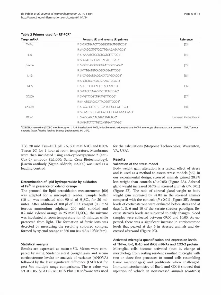
Table 2 Primers used for RT-PCRa
Target mRNA Forward (F) and reverse (R) primers Reference
TNF-α F: 5′-TACTGAACTTCGGGGTGATTGGTCC-3′ [53]
R: 5′-CAGCCTTGTCCCTTGAAGAGAACC-3′
IL-6 F: 5′-AAAATCTGCTCTGGTCTTCTGG-3′ [54]
R: 5′-GGTTTGCCGAGTAGACCTCA-3′
β-actin F: 5′-TGTGATGGTGGGAATGGGTCAG-3′ [55]
R: 5′-TTTGATGTCACGCACGATTTCC-3′
IL-1β F: 5′-CAGGATGAGGACATGAGCACC-3′ [55]
R: 5′-CTCTGCAGACTCAAACTCCAC-3′
iNOS F: 5′-CCTCCTCCACCCTACCAAGT-3′ [56]
R: 5′-CACCCAAAGTGCTTCAGTCA-3′
CD200 F: 5′-TGTTCCGCTGATTGTTGGC-3′ [57]
R: 5′- ATGGACACATTACGGTTGCC-3′
CX3CR1 F: 5′-GGC CTT GTC TGA TCT GCT GTT TG-3′ [58]
R: 5′- AAT GCT GAT GAC GGT GAT GAA GAA-3′
MCP-1 F: 5′-AGCATCCACGTGCTGTCTC-3′ Universal ProbeLibraryb
R: 5′-GATCATCTTGCCAGTGAATGAG-3′aCX3CR1, chemokine (C-X3-C motif) receptor 1; IL-6, Interleukin 6; iNOS, inducible nitric oxide synthase; MCP-1, monocyte chemoattractant protein 1; TNF, Tumournecrosis factor. bRoche Applied Science (Indianapolis, IN, USA).
de Pablos et al. Journal of Neuroinflammation 2014, 11:34 Page 6 of 18http://www.jneuroinflammation.com/content/11/1/34
TBS: 20 mM Tris–HCl, pH 7.5, 500 mM NaCl and 0.05%Tween 20) for 1 hour at room temperature. Membraneswere then incubated using anti-cyclooxygenase 2 (anti-Cox-2) antibody (1:1,000; Santa Cruz Biotechnology).β-actin antibody (Sigma-Aldrich; 1:2,000) was used as aloading control.
Determination of lipid hydroperoxide by oxidationof Fe2+ in presence of xylenol orangeThe protocol for lipid peroxidation measurements [60]was adapted for a microplate reader. Sample buffer(10 μl) was incubated with 90 μl of H2SO4 for 30 mi-nutes. After addition of 100 μl of FOX reagent (0.5 mMferrous ammonium sulphate, 200 mM sorbitol and0.2 mM xylenol orange in 25 mM H2SO4), the mixturewas incubated at room temperature for 45 minutes whileprotected from light. The formation of ferric ions wasdetected by measuring the resulting coloured complexformed by xylenol orange at 560 nm (ε = 4.3 × 10⁴/M/cm).
Statistical analysisResults are expressed as mean ± SD. Means were com-pared by using Student’s t-test (weight gain and serumcorticosterone levels) or analysis of variance (ANOVA)followed by the least significant difference (LSD) test forpost hoc multiple range comparisons. The α value wasset at 0.05. STATGRAPHICS Plus 3.0 software was used
for the calculations (Statpoint Technologies, Warrenton,VA, USA).
ResultsValidation of the stress modelBody weight gain alteration is a typical effect of stressand is used as a method to assess stress models [46]. Inour experimental design, stressed animals gained 28.8%less weight than controls (P < 0.05) (Figure 2A). Adrenalgland weight increased 34.7% in stressed animals (P < 0.01)(Figure 2B). The ratio of adrenal gland weight to bodyweight gain increased by 94.0% in the stressed animalscompared with the controls (P < 0.01) (Figure 2B). Serumlevels of corticosterone were evaluated before stress and atdays 1, 3, 6 and 10 of the variate stressor paradigm. Be-cause steroids levels are subjected to daily changes, bloodsamples were collected between 09:00 and 10:00. As ex-pected, there was a significant increase in corticosteronelevels that peaked at day 6 in stressed animals and de-creased afterward (Figure 2C).
Activated microglia quantification and expression levelsof TNF-α, IL-6, IL-1β and iNOS mRNAs and COX-2 proteinMicroglial cells become activated (that is, change ofmorphology from resting resident ramified microglia withtwo or three fine processes to round cells resemblingtissue macrophages) and proliferate when challenged.Immunohistochemistry of Iba-1 and OX-6 showed thatinjection of vehicle in nonstressed animals (controls)

Figure 2 Validation of the stress model. (A) Body weight gain(in grams). *P < 0.05. (B) Adrenal gland weight (in milligrams; bars)and ratio between adrenal gland weight and body weight gain(scatterplot and line). **P < 0.01, #P < 0.01 (for adrenal gland weight/body weight gain ratio). (C) Serum corticosterone (percentage ofcontrol animals). *P < 0.01 compared with control, aP < 0.01 comparedwith previous time point (S1d to S10d indicates days subjected tovariate stress). Statistical significance was calculated by using Student’st-test to compare data before (C) and after 10 days (S10d) of variatestress. Data were derived from one-way analysis of variance followedby the least significant difference (LSD) post hoc test for multiplerange comparisons.
de Pablos et al. Journal of Neuroinflammation 2014, 11:34 Page 7 of 18http://www.jneuroinflammation.com/content/11/1/34
produced a slight microglial reaction that was moreintense in stressed animals (approximately fivefold thatof controls; P < 0.001) (Figures 3 and 4). LPS injectioninduced stronger activation in nonstressed animals (abouteightfold compared with control animals; P < 0.001). Com-bination of stress and LPS produced an additive effect
(nearly 15-fold increase over controls; P < 0.001) thatwas reduced by RU486 (decreased ninefold from con-trol values; P < 0.001).Microglia in the SN were also studied at the molecular
level by RT-PCR to evaluate their activation under stress,brain inflammation or both. Activated microglia producevarious cytokines and proinflammatory substances withdifferent actions, depending on the inductor of their ac-tivation. Previous short-term temporal analysis revealedthat the highest induction of cytokines occurred 6 hoursafter the LPS injection and decreased at 48 hours [61].Hence, we measured the differences in the expressionprofile of the cytokines in the ventral mesencephalon at6 hours after the injection of LPS. Stress produced noeffect on TNF-α (Figure 5A), whereas LPS induced anincrease in its expression in nonstressed animals sevenfoldover control values (P < 0.001). When LPS was injectedinto stressed animals, expression levels of TNF-α wereabout 13-fold greater than control values (P < 0.001). Theexpression levels of IL-1β in SN were not affected bystress (Figure 5B) and increased after the injection of LPSin both nonstressed and stressed animals (approximately9- and 18-fold greater than control values, respectively;P < 0.001). Similarly, the expression levels of IL-6 in SNwere not affected by stress (Figure 5C) and increasedafter the injection of LPS in both nonstressed andstressed animals (about 10- and 25-fold above controlvalues, respectively; P < 0.05). The expression levels ofiNOS in SN were not affected by stress (Figure 5D) andincreased after the injection of LPS in both nonstressedand stressed animals (approximately 30- and 60-fold overcontrol values, respectively; P < 0.05). RU486 reduced theexpression levels of all parameters assayed to values closeto those induced by LPS in nonstressed animals.The protein levels of COX-2 were measured by Western
blot analysis. As expected, LPS treatment increased theexpression levels of COX-2, by 33.1% (P < 0.05). Stress in-duced a nonsignificant tendency to further increase theprotein levels of COX-2 in both vehicle- and LPS-injectedanimals (data not shown).In order to obtain direct proof that microglia are acti-
vated in a proinflammatory state and release proinflamma-tory mediators, we performed double-immunofluorescencewith Iba-1 and IKKβ. As shown in Figure 6, most activatedmicroglial cells colocalized with IKKβ in the ventralmesencephalon.
Effect lipopolysaccharide and stress on lipid peroxidationTo investigate the production of reactive oxygen species(ROS) induced by stress, we determined lipid peroxidationusing a FOX assay. As shown in Figure 7, stress increasedthe amount of lipid peroxides in both vehicle- and LPS-injected animals (127 ± 6.18% and 165 ± 3.48% of controlvalues; P < 0.001).

Figure 3 Effect of chronic stress on the lipopolysaccharide-induced activation of microglia in the ventral mesencephalon. Midbrainmicroglia were evaluated by immunohistochemistry with Iba-1 (left and middle columns) and OX-6 antibodies (right column) in vehicle-injectedanimals (A) through (F) and lipopolysaccharide (LPS)-injected animals (G) through (L) under nonstressed conditions ((A) through C) and (G)through (I)) and stressed conditions ((D) through (F) and (J) through (L)). Iba-1 immunohistochemistry is shown at low and high magnification(left and middle columns, respectively). (M) through (O) The effect of RU486 (mifepristone (11β-[p-(dimethylamino)phenyl]-17β-hydroxy-17-(1-propynyl)estra-4,9-dien-3-one)) on the microglia population in response to LPS injection in stressed animals. Note that stress highly increasedthe microglial activation response to LPS injection (J) through (L) compared with nonstressed conditions (G) through (I). Note that, after LPS injection,most microglial cells display a round morphology typical of macrophages, whose density significantly increases under conditions of chronic stress. Alsonote how RU486 treatment strongly prevents the stress-induced sensitisation of microglia to subsequent LPS injection (M) through (O). Theblue staining in all panels is the Monastral Blue inert tracer contained in the vehicle. Scale bars: 500 μm (A, D, G, J and M); 100 μm (all otherpanels). Abbreviations: V, Vehicle; S, Stress; SL, Lipopolysaccharide injected into stressed animals; SLR, Lipopolysaccharide injected into stressedanimals treated with RU486.
de Pablos et al. Journal of Neuroinflammation 2014, 11:34 Page 8 of 18http://www.jneuroinflammation.com/content/11/1/34
Quantification of CD200, CX3CR1 and MCP-1 mRNAexpression levelsThere is evidence that several molecules associate withinhibitory actions on microglia, including CD200 andCX3CR1. Hence, we decided to monitor these moleculesin our experimental conditions to seek further explanations
of how stress triggers an exacerbated response. The ex-pression levels of CD200 mRNA in SN were reducedafter the injection of LPS into nonstressed animals(29.6 ± 17% of control values; P < 0.05) (Figure 8A),whereas these levels were increased after the injectionof LPS into stressed animals (185 ± 69.2% of control

Figure 4 Effect of lipopolysaccharide and stress on the numberof activated microglial cells in the substantia nigra. Quantificationof changes on the reactive microglial population in the substantianigra at the end of the treatments. Results are mean ± SD of fourindependent experiments expressed as OX-6-positive cells/mm2.P < 0.001 by one-way analysis of variance followed by the leastsignificant difference post hoc test for multiple range comparisons:a, compared with vehicle (V); b, compared with stress (S); c, comparedwith lipopolysaccharide (L); d, compared with stress + lipopolysaccharide(SL). SLR, Lipopolysaccharide injected into stressed animals treated withRU486 (mifepristone (11β-[p-(dimethylamino)phenyl]-17β-hydroxy-17-(1-propynyl)estra-4,9-dien-3-one)).
Figure 5 Effect of lipopolysaccharide and stress on expression of tumooxide synthase mRNAs in substantia nigra. mRNA expression was quaanimals. As expected, lipopolysaccharide (LPS) injection increased the expresscombined, whereas treatment with RU486 (mifepristone (11β-[p-(dimethylamthis effect. Results are mean ± SD of at least three independent experiments ecalculated by one-way analysis of variance followed by the least significant diffvehicle (V); b, compared with stress (S); c, compared with lipopolysaccharide (L)injected into stressed animals treated with RU486 (mifepristone (11β-[p-(dime(A) Tumour necrosis factor α (TNF-α), P < 0.001. (B) Interleukin 1β (IL-1β),synthase (iNOS), P < 0.01.
de Pablos et al. Journal of Neuroinflammation 2014, 11:34 Page 9 of 18http://www.jneuroinflammation.com/content/11/1/34
values; P < 0.05). However, the expression levels of CX3CR1mRNA in SN were reduced after the injection of LPS inboth nonstressed and stressed animals (48.7 ± 21.1% and17.4 ± 10.2% of control values, respectively; P < 0.001)(Figure 8B).The MCP-1–CCR2 chemokine axis is an important
mediator of the migration of monocytes, memory T lym-phocytes and natural killer cells into affected areas indiseases such as multiple sclerosis, rheumatoid arthritis,type 2 diabetes and Alzheimer’s disease [62,63]. Hence,we decided to study the changes in the expression levelsof the chemokine MCP-1 in the SN of stressed and non-stressed animals. Our results show that chronic stressinduces a dramatic increase in the mRNA expression ofMCP-1 in the SN of LPS-injected animals (P < 0.001)(Figure 8C).
Astroglia populationWe have previously shown that the intranigral injectionof LPS induces the loss of astroglia through a mech-anism that is not yet well-known [26-28]. In ourpresent study, we found that there is slight astroglio-sis around the vehicle injection site, without loss ofGFAP immunostaining, in nonstressed and stressed
ur necrosis factor α, interleukin 1β, interleukin 6 and inducible nitricntified by real-time RT-PCR. Stress had no effect in the vehicle-injectedion levels of mRNA. This induction was higher when LPS and stress wereino)phenyl]-17β-hydroxy-17-(1-propynyl)estra-4,9-dien-3-one)) preventedxpressed as percentage of control values. Statistical significance waserence post hoc test for multiple range comparisons a, compared with; d, compared with stress + lipopolysaccharide (SL). SLR, Lipopolysaccharidethylamino)phenyl]-17β-hydroxy-17-(1-propynyl)estra-4,9-dien-3-one)).P < 0.001. (C) Interleukin 6 (IL-6), P < 0.01. (D) Inducible nitric oxide

Figure 6 Effect of stress and lipopolysaccharide on inhibitor of nuclear factor κΒ kinase, subunit β, and Iba-1 in the substantia nigra.Iba-1 immunofluorescence (A), (D) and (G) and inhibitor of nuclear factor κΒ kinase, subunit β (IKKβ), immunofluorescence (B), (E) and (H) showrobust induction of IKKβ in Iba-1-labelled microglial cells in the merged images (C), (F) and (I). Images (A), (B) and (C) were taken of an animalinjected with lipopolysaccharide (LPS; L). Images (D) through (F) were taken of a stressed animal injected with LPS (SL). Images (G), (H) and (I)are high-magnification photomicrographs showing one representative cell. Scale bars: (A) through (F), 200 μm; (G) through (I), 25 μm. Arrowsindicate colocalizing cells.
Figure 7 Effect of stress and lipopolysaccharide on lipidperoxidation in the substantia nigra. Lipid peroxidation increasedin stressed animals as well as after the injection of lipopolysaccharide(LPS) in nonstressed animals. When combined, stress and LPS had anadditive effect. P < 0.01 by one-way analysis of variance followed byleast significant difference post hoc test for multiple range comparisons.a, compared with vehicle (V); b, compared with stress (S); c, comparedwith lipopolysaccharide (L). SL, stressed animals injected withlipopolysaccharide.
de Pablos et al. Journal of Neuroinflammation 2014, 11:34 Page 10 of 18http://www.jneuroinflammation.com/content/11/1/34
animals (Figures 9A, 9B and 9G). Astroglia disappearedaround the LPS injection site, an area absent of GFAP-positive structures (0.67 mm2; P < 0.001 compared withcontrol animals) but surrounded by hyperreactive astro-cytes (Figures 9C, 9D and 9G). Stress and LPS interact ina synergistic manner, producing an increase in the arealacking astrocytes (1.15 mm2; P < 0.001 compared withcontrol animals) (Figures 9E, 9F and 9G). This effect wasnot reduced by RU486 (1.17 mm2 (Figure 9G).
Dopaminergic neuronsTH immunostaining was carried out to detect DAergicneurons. An even distribution was seen in the SN ofnonstressed and stressed animals injected with vehicle(Figures 10A and 10B). As we have previously shown,the intranigral injection of LPS produced a decrease inthe number of DAergic neurons around the injectionsite (55.6% of controls; P < 0.001) (Figure 10C). WhenLPS was injected into stressed rats, its effect was stronger,decreasing the number of neurons to 27.1% of that of con-trols (P < 0.001) (Figure 10D). This effect was reduced byRU486 (69.0% of controls; P < 0.001) (Figure 10E). Inorder to demonstrate the presence of morphologically

Figure 8 Effect of stress and lipopolysaccharide on the expressionof CD200, chemokine (C-X3-C motif) receptor 1 and monocytechemoattractant protein 1 mRNAs in the substantia nigra.mRNA expression was quantified by real-time RT-PCR. (A) CD200.Lipopolysaccharide (LPS) decreased CD200 expression in nonstressedanimals and increased it in stressed rats. (B) Chemokine (C-X3-Cmotif) receptor 1 (CX3CR1). LPS decreased CX3CR1expression innonstressed animals, and in stressed animals the decrease waseven greater. (C) Monocyte chemoattractant protein 1 (MCP-1). Nosignificant change was observed after injection of LPS into nonstressedrats. However, the effect of LPS on MCP-1 expression in stressedanimals was massive. P < 0.01 by one-way analysis of variancefollowed by least significant difference post hoc test for multiplerange comparisons. a, compared with vehicle (V); b, compared withlipopolysaccharide (L). SL, stressed animals injectedwith lipopolysaccharide.
de Pablos et al. Journal of Neuroinflammation 2014, 11:34 Page 11 of 18http://www.jneuroinflammation.com/content/11/1/34
active microglial cells in the areas of less DAergic neur-onal density, we performed double-immunostaining ofTH and Iba-1 (Figures 10G, 10H and 10I). Our results
show that, in the areas with activated microglia, the TH-immunopositive cells are scarce and have a degenerativemorphology.
DiscussionIn our present study, we show that chronic stress ex-acerbates microglial activation after injection of a pro-inflammatory stimulus such as LPS in the ventralmesencephalon, leading to an increase in the death ofDAergic neurons in the SN. This effect was glucocorticoid(GC)-dependent because treatment with the GR antag-onist RU486 prevented stress-induced microglial over-activation and the subsequent higher neuronal death inresponse to LPS.In a previous study, our group showed that chronic
stress strengthened the inflammatory stimulus associatedwith a single LPS injection in limbic areas such as thePFC, and, more important, induced extensive neuronalloss [44]. The injection of LPS into the PFC induced amoderate inflammatory response compared with injec-tion into the SN [26,44]. Similar results were foundwhen LPS was injected into the hippocampi of stressedanimals [43]. However, contrary to the effect observed inthe PFC, the hippocampus was totally resistant to theproinflammatory reaction induced by LPS in the absenceof stress. Consistently, our study results suggest thatstress strongly sensitises microglial cells to proinflamma-tory stimuli in limbic areas, which express high levels ofGR [64]. It is unknown if this is a general effect in theCNS. Consequently, the aim of the present study was toelucidate whether microglial sensitisation by stress inthe hippocampus and the PFC is extensible to otherCNS structures, especially those involved in neurodegen-erative disorders in which inflammation seems to playan important role [65]. To test this hypothesis, we per-formed intracerebral injection of LPS into the SN, whichis characterized by a high density of microglia [66]. Thisfeature makes the SN highly reactive to proinflammatorystimuli. The degeneration of the nigral DAergic systemis the most important distinguishing characteristic of PD.Our study shows that there is an important neurode-
generative process in the SN of stressed animals afterthe injection of LPS. Stereological analysis revealed a sig-nificant effect of chronic stress, reinforcing the loss ofTH-immunopositive neurons induced by LPS. In a pre-vious study, Smith et al. [67], by using the 6-OHDAmodel of PD, showed that chronic psychological stressaccelerates neural degeneration, suggesting that stresscould be an aggravating factor in DAergic degeneration.The question that remains is how chronic stress sensi-tises DAergic neurons to further damage. To answer thisquestion, we used a model of DAergic degenerationbased on brain inflammation in the ventral mesenceph-alon induced by a single intranigral injection of LPS.

Figure 9 Effect of stress and lipopolysaccharide in astroglia in the substantia nigra. (A) Coronal section showing glial fibrillary acidic protein(GFAP) immunoreactivity in a vehicle-injected nonstressed animal (arrow points to injection site). A limited alteration restricted to the needle tractis observed. (B) High-magnification image of the area within the box in (A). (C) GFAP immunoreactivity in a lipopolysaccharide (LPS)-injectednonstressed animal. There is an area lacking GFAP immunoreactivity around the injection track (dotted encircled area). (D) High-magnificationimage of the square box in (C); the arrow shows the injection site. (E) GFAP immunoreactivity in a LPS-injected stressed animal. The area lackingGFAP immunoreactivity is bigger (dotted encircled area). (F) High magnification of the square box in panel E showing hypertrophic astrocytessurrounding the injection site. Scale bars: (A), (C) and (E), 500 μm; (B), (D) and (F), 100 μm. Abbreviations: V, vehicle; S, stress; L, lipopolysaccharide; SL,lipopolysaccharide injected into stressed animals; SLR, lipopolysaccharide injected into stressed animals treated with RU486 (mifepristone(11β-[p-(dimethylamino)phenyl]-17β-hydroxy-17-(1-propynyl)estra-4,9-dien-3-one)). (G) Quantification of the areas lacking GFAP immunoreactivity onthe substantia nigra at the end of the treatments. Results are mean ± SD of at least four independent experiments expressed in millimetres squared.P < 0.001 by analysis of variance followed by least significant difference post hoc test for multiple comparisons. a, compared with vehicle (V); b,compared with stress (S); c, compared with lipopolysaccharide (L). SL, stressed animals injected with lipopolysaccharide; SLR, lipopolysaccharideinjected into stressed animals treated with RU486.
de Pablos et al. Journal of Neuroinflammation 2014, 11:34 Page 12 of 18http://www.jneuroinflammation.com/content/11/1/34
Our long-term analysis shows that, when combined,stress and LPS significantly increased microglial activa-tion compared with LPS alone, an indication that stress
sensitises microglia in brain areas other than limbicstructures such as the hippocampus and the PFC. A typ-ical feature of Toll-like receptor ligation is activation of

Figure 10 Chronic stress increases the lipopolysaccharide-induced loss of dopaminergic neurons in the substantia nigra. (A) Coronalsection showing tyrosine hydroxylase (TH) immunoreactivity after the injection of vehicle (arrow) in nonstressed animals. (B) TH immunoreactivityafter the injection of vehicle (arrow) in stressed animals. No significant changes can be observed. (C) TH immunoreactivity after the injection of2 μg of lipopolysaccharide (LPS) into the substantia nigra (SN) of nonstressed rats. There is a loss of dopaminergic neurons around the injectiontrack (arrow). (D) TH immunoreactivity after the injection of 2 μg of LPS into the SN of stressed rats. The loss of neurons is higher around theinjection track (arrow). (E) RU486 (mifepristone (11β-[p-(dimethylamino)phenyl]-17β-hydroxy-17-(1-propynyl)estra-4,9-dien-3-one)) diminished the lossof TH-positive neurons caused by the combined action of LPS and stress. Scale bar: 500 μm. Abbreviations: V, vehicle; S, stress; L, lipopolysaccharide; SL,lipopolysaccharide injected into stressed animals; SLR, lipopolysaccharide injected into stressed animals treated with RU486. (F) Quantification of thenumber of TH-positive cells. Results are mean ± SD of four independent experiments expressed as TH-positive cells within the bounded area of the SN.P < 0.001 by analysis of variance followed by least significant difference post hoc test for multiple comparisons. a, compared with V; b, compared withS; c, compared with L; d, compared with SL. (G) Immunofluorescence of TH after the injection of 2 μg of LPS into the SN of stressed rats.(H) Immunofluorescence of Iba-1 after the injection of 2 μg of LPS into the SN of stressed rats. (I) Merged image of (G) and (H) showingactivated microglia around the dopaminergic neurons. Scale bars in (G) through (I): 100 μm.
de Pablos et al. Journal of Neuroinflammation 2014, 11:34 Page 13 of 18http://www.jneuroinflammation.com/content/11/1/34

de Pablos et al. Journal of Neuroinflammation 2014, 11:34 Page 14 of 18http://www.jneuroinflammation.com/content/11/1/34
the transcription factor NF-κB, leading to transcriptionof proinflammatory genes [65]. Activation of NF-κB re-lies on activation of the IKK complex, in which IKKβtriggers the canonical pathway. We have previously shownthat LPS induces IKKβ expression in reactive microgliasimilarly to iNOS, as revealed by Western blot analysisand quantitative PCR [68]. In our present study, we per-formed dual-immunofluorescence detection of Iba-1 andIKKβ in the ventral mesencephalon in response to intrani-gral LPS injection to further test whether microglia wereindeed activated and thus capable of releasing proinflam-matory mediators responsible for the death of DAergicneurons. Our immunohistochemical data demonstrate ro-bust induction of IKKβ in Iba-1-labelled microglia in theventral mesencephalon in response to LPS injection in theabsence or presence of chronic stress, thus demonstratingproinflammatory gene expression in LPS-induced reactivemicroglia. ROS generation is another hallmark of neuro-toxic microglia, which readily attack numerous biomole-cules, including lipids, nucleic acids and proteins. Wewanted to know whether LPS would increase the oxida-tion status of mesencephalic tissue and whether chronicstress would alter the expected LPS-induced free radicalgeneration. Toward this end, we quantified lipid peroxida-tion in mesencephalic tissue using a FOX assay. As ex-pected, LPS significantly increased lipid peroxidation, and,more important, chronic stress further increased thiseffect, in keeping with its deleterious effect on the DAergicsystem. Real-time PCR shows an increase in the mRNAexpression levels of different inflammatory mediators, in-cluding IL-6, TNF-α, IL-1β and iNOS, 6 hours after thetreatment with LPS, which was further elevated in theLPS-injected animals under chronic stress. These findingsare suggestive of microglia priming. When priming doesoccur in microglia or peripheral macrophages, thesesensitised cells do not produce proinflammatory or anti-inflammatory products, but, if further stimulated, theyproduce high levels of proinflammatory products [69-75].GCs are generally regarded as anti-inflammatory and
indeed have a variety of actions that inhibit inflamma-tion. However, it is known that the temporal relationshipbetween GC treatment and immune challenge may bean important factor in determining whether GCs exhibitpro- or anti-inflammatory properties. To shed light onthis issue, we studied the mRNA expression of CD200and CX3CR1 (also known as fractalkine receptors),which have been associated with inhibitory actions onbrain microglia [76], and MCP-1, a potent chemokinewhich has been implicated in different neurologicaldisorders [77]. CD200 is an extrinsic factor widelyexpressed not only on neurons but also on astrocytesand oligodendrocytes [78]. Its receptor, CD200R, isexpressed exclusively on macrophages in the CNS, in-cluding microglia. The interaction of neuronal CD200
with CD200R leads to inactivation of microglia andkeeps them in a resting state [79,80]. As expected, intra-nigral LPS injection led to significant downregulation ofCD200 mRNA expression in the ventral mesencephalon.However, chronic stress turned the LPS-induced down-regulation of CD200 expression into significant upregu-lation of this extrinsic microglia regulator, thus excludingCD200 as a physiological stress-related molecule respon-sible for activating microglia. In the CNS, microglia arethe only cells that express CX3CR1 [81]. Cardona et al.[81], using CX3CR1+/− and CX3CR1−/− mice, demon-strated an inverse relationship between CX3CR1 expres-sion and neurotoxic activation of microglia in threedifferent models of neurodegeneration, including theMPTP model of PD, a transgenic model of amyotrophiclateral sclerosis, and systemic LPS injections. Our datademonstrate that intranigral LPS injection significantlydecreased CX3CR1 expression. Strikingly, chronic stressfurther strengthened LPS-induced CX3CR1 downregu-lation. It will be very important to discover whetherphysiological stress downregulates CX3CR1 expressionin different animal models of neurodegeneration, takinginto consideration the role of CX3CR1 as a selectiveregulator of microglial neurotoxicity in vivo.MCP-1 is the most potent activator of signal transduc-
tion pathways leading to monocyte transmigration [82].There is strong evidence that MCP-1 is involved in therecruitment of monocytes, macrophages and activatedlymphocytes into the CNS [83]. Of note, chronic stressdramatically increased the LPS-induced expression ofMCP-1 in the mesencephalon, which may facilitate theinfiltration of peripheral immune cells. In support of thisobservation, chronic stress has been shown to stimulatethe expression of MCP-1 along with infiltration of bonemarrow-derived microglia into the paraventricular nu-cleus [84]. Taken together, our results demonstrate thatchronic stress modulates key molecules regulating braininflammation, which opens the potential for strategiesaimed at minimising its deleterious effects.Frank et al. [69] showed that, when corticosterone was
administered 2 hours before LPS injection, corticosteronewas unable to inhibit hippocampal cytokine responses po-tentiating the production of IL-6. In our experimentalconditions, LPS was injected 2 hours after the introduc-tion of the first stressor (forced swimming). It is knownthat, immediately after acute forced swimming stress, cor-ticosterone levels increase in plasma [85]. This suggeststhat stress sensitises (that is, primes) midbrain microgliato subsequent proinflammatory stimuli. Consistent withour results, other studies have shown that exposure tostressors increases the immune activation state in theCNS. For example, Nair and Bonneau [86] showed thatchronic restraint stress increased microglial proliferationand microglial activation. Taking into consideration our

de Pablos et al. Journal of Neuroinflammation 2014, 11:34 Page 15 of 18http://www.jneuroinflammation.com/content/11/1/34
findings in previous studies on limbic areas [43,44], ourpresent results are consistent with the view that stressorssensitise the neuroinflammatory response to central im-munological challenges.Ros-Bernal et al. [87] studied the involvement of GC-
GR in MPTP-lesioned mice in terms of DAergic lossand microglial activation. Using conditional GC-CR-knockout mice in the myeloid cell lineage, includingmicroglia and peripheral macrophages, they foundhigher loss of TH-positive neurons in the SN of knock-out mice, as well as higher reactive microgliosis thanthat in wild-type mice. Further, proinflammatory geneswere significantly upregulated in the conditional GC-GR-knockout mice [87]. These findings are good exam-ples of how acute and chronic stress may have oppositeeffects in terms of brain inflammation and associatedneurodegeneration. Our model of chronic stress was val-idated in three different ways; time course of cortico-sterone levels, body weight loss and hypertrophy ofadrenal glands. Besides, we had previously measureddopamine release in the PFC under the same stress para-digm [44]. Overall, our data demonstrate a persistentelevation of GC (9 days) covering the period from theintranigral LPS injection to the immunohistochemicaldata analysis. In fact, the most plausible mechanismassociated with our chronic stress paradigm is the long-lasting increase in systemic GC associated with activa-tion of the HPA axis. To prove this hypothesis, we testedthe ability of RU486 (mifepristone, a potent inhibitor ofGR activation and also an antagonist of the progesteronereceptor) to counteract the aggravating effect of chronicstress on the LPS-induced death of nigral DAergic neu-rons. We have used a dose of 20 mg/kg RU486, as we andothers have reported previously [43,44,88-90]. Treatmentwith RU486 significantly protects nigral TH-positiveneurons from the damage observed in stressed animalsinjected with LPS. Importantly, treatment with RU486prevented the “priming effect” associated with stress interms of activated microglia and expression of proin-flammatory markers in response to intranigral LPS.Altogether, these data show that GCs play a critical rolein the activation of microglia in response to LPS, lead-ing to increased neuronal death in the SN.There is no clear evidence of a causal relationship be-
tween stress (and stress hormones) and PD. However, anincreasing base of data suggests that they could be animportant factor in its pathogenesis. GR density is notequal in all brain structures, and, interestingly, it ishigher in regions that are involved in motor control,such as the motor cortex, basal ganglia and cerebellum[91,92]. This makes these areas more susceptible to theeffects of stress, so that stress and stress hormones affectthe function of intact (that is, undamaged) motor sys-tems in both humans [93] and rats [94,95]. This is also
observed in PD patients, with a positive association be-tween cortisol and gait deficits [96]. It has been shownthat immobilization stress increases oxidative stress andthat this selectively damages the DAergic system [97].This is consistent with studies showing that the DAergicsystem is particularly sensitive to stress [98-100]. How-ever, it seems that the greatest risk factor for the devel-opment of PD is age, which could contribute to adeterioration of the compensatory mechanisms that ex-tends along with the preclinical stages of the disease[101,102]. Also, there is an alteration in the response tostress during aging. For example, the response of theHPA axis to stress is higher, and its return to homeo-static conditions is slower, so that cells are exposed tohigh levels of GCs for a longer period (see [103] for areview). The deregulation of these processes could berelated to the higher sensitivity of the DAergic neuronsto damage, causing neurons of certain brain areas to bemore susceptible to degeneration during stress episodes.
ConclusionsOur data show that chronic stress exacerbates microglialactivation and the death of DAergic neurons after aninflammatory challenge to the ventral midbrain, suggest-ing that stress may be an important factor in the degen-erative processes in and the development of symptomsof PD. Therefore, new strategies to reduce the effect ofstress should be explored for the prevention and treat-ment of PD and other neurodegenerative conditions.
Abbreviations6-OHDA: 6-hydroxydopamine; CNS: Central nervous system;GC: Glucocorticoid; GFAP: Glial fibrillary acidic protein; GR: Glucocorticoidreceptor; HPA: Hypothalamic–pituitary–adrenal axis; IL: Interleukin;LPS: Lipopolysaccharide; MPTP: 1-methyl-4-phenyl-1,2,3,6-tetrahydropyridine;MR: Mineralocorticoid receptor; PD: Parkinson’s disease; PFC: Prefrontalcortex; SNpc: Substantia nigra pars compacta; TH: Tyrosine hydroxylase;TNF: Tumour necrosis factor; TBS: Tris-buffered saline.
Competing interestsThe authors declare that they have no competing interests.
Authors’ contributionsRMP performed the surgery, carried out the immunohistochemistry and PCRexperiments, analysed and interpreted data, and contributed to the writingof the manuscript. AJH contributed to the design of the experiments, analysisof data and writing of the manuscript. AMEO performed the enzyme-linkedimmunosorbent assays and analysed data. MS carried out the perfusions. MFMcarried out Western blots. AM was involved in drafting the manuscript and re-vising it critically. JLV conceived the study, contributed to its experimental de-sign and coordination and helped with the writing. All authors read andapproved the final manuscript.
AcknowledgementsThis work was supported by grants from Junta de Andalucía (P10-CTS-6494and P09-CTS-5244).
Author details1Department of Biochemistry and Molecular Biology, Faculty of Pharmacy,University of Seville, E-41012 Seville, Spain. 2Institute of Biomedicine ofSeville, Virgen del Rocio University Hospital/CSIC/University of Seville, E-41013

de Pablos et al. Journal of Neuroinflammation 2014, 11:34 Page 16 of 18http://www.jneuroinflammation.com/content/11/1/34
Seville, Spain. 3Present address: Gray Institute for Radiation Oncology andBiology, Department of Oncology, University of Oxford, Oxford OX3 7LJ, UK.
Received: 23 October 2013 Accepted: 7 February 2014Published: 24 February 2014
References1. Olanow CW, Shapira AHV, Agid Y: Neuroprotection for Parkinson’s disease:
prospects and promises. Ann Neurol 2003, 53(Suppl 3):S1–S2.2. Dauer W, Przedborski S: Parkinson’s disease: mechanisms and models.
Neuron 2003, 39:889–909.3. Di Monte DA, Langston JW: Idiopathic and 1-methyl-4phenyl-1,2,3,6-
tetrahydropyridine (MPTP)-induced Parkinsonism. In Neuroglia. Editedby Kettenmann H, Ransom BR. New York: Oxford University Press;1995:989–997.
4. Lesage S, Brice A: Parkinson’s disease: from monogenic forms to geneticsusceptibility factors. Hum Mol Genet 2009, 18(R1):R48–R59.
5. Gao HM, Liu B, Zhang W, Hong JS: Synergistic dopaminergic neurotoxicityof MPTP and inflammogen lipopolysaccharide: relevance to the etiologyof Parkinson’s disease. FASEB J 2003, 17:1957–1959.
6. Hirsch EC, Hunot S: Neuroinflammation in Parkinson’s disease: a target forneuroprotection? Lancet Neurol 2009, 8:382–397.
7. Jenner P: Oxidative stress in Parkinson’s disease. Ann Neurol 2003,53(Suppl 3):S26–S38.
8. Marchetti B, Abbracchio MP: To be or not to be (inflamed): Is that thequestion in anti-inflammatory drug therapy of neurodegenerativedisorders? Trends Pharmacol Sci 2005, 26:517–525.
9. Marchetti B, Serra PA, L’Episcopo F, Tirolo C, Caniglia S, Testa N, Cioni S,Gennuso F, Rocchitta G, Desole MS, Mazzarino MC, Miele E, Morale MC:Hormones are key actors in gene × environment interactionsprogramming vulnerability to Parkinson’s disease: glia as a commonfinal pathway. Ann N Y Acad Sci 2005, 1057:296–318.
10. Marchetti B, Serra PA, Tirolo C, ’L’Episcopo F, Caniglia S, Gennuso F,Testa N, Miele E, Desole S, Barden N, Morale MC: Glucocorticoidreceptor–nitric oxide crosstalk and vulnerability to experimentalParkinsonism: pivotal role for glia–neuron interactions. Brain Res Rev2005, 48:302–321.
11. McGeer PL, McGeer EG: Inflammation and neurodegeneration inParkinson’s disease. Parkinsonism Relat Disord 2004, 10(Suppl 1):S3–S7.
12. McGeer PL, Itagaki S, Boyes BE, McGeer EG: Reactive microglia are positivefor HLA-DR in the substantia nigra of Parkinson’s and Alzheimer’s diseasebrains. Neurology 1988, 38:1285–1291.
13. Klegeris A, McGeer EG, McGeer PL: Therapeutic approaches toinflammation in neurodegenerative disease. Curr Opin Neurol 2007,20:351–357.
14. McGeer PL, McGeer EG: Glial reactions in Parkinson’s disease. Mov Disord2008, 23:474–483.
15. Tansey MG, McCoy MK, Frank-Cannon TC: Neuroinflammatory mechanismsin Parkinson’s disease: potential environmental triggers, pathways, andtargets for early therapeutic intervention. Exp Neurol 2007, 208:1–25.
16. Cicchetti F, Brownell AL, Williams K, Chen YI, Livni E, Isacson O:Neuroinflammation of the nigrostriatal pathway during progressive 6-OHDAdopamine degeneration in rats monitored by immunohistochemistry andPET imaging. Eur J Neurosci 2002, 15:991–998.
17. Gao HM, Jiang J, Wilson B, Zhang W, Hong JS, Liu B: Microglial activation-mediated delayed and progressive degeneration of rat nigral dopamin-ergic neurons: relevance to Parkinson’s disease. J Neurochem 2002,81:1285–1297.
18. Liberatore GT, Jackson-Lewis V, Vukosavic S, Mandir AS, Vila M, McAuliffe WG,Dawson VL, Dawson TM, Przedborski S: Inducible nitric oxide synthasestimulates dopaminergic neurodegeneration in the MPTP model ofParkinson disease. Nat Med 1999, 5:1403–1409.
19. Miklossy J, Doudet DD, Schwab C, Yu S, McGeer EG, McGeer PL: Role ofICAM-1 in persisting inflammation in Parkinson disease and MPTPmonkeys. Exp Neurol 2006, 197:275–283.
20. Holst O, Ulmer AJ, Brade H, Flad HD, Rietschel ET: Biochemistry and cellbiology of bacterial endotoxins. FEMS Immunol Med Microbiol 1996,16:83–104.
21. Kielian TL, Blecha F: CD14 and other recognition molecules forlipopolysaccharide: a review. Immunopharmacology 1995, 29:187–205.
22. Godoy M, Tarelli R, Ferrari CC, Sarchi MI, Pitossi FJ: Central and systemicIL-1 exacerbates neurodegeneration and motor symptoms in a model ofParkinson’s disease. Brain 2008, 131:1880–1894.
23. Chen H, Jacobs E, Schwarzschild MA, McCullough ML, Calle EE, Thun MJ,Ascherio A: Nonsteroidal antiinflammatory drug use and the risk forParkinson’s disease. Ann Neurol 2005, 58:963–967.
24. Chen H, Zhang S, Hernán MA, Schwarzschild MA, Willett WC, Colditz GA,Speizer FE, Ascherio A: Nonsteroidal anti-inflammatory drugs and the riskof Parkinson disease. Arch Neurol 2003, 60:1059–1064.
25. Esposito E, Di Matteo V, Benigno A, Pierucci M, Crescimanno G, Di Giovanni G:Non-steroidal anti-inflammatory drugs in Parkinson’s disease. Exp Neurol2007, 205:295–312.
26. Castaño A, Herrera AJ, Cano J, Machado A: Lipopolysaccharide intranigralinjection induces inflammatory reaction and damage in nigrostriataldopaminergic system. J Neurochem 1998, 70:1584–1592.
27. Castaño A, Herrera AJ, Cano J, Machado A: The degenerative effect of asingle intranigral injection of LPS on the dopaminergic system isprevented by dexamethasone, and not mimicked by rh-TNF-α, IL-1β andIFN-γ. J Neurochem 2002, 81:150–157.
28. Herrera AJ, Castaño A, Venero JL, Cano J, Machado A: The single intranigralinjection of LPS as a new model for studying the selective effects ofinflammatory reactions on dopaminergic system. Neurobiol Dis 2000,7:429–447.
29. Kim WG, Mohney RP, Wilson B, Jeohn GH, Liu B, Hong JS: Regionaldifference in susceptibility to lipopolysaccharide-induced neurotoxicityin the rat brain: role of microglia. J Neurosci 2000, 20:6309–6316.
30. Gayle DA, Ling Z, Tong C, Landers T, Lipton JW, Carvey PM:Lipopolysaccharide (LPS)-induced dopamine cell loss in culture: roles oftumor necrosis factor-α, interleukin-1β, and nitric oxide. Brain Res DevBrain Res 2002, 133:27–35.
31. Jeohn GH, Cooper CL, Wilson B, Chang RC, Jang KJ, Kim HC, Liu B, Hong JS:p38 MAP kinase is involved in lipopolysaccharide-induced dopaminergicneuronal cell death in rat mesencephalic neuron-glia cultures. Ann N YAcad Sci 2002, 962:332–346.
32. Liu B, Du L, Hong JS: Naloxone protects rat dopaminergic neuronsagainst inflammatory damage through inhibition of microglia activationand superoxide generation. J Pharmacol Exp Ther 2000, 293:607–617.
33. Liu Y, Qin L, Li G, Zhang W, An L, Liu B, Hong JS: Dextromethorphanprotects dopaminergic neurons against inflammation-mediateddegeneration through inhibition of microglial activation. J PharmacolExp Ther 2003, 305:212–218.
34. Foley P, Gerlach M, Double KL, Riederer P: Dopamine receptor agonists inthe therapy of Parkinson’s disease. J Neural Transm 2004, 111:1375–1446.
35. Caspi A, Sugden K, Moffitt TE, Taylor A, Craig IW, Harrington H, McClay J,Mill J, Martin J, Braithwaite A, Poulton R: Influence of life stress ondepression: moderation by a polymorphism in the 5-HTT gene.Science 2003, 301:386–389.
36. Kendler KS, Karkowski-Shuman L: Stressful life events and genetic liabilityto major depression: genetic control of exposure to the environment?Psychol Med 1997, 27:539–547.
37. Charcot JM: Lectures on the Diseases of the Nervous System, Volume 1. London:The New Sydenham Society; 1878 (English translation by Sigerson G).
38. Gowers WR: Diseases of the Nervous System, (American edition). Philadelphia,PA: Blakiston, Son & Co; 1888.
39. McEwen BS: Protective and damaging effects of stress mediators: thegood and bad sides of the response to stress. Metabolism 2002,51(6 Suppl 1):2–4.
40. McEwen BS: The neurobiology of stress: from serendipity to clinicalrelevance. Brain Res 2000, 886:172–189.
41. de Kloet ER, Joles M, Holsboer F: Stress and the brain: from adaptation todisease. Nat Rev Neurosci 2005, 6:463–475.
42. Joëls M, Krugers H, Karst H: Stress-induced changes in hippocampalfunction. Prog Brain Res 2008, 167:3–15.
43. Espinosa-Oliva AM, de Pablos RM, Villarán RF, Arguelles S, Venero JL,Machado A, Cano J: Stress is critical for LPS-induced activation of microgliaand damage in the rat hippocampus. Neurobiol Aging 2011, 32:85–102.
44. de Pablos RM, Villaran RF, Argüelles S, Herrera AJ, Venero JL, Ayala A, Cano J,Machado A: Stress increases vulnerability to inflammation in the ratprefrontal cortex. J Neurosci 2006, 26:5709–5719.
45. Paxinos G, Watson C: The Rat Brain in Stereotaxic Coordinates. London:Academic Press; 1986.

de Pablos et al. Journal of Neuroinflammation 2014, 11:34 Page 17 of 18http://www.jneuroinflammation.com/content/11/1/34
46. Gamaro GD, Manoli P, Torres IL, Silveira R, Dalmaz C: Effects of chronicvariate stress on feeding behavior and on monoamine levels in differentrat brain structures. Neurochem Int 2003, 42:107–114.
47. Konarska M, Stewart RE, McCarty R: Predictability of chronic intermittentstress: effects on sympathetic-adrenal medullary responses of laboratoryrats. Behav Neural Biol 1990, 53:231–243.
48. Murua VS, Molina VA: Effects of chronic variable stress and antidepressantdrugs on behavioral inactivity during an uncontrollable stress:interaction between both treatments. Behav Neural Biol 1992, 57:87–89.
49. Muscat R, Papp M, Willner P: Reversal of stress-induced anhedonia by theatypical antidepressants, fluoxetine and maprotiline. Psychopharmacology(Berl) 1992, 109:433–438.
50. Papp M, Willner P, Muscat R: An animal model of anhedonia: attenuationof sucrose consumption and place preference conditioning by chronicunpredictable mild stress. Psychopharmacology (Berl) 1991, 104:255–259.
51. Willner P, Towell A, Sampson D, Sophokleous S, Muscat R: Reduction ofsucrose preference by chronic unpredictable mild stress, and itsrestoration by a tricyclic antidepressant. Psychopharmacology (Berl) 1987,93:358–364.
52. Gundersen HGJ, Bagger P, Bendtsen TF, Evans SM, Korbo L, Marcussen N,Møller A, Nielsen K, Nyengaard JR, Pakkenberg B, Sørensen FB, Vesterby A,West MJ: The new stereological tools: disector, fractionator, nucleatorand point sampled intercepts and their use in pathological research anddiagnosis. APMIS 1988, 96:857–881.
53. Meltzer JC, Sanders V, Grimm PC, Stern E, Rivier C, Lee S, Rennie SL, Gietz RD,Hole AK, Watson PH, Greenberg AH, Nance DM: Production of digoxigenin-labelled RNA probes and the detection of cytokine mRNA in rat spleen andbrain by in situ hybridization. Brain Res Brain Res Protoc 1998, 2:339–351.
54. Lee DY, Oh YJ, Jin BK: Thrombin-activated microglia contributes to deathof dopaminergic neurons in rat mesencephalic cultures: dual roles ofmitogen-activated protein kinase signaling pathways. Glia 2005,51:98–110.
55. Rostworowski M, Balasingam V, Chabot S, Owens T, Yong VW: Astrogliosisin the neonatal and adult murine brain post-trauma: elevation ofinflammatory cytokines and the lack of requirement for endogenousinterferon-γ. J Neurosci 1997, 17:3664–3674.
56. Kim YW, Zhao RJ, Park SJ, Lee JR, Cho IJ, Yang CH, Kim SG, Kim SC: Anti-inflammatory effects of liquiritigenin as a consequence of the inhibitionof NF-κB-dependent iNOS and pro-inflammatory cytokines production.Br J Pharmacol 2008, 154:165–173.
57. Bland ST, Beckley JT, Watkins LR, Maier SF, Bilbo SD: Neonatal Escherichiacoli infection alters glial, cytokine, and neuronal gene expression inresponse to acute amphetamine in adolescent rats. Neurosci Lett 2010,474:52–57.
58. Zhu J, Zhou Z, Liu Y, Zheng J: Fractalkine and CX3CR1 are involved in themigration of intravenously grafted human bone marrow stromal cellstoward ischemic brain lesion in rats. Brain Res 2009, 1287:173–183.
59. Fryer HJ, Davis GE, Manthorpe M, Varon S: Lowry protein assay using anautomatic microtiter plate spectrophotometer. Anal Biochem 1986,153:262–266.
60. Jiang ZY, Woollard AC, Wolff SP: Lipid hydroperoxide measurement byoxidation of Fe2+ in the presence of xylenol orange: comparison withthe TBA assay and an iodometric method. Lipids 1991, 26:853–856.
61. Tomás-Camardiel M, Venero JL, Herrera AJ, de Pablos RM, Pintor-Toro JA,Machado A, Cano J: Blood–brain barrier disruption highly inducesaquaporin-4 mRNA and protein in perivascular and parenchymalastrocytes: protective effect by estradiol treatment in ovariectomizedanimals. J Neurosci Res 2005, 80:235–246.
62. Deshmane SL, Kremlev S, Amini S, Sawaya BE: Monocyte chemoattractantprotein-1 (MCP-1): an overview. J Interferon Cytokine Res 2009, 29:313–326.
63. Hickman SE, El Khoury J: Mechanisms of mononuclear phagocyte recruitmentin Alzheimer’s disease. CNS Neurol Disord Drug Targets 2010, 9:168–173.
64. Morimoto M, Morita N, Ozawa H, Yokoyama K, Kawata M: Distribution ofglucocorticoid receptor immunoreactivity and mRNA in the rat brain: animmunohistochemical and in situ hybridization study. Neurosci Res 1996,26:235–269.
65. Venero JL, Burguillos MA, Brundin P, Joseph B: The executioners sing a newsong: killer caspases activate microglia. Cell Death Differ 2011, 18:1679–1691.
66. Lawson LJ, Perry VH, Dri P, Gordon S: Heterogeneity in the distributionand morphology of microglia in the normal adult mouse brain.Neuroscience 1990, 39:151–170.
67. Smith LK, Jadavji NM, Colwell KL, Katrina Perehudoff S, Metz GA: Stressaccelerates neural degeneration and exaggerates motor symptoms in arat model of Parkinson’s disease. Eur J Neurosci 2008, 27:2133–2146.
68. Burguillos MA, Deierborg T, Kavanagh E, Persson A, Hajji N,Garcia-Quintanilla A, Cano J, Brundin P, Englund E, Venero JL, Joseph B:Caspase signalling controls microglia activation and neurotoxicity.Nature 2011, 472:319–324.
69. Frank MG, Miguel ZD, Watkins L, Maier SF: Prior exposure toglucocorticoids sensitizes the neuroinflammatory and peripheralinflammatory responses to E. coli lipopolysaccharide. Brain Behav Immun2010, 24:19–30.
70. Frank MG, Thompson BM, Watkins LR, Maier SF: Glucocorticoids mediatestress-induced priming of microglial pro-inflammatory responses.Brain Behav Immun 2012, 26:337–345.
71. Frank MG, Watkins LR, Maier SF: Stress- and glucocorticoid-inducedpriming of neuroinflammatory responses: potential mechanisms ofstress-induced vulnerability to drugs of abuse. Brain Behav Immun 2011,25(Suppl 1):S21–S28.
72. Johnson JD, O’Connor KA, Deak T, Stark M, Watkins LR, Maier SF: Priorstressor exposure sensitizes LPS-induced cytokine production.Brain Behav Immun 2002, 16:461–476.
73. Perry VH: The influence of systemic inflammation on inflammation in thebrain: implications for chronic neurodegenerative disease. Brain BehavImmun 2004, 18:407–413.
74. Smyth GP, Stapleton PP, Freeman TA, Concannon EM, Mestre JR, Duff M,Maddali S, Daly JM: Glucocorticoid pretreatment induces cytokineoverexpression and nuclear factor-κB activation in macrophages.J Surg Res 2004, 116:253–261.
75. Sorrells SF, Sapolsky RM: An inflammatory review of glucocorticoid actionsin the CNS. Brain Behav Immun 2007, 21:259–272.
76. Kierdorf K, Prinz M: Factors regulating microglia activation. Front CellNeurosci 2013, 7:44.
77. Conductier G, Blondeau N, Guyon A, Nahon JL, Rovère C: The role ofmonocyte chemoattractant protein MCP1/CCL2 in neuroinflammatorydiseases. J Neuroimmunol 2010, 224:93–100.
78. Barclay AN, Wright GJ, Brooke G, Brown MH: CD200 and membraneprotein interactions in the control of myeloid cells. Trends Immunol 2002,23:285–290.
79. Hoek RM, Ruuls SR, Murphy CA, Wright GJ, Goddard R, Zurawski SM, Blom B,Homola ME, Streit WJ, Brown MH, Barclay AN, Sedgwick JD: Down-regulationof the macrophage lineage through interaction with OX2 (CD200).Science 2000, 290:1768–1771.
80. Biber K, Neumann H, Inoue K, Boddeke HW: Neuronal ‘On’ and ‘Off’ signalscontrol microglia. Trends Neurosci 2007, 30:596–602.
81. Cardona AE, Pioro EP, Sasse ME, Kostenko V, Cardona SM, Dijkstra IM, Huang D,Kidd G, Dombrowski S, Dutta R, Lee JC, Cook DN, Jung S, Lira SA, Littman DR,Ransohoff RM: Control of microglial neurotoxicity by the fractalkinereceptor. Nat Neurosci 2006, 9:917–924.
82. Sozzani S, Zhou D, Locati M, Rieppi M, Proost P, Magazin M, Vita N,van Damme J, Mantovani A: Receptors and transduction pathways formonocyte chemotactic protein-2 and monocyte chemotactic protein-3:Similarities and differences with MCP-1. J Immunol 1994, 152:3615–3622.
83. Stamatovic SM, Keep RF, Kunkel SL, Andjelkovic AV: Potential role of MCP-1in endothelial cell tight junction ‘opening’: signaling via Rho and Rhokinase. J Cell Sci 2003, 116:4615–4628.
84. Ataka K, Asakawa A, Nagaishi K, Kaimoto K, Sawada A, Hayakawa Y,Tatezawa R, Inui A, Fujimiya M: Bone marrow-derived microglia infiltrateinto the paraventricular nucleus of chronic psychological stress-loadedmice. PLoS One 2013, 8:e81744.
85. Calvez J, Fromentin G, Nadkarni N, Darcel N, Even P, Tomé D, Ballet N,Chaumontet C: Inhibition of food intake induced by acute stress in rats isdue to satiation effects. Physiol Behav 2011, 104:675–683.
86. Nair A, Bonneau RH: Stress-induced elevation of glucocorticoids increasesmicroglia proliferation through NMDA receptor activation.J Neuroimmunol 2006, 171:72–85.
87. Ros-Bernal F, Hunot S, Herrero MT, Parnadeau S, Corvol JC, Lu L,Alvarez-Fischer D, Carrillo-de Sauvage MA, Saurini F, Coussieu C, Kinugawa K,Prigent A, Höglinger G, Hamon M, Tronche F, Hirsch EC, Vyas S: Microglialglucocorticoid receptors play a pivotal role in regulating dopaminergicneurodegeneration in parkinsonism. Proc Natl Acad Sci USA 2011,108:6632–6637.

de Pablos et al. Journal of Neuroinflammation 2014, 11:34 Page 18 of 18http://www.jneuroinflammation.com/content/11/1/34
88. Kim YM, Lee JY, Choi SH, Kim DG, Jahng JW: RU486 blocks fasting-induceddecrease of neuronal nitric oxide synthase in the rat paraventricularnucleus. Brain Res 2004, 1018:221–226.
89. Mailliet F, Qi H, Rocher C, Spedding M, Svenningsson P, Jay TM: Protectionof stress-induced impairment of hippocampal/prefrontal LTP throughblockade of glucocorticoid receptors: implication of MEK signaling.Exp Neurol 2008, 211:593–596.
90. Sharrett-Field L, Butler TR, Berry JN, Reynolds AR, Prendergast MA:Mifepristone pretreatment reduces ethanol withdrawal severity in vivo.Alcohol Clin Exp Res 2013, 37:1417–1423.
91. Ahima RS, Harlan RE: Charting of type II glucocorticoid receptor-likeimmunoreactivity in the rat central nervous system. Neuroscience 1990,39:579–604.
92. Ahima RS, Tagoe CNB, Harlan RE: Type II corticosteroid receptor-likeimmunoreactivity in the rat cerebellar cortex: differential regulation bycorticosterone. Neuroendocrinology 1992, 55:683–694.
93. Maki BE, McIlroy WE: Influence of arousal and attention on the control ofpostural sway. J Vestib Res 1996, 6:53–59.
94. Metz GA, Jadavji NM, Smith LK: Modulation of motor function by stress: anovel concept of the effects of stress on behavior. Eur J Neurosci 2005,22:1190–1200.
95. Metz GAS, Schwab ME, Welzl H: The effects of acute and chronic stress onmotor and sensory performance in male Lewis rats. Physiol Behav 2001,72:29–35.
96. Charlett A, Dobbs RJ, Purkiss AG, Wright DJ, Peterson DW, Weller C, Dobbs SM:Cortisol is higher in parkinsonism and associated with gait deficit.Acta Neurol Scand 1998, 97:77–85.
97. Kim ST, Choi JH, Chang JW, Kim SW, Hwang O: Immobilization stresscauses increases in tetrahydrobiopterin, dopamine, and neuromelaninand oxidative damage in the nigrostriatal system. J Neurochem 2005,95:89–98.
98. Finlay JM, Zigmond MJ: The effects of stress on central dopaminergicneurons: possible clinical implications. Neurochem Res 1997, 22:1387–1394.
99. Izzo E, Sanna PP, Koob GF: Impairment of dopaminergic system functionafter chronic treatment with corticotropin-releasing factor. PharmacolBiochem Behav 2005, 81:701–708.
100. Pani L, Porcella A, Gessa GL: The role of stress in the pathophysiology ofthe dopaminergic system. Mol Psychiatry 2000, 5:14–21.
101. Smith AD, Castro SL, Zigmond MJ: Stress-induced Parkinson’s disease: aworking hypothesis. Physiol Behav 2002, 77:527–531.
102. Zigmond MJ, Abercrombie ED, Berger TW, Grace AA, Stricker EM:Compensatory responses to partial loss of dopaminergic neurons:studies with 6-hydroxydopamine. In Current Concepts in Parkinson’s DiseaseResearch. Edited by Schneider JS, Gupta M. Toronto: Hogrefe & Huber;1993:99–140.
103. Stein-Behrens BA, Elliot EM, Miller CA, Schilling JW, Newcombe R, Sapolsky RM:Glucocorticoids exacerbate kainic acid-induced extracellular accumulationof excitatory amino acids in the rat hippocampus. J Neurochem 1992,58:1730–1735.
doi:10.1186/1742-2094-11-34Cite this article as: de Pablos et al.: Chronic stress enhances microgliaactivation and exacerbates death of nigral dopaminergic neurons underconditions of inflammation. Journal of Neuroinflammation 2014 11:34.
Submit your next manuscript to BioMed Centraland take full advantage of:
• Convenient online submission
• Thorough peer review
• No space constraints or color figure charges
• Immediate publication on acceptance
• Inclusion in PubMed, CAS, Scopus and Google Scholar
• Research which is freely available for redistribution
Submit your manuscript at www.biomedcentral.com/submit
Recommended


