Deformable Registration of BrainTumor Images Via a Statistical Model
of Tumor-Induced Deformation
Ashraf Mohamed1,2, Dinggang Shen1,2, and Christos Davatzikos1,2
1 CISST NSF Engineering Research Center,Department of Computer Science, Johns Hopkins University
2 Section for Biomedical Image Analysis, Department of Radiology,University of Pennsylvania School of Medicine{ashraf, dgshen, christos}@rad.upenn.edu
Abstract. An approach to deformable registration of three-dimensionalbrain tumor images to a normal brain atlas is presented. The approachinvolves the integration of three components: a biomechanical model oftumor mass-effect, a statistical approach to estimate the model’s param-eters, and a deformable image registration method. Statistical propertiesof the desired deformation map are first obtained through tumor mass-effect simulations on normal brain images. This map is decomposed intothe sum of two components in orthogonal subspaces, one representinginter-individual differences, and the other involving tumor-induced de-formation. For a new tumor case, a partial observation of the desireddeformation map is obtained via deformable image registration and isdecomposed into the aforementioned spaces in order to estimate themass-effect model parameters. Using this estimate, a simulation of tumormass-effect is performed on the atlas to generate an image that is moresimilar to brain tumor image, thereby facilitating the atlas registrationprocess. Results for a real and a simulated tumor case indicate signifi-cant reduction in the registration error due to the presented approach ascompared to the direct use of deformable image registration.
1 Introduction
Deformable registration of normal brain images into a common stereotactic spacemakes possible the construction of statistical atlases that are based on collectivemorphological, functional, and pathological information [1]. Similar atlases con-structed from tumor patients’ images can act as tools for optimal planning oftherapeutic and neuro-surgical approaches that deal with tumors by statisticallylinking functional, and structural neuroanatomy to variables such as tumor size,location, and grade to the surgical or treatment approach and outcomes [2,3,4,5].
A major hurdle preventing the construction of such brain tumor atlases is theunsuitability of currently available deformable registration methods for adapt-ing a tumor-bearing image to the stereotactic space of a normal neuro-anatomyatlas image. This is due to the substantial dissimilarity between the two images
J. Duncan and G. Gerig (Eds.): MICCAI 2005, LNCS 3750, pp. 263–270, 2005.c© Springer-Verlag Berlin Heidelberg 2005
264 A. Mohamed, D. Shen, and C. Davatzikos
resulting from topological differences, tissue death and resorption, the confound-ing effects of edema and tissue infiltration, and severe deformation in the vicinityof the tumor beyond natural anatomical variability.
To account for topological differences between the atlas and the patient’simages, Dawant et al. [4] proposed the introduction of a tumor “seed” in theatlas image and relied on image features to drive the registration. Bach Cuadraet al. [5] extended this idea with the use of a radially symmetric model of tumorgrowth. The lack of a physically realistic model of tumor-induced deformation,as well as the approximate determination of the seed location results in limitedaccuracy of these approaches for large tumor cases. Kyriacou et al. [2] used abiomechanical model of the deformation caused by tumors to register images oftumor patients to anatomical atlases. However, this approach was only imple-mented in 2D and relied on a computationally expensive regression procedureto solve the inverse problem of estimating the tumor location in the atlas.
In order to register brain tumor images to a normal anatomical brain atlas,here we present an approach that requires the integration of 3 components. Thefirst, is a biomechanical 3D model for the soft-tissue deformation caused bythe bulk tumor and peri-tumor edema. This model is implemented using thefinite element (FE) method and is used to generate a number of examples ofdeformed brain anatomies due to tumors starting from normal brain images.The second component is a statistical model of the desired deformation mapwhich approximates this map via the sum of two components in orthogonalsubspaces with different statistical properties. For any particular tumor case thatshould be registered to the atlas, a partial observation of the desired deformationmap is obtained via a deformable image registration method, which is the thirdcomponent of the presented approach. Based on the constructed statistical modelof the deformation, this partial observation is used to estimate the correspondingmass-effect model parameters that would have produced such a deformation.Finally, the desired deformation is obtained by applying the mass-effect modelto the atlas image and the use of deformable image registration to match it tothe subject’s image. Details of the proposed approach are presented in Sect. 2.
In Sect. 3, we demonstrate our approach on real and simulated tumor cases,and we show that the registration error decreases significantly with our approachcompared to the direct use of a readily available image registration method. Thepaper is concluded with a discussion of future work in Sect. 4.
2 Methods
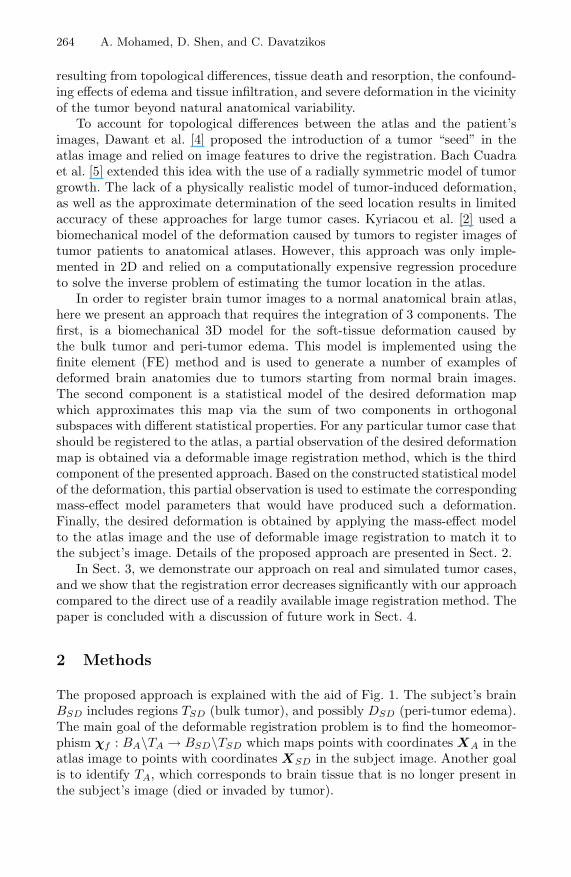
The proposed approach is explained with the aid of Fig. 1. The subject’s brainBSD includes regions TSD (bulk tumor), and possibly DSD (peri-tumor edema).The main goal of the deformable registration problem is to find the homeomor-phism χf : BA\TA → BSD\TSD which maps points with coordinates XA in theatlas image to points with coordinates XSD in the subject image. Another goalis to identify TA, which corresponds to brain tissue that is no longer present inthe subject’s image (died or invaded by tumor).
Deformable Registration of Brain Tumor Images 265
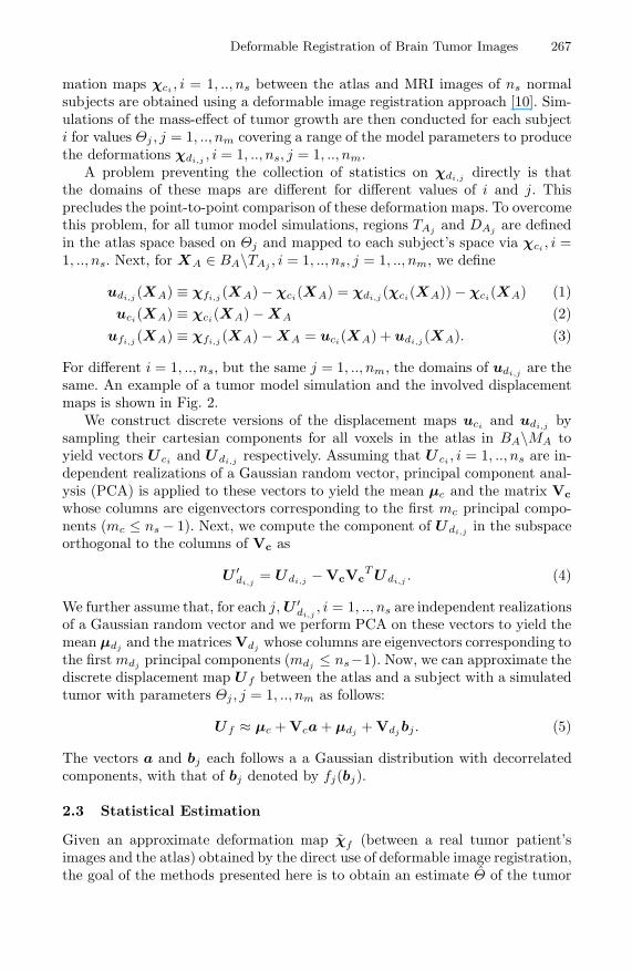
Fig. 1. Illustration of the deformation maps involved in the proposed approach. χf
is the map from the atlas to a subject’s tumor-bearing image. Regions TSD and DSD
denote the bulk tumor and edema regions in the subject’s images, and TA, DA are thecorresponding regions in the atlas. χc is the mapping from the atlas to the subject’simage before tumor mass-effect simulation (BS is not known for non-simulated cases),and χd is that obtained through the simulation of tumor mass-effect. Simulating thetumor mass-effect on the atlas results in χa and a deformed atlas image which canthen be registered to the deformed subject’s image through χb.
If an accurate model of the deformation induced by the tumor is available,it can be used to simulate this deformation in the atlas and obtain χa, followedby the application of deformable image registration to get χb, and thereforeχf = χb ◦ χa. A model of the mass-effect caused by tumor growth is describedin Sect. 2.1. Estimates of region TA as well as the other parameters affecting themodel’s behavior, such as the extent of peri-tumor edema and the mass-effect ofthe bulk tumor, are still needed in order to apply this approach. Here, we solvethis inverse estimation problem by exploiting the statistical dependency betweenχf and the mass-effect model parameters. Although an approximation of χf
obtained by the direct application of deformable image registration is incorrectin and around the tumor (region MA in Fig. 1), the pattern of this deformationoutside that region can guide the estimation of the tumor model parameters. InSect. 2.2, we explain the collection of the statistics on χf = χd ◦ χc throughtumor mass-effect simulations on images of normal subjects. Estimation of themass-effect model parameters is explained in Sect. 2.3.
2.1 Tumor Mass-Effect Model
This model is initialized with a 3D normal brain image (free of tumor) and itproduces an estimate of the deformation due to the mass-effect of a simulatedtumor. We explain the model by assuming that it is applied to the atlas image,although as explained later, the model may also be applied to other normalimages for statistical training.
266 A. Mohamed, D. Shen, and C. Davatzikos
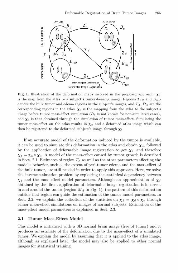
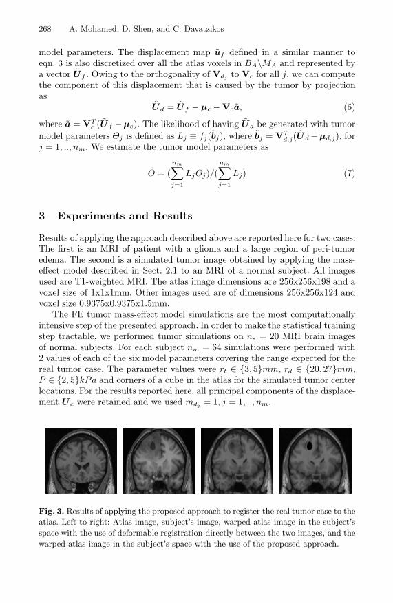
Fig. 2. Illustration of a tumor mass-effect simulation and the associated displacementmaps. Upper row (left to right): atlas image, normal subject’s MRI with an introducedsmall tumor, and resulting image after simulation of tumor mass-effect. Lower row(Left to right): displacement map uc, displacement map χd − XS , displacement mapuf , and displacement map ud.
With the assumption that the mass-effect is due to the bulk tumor andthe peri-tumor edema only, regions TA and DA are defined in the undeformed(normal) atlas image. These correspond to the bulk tumor and peri-tumor edemaregions in the deformed atlas at the end of the simulation. Although these regionsare highly variable for different tumor cases and are not known in general, here fortractability, we assume that TA and DA are spherical and concentric with centerct and radii rt and rd respectively. It is important to note that this does notrestrict our approach to dealing with spherical tumors only since final simulatedtumors need not be spherical (see Fig. 2) and also these regions are later refinedthrough the deformable image registration component of our approach.
Brain tissue swelling due to edema is restricted to white matter in DA and avolume expansion of 250% is used. Swelling is simulated by analogy to thermalexpansion. We further assume that the expansive force of the bulk tumor may beapproximated with a pressure P normal to the boundary of TA [6]. With theseassumptions, appending the necessary boundary conditions at the falx cerebriand the brain surface [7], and using the material constitutive model suggestedin [8] for brain tissues, a mechanical problem is formulated and solved usingthe FE method. More details on this tumor mass-effect model can be foundin [9]. The model parameters are collectively referred to by Θ ≡ (ct, rt, rd, P ).The values of these parameters are not known for a real tumor case, but areestimated using the statistical model of the deformation explained next.
2.2 Statistical Model Training
The goal of this step is to create a statistical model for the deformation χf thatwill aid in the estimation of Θ for a particular tumor image. First, the defor-
Deformable Registration of Brain Tumor Images 267
mation maps χci , i = 1, .., ns between the atlas and MRI images of ns normalsubjects are obtained using a deformable image registration approach [10]. Sim-ulations of the mass-effect of tumor growth are then conducted for each subjecti for values Θj , j = 1, .., nm covering a range of the model parameters to producethe deformations χdi,j , i = 1, .., ns, j = 1, .., nm.
A problem preventing the collection of statistics on χdi,j directly is thatthe domains of these maps are different for different values of i and j. Thisprecludes the point-to-point comparison of these deformation maps. To overcomethis problem, for all tumor model simulations, regions TAj and DAj are definedin the atlas space based on Θj and mapped to each subject’s space via χci , i =1, .., ns. Next, for XA ∈ BA\TAj , i = 1, .., ns, j = 1, .., nm, we define
udi,j (XA) ≡ χfi,j (XA) − χci(XA) = χdi,j (χci(XA)) − χci(XA) (1)uci(XA) ≡ χci(XA) − XA (2)
ufi,j (XA) ≡ χfi,j (XA) − XA = uci(XA) + udi,j (XA). (3)
For different i = 1, .., ns, but the same j = 1, .., nm, the domains of udi,j are thesame. An example of a tumor model simulation and the involved displacementmaps is shown in Fig. 2.
We construct discrete versions of the displacement maps uci and udi,j bysampling their cartesian components for all voxels in the atlas in BA\MA toyield vectors U ci and Udi,j respectively. Assuming that U ci , i = 1, .., ns are in-dependent realizations of a Gaussian random vector, principal component anal-ysis (PCA) is applied to these vectors to yield the mean µc and the matrix Vcwhose columns are eigenvectors corresponding to the first mc principal compo-nents (mc ≤ ns − 1). Next, we compute the component of Udi,j in the subspaceorthogonal to the columns of Vc as
U ′di,j
= Udi,j − VcVcT Udi,j . (4)
We further assume that, for each j, U ′di,j
, i = 1, .., ns are independent realizationsof a Gaussian random vector and we perform PCA on these vectors to yield themean µdj and the matrices Vdj whose columns are eigenvectors corresponding tothe first mdj principal components (mdj ≤ ns−1). Now, we can approximate thediscrete displacement map Uf between the atlas and a subject with a simulatedtumor with parameters Θj , j = 1, .., nm as follows:
Uf ≈ µc + Vca + µdj + Vdj bj . (5)
The vectors a and bj each follows a a Gaussian distribution with decorrelatedcomponents, with that of bj denoted by fj(bj).
2.3 Statistical Estimation
Given an approximate deformation map χf (between a real tumor patient’simages and the atlas) obtained by the direct use of deformable image registration,the goal of the methods presented here is to obtain an estimate Θ of the tumor
268 A. Mohamed, D. Shen, and C. Davatzikos
model parameters. The displacement map uf defined in a similar manner toeqn. 3 is also discretized over all the atlas voxels in BA\MA and represented bya vector Uf . Owing to the orthogonality of Vdj to Vc for all j, we can computethe component of this displacement that is caused by the tumor by projectionas
Ud = Uf − µc − Vca, (6)
where a = VTc (Uf − µc). The likelihood of having Ud be generated with tumor
model parameters Θj is defined as Lj ≡ fj(bj), where bj = VTd,j(U d −µd,j), for
j = 1, .., nm. We estimate the tumor model parameters as
Θ = (nm∑
j=1
LjΘj)/(nm∑
j=1
Lj) (7)
3 Experiments and Results
Results of applying the approach described above are reported here for two cases.The first is an MRI of patient with a glioma and a large region of peri-tumoredema. The second is a simulated tumor image obtained by applying the mass-effect model described in Sect. 2.1 to an MRI of a normal subject. All imagesused are T1-weighted MRI. The atlas image dimensions are 256x256x198 and avoxel size of 1x1x1mm. Other images used are of dimensions 256x256x124 andvoxel size 0.9375x0.9375x1.5mm.
The FE tumor mass-effect model simulations are the most computationallyintensive step of the presented approach. In order to make the statistical trainingstep tractable, we performed tumor simulations on ns = 20 MRI brain imagesof normal subjects. For each subject nm = 64 simulations were performed with2 values of each of the six model parameters covering the range expected for thereal tumor case. The parameter values were rt ∈ {3, 5}mm, rd ∈ {20, 27}mm,P ∈ {2, 5}kPa and corners of a cube in the atlas for the simulated tumor centerlocations. For the results reported here, all principal components of the displace-ment U c were retained and we used mdj = 1, j = 1, .., nm.
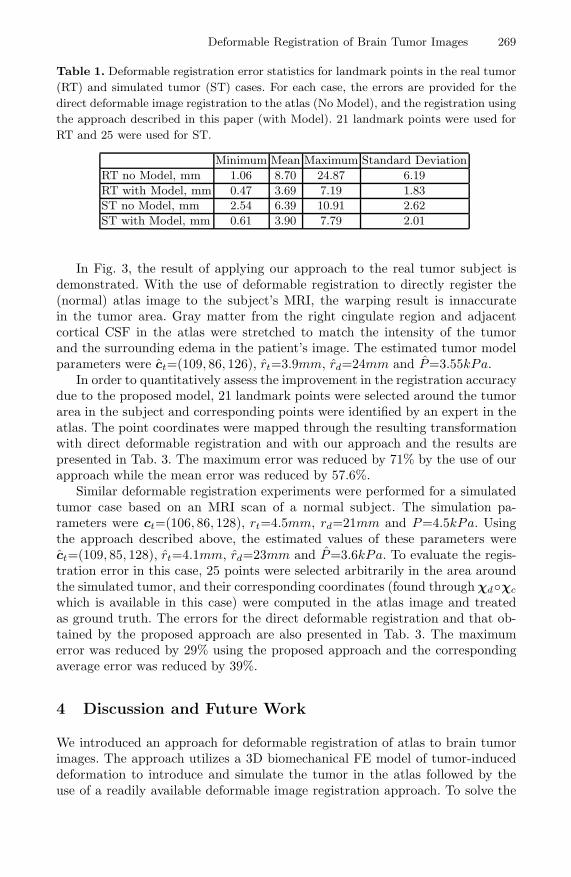
Fig. 3. Results of applying the proposed approach to register the real tumor case to theatlas. Left to right: Atlas image, subject’s image, warped atlas image in the subject’sspace with the use of deformable registration directly between the two images, and thewarped atlas image in the subject’s space with the use of the proposed approach.
Deformable Registration of Brain Tumor Images 269
Table 1. Deformable registration error statistics for landmark points in the real tumor(RT) and simulated tumor (ST) cases. For each case, the errors are provided for thedirect deformable image registration to the atlas (No Model), and the registration usingthe approach described in this paper (with Model). 21 landmark points were used forRT and 25 were used for ST.
Minimum Mean Maximum Standard DeviationRT no Model, mm 1.06 8.70 24.87 6.19RT with Model, mm 0.47 3.69 7.19 1.83ST no Model, mm 2.54 6.39 10.91 2.62ST with Model, mm 0.61 3.90 7.79 2.01
In Fig. 3, the result of applying our approach to the real tumor subject isdemonstrated. With the use of deformable registration to directly register the(normal) atlas image to the subject’s MRI, the warping result is innaccuratein the tumor area. Gray matter from the right cingulate region and adjacentcortical CSF in the atlas were stretched to match the intensity of the tumorand the surrounding edema in the patient’s image. The estimated tumor modelparameters were ct=(109, 86, 126), rt=3.9mm, rd=24mm and P=3.55kPa.
In order to quantitatively assess the improvement in the registration accuracydue to the proposed model, 21 landmark points were selected around the tumorarea in the subject and corresponding points were identified by an expert in theatlas. The point coordinates were mapped through the resulting transformationwith direct deformable registration and with our approach and the results arepresented in Tab. 3. The maximum error was reduced by 71% by the use of ourapproach while the mean error was reduced by 57.6%.
Similar deformable registration experiments were performed for a simulatedtumor case based on an MRI scan of a normal subject. The simulation pa-rameters were ct=(106, 86, 128), rt=4.5mm, rd=21mm and P=4.5kPa. Usingthe approach described above, the estimated values of these parameters werect=(109, 85, 128), rt=4.1mm, rd=23mm and P=3.6kPa. To evaluate the regis-tration error in this case, 25 points were selected arbitrarily in the area aroundthe simulated tumor, and their corresponding coordinates (found through χd◦χc
which is available in this case) were computed in the atlas image and treatedas ground truth. The errors for the direct deformable registration and that ob-tained by the proposed approach are also presented in Tab. 3. The maximumerror was reduced by 29% using the proposed approach and the correspondingaverage error was reduced by 39%.
4 Discussion and Future Work
We introduced an approach for deformable registration of atlas to brain tumorimages. The approach utilizes a 3D biomechanical FE model of tumor-induceddeformation to introduce and simulate the tumor in the atlas followed by theuse of a readily available deformable image registration approach. To solve the
270 A. Mohamed, D. Shen, and C. Davatzikos
inverse problem of determining the model parameters, we proposed a statisticalapproach that relies on the decomposition of the desired deformation map intothe sum of two maps defined on the same domain, but with different statisticalproperties that are learned via PCA from a number of training samples. Thesemaps are modeled via two orthogonal subspaces which allows the estimation ofthe tumor model parameters via projection of a rough estimate of the requireddeformation map on the subspace representing tumor induced-deformation.
The results of applying the proposed approach on a real tumor case anda simulated one indicate significant reduction in the registration error. Theseexperiments should be regarded as a proof-of-concept study. More validationexperiments are need to asses the viability of the proposed approach for a varietyof tumor cases of different grades, types and sizes. In addition, the sensitivity ofthe statistical estimator of the model parameters to the number of used principalcomponents and the number of training samples also will be investigated.
Acknowledgments
The authors would like to thank Dr. Nick Fox at the University College London,UK for providing the tumor patient’s images. This work was supported in partby the National Science Foundation under Engineering Research Center grantEEC9731478, and by the National Institutes of Health grant R01NS42645.
References
1. Davatzikos, C.: Spatial transformation and registration of brain images using elas-tically deformable models. CVIU, Spec. iss. on Medical Imaging 66 (1997) 207–222
2. Kyriacou, S.K., Davatzikos, C., Zinreich, S.J., Bryan, R.N.: Nonlinear elastic regis-tration of brain images with tumor pathology using a biomechanical model. IEEETrans. Med. Imag. 18 (1999) 580–592
3. Mohamed, A., Kyriacou, S.K., Davatzikos, C.: A statistical approach for estimatingbrain tumor induced deformation. Proc. of IEEE Workshop on MMBIA (2001) 52–59
4. Dawant, B.M., Hartmann, S.L., Pan, S., Gadamsetty, S.: Brain atlas deformation inthe presence of small and large space-occupying tumors. Computer Aided Surgery7 (2002) 1–10
5. Cuadra, M.B., Pollo, C., Bardera, A., Cuisenaire, O., Villemure, J.G., Thiran,J.P.: Atlas-based segmentation of pathological MR brains using a model of lesiongrowth. IEEE Trans. Med. Imag. 23 (2004) 1301–1314
6. Wasserman, R., Acharya, R.: A patient-specific in vivo tumor model. MathematicalBiosciences 136 (1996) 111–140
7. Miga, M., Paulsen, K., Kennedy, F.E., Hartov, A., Roberts, D.: Model-updatedimage-guided neurosurgery using the finite element method: Incorporation of thefalx cerebri. In: MICCAI 1999. (1999)
8. Miller, K., Chinzei, K.: Mechanical properties of brain tissue in tension. Journalof Biomechanics 35 (2002) 483–490
9. Mohamed, A., Davatzikos, C.: Finite element modeling of brain tumor mass-effectfrom 3D medical images. In: Proceedings of MICCAI 2005. (2005)
10. Shen, D., Davatzikos, C.: HAMMER: Hierarchical attribute matching mechanismfor elastic registration. IEEE Trans. Med. Imag. 21 (2002) 1421–1439
Recommended


