Determinants for the efficiency of anticancer drugstargeting either Aurora-A or Aurora-B kinases inhuman colon carcinoma cells
Phillip Kaestner, Ailine Stolz, and Holger Bastians
Institute for Molecular Biology and Tumor Research, PhilippsUniversity Marburg, Marburg, Germany
AbstractThemitotic Aurora kinases, including Aurora-A and Aurora-B, are attractive novel targets for anticancer therapy,and inhibitory drugs have been developed that are current-ly undergoing clinical trials. However, the molecular me-chanisms how these drugs induce tumor cell death arepoorly understood. We have addressed this question bycomparing the requirements for an efficient induction ofapoptosis in response to MLN8054, a selective inhibitorof Aurora-A, and the selective Aurora-B inhibitor ZM44‐7439 in human colon carcinoma cells. By using variousisogenic knockout as well as inducible colon carcinomacell lines, we found that treatment with MLN8054 in-duces defects in mitotic spindle assembly, which causesa transient spindle checkpoint–dependent mitotic arrest.This cell cycle arrest is not maintained due to the activityof MLN8054 to override the spindle checkpoint. Subse-quently, MLN8054-treated cells exit from mitosis and ac-tivate a p53-dependent postmitotic G1 checkpoint, whichsubsequently induces p21 and Bax, leading to G1 arrestfollowed by the induction of apoptosis. In contrast, inhibi-tion of Aurora-B by ZM447439 also interferes with normalchromosome alignment during mitosis and overrides themitotic spindle checkpoint but allows a subsequent endo-reduplication, although ZM447439 potently activates thep53-dependent postmitotic G1 checkpoint. Moreover, theZM447439-induced endoreduplication is a prerequisite forthe efficiency of the drug. Thus, our results obtained in hu-man colon carcinoma cells indicate that although both Au-rora kinase inhibitors are potent inducers of tumor celldeath, the pathways leading to the induction of apoptosis
in response to these drugs are distinct. [Mol Cancer Ther2009;8(7):2046–56]
IntroductionTargeting the progression of mitosis is a highly successfulstrategy for anticancer treatment (1). Recently, much atten-tion has been drawn to the Aurora kinases, which comprisethree family members, Aurora-A, Aurora-B, and Aurora-C,as novel mitotic drug targets. At least Aurora-A and Auro-ra-B function as key regulators of mitosis and they are fre-quently overexpressed in human cancer (2–5), whichprovides the basis for their importance as chemotherapeuticdrug targets.Aurora-A is localized on duplicated centrosomes and
spindle poles during mitosis and is required for the timelyentry into mitosis and proper formation of a bipolar mitoticspindle by regulating centrosome maturation, separation,and microtubule nucleation activity (6). In contrast, Auro-ra-B is a chromosomal passenger protein, which is, togetherwith INCENP, borealin, and survivin, part of the chromo-somal passenger complex (7). This complex changes its local-ization during mitotic progression from centromeres in earlymitosis to the spindle midzone in anaphase and finally to thecleavage furrow and the midbody during cytokinesis. Ac-cording to the different localizations, Aurora-B is requiredfor phosphorylation of histone H3 (8), the proper biorienta-tion and alignment of chromosomes by correcting faulty mi-crotubule-kinetochore attachments (9, 10), and the executionof cytokinesis (11). It has also been suggested that Aurora-Bmight contribute to spindle checkpoint function, whichmonitors proper chromosome alignment during mitosis (9,12), although it is not clear whether this role is related toits function to resolve incorrect kinetochore attachments.The third member of the Aurora kinase family, Aurora-C,might have functions during meiosis rather than during mi-tosis (13) and it seems not to be aberrantly expressed in hu-man cancer (2).Extensive efforts have been made to develop inhibitors
for Aurora-A and Aurora-B, and several highly potent in-hibitory compounds have been identified. Examples are He-speradin (10), ZM447439 (9), and AZD1152 (14), whichshow selectivity for Aurora-B in vivo and VX-680 (MK-0547), which inhibits both Aurora-A and Aurora-B (15,16). Most recently, the first Aurora-A selective inhibitor,MLN8054, has been introduced (17). It has been shown thattreatment of cells with Hesperadin, ZM447439, AZD1152, orVX-680 inhibits the phosphorylation of histone H3 and cy-tokinesis and induces polyploidization (9, 10, 14), indicatingthat these drugs inhibit Aurora-B in vivo. In contrast, VX-680and MLN8054 treatment abolishes the activation-specific
Received 2/5/09; revised 4/7/09; accepted 4/23/09;published OnlineFirst 7/7/09.
Grant support: Deutsche Forschungsgemeinschaft andvon Behring-Röntgen Stiftung.
The costs of publication of this article were defrayed in part by thepayment of page charges. This article must therefore be hereby markedadvertisement in accordance with 18 U.S.C. Section 1734 solely toindicate this fact.
Requests for reprints: Holger Bastians, Institute for Molecular Biology andTumor Research, Philipps University Marburg, Emil-Mannkopff-Strasse 2,D-35037 Marburg, Germany. Phone: 49-6421-2863113;Fax: 49-6421-2865932. E-mail: [email protected]
Copyright © 2009 American Association for Cancer Research.
doi:10.1158/1535-7163.MCT-09-0323
Mol Cancer Ther 2009;8(7). July 2009
2046

autophosphorylation of Aurora-A on Thr288 (16, 17). At thesame time, MLN8054 treatment leaves the histone H3 phos-phorylation intact (17). For some inhibitors (e.g., AZD1152,VX-680, and MLN8054), an efficacy in vivo on human tumorxenografts has been shown and several compounds are cur-rently investigated in clinical trials (1).Thus far, little is known about how Aurora inhibition
causes tumor cell death. In fact, a detailed knowledge ofthe molecular pathways involved in the induction of apo-ptosis in response to inhibition of either Aurora-A or Auro-ra-B is essential to improve therapeutic strategies, tocircumvent drug resistance, and to answer the long-stand-ing question about what the better target is, Aurora-A orAurora-B. Here, we used the pharmacologic inhibitorsMLN8054 and ZM447439 that selectively target the Auro-ra-A or Aurora-B kinases, respectively. To define the molec-ular pathways that are activated and required for anefficient induction of apoptosis after treatment with thesedrugs, we took advantage of the well-established and ge-netically defined HCT116 human colon carcinoma cell sys-tem and a set of different isogenic knockout derivativesthereof.
Materials and MethodsCell Culture
HCT116, HCT-p53−/−, HCT-p21−/− (18), HCT-BAX−/− (19),and HCT-MAD2+/− (20) cell lines were grown in RPMI 1640,10% FCS, 1% glutamine, 100 μg/mL streptomycin, and 100units/mL penicillin (Invitrogen). RKO-p21 and RKO-p27cells (21) were maintained in DMEM plus 10% FCS,500 μg/mL G418, and 200 μg/mL zeocin (Invitrogen). p21and p27 expression was induced by addition of 10 μmol/Lponasteron-A (Sigma). RKO cells were synchronized atG1-S by treatment with 1 μg/mL aphidicolin (Alexis) for24 h followed by release into medium.Drug Treatments
Cells were treated with 0.1 to 2.0 μmol/L of ZM447439(Biomol), 0.1 to 2.0 μmol/L of MLN8054 (a kind gift fromMillennium Pharmaceuticals, Inc.), 68 μmol/L monastrol(Biomol), 300 nmol/L nocodazole (Sigma), 100 nmol/LTaxol (Sigma), and 20 μmol/L MG132 (Calbiochem).Flow Cytometry
Harvested cells were fixed in 70% (v/v) ethanol at 4°C for16 h. The DNA content was determined by staining cellswith 50 μg/mL propidium iodide (Sigma). The mitotic in-dex was determined by doing intracellular staining with an-ti-MPM2 antibodies (Upstate) as described before (22).Apoptotic cells were identified as cells with a sub-G1DNA content. Cells were analyzed on a FACSCalibur (Bec-ton Dickinson) and data analysis was carried out using theCellQuest Pro software.Western Blotting
Cell lysates were prepared in lysis buffer [50 mmol/L Tris(pH 7.4), 150 mmol/L NaCl, 5 mmol/L EDTA, 5 mmol/LEGTA, 1% (v/v) NP40, 0.1% (w/v) sodium deoxycholate,0.1% (w/v) SDS, 20 mmol/L sodium orthovanadate,0.5 μmol/L microcystin, complete protease inhibitors
(Roche)]. SDS-PAGE and semidry Western blotting and de-tection were done using standard protocols. The followingantibodies were used for Western blotting: anti–Aurora-A(Santa Cruz Biotechnology), anti–Aurora-A-pT288 (CellSignaling), anti–Aurora-B (BD Transduction Laboratories),anti–Aurora-B-pT232 (Cell Signaling), anti-actin (Sigma),anti–cyclin B (Santa Cruz Biotechnology), anti-securin(Lab Vision), anti-Bub1 (a gift from Stephen Taylor, Univer-sity of Manchester, Manchester, UK), anti-BubR1 (Chemi-con), anti-Bub3 (BD Transduction Laboratories), anti-p53(Oncogene), anti-p21 (Oncogene), anti-p27 (Santa Cruz Bio-technology), and anti-Bax (Cell Signaling). Secondary anti-bodies conjugated to horseradish peroxidase were fromJackson. The enhanced chemiluminescence system was usedfor detection.Microscopy
Cells were grown on poly-lysine–coated coverslips andfixed in 2% paraformaldehyde for 5 min followed by per-meabilization in methanol at −20°C. Primary antibodies[α-tubulin (Sigma), CREST (Europa Bioproducts), phos-pho-histone H3 (Ser10; Cell Signaling), Bub1, and BubR1]were incubated for 2 h at room temperature followed by a2-h incubation with Alexa Fluor–conjugated secondary anti-bodies (Molecular Probes). DNA was stained with 4′,6-dia-midino-2-phenylindole and microscopy was carried outusing a Leica DM6000B microscope. Z-optical stacks witha spacing of 0.2 μm were recorded, and deconvolution ofimages and quantitation of fluorescence intensities were car-ried out using the Leica LAS-AF software.
ResultsMLN8054 Is a Specific Inhibitor for Aurora-A and
ZM447439 Inhibits Aurora-B Selectively in Human
Colon Carcinoma Cells
To determine the specific requirements for the efficacyof Aurora kinase inhibitors in human colon carcinomacells, we selected MLN8054 and ZM447439. MLN8054represents a compound that displays a high selectivityfor Aurora-A over Aurora-B (IC50: 4 and 172 nmol/L, re-spectively; ref. 17), whereas ZM447439 shows a clearselectivity for Aurora-B over Aurora-A (IC50: 50 and1,000 nmol/L, respectively; ref. 23). The selectivity of thesedrugs in our experimental system based on HCT116 cellswas verified by analyzing the autophosphorylation statuswithin the activation loops of Aurora-A and Aurora-B onWestern blots (Fig. 1A), by evaluating the Aurora-B–medi-ated phosphorylation of histone H3 on Ser10 in mitoticcells in immunofluorescence studies (SupplementaryFig. S1A),1 and by investigating the inhibition of cytokine-sis that is dependent on Aurora-B by determining theDNA content in fluorescence-activated cell sorting (FACS)analyses (Supplementary Fig. S1B).1 All these cellular as-says confirmed the selectivity of the drugs for Aurora-Aor Aurora-B when used at 0.5 and 2 μmol/L, respectively.
1 Supplementary material for this article is available at Molecular CancerTherapeutics Online (http://mct.aacrjournals.org/).
Molecular Cancer Therapeutics
Mol Cancer Ther 2009;8(7). July 2009
2047
In addition, we analyzed the formation of the mitoticspindle after releasing cells from a monastrol block in thepresence or absence of the drugs (Supplementary Fig. S1C).1
In agreement with previous results, inhibition of Aurora-Ainhibits bipolar mitotic spindle assembly, whereas inhibitionof Aurora-B prevents the correct microtubule-kinetochoreattachment causing severe chromosome alignment defects(9, 10, 17, 23). Together, these results verify that MLN8054and ZM447439 are indeed well suited to discriminate be-tween inhibition of Aurora-A and Aurora-B in our experi-mental cell system.Inhibition of Aurora-A, but not of Aurora-B, Causes a
Mitotic Arrest That Cannot Be Maintained
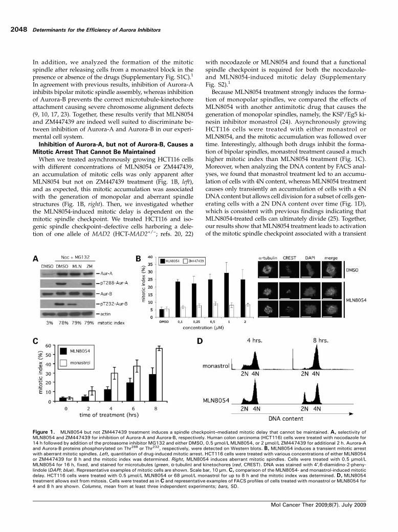
When we treated asynchronously growing HCT116 cellswith different concentrations of MLN8054 or ZM447439,an accumulation of mitotic cells was only apparent afterMLN8054 but not on ZM447439 treatment (Fig. 1B, left),and as expected, this mitotic accumulation was associatedwith the generation of monopolar and aberrant spindlestructures (Fig. 1B, right). Then, we investigated whetherthe MLN8054-induced mitotic delay is dependent on themitotic spindle checkpoint. We treated HCT116 and iso-genic spindle checkpoint–defective cells harboring a dele-tion of one allele of MAD2 (HCT-MAD2+/−; refs. 20, 22)
with nocodazole or MLN8054 and found that a functionalspindle checkpoint is required for both the nocodazole-and MLN8054-induced mitotic delay (SupplementaryFig. S2).1
Because MLN8054 treatment strongly induces the forma-tion of monopolar spindles, we compared the effects ofMLN8054 with another antimitotic drug that causes thegeneration of monopolar spindles, namely, the KSP/Eg5 ki-nesin inhibitor monastrol (24). Asynchronously growingHCT116 cells were treated with either monastrol orMLN8054, and the mitotic accumulation was followed overtime. Interestingly, although both drugs inhibit the forma-tion of bipolar spindles, monastrol treatment caused a muchhigher mitotic index than MLN8054 treatment (Fig. 1C).Moreover, when analyzing the DNA content by FACS anal-yses, we found that monastrol treatment led to an accumu-lation of cells with 4N content, whereas MLN8054 treatmentcauses only transiently an accumulation of cells with a 4NDNA content but allows cell division for a subset of cells gen-erating cells with a 2N DNA content over time (Fig. 1D),which is consistent with previous findings indicating thatMLN8054-treated cells can ultimately divide (25). Together,our results show that MLN8054 treatment leads to activationof the mitotic spindle checkpoint associated with a transient
Figure 1. MLN8054 but not ZM447439 treatment induces a spindle checkpoint–mediated mitotic delay that cannot be maintained. A, selectivity ofMLN8054 and ZM447439 for inhibition of Aurora-A and Aurora-B, respectively. Human colon carcinoma (HCT116) cells were treated with nocodazole for14 h followed by addition of the proteasome inhibitor MG132 and either DMSO, 0.5 μmol/L MLN8054, or 2 μmol/L ZM447439 for additional 2 h. Aurora-Aand Aurora-B proteins phosphorylated on Thr288 or Thr232, respectively, were detected on Western blots. B, MLN8054 induces a transient mitotic arrestwith aberrant mitotic spindles. Left, quantitation of drug-induced mitotic arrest. HCT116 cells were treated with various concentrations of either MLN8054or ZM447439 for 8 h and the mitotic index was determined. Right, MLN8054 induces aberrant mitotic spindles. Cells were treated with 0.5 μmol/LMLN8054 for 16 h, fixed, and stained for microtubules (green, α-tubulin) and kinetochores (red, CREST). DNA was stained with 4′,6-diamidino-2-pheny-lindole (DAPI; blue). Representative examples of mitotic cells are shown. Scale bar, 10 μm.C, comparison of the MLN8054- and monastrol-induced mitoticdelay. HCT116 cells were treated with 0.5 μmol/L MLN8054 or 68 μmol/L monastrol for up to 8 h and the mitotic index was determined. D, MLN8054treatment allows exit from mitosis. Cells were treated as in C and representative examples of FACS profiles of cells treated with monastrol or MLN8054 for4 and 8 h are shown. Columns, mean from at least three independent experiments; bars, SD.
Determinants for the Efficiency of Aurora Inhibitors
Mol Cancer Ther 2009;8(7). July 2009
2048
mitotic arrest, which cannot be maintained over time, allow-ing an unscheduled exit from mitosis and the generation ofcells with a 2N DNA content.Both MLN8054 and ZM447439 Override a Spindle
Checkpoint–Mediated Mitotic Arrest
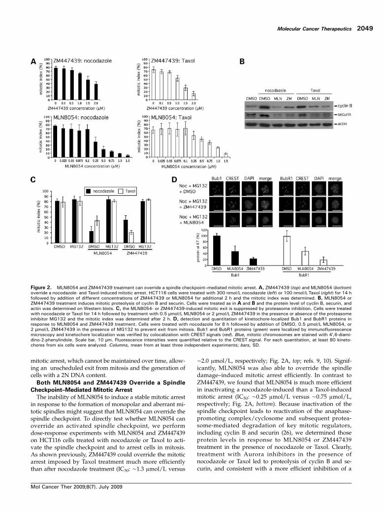
The inability of MLN8054 to induce a stable mitotic arrestin response to the formation of monopolar and aberrant mi-totic spindles might suggest that MLN8054 can override thespindle checkpoint. To directly test whether MLN8054 canoverride an activated spindle checkpoint, we performdose-response experiments with MLN8054 and ZM447439on HCT116 cells treated with nocodazole or Taxol to acti-vate the spindle checkpoint and to arrest cells in mitosis.As shown previously, ZM447439 could override the mitoticarrest imposed by Taxol treatment much more efficientlythan after nocodazole treatment (IC50: ∼1.3 μmol/L versus
∼2.0 μmol/L, respectively; Fig. 2A, top; refs. 9, 10). Signif-icantly, MLN8054 was also able to override the spindledamage–induced mitotic arrest efficiently. In contrast toZM447439, we found that MLN8054 is much more efficientin inactivating a nocodazole-induced than a Taxol-inducedmitotic arrest (IC50: ∼0.25 μmol/L versus ∼0.75 μmol/L,respectively; Fig. 2A, bottom). Because inactivation of thespindle checkpoint leads to reactivation of the anaphase-promoting complex/cyclosome and subsequent protea-some-mediated degradation of key mitotic regulators,including cyclin B and securin (26), we determined thoseprotein levels in response to MLN8054 or ZM447439treatment in the presence of nocodazole or Taxol. Clearly,treatment with Aurora inhibitors in the presence ofnocodazole or Taxol led to proteolysis of cyclin B and se-curin, and consistent with a more efficient inhibition of a
Figure 2. MLN8054 and ZM447439 treatment can override a spindle checkpoint–mediated mitotic arrest. A, ZM447439 (top) and MLN8054 (bottom)override a nocodazole- and Taxol-induced mitotic arrest. HCT116 cells were treated with 300 nmol/L nocodazole (left) or 100 nmol/L Taxol (right) for 14 hfollowed by addition of different concentrations of ZM447439 or MLN8054 for additional 2 h and the mitotic index was determined. B, MLN8054 orZM447439 treatment induces mitotic proteolysis of cyclin B and securin. Cells were treated as in A and B and the protein level of cyclin B, securin, andactin was determined on Western blots. C, the MLN8054- or ZM447439-induced mitotic exit is suppressed by proteasome inhibition. Cells were treatedwith nocodazole or Taxol for 14 h followed by treatment with 0.5 μmol/L MLN8054 or 2 μmol/L ZM447439 in the presence or absence of the proteasomeinhibitor MG132 and the mitotic index was determined after 2 h. D, detection and quantitation of kinetochore-localized Bub1 and BubR1 proteins inresponse to MLN8054 and ZM447439 treatment. Cells were treated with nocodazole for 8 h followed by addition of DMSO, 0.5 μmol/L MLN8054, or2 μmol/L ZM447439 in the presence of MG132 to prevent exit from mitosis. Bub1 and BubR1 proteins (green) were localized by immunofluorescencemicroscopy and kinetochore localization was verified by colocalization with CREST signals (red). Blue, mitotic chromosomes are stained with 4′,6-diami-dino-2-phenylindole. Scale bar, 10 μm. Fluorescence intensities were quantified relative to the CREST signal. For each quantitation, at least 80 kineto-chores from six cells were analyzed. Columns, mean from at least three independent experiments; bars, SD.
Molecular Cancer Therapeutics
Mol Cancer Ther 2009;8(7). July 2009
2049
nocodazole-induced mitotic arrest, MLN8054-treated cellsdisplayed a lower level of cyclin B in the presence of noco-dazole than Taxol (Fig. 2B). Moreover, the Aurora inhibi-tor–mediated escape from the mitotic arrest wassuppressed by cotreatment with the proteasome inhibitorMG132 (Fig. 2C). This indicates that inhibition of Aurora-A or Aurora-B relieves the inhibition of the proteasome-dependent mitotic proteolysis machinery and supports
our finding that Aurora kinase inhibitors can over‐ridethe spindle checkpoint.Activation of the spindle checkpoint is associated with a
recruitment of spindle checkpoint proteins to kinetochoresthat are not attached to microtubules or that are not undertension (26). We investigated the kinetochore localization ofthe spindle checkpoint proteins Bub1 and BubR1 after treat-ment of mitotic cells with MLN8054 or ZM447439 and
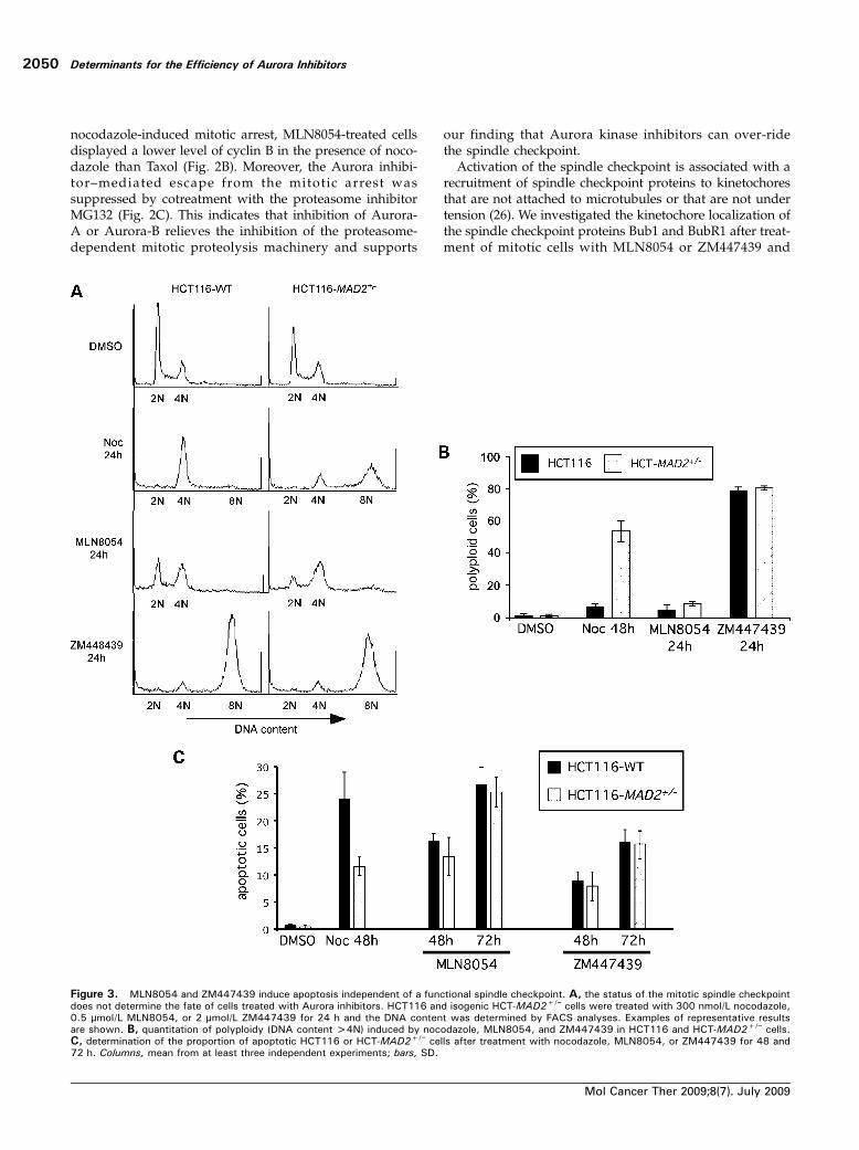
Figure 3. MLN8054 and ZM447439 induce apoptosis independent of a functional spindle checkpoint. A, the status of the mitotic spindle checkpointdoes not determine the fate of cells treated with Aurora inhibitors. HCT116 and isogenic HCT-MAD2+/− cells were treated with 300 nmol/L nocodazole,0.5 μmol/L MLN8054, or 2 μmol/L ZM447439 for 24 h and the DNA content was determined by FACS analyses. Examples of representative resultsare shown. B, quantitation of polyploidy (DNA content >4N) induced by nocodazole, MLN8054, and ZM447439 in HCT116 and HCT-MAD2+/− cells.C, determination of the proportion of apoptotic HCT116 or HCT-MAD2+/− cells after treatment with nocodazole, MLN8054, or ZM447439 for 48 and72 h. Columns, mean from at least three independent experiments; bars, SD.
Determinants for the Efficiency of Aurora Inhibitors
Mol Cancer Ther 2009;8(7). July 2009
2050
found that treatment with both Aurora kinase inhibitors re-duced the kinetochore localization of both spindle check-point proteins. Quantitation of the amount of Bub1 andBubR1 at kinetochores relative to a kinetochore marker(CREST) revealed a ∼80% reduction of Bub1 and BubR1in response to ZM447439 treatment and a ∼60% reductionafter MLN8054 treatment (Fig. 2D), whereas the overalllevel of the spindle checkpoint proteins was not impaired(Supplementary Fig. S3).1 Thus, the reduced amount ofspindle checkpoint proteins at kinetochores might explainthe inhibition of the spindle checkpoint on treatment witheither MLN8054 or ZM447439. The exact mechanism howthe Aurora inhibitors can override the spindle checkpointleading to loss of checkpoint proteins at kinetochores is cur-rently not known and deserves further detailed analyses.The Induction of Apoptosis on Aurora-A or Aurora-B
Inhibition Does Not Require a Functional Spindle
Checkpoint
Recent evidence has indicated that the induction of ap-optosis in response to various antimitotic drug treatmentsis dependent on a functional spindle checkpoint (22, 27–
30). Therefore, we investigated whether the efficiency ofMLN8054 or ZM447439 relies on a functional spindlecheckpoint. HCT116 and isogenic spindle checkpoint–com-promised HCT-MAD2+/− cells were treated with nocoda-zole, MLN8054, or ZM447439 for up to 72 hours, andcell cycle profiles and the induction of cell death were de-termined. As shown previously and consistent with an im-paired function of the spindle checkpoint, MAD2+/− cellsexhibited a reduced mitotic arrest (SupplementaryFig. S2; ref. 20), severe endoreduplication (Fig. 3A and B;ref. 31), and reduced apoptosis (Fig. 3C; ref. 22) after pro-longed treatment with nocodazole. Interestingly, HCT-MAD2+/− cells treated with MLN8054 showed a reducedmitotic arrest (Supplementary Fig. S2) but no endoredupli-cation (Fig. 3A and B) and no alteration in the rate of ap-optosis (Fig. 3C). However, treatment with ZM447439induced polyploidization in wild-type as well as in spindlecheckpoint–impaired cells (Fig. 3A and B) and the induc-tion of apoptosis was not dependent on a functional spin-dle checkpoint (Fig. 3C). Thus, a functional spindlecheckpoint is not essential for the induction of cell death
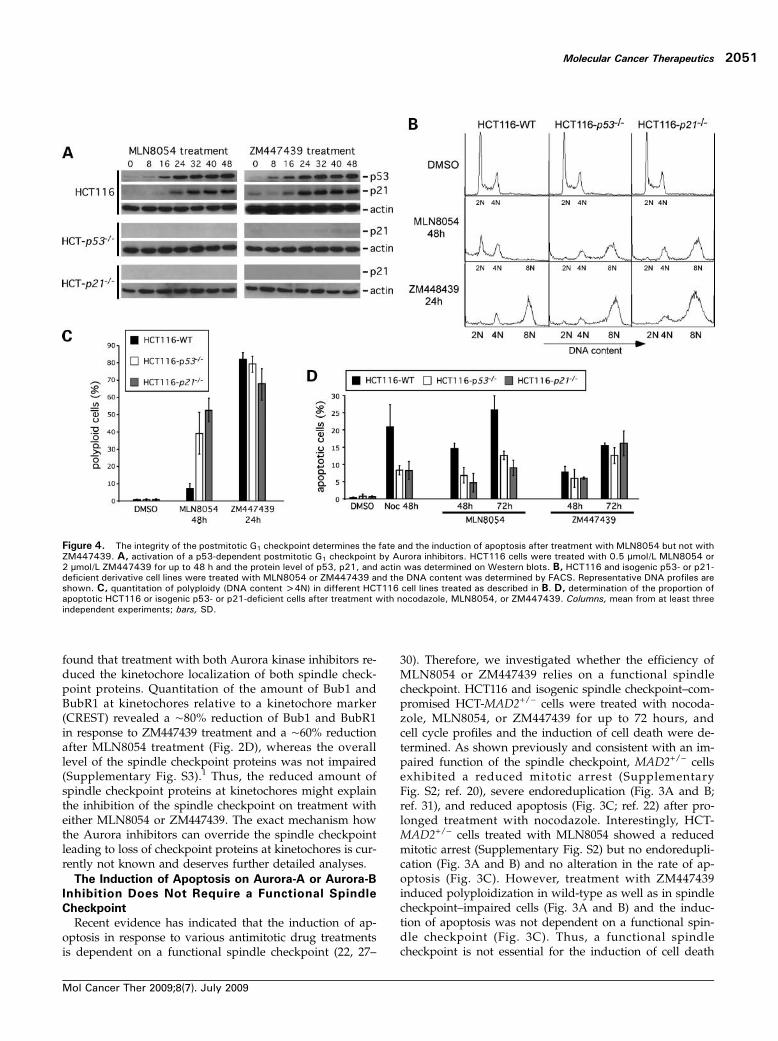
Figure 4. The integrity of the postmitotic G1 checkpoint determines the fate and the induction of apoptosis after treatment with MLN8054 but not withZM447439. A, activation of a p53-dependent postmitotic G1 checkpoint by Aurora inhibitors. HCT116 cells were treated with 0.5 μmol/L MLN8054 or2 μmol/L ZM447439 for up to 48 h and the protein level of p53, p21, and actin was determined on Western blots. B, HCT116 and isogenic p53- or p21-deficient derivative cell lines were treated with MLN8054 or ZM447439 and the DNA content was determined by FACS. Representative DNA profiles areshown. C, quantitation of polyploidy (DNA content >4N) in different HCT116 cell lines treated as described in B. D, determination of the proportion ofapoptotic HCT116 or isogenic p53- or p21-deficient cells after treatment with nocodazole, MLN8054, or ZM447439. Columns, mean from at least threeindependent experiments; bars, SD.
Molecular Cancer Therapeutics
Mol Cancer Ther 2009;8(7). July 2009
2051
on treatment with the Aurora kinase inhibitors MLN8054and ZM447439 in human colon carcinoma cells.A p53-Dependent Postmitotic Checkpoint Is Required
for the Induction of Apoptosis after MLN8054 but not
after ZM447439 Treatment
On prolonged treatment with spindle poisons, cells canescape from mitosis and activate a postmitotic G1 check-point, which arrests cells in a p53-dependent manner be-fore S phase. This checkpoint is thought to act as asecond fail-safe mechanism for cells that exited aberrantlyfrom mitosis (31, 32). Because treatment with Aurora ki-nase inhibitors causes an aberrant progression and exitfrom mitosis, we investigated if this is associated withthe activation of the postmitotic G1 checkpoint. Indeed,treatment with MLN8054 or ZM447439 strongly activatedp53 and induced p21 in a p53-dependent manner in ourHCT116 cell system (Fig. 4A). Interestingly, the activationof the postmitotic checkpoint caused a G1 arrest onMLN8054 but not after ZM447439 treatment (Fig. 4B andC). Moreover, p53 and p21 are required to maintain a post-mitotic G1 checkpoint triggered by MLN8054 but not afterZM447439 treatment. Isogenic HCT116 cells deficient forp53 or p21 (HCT-p53−/− and HCT-p21−/−; ref. 18) exhibitedsevere endoreduplication after treatment with MLN8054,whereas ZM447439-mediated polyploidization was notsignificantly affected in the different knockout cell lines(Fig. 4B and C). Thus, both MLN8054 and ZM447439 acti-
vate a p53-dependent postmitotic G1 checkpoint, but itsgenetic inactivation has only an effect on the fate ofMLN8054-treated HCT116 cells.To examine whether the postmitotic G1 checkpoint is
important for the induction of cell death after Aurora in-hibition, we treated HCT116 and isogenic TP53- and p21-deficient cells with nocodazole, MLN8054, or ZM447439and determined the proportion of apoptotic cells. Signifi-cantly, loss of either p53 or p21 protected cells from apo-ptosis after treatment with MLN8054 or nocodazole,whereas ZM447439-induced apoptosis was not altered(Fig. 4D). Thus, the p53-dependent postmitotic G1 check-point is required for MLN8054-induced but notZM447439-induced cell death, although both drugsstrongly activate the postmitotic p53 response in humancolon carcinoma cells.Endoreduplication Is an Important Trigger for the
Induction of Apoptosis after Aurora-B Inhibition
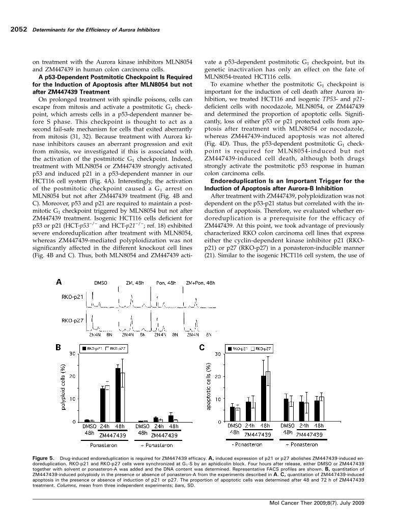
After treatment with ZM447439, polyploidization was notdependent on the p53-p21 status but correlated with the in-duction of apoptosis. Therefore, we evaluated whether en-doreduplication is a prerequisite for the efficacy ofZM447439. At this point, we took advantage of previouslycharacterized RKO colon carcinoma cell lines that expresseither the cyclin-dependent kinase inhibitor p21 (RKO-p21) or p27 (RKO-p27) in a ponasteron-inducible manner(21). Similar to the isogenic HCT116 cell system, the use of
Figure 5. Drug-induced endoreduplication is required for ZM447439 efficacy. A, induced expression of p21 or p27 abolishes ZM447439-induced en-doreduplication. RKO-p21 and RKO-p27 cells were synchronized at G1-S by an aphidicolin block. Four hours after release, either DMSO or ZM447439together with solvent or ponasteron-A was added and the DNA content was determined. Representative FACS profiles are shown. B, quantitation ofZM447439-induced polyploidy in the presence or absence of ponasteron-A from the experiments described in A. C, quantitation of ZM447439-inducedapoptosis in the presence or absence of induction of p21 or p27. The proportion of apoptotic cells was determined after 48 and 72 h of ZM447439treatment. Columns, mean from three independent experiments; bars, SD.
Determinants for the Efficiency of Aurora Inhibitors
Mol Cancer Ther 2009;8(7). July 2009
2052
these inducible RKO colon carcinoma cells allows a definedphenotypic analysis in an isogenic genetic background. Asexpected, after addition of ponasteron-A, expression of p21or p27 was induced and caused a cell cycle arrest in G1(Supplementary Fig. S4).1 Both RKO cell lines were sensitivetoward ZM447439 treatment and exhibited endoreduplica-tion in response to ZM447439 treatment (SupplementaryFig. S5).1 To test whether induced expression of either p21or p27 would suppress the endoreduplication induced byZM447439 treatment, cells were synchronized at G1-S (Sup-plementary Fig. S6).1 On release from the block, cells weretreated with ZM447439 and ponasteron to induce the ex-pression of p21 or p27 and endoreduplication and the in-duction of apoptosis were analyzed after progressionthrough mitosis. Significantly, the induced expression ofp21 or p27 in the presence of ZM447439 led to an accu-mulation of tetraploid cells and endoreduplication wassuppressed (Fig. 5A and B). Importantly, the forced postmi-totic G1 arrest also protected RKO cells from ZM447439-induced apoptosis (Fig. 5C). Thus, the endoreduplicationinduced by ZM447439-mediated Aurora-B inhibition is animportant trigger for the efficacy of the drug in these coloncarcinoma cells.
Bax Is Required for the Induction of Apoptosis after
MLN8054 or ZM447439 Treatment
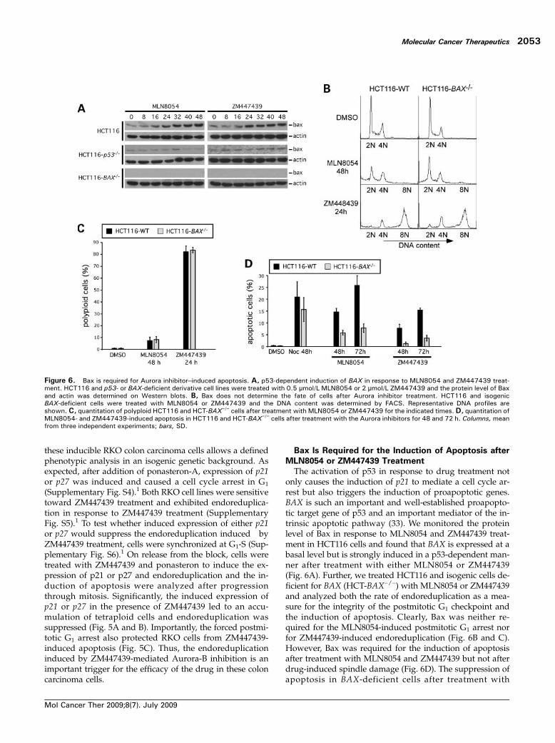
The activation of p53 in response to drug treatment notonly causes the induction of p21 to mediate a cell cycle ar-rest but also triggers the induction of proapoptotic genes.BAX is such an important and well-established proapopto-tic target gene of p53 and an important mediator of the in-trinsic apoptotic pathway (33). We monitored the proteinlevel of Bax in response to MLN8054 and ZM447439 treat-ment in HCT116 cells and found that BAX is expressed at abasal level but is strongly induced in a p53-dependent man-ner after treatment with either MLN8054 or ZM447439(Fig. 6A). Further, we treated HCT116 and isogenic cells de-ficient for BAX (HCT-BAX−/−) with MLN8054 or ZM447439and analyzed both the rate of endoreduplication as a mea-sure for the integrity of the postmitotic G1 checkpoint andthe induction of apoptosis. Clearly, Bax was neither re-quired for the MLN8054-induced postmitotic G1 arrest norfor ZM447439-induced endoreduplication (Fig. 6B and C).However, Bax was required for the induction of apoptosisafter treatment with MLN8054 and ZM447439 but not afterdrug-induced spindle damage (Fig. 6D). The suppression ofapoptosis in BAX-deficient cells after treatment with
Figure 6. Bax is required for Aurora inhibitor–induced apoptosis. A, p53-dependent induction of BAX in response to MLN8054 and ZM447439 treat-ment. HCT116 and p53- or BAX-deficient derivative cell lines were treated with 0.5 μmol/L MLN8054 or 2 μmol/L ZM447439 and the protein level of Baxand actin was determined on Western blots. B, Bax does not determine the fate of cells after Aurora inhibitor treatment. HCT116 and isogenicBAX-deficient cells were treated with MLN8054 or ZM447439 and the DNA content was determined by FACS. Representative DNA profiles areshown. C, quantitation of polyploid HCT116 and HCT-BAX−/− cells after treatment with MLN8054 or ZM447439 for the indicated times.D, quantitation ofMLN8054- and ZM447439-induced apoptosis in HCT116 and HCT-BAX−/− cells after treatment with the Aurora inhibitors for 48 and 72 h. Columns,meanfrom three independent experiments; bars, SD.
Molecular Cancer Therapeutics
Mol Cancer Ther 2009;8(7). July 2009
2053
MLN8054 was associated with an enhanced survival of G1-arrested cells (Fig. 6B), further supporting our result that thepostmitotic G1 arrest is required for the induction of apopto-sis after MLN8054 treatment (Fig. 4).
DiscussionAurora inhibitors target the mitotic functions of the Aurorakinases and induce apoptosis in cultured tumor cells as wellas in human xenografts (2, 9, 10, 14, 15, 34–36). However,the molecular mechanisms leading to the induction of tu-mor cell death are poorly understood, although a detailedknowledge about these mechanisms is most important toimprove therapeutic strategies and drug combinations andto explain resistance on a molecular level.By the use of various isogenic somatic knockout as well as
inducible cell lines, we investigated the requirements ofapoptosis after treatment with selective Aurora-A and Auro-ra-B inhibitors in human colon carcinoma cells. From our re-sults, we suggest the following model describing theinduction of apoptosis after treatment with these drugs.The abrogation of normal chromosome alignment byMLN8054 treatment leads to a transient mitotic arrest thatis not maintained, possibly due to the ability of MLN8054 tooverride an activated spindle checkpoint. Subsequently,MLN8054-treated cells exit from mitosis and activate ap53- and p21-dependent postmitotic G1 checkpoint that isrequired not only for G1 arrest but also for the inductionof apoptosis. In addition, the p53-dependent BAX inductionis also required for the execution of apoptosis. Thus, impor-tant determinants for the efficacy of MLN8054 are an un-scheduled exit from mitosis, an intact postmitotic G1checkpoint, and a functional Bax-dependent apoptotic path-way in human colon carcinoma cells.In comparison, ZM447439 treatment causes severe chro-
mosome alignment defects, which do not result in a mitoticarrest, probably due to the abrogation of the spindle check-point by ZM447439. Similar to MLN8054 treatment, inhibi-tion of Aurora-B causes a strong activation of the postmitoticG1 checkpoint, which does, surprisingly, not result in arrestin G1. ZM447439-treated cells endoreduplicate, and this is re-quired for the subsequent induction of apoptosis, which isdependent on basal levels of Bax. Thus, efficacy ofZM447439 in human colon carcinoma cells requires an aber-rant exit from mitosis in the presence of misaligned chromo-somes but not an intact spindle checkpoint, nor a functionalpostmitotic G1 checkpoint, nor an accumulation of Bax.Our data indicate that overriding the spindle checkpoint
by both Aurora inhibitors is required for their efficacy.Consequently, the induction of apoptosis on Aurora kinaseinhibition was found to be independent of the spindlecheckpoint in human colon carcinoma cells and mightrather depend on the induction of severe aneuploidy assuggested before (25) than on inducing a prolonged mito-tic arrest as observed after treatment with chemotherapeu-tic spindle-damaging drugs such as Taxol or various Vincaalkaloids (1). It has been shown previously that inhibitionof Aurora-B leads to silencing of the mitotic checkpoint
(9, 10), although it is still unclear whether Aurora-B hasa direct function in spindle checkpoint signaling or wheth-er checkpoint silencing is just the consequence of the func-tion of Aurora-B to resolve faulty kinetochore attachments(12). On the other hand, it was surprising to find that theselective inhibition of Aurora-A by MLN8054 also causesan override of the spindle checkpoint, a result that hasbeen also shown most recently by Wysong and colleagues(37). At present, it is unclear whether Aurora-A has a di-rect role in regulating the spindle checkpoint and it is notclear whether a weak inhibition of Aurora-B by MLN8054might contribute to the checkpoint override. We found thattreatment with both ZM447439 and MLN8054 led to di-minished amounts of Bub1 and BubR1 at kinetochores,suggesting that both Aurora-A and Aurora-B either mightcontribute to spindle checkpoint function or might be re-quired to activate the checkpoint indirectly by generatingunattached kinetochores. Clearly, more work is required tofurther investigate the roles of the Aurora kinases in thespindle checkpoint.Nevertheless, because the spindle checkpoint is overrid-
den by Aurora inhibitors, it is conceivable that the inductionof apoptosis occurs independent of a functional spindlecheckpoint. This is in contrast to spindle-damaging drugsincluding Taxol or Vinca alkaloids that are frequently usedin the clinic and whose efficacy depends on a functionalspindle checkpoint (22, 27, 28). Thus, based on our resultsobtained in colon carcinoma cells, we expect that inhibitionof Aurora-A or Aurora-B might be efficient even in tumorsharboring an impaired spindle checkpoint. It will be inter-esting to see whether this holds true in other human tumorentities.A general feature of the mechanism of antimitotic drugs
seems to be the activation of a p53-dependent postmitoticG1 checkpoint (32, 38). On prolonged treatment with spin-dle damaging agents, cells first activate the spindle check-point and delay in mitosis, which is followed by anunscheduled exit from mitosis leading to the activationof the postmitotic checkpoint, which arrests cells in ap53- and p21-dependent manner in G1. Failure of thischeckpoint (e.g., on inactivation of p53) results in endore-duplication. Thus, the postmitotic G1 checkpoint might actas a second fail-safe mechanism following a faulty mitosis(32, 38). We showed that inhibition of either Aurora-A orAurora-B activates the postmitotic checkpoint. Surprising-ly, the checkpoint activation causes a G1 arrest only afterMLN8054, which is required to induce apoptosis, but notafter ZM447439 treatment, whose efficacy even requires asevere polyploidization. Interestingly, the latter might berelated to the most recently discovered phosphorylationof the Rb tumor suppressor protein by Aurora-B, whichmight contribute to the cell cycle arrest in the postmitoticG1 phase on unscheduled exit from mitosis (39). In agree-ment with this, we found that endoreduplication, and thusapoptosis, after Aurora-B inhibition by ZM447439 is notdependent on p53. Thus, based on these results in coloncarcinoma cells, Aurora-B inhibitors are expected to be ef-ficient in TP53-deficient tumors. In fact, this was seen in
Determinants for the Efficiency of Aurora Inhibitors
Mol Cancer Ther 2009;8(7). July 2009
2054
TP53-deficient PC-3 prostate cancer xenografts as well asin human lung and esophageal adenocarcinoma cell linesafter treatment with high concentrations of MLN8054 com-patible with Aurora-B inhibition (17, 40). Similarly, apopto-sis is readily induced in TP53-deficient cancer cell linesderived from different tumor entities after treatment withVX-680 (41).In contrast, selective inhibition of Aurora-A (at MLN8054
concentrations of 0.5 μmol/L) causes a postmitotic G1 ar-rest, which is required for the induction of apoptosis inour colon carcinoma cell system. Thus, based on this, selec-tive Aurora-A inhibitors might be more efficient in TP53-proficient tumor cells. In fact, low concentrations ofMLN8054 that mediate selective inhibition of Aurora-A in-duced apoptosis more efficiently in TP53-proficient HCT116cells than in TP53-deficient PC-3 cells (17).In our work presented here, we focused on pathways that
are activated in response to Aurora kinase inhibition. Thus,we define a functional spindle checkpoint, the p53-depen-dent postmitotic G1 checkpoint, and Bax as critical regula-tors of apoptosis after Aurora kinase inhibition in coloncarcinoma cells. Additional molecular parameters are alsocritical for the efficacy of Aurora kinase inhibitors. For in-stance, for the Aurora-B inhibitor AZD1152, the avidin-bio-tin complex method transporters multidrug resistance 1 andbreast cancer resistance protein were found to mediate resis-tance, indicating that this compound is a substrate for effluxpumps (42).In addition, the status of the target kinases itself as well as
proteins regulating the function of the Aurora kinases mightalso represent important determinants for the efficacy ofAurora kinase inhibitors. In fact, it has been shown thatforced expression of Aurora-A decreases and its down-reg-ulation increases the sensitivity toward Aurora inhibitors inmultiple myeloma (36). Remarkably, a high expression ofthe hyaluronan-mediated motility receptor (HMMR,RHAMM), which occurs frequently in multiple myeloma(43) and in breast cancer (44), has been correlated with in-creased sensitivity for Aurora inhibitors (36). Interestingly,like Aurora-A, HMMR/RHAMM localizes to centrosomeswhere it regulates the assembly of the mitotic spindle.Moreover, HMMR/RHAMM interacts with TPX2, an im-portant coregulator of Aurora-A (45). Thus, overexpressionof HMMR/RHAMM in cancer cells might negatively inter-fere with Aurora-A activity, leading to a greater sensitivitytoward Aurora kinase inhibitors.Furthermore, it has been shown that mutations within the
Aurora-B gene are generated on drug treatment and this canlead to resistance toward Aurora-B kinase inhibitors, includ-ing ZM447439 (46).Together, it should be taken into account that multiple
parameters can mediate resistance toward Aurora kinaseinhibitors. Clearly, much more work on the mechanismsof action of Aurora kinase inhibitors in various human tu-mor entities is needed to fully understand the differentroutes of resistance. The ultimate goal is a tailored treat-ment using various chemotherapeutic drugs or even acombination of different drugs depending on the genetic
predisposition of the tumor to circumvent the mechanismsof resistance.
Disclosure of Potential Conflicts of Interest
No potential conflicts of interest were disclosed.
Acknowledgments
We thank Millennium Pharmaceuticals for the gift of MLN8054; StephenTaylor for antibodies; Bert Vogelstein, Robert Benezra, Mathias Schmidt,and Klaus Roemer for cell lines; Rolf Müller for support; and Heike Krebberand members of the Bastians lab for continuous help and comments on themanuscript.
References
1. Schmidt M, Bastians H. Mitotic drug targets and the development ofnovel anti-mitotic anticancer drugs. Drug Resist Updat 2007;10:162–81.
2. Keen N, Taylor SS. Aurora-kinase inhibitors as anticancer agents. NatRev Cancer 2004;4:927–36.
3. Zhou H, Kuang J, Kuo W, et al. Tumor amplified kinase STK15/BTAKinduces centrosome amplification, aneuploidy and transformation. NatGenet 1998;20:189–93.
4. Bischoff JR, Anderson L, Zhu Y, et al. A homologue of Drosophila au-rora kinase is oncogenic and amplified in human colorectal cancers. EMBOJ 1998;17:3052–65.
5. Anand S, Penrhyn-Lowe S, Venkitaraman AR. AURORA-A amplificationoverrides the mitotic spindle assembly checkpoint, inducing resistance toTaxol. Cancer Cell 2003;3:51–62.
6. Marumoto T, Zhang D, Saya H. Aurora-A—a guardian of poles. Nat RevCancer 2005;5:42–50.
7. Ruchaud S, Carmena M, Earnshaw WC. Chromosomal passengers:conducting cell division. Nat Rev Mol Cell Biol 2007;8:798–812.
8. Hirota T, Lipp JJ, Toh BH, Peters JM. Histone H3 serine 10 phosphor-ylation by Aurora B causes HP1 dissociation from heterochromatin. Nature2005;438:1176–80.
9. Ditchfield C, Johnson VL, Tighe A, et al. Aurora B couples chromosomealignment with anaphase by targeting BubR1, Mad2, and Cenp-E to kine-tochores. J Cell Biol 2003;161:267–80.
10. Hauf S, Cole RW, LaTerra S, et al. The small molecule Hesperadin re-veals a role for Aurora B in correcting kinetochore-microtubule attachmentand in maintaining the spindle assembly checkpoint. J Cell Biol 2003;161:281–94.
11. Terada Y, Tatsuka M, Suzuki F, Yasuda Y, Fujita S, Otsu M. AIM-1: amammalian midbody-associated protein required for cytokinesis. EMBO J1998;17:667–76.
12. Vader G, Maia AF, Lens SM. The chromosomal passenger complexand the spindle assembly checkpoint: kinetochore-microtubule error cor-rection and beyond. Cell Division 2008;3:10.
13. Tang CJ, Lin CY, Tang TK. Dynamic localization and functional impli-cations of Aurora-C kinase during male mouse meiosis. Dev Biol 2006;290:398–410.
14. Wilkinson RW, Odedra R, Heaton SP, et al. AZD1152, a selectiveinhibitor of Aurora B kinase, inhibits human tumor xenograft growth byinducing apoptosis. Clin Cancer Res 2007;13:3682–8.
15. Harrington EA, Bebbington D, Moore J, et al. VX-680, a potent andselective small-molecule inhibitor of the Aurora kinases, suppresses tumorgrowth in vivo. Nat Med 2004;10:262–7.
16. Tyler RK, Shpiro N, Marquez R, Eyers PA. VX-680 inhibits Aurora Aand Aurora B kinase activity in human cells. Cell Cycle 2007;6:2846–54.
17. Manfredi MG, Ecsedy JA, Meetze KA, et al. Antitumor activity ofMLN8054, an orally active small-molecule inhibitor of Aurora A kinase.Proc Natl Acad Sci U S A 2007;104:4106–11.
18. Bunz F, Dutriaux A, Lengauer C, et al. Requirement for p53 and p21 tosustain G2 arrest after DNA damage. Science 1998;282:1497–501.
19. Zhang L, Yu J, Park BH, Kinzler KW, Vogelstein B. Role of BAX in theapoptotic response to anticancer agents. Science 2000;290:989–92.
20. Michel LS, Liberal V, Chatterjee A, et al. MAD2 haplo-insufficiency
Molecular Cancer Therapeutics
Mol Cancer Ther 2009;8(7). July 2009
2055
causes premature anaphase and chromosome instability in mammaliancells. Nature 2001;409:355–9.
21. Schmidt M, Lu Y, Liu B, Fang M, Mendelsohn J, Fan Z. Differentialmodulation of paclitaxel-mediated apoptosis by p21Waf1 and p27Kip1.Oncogene 2000;19:2423–9.
22. Kienitz A, Vogel C, Morales I, Muller R, Bastians H. Partial downregu-lation of MAD1 causes spindle checkpoint inactivation and aneuploidy, butdoes not confer resistance toward taxol. Oncogene 2005;24:4301–10.
23. Girdler F, Gascoigne KE, Eyers PA, et al. Validating Aurora B as an anti-cancer drug target. J Cell Sci 2006;119:3664–75.
24. Mayer TU, Kapoor TM, Haggarty SJ, King RW, Schreiber SL, MitchisonTJ. Small molecule inhibitor of mitotic spindle bipolarity identified in a phe-notype-based screen. Science 1999;286:971–4.
25. Hoar K, Chakravarty A, Rabino C, et al. MLN8054, a small-moleculeinhibitor of Aurora A, causes spindle pole and chromosome congressiondefects leading to aneuploidy. Mol Cell Biol 2007;27:4513–25.
26. Musacchio A, Salmon ED. The spindle-assembly checkpoint in spaceand time. Nat Rev Mol Cell Biol 2007;8:379–93.
27. Tao W, South VJ, Zhang Y, et al. Induction of apoptosis by an inhibitorof the mitotic kinesin KSP requires both activation of the spindle assemblycheckpoint and mitotic slippage. Cancer Cell 2005;8:49–59.
28. Sudo T, Nitta M, Saya H, Ueno NT. Dependence of paclitaxel sensitiv-ity on a functional spindle assembly checkpoint. Cancer Res 2004;64:2502–8.
29. Vogel C, Kienitz A, Muller R, Bastians H. The mitotic spindle check-point is a critical determinant for topoisomerase-based chemotherapy.J Biol Chem 2005;280:4025–8.
30. Vogel C, Hager C, Bastians H. Mechanisms of mitotic cell death in-duced by chemotherapy-mediated G2 checkpoint abrogation. Cancer Res2007;67:339–45.
31. Vogel C, Kienitz A, Hofmann I, Muller R, Bastians H. Crosstalk of themitotic spindle assembly checkpoint with p53 to prevent polyploidy. On-cogene 2004;23:6845–53.
32. Margolis RL, Lohez OD, Andreassen PR. G1 tetraploidy checkpointand the suppression of tumorigenesis. J Cell Biochem 2003;88:673–83.
33. Wu X, Deng Y. Bax and BH3-domain-only proteins in p53-mediatedapoptosis. Front Biosci 2002;7:d151–6.
34. Carpinelli P, Ceruti R, Giorgini ML, et al. PHA-739358, a potent inhib-
itor of Aurora kinases with a selective target inhibition profile relevant tocancer. Mol Cancer Ther 2007;6:3158–68.
35. Long ZJ, Xu J, Yan M, et al. ZM 447439 inhibition of aurora kinaseinduces Hep2 cancer cell apoptosis in three-dimensional culture. CellCycle 2008;7:1473–9.
36. Shi Y, Reiman T, Li W, et al. Targeting aurora kinases as therapy inmultiple myeloma. Blood 2007;109:3915–21.
37. Wysong DR, Chakravarty A, Hoar K, Ecsedy JA. The inhibition of Au-rora A abrogates the mitotic delay induced by microtubule perturbingagents. Cell Cycle 2009;8:876–88.
38. Rieder CL, Maiato H. Stuck in division or passing through: what hap-pens when cells cannot satisfy the spindle assembly checkpoint. Dev Cell2004;7:637–51.
39. Nair JS, Ho AL, Tse AN, et al. Aurora B kinase regulates the post-mi-totic endoreduplication checkpoint via phosphorylation of the retinoblas-toma protein at serine 780. Mol Biol Cell 2009.
40. Dar AA, Belkhiri A, Ecsedy J, Zaika A, El-Rifai W. Aurora kinase Ainhibition leads to p73-dependent apoptosis in p53-deficient cancer cells.Cancer Res 2008;68:8998–9004.
41. Gizatullin F, Yao Y, Kung V, Harding MW, Loda M, Shapiro GI. TheAurora kinase inhibitor VX-680 induces endoreduplication and apoptosispreferentially in cells with compromised p53-dependent postmitoticcheckpoint function. Cancer Res 2006;66:7668–77.
42. Guo J, Anderson MG, Tapang P, et al. Identification of genes that con-fer tumor cell resistance to the Aurora B kinase inhibitor, AZD1152. Phar-macogenomics J 2009;9:90–102.
43. Maxwell CA, Rasmussen E, Zhan F, et al. RHAMM expression and iso-form balance predict aggressive disease and poor survival in multiple my-eloma. Blood 2004;104:1151–8.
44. Pujana MA, Han JD, Starita LM, et al. Network modeling links breastcancer susceptibility and centrosome dysfunction. Nat Genet 2007;39:1338–49.
45. Maxwell CA, Keats JJ, Belch AR, Pilarski LM, Reiman T. Receptor forhyaluronan-mediated motility correlates with centrosome abnormalities inmultiple myeloma and maintains mitotic integrity. Cancer Res 2005;65:850–60.
46. Girdler F, Sessa F, Patercoli S, Villa F, Musacchio A, Taylor SS. Mo-lecular basis of drug resistance in aurora kinases. Chem Biol 2008;15:552–62.
Determinants for the Efficiency of Aurora Inhibitors
Mol Cancer Ther 2009;8(7). July 2009
2056
Recommended


