Biophysical Chemistry xxx (2013) xxx–xxx
BIOCHE-05692; No of Pages 7
Contents lists available at ScienceDirect
Biophysical Chemistry
j ourna l homepage: ht tp : / /www.e lsev ie r .com/ locate /b iophyschem
Different effects of Alzheimer's peptide Aβ(1–40) oligomers and fibrils on supportedlipid membranes
Claudio Canale a, Silvia Seghezza a, Silvia Vilasi b, Rita Carrotta b,⁎, Donatella Bulone b, Alberto Diaspro a,Pier Luigi San Biagio b, Silvia Dante a,c
a Nanophysics, Istituto Italiano di Tecnologia, Via Morego 30, 16163 Genova, Italyb Institute of Biophysics, CNR, Via Ugo La Malfa 153, 90146 Palermo, Italyc Neuroscience and Brain Technologies, Istituto Italiano di Tecnologia, Via Morego 30, 16163 Genova, Italy
H I G H L I G H T S G R A P H I C A L A B S T R A C T
• The supported lipid bilayer is a mem-brane model system sensitive to Aβaggregates.
• The approach allows to point on inter-action effects without any aggregation.
• Aβ oligomers produce large holes in thedouble layer.
⁎ Corresponding author.E-mail address: [email protected] (R. Carrotta)
0301-4622/$ – see front matter © 2013 Elsevier B.V. All rihttp://dx.doi.org/10.1016/j.bpc.2013.07.010
Please cite this article as: C. Canale, et al., DifBiophysical Chemistry (2013), http://dx.doi.
a b s t r a c t
a r t i c l e i n f oArticle history:Received 24 April 2013Received in revised form 18 July 2013Accepted 19 July 2013Available online xxxx
Keywords:Aβ toxicitySupported lipid bilayersIn liquid AFMForce spectroscopy
Beta-amyloid (1–40) is one of the two most abundant species of amyloid-beta peptides present as fibrils in theextracellular senile plaques in the brain of Alzheimer's patients. Recently, the molecular aggregates constitutingthe early stage of fibril formation, i.e., oligomers and protofibrils, have been investigated as the main responsiblefor amyloid-beta cytotoxic effect. The molecular mechanism leading to neurodegeneration is still under debate,and it is common opinion that it may reside in the interaction between amyloid species and the neural mem-brane. In this investigation Atomic ForceMicroscopy and spectroscopy have been used to understand how struc-tural (and mechanical) properties of POPC/POPS lipid bilayers, simulating the phospholipid composition andnegative net charge of neuritic cell membranes, are influenced by the interaction with Aβ(1–40), in differentstages of the peptide aggregation. Substantial differences in the damage caused to the lipid bilayers have beenobserved, confirming the toxic effect exerted especially by Aβ(1–40) prefibrillar oligomers.
© 2013 Elsevier B.V. All rights reserved.
1. Introduction
Alzheimer's disease (AD) is a chronic and progressive syndrome,which affects about 5% of the population over age 65. It represents the
.
ghts reserved.
ferent effects of Alzheimer's porg/10.1016/j.bpc.2013.07.01
most common cause of dementia in the elderly population. From a mo-lecular point of view, AD is characterized by the accumulation of a39–42 amino acid peptide, the amyloid-beta peptide (Aβ), in insolublecerebral plaques, known as amyloid fibrils [1]. The amyloid aggregationprocess in solution follows typical nucleation-polymerization kinetics,characterized, in each phase, by structural intermediates presenting
eptide Aβ(1–40) oligomers and fibrils on supported lipid membranes,0
2 C. Canale et al. / Biophysical Chemistry xxx (2013) xxx–xxx
specific dimensions, morphologies and cytotoxic activity. Substantialevidence suggests that small, prefibrillar oligomers that form at the be-ginning of the aggregation path represent the most toxic amyloid spe-cies [2–4]. Experimental evidences connect the interaction betweenAβ peptides and the neural membranes as the trigger of the neurotoxicmechanism [5–7]. Nevertheless, the specific molecularmechanisms un-derlying Aβ/membrane interaction remain to be elucidated. The recip-rocal action of Aβ and membranes can be analyzed in two differentperspectives. On one hand, Aβ species have been reported to damagethe membrane structure [8,9] and to perturb its ionic balance [10] aswell as its mechanical stability [11]. On the other hand, the membranesurface itself, depending on its chemistry, may locally act as a catalyzerfor thepeptidemisfolding, producingdangerous intermediates and trig-gering the fibrillogenesis [12]: the membrane acts as a bidimensionaltemplate for the aggregation steps of the Aβ peptide. In this scenario,the use of supported lipid bilayers (SLBs), obtained throughLangmuir–Blodgett deposition or through fusion of unilamellar vesicleson a flat surface [13,14], andmimicking the composition of natural neu-ral membranes is of particular appeal in order to select and elucidateparticular aspects of this interaction at a molecular level. In particular,Atomic ForceMicroscopy (AFM) is an ideal tool to study the effect of ex-ogenous molecules on lipid bilayers, especially because it allows the insitu investigation in physiological conditions; AFM has therefore be-come a well-established technique for imaging SLBs at the nanoscoperesolution and, in the spectroscopy mode, for the analysis of the mem-brane nanomechanics; this latter is analyzed through the acquisitionof force–distance curves and the determination of the breakthroughforce Fb for SLBs [15,16] and is directly related to lipid packing andlipid order in the membrane.
In this investigation we have studied the effects of differentAβ(1–40) intermediates on SLBs by AFM imaging and forcespectroscopy. As model membrane, SLBs made of a mixture of9:1 mol/mol of 1-palmitoyl-2-oleoyl-phosphatidylcholine (POPC)and 1-palmitoyl-2-oleoyl-phosphatidylserine (POPS) were used,in order to mimic, as closely as possible, the composition and thenegative charge state of neuritic cell membranes without increas-ing the complexity of the system too much. The same lipid mixturewas used in our preliminary studies showing that the interactionand intercalation of different Aβ species are very dependent on an-ionic state and composition of the membrane [17,18].
The different Aβ species at the various instants of the aggregation ki-netics have been found to produce different effects not only on thestructure but also on the mechanical properties of the SLBs. A peculiarinstant of transition between membrane active and membrane-non-active Aβ peptides has been also evidenced.
2. Materials and methods
2.1. Aβ aggregation by fluorescence spectroscopy
The synthetic peptideAβ(1–40) (PolyPeptide Laboratories, Strasbourg,France) was pretreated according to the procedure of Fezoui et al. [19] forimproving the reliability of experiments at neutral pH. Stock aliquots(200 μg each) were stored at−80 °C.
Aβ(1–40) samples were prepared by dissolving the lyophilized pep-tide in 50 mM phosphate buffer, pH 7.4, at a concentration of about70 μM. The solution was serially filtered through 0.22 μm (Millex-LG)and 20 nm (Anotop-Whatman) filters into a fluorescence quartz cu-vette containing a small magnetic stirring bar. Aβ(1–40) concentrationwas determined by tyrosine absorption at 276 nm using an extinctioncoefficient of 1390 cm−1 M−1 [20]. The sample was then diluted tothe working Aβ(1–40)concentration of 48 μM, by adding the appropri-ate amount of buffer and tiny amount of a concentrated solution ofThioflavin T (ThT) to have a final ThT concentration of 12 μM.
The change in ThT fluorescence emission during the kinetics of Aβaggregation was monitored by using a JASCO FP-6500 spectrometer.
Please cite this article as: C. Canale, et al., Different effects of Alzheimer's pBiophysical Chemistry (2013), http://dx.doi.org/10.1016/j.bpc.2013.07.01
The excitation and emission wavelengths were 450 and 485 nm, re-spectively and the slit width was set to 3 nm both in excitation andemission. The samplewas placed in the cell compartment thermostatedat 37 °C, and continuously sheared at 200 rpm by using amagnetic stir-rer (mod. 300, Rank Brothers Ltd., Cambridge). Each experimentwas re-peated at least three times. Aliquots of 100 μL of solution were takenaway at different times to be used for AFM characterization and bilayerinteraction studies.
2.2. Supported lipid bilayers
The phospholipids used for vesicle preparation were the zwitterion-ic 1-palmitoyl-2-oleoyl-phosphatidylcholine (POPC) and the negativelycharged 1-palmitoyl-2-oleoyl-phospatidylserine (POPS) from AvantiPolar Lipids (Alabaster, Alabama, US). The lipids were dissolved in chlo-roform/methanol (Sigma-Aldrich) 2:1, mixed in a ratio POPC/POPS9:1 mol/mol and gently dried under a nitrogen flux. Solvent traceswere removed under vacuum, overnight. The lipid mixtures were thenresuspended in Phosphate Buffer Saline (PBS 1×), in a concentrationof 1 mg/ml, vortexed and let to swell for at least 30 min. The resultingopalescent suspension was extruded at least 11 times through polycar-bonate membranes with 100 nm pores using a commercial extruder(Avanti Polar Lipids). The obtained large unilamellar vesicles (LUVs)were then diluted 10 fold and 50 μL of the vesicle suspension was ad-ministered to the solid support for AFM investigation. Square siliconsupports 5 mm × 5 mmwere used as substrates; before use, the Si sur-face was cleaned with a sodium dodecyl sulfate (SDS) solution undersonication (30 min), rinsed with Milli-Q water several times and putin an UV/Ozone chamber (Bioforce, US) for at least 30 min to removeany organic contamination. The vesicles were let in incubation over-night at ambient temperature at high relative humidity to allow vesiclefusion and to achieve uniform lipid bilayer formation. The substratewasthen gently rinsed with Milli-Q water to remove the vesicle in excess.
2.3. Aβ peptide
After imaging the lipid bilayer to acquire a topographical view of thesurface and collecting force curvemaps in at least 5 different areas, so toensure homogeneity of the sample, fresh solutions of Aβwere preparedby suspending in phosphate buffer the aliquots, collected at the differ-ent stages of fibrillization. The final concentrationwas 1 μM. Thereafter,40 μL of 1 μM Aβ solutions was administered to the liquid subphase incontact to the lipid bilayer and let to incubate 20 min and carefullyrinsed. The samplewas then placed again under the AFM for topograph-ical inspection.
2.4. AFM imaging
All AFM measurements were performed by using a Nanowizard III(JPK Instruments, Germany) mounted on an Axio Observer D1 (CarlZeiss, Germany) inverted optical microscope. V-shaped silicon nitridecantilevers (SNL, USA), with a nominal spring constant ranging from0.12 N/m to 0.48 N/m, with a resonance frequency in air ranging from40 kHz to 75 kHz and tip with typical curvature radius of 2–12 nmwere used. The actual spring constant of each cantilever was deter-mined in situ, using the thermal noise method [21].
Aliquots of protein solutions (1 μM) were deposited onto freshlycleaved mica surfaces (Agar Scientific, Assing, Italy) and incubated forup to 20 min before rinsing with deionized water and drying under alowpressure nitrogen flow. Imaging of the proteinwas carried out in in-termittent contact mode in air.
To image the supported lipid bilayers, the silicon substrates werefixed to a glass coverslip by using a two-component epoxy glue. Thesamples were always kept in an aqueous subphase. AFM images werecollected using Quantitative Imaging mode (QI mode, JPK), working inliquid environment. In QI mode the height information is extracted
eptide Aβ(1–40) oligomers and fibrils on supported lipid membranes,0

Fig. 1. Time course aggregation of a 48 μM solution of Aβ(1–40) at 37 °C, monitored byThT fluorescence increase. The emission and excitation wavelengths were 485 and450 nm, respectively, and 3 nm the emission and excitation slit width. ThT concentrationwas 12 μM. Aliquots of 100 μL of solution were taken away at different time processes, asmarked by numbers in the plot. The inset reports on the kinetic data monitored in a platereader setup. Experiments are conducted in triplicate under very similar conditions (agi-tation, T = 37 °C, 48 μM peptide concentration and 12 μMThT concentration, same buff-er) to the ones reported in the main panel, monitored in a spectrofluorimeter.
3C. Canale et al. / Biophysical Chemistry xxx (2013) xxx–xxx
froma force–distance curve computed fromeach pixel of the image. Theforce curve set point was in the range of 0.4–0.8 nN, with a tip velocityof 30 μm/s.
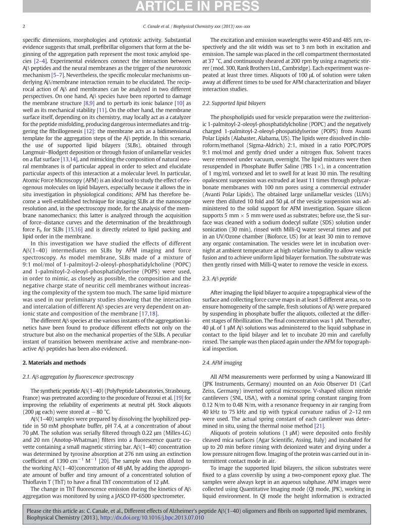
Fig. 2.AFM images of the different Aβ(1–40) intermediate species desiccated onmica at differenthe insets (scale bar 1 μm, z-range 2 nm) at t0 (A) the peptide aggregates are very small, aboutthe aggregation time, the dimension and surface density of the globular aggregates increase, askinetics (t4, D), only fibers are detected.
Please cite this article as: C. Canale, et al., Different effects of Alzheimer's pBiophysical Chemistry (2013), http://dx.doi.org/10.1016/j.bpc.2013.07.01
2.5. AFM force spectroscopy
Force spectroscopy maps were acquired in contact mode. The samecantilevers used for imaging were employed. In force mapping, a se-quence of force–distance curves was collected over at least 5 differentareas of the supported membrane. The areas had a dimension of10 μm × 10 μm and were divided in grids of 20 × 20 sampling points.In each point an extension–retraction force–distance curve wasrecorded. The applied load was in the range of 10–25 nN, the curvelength was 300 nm and the tip velocity of 1 μm/s was maintainedconstant.
For each sample at least 5 maps consisting of 400 force–distancecurves were collected and analyzed using a home built algorithm.
The maps were collected on the same SLB before and after interac-tion with Aβ(1–40) for each of the sampled instant of the aggregationkinetics.
3. Results and discussion
3.1. Aβ(1–40) fibrillogenesis
ThT fluorescence increase was used to monitor the fibrillizationkinetics of a 48 μM Aβ(1–40) solution at 37 °C, under stirring. Sam-ples were continuously sheared at a 200 rpm rate. ThT fluorescenceof the sheared sample was measured in situ in the cuvette cell. Ali-quots (t0, t1, t2, t3, t4) were removed at chosen times and kept at4 °C, in order to significantly slow down the self-assembling process.Freezing was avoided on purpose for preventing Aβ(1–40) solution
t times of the aggregation kinetics (scale bars A–D2 μm, z-range 10 nm). As highlighted infew tens of nanometers in lateral size and in the nanometer range in thickness. Increasingvisible in panels B and C, corresponding to t1 and t2, respectively. At the final time of the
eptide Aβ(1–40) oligomers and fibrils on supported lipid membranes,0
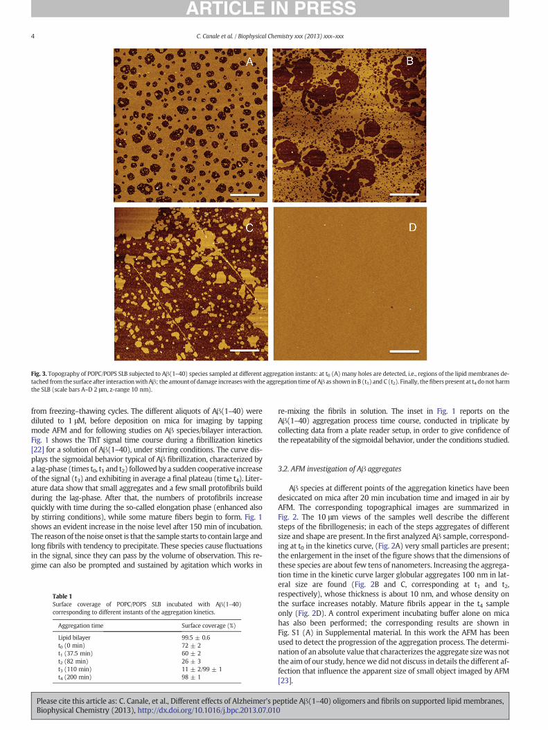
Fig. 3. Topography of POPC/POPS SLB subjected to Aβ(1–40) species sampled at different aggregation instants: at t0 (A) many holes are detected, i.e., regions of the lipid membranes de-tached from the surface after interactionwith Aβ; the amount of damage increaseswith the aggregation time of Aβ as shown in B (t1) and C (t2). Finally, thefibers present at t4 donot harmthe SLB (scale bars A–D 2 μm, z-range 10 nm).
4 C. Canale et al. / Biophysical Chemistry xxx (2013) xxx–xxx
from freezing–thawing cycles. The different aliquots of Aβ(1–40) werediluted to 1 μM, before deposition on mica for imaging by tappingmode AFM and for following studies on Aβ species/bilayer interaction.Fig. 1 shows the ThT signal time course during a fibrillization kinetics[22] for a solution of Aβ(1–40), under stirring conditions. The curve dis-plays the sigmoidal behavior typical of Aβ fibrillization, characterized bya lag-phase (times t0, t1 and t2) followedby a sudden cooperative increaseof the signal (t3) and exhibiting in average a final plateau (time t4). Liter-ature data show that small aggregates and a few small protofibrils buildduring the lag-phase. After that, the numbers of protofibrils increasequickly with time during the so-called elongation phase (enhanced alsoby stirring conditions), while some mature fibers begin to form. Fig. 1shows an evident increase in the noise level after 150 min of incubation.The reason of the noise onset is that the sample starts to contain large andlong fibrils with tendency to precipitate. These species cause fluctuationsin the signal, since they can pass by the volume of observation. This re-gime can also be prompted and sustained by agitation which works in
Table 1Surface coverage of POPC/POPS SLB incubated with Aβ(1–40)corresponding to different instants of the aggregation kinetics.
Aggregation time Surface coverage (%)
Lipid bilayer 99.5 ± 0.6t0 (0 min) 72 ± 2t1 (37.5 min) 60 ± 2t2 (82 min) 26 ± 3t3 (110 min) 11 ± 2/99 ± 1t4 (200 min) 98 ± 1
Please cite this article as: C. Canale, et al., Different effects of Alzheimer's pBiophysical Chemistry (2013), http://dx.doi.org/10.1016/j.bpc.2013.07.01
re-mixing the fibrils in solution. The inset in Fig. 1 reports on theAβ(1–40) aggregation process time course, conducted in triplicate bycollecting data from a plate reader setup, in order to give confidence ofthe repeatability of the sigmoidal behavior, under the conditions studied.
3.2. AFM investigation of Aβ aggregates
Aβ species at different points of the aggregation kinetics have beendesiccated on mica after 20 min incubation time and imaged in air byAFM. The corresponding topographical images are summarized inFig. 2. The 10 μm views of the samples well describe the differentsteps of the fibrillogenesis; in each of the steps aggregates of differentsize and shape are present. In the first analyzed Aβ sample, correspond-ing at t0 in the kinetics curve, (Fig. 2A) very small particles are present;the enlargement in the inset of the figure shows that the dimensions ofthese species are about few tens of nanometers. Increasing the aggrega-tion time in the kinetic curve larger globular aggregates 100 nm in lat-eral size are found (Fig. 2B and C, corresponding at t1 and t2,respectively), whose thickness is about 10 nm, and whose density onthe surface increases notably. Mature fibrils appear in the t4 sampleonly (Fig. 2D). A control experiment incubating buffer alone on micahas also been performed; the corresponding results are shown inFig. S1 (A) in Supplemental material. In this work the AFM has beenused to detect the progression of the aggregation process. The determi-nation of an absolute value that characterizes the aggregate sizewas notthe aim of our study, hencewe did not discuss in details the different af-fection that influence the apparent size of small object imaged by AFM[23].
eptide Aβ(1–40) oligomers and fibrils on supported lipid membranes,0
5C. Canale et al. / Biophysical Chemistry xxx (2013) xxx–xxx
3.3. Aβ interaction with supported lipid bilayers
3.3.1. AFM imagingAβ samples homologous to those desiccated on mica and inspected
by AFM have been administered to POPC/POPS lipid bilayers; Aβ hasbeen let in incubation in phosphate buffer for 20 min, after administra-tion to the lipid bilayer, and therefore gently removed and substitutedwith the same buffer, with special care not to dehydrate the lipid sam-ple. The topography of the lipid bilayer was controlled by AFM beforeAβ administration and only defect-free bilayer, i.e., topographicallyflat, was used for further investigation; an example of SLB producedafter overnight vesicle incubation is shown in Fig. S1 (B) in Supplemen-tal material. Since in most of cases SLB topography before peptide incu-bation does not display significant defects, the presence of a bilayer onthe substrate cannot be easily proven only through imaging. For thisreason, we decided to verify the actual SLB coverage by analyzingforce spectroscopy data recorded overall the 100 μm × 100 μm AFMscanning area. As it will be described in details in the next section,each force curve acquired on a bilayer covered region presents a charac-teristic breakthrough event that is not observed in correspondence ofholes, where the tip interacts directly with the substrate (Fig. 5A);thus, the percentage of curves containing a breakthrough event in the5 sets of 400 curves acquired for each sample can be used as ameasure-ment of SLB coverage. A surface coverage of 99.5 ± 0.6% has been deter-mined for SLBs formed after overnight incubation, averaging the resultsof 13 different samples.
Fig. 3 reports the topography of the lipid bilayers after exposure tothe different investigated Aβ species. Already at t0 the bilayer appearsto be heavily damaged: roundish areas inwhich the bilayer has been re-moved from the surface are uniformly distributed on the samples; at t1the size of the holes has considerably increased and bigger empty areasare surrounded by a great number of smaller ones. At t2 the SLB is al-most completely destroyed by the action of Aβ(1–40) and only separat-ed islands of intact membrane are left on the surface. The surfacecoverage has been quantified in each of the steps and is reported inTable 1. On the contrary, when the last Aβ species, obtained at t4, havebeen incubatedwith the SLB, the topography of the lipid bilayer appearsto be unperturbed: the bilayer is uniformly flat and without holes. Con-trol measurements, incubating the sample with pure buffer and rinsing,were carried on. In this case, no effect on the bilayer topographywas ob-served (Fig. S1 (B–C) Supplemental material).
These results unequivocally demonstrates that Aβ(1–40) fibrils,i.e. the preponderant species at t4, do not harm the membrane andthat the oligomers or proto-fibrils present at the early stage of theaggregation path are the most dangerous forms. They actively inter-act with the bilayer and presumably form aggregates with the lipids,which are removed from the surface in the rinsing step.
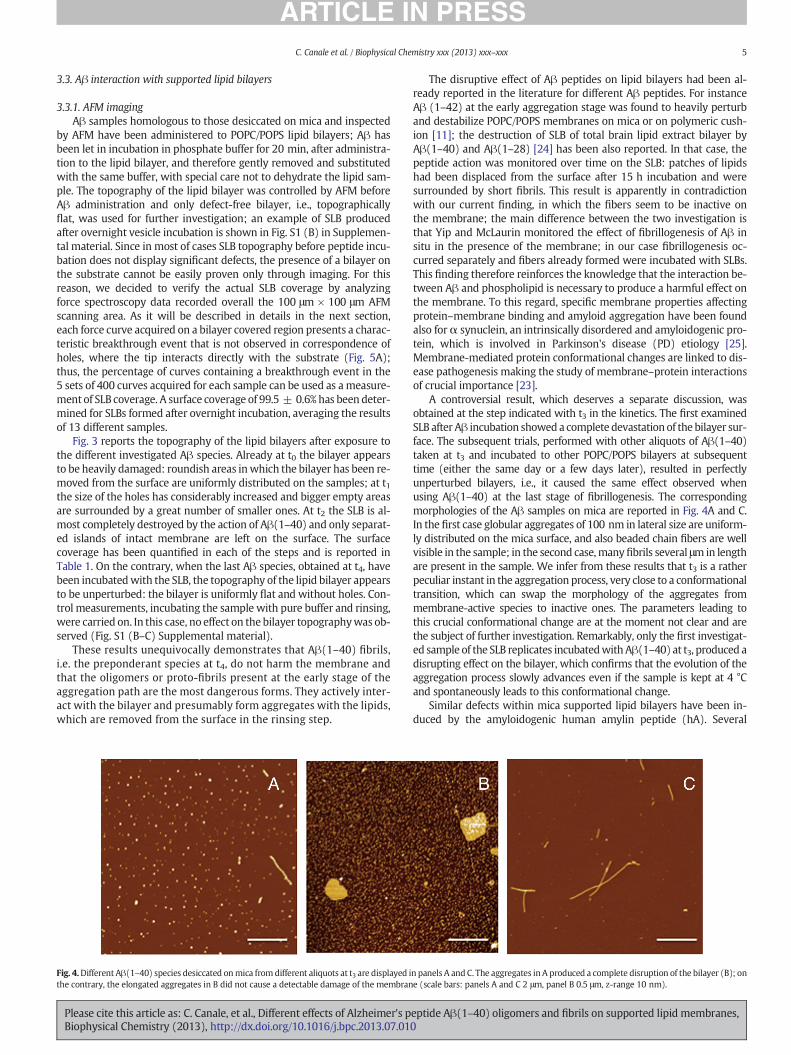
Fig. 4.Different Aβ(1–40) species desiccated onmica from different aliquots at t3 are displayed ithe contrary, the elongated aggregates in B did not cause a detectable damage of the membran
Please cite this article as: C. Canale, et al., Different effects of Alzheimer's pBiophysical Chemistry (2013), http://dx.doi.org/10.1016/j.bpc.2013.07.01
The disruptive effect of Aβ peptides on lipid bilayers had been al-ready reported in the literature for different Aβ peptides. For instanceAβ (1–42) at the early aggregation stage was found to heavily perturband destabilize POPC/POPS membranes on mica or on polymeric cush-ion [11]; the destruction of SLB of total brain lipid extract bilayer byAβ(1–40) and Aβ(1–28) [24] has been also reported. In that case, thepeptide action was monitored over time on the SLB: patches of lipidshad been displaced from the surface after 15 h incubation and weresurrounded by short fibrils. This result is apparently in contradictionwith our current finding, in which the fibers seem to be inactive onthe membrane; the main difference between the two investigation isthat Yip and McLaurin monitored the effect of fibrillogenesis of Aβ insitu in the presence of the membrane; in our case fibrillogenesis oc-curred separately and fibers already formed were incubated with SLBs.This finding therefore reinforces the knowledge that the interaction be-tween Aβ and phospholipid is necessary to produce a harmful effect onthe membrane. To this regard, specific membrane properties affectingprotein–membrane binding and amyloid aggregation have been foundalso forα synuclein, an intrinsically disordered and amyloidogenic pro-tein, which is involved in Parkinson's disease (PD) etiology [25].Membrane-mediated protein conformational changes are linked to dis-ease pathogenesis making the study of membrane–protein interactionsof crucial importance [23].
A controversial result, which deserves a separate discussion, wasobtained at the step indicated with t3 in the kinetics. The first examinedSLB after Aβ incubation showed a complete devastationof the bilayer sur-face. The subsequent trials, performed with other aliquots of Aβ(1–40)taken at t3 and incubated to other POPC/POPS bilayers at subsequenttime (either the same day or a few days later), resulted in perfectlyunperturbed bilayers, i.e., it caused the same effect observed whenusing Aβ(1–40) at the last stage of fibrillogenesis. The correspondingmorphologies of the Aβ samples on mica are reported in Fig. 4A and C.In the first case globular aggregates of 100 nm in lateral size are uniform-ly distributed on the mica surface, and also beaded chain fibers are wellvisible in the sample; in the second case,manyfibrils several μmin lengthare present in the sample. We infer from these results that t3 is a ratherpeculiar instant in the aggregation process, very close to a conformationaltransition, which can swap the morphology of the aggregates frommembrane-active species to inactive ones. The parameters leading tothis crucial conformational change are at the moment not clear and arethe subject of further investigation. Remarkably, only the first investigat-ed sample of the SLB replicates incubatedwithAβ(1–40) at t3, produced adisrupting effect on the bilayer, which confirms that the evolution of theaggregation process slowly advances even if the sample is kept at 4 °Cand spontaneously leads to this conformational change.
Similar defects within mica supported lipid bilayers have been in-duced by the amyloidogenic human amylin peptide (hA). Several
n panels A and C. The aggregates in A produced a complete disruption of the bilayer (B); one (scale bars: panels A and C 2 μm, panel B 0.5 μm, z-range 10 nm).
eptide Aβ(1–40) oligomers and fibrils on supported lipid membranes,0
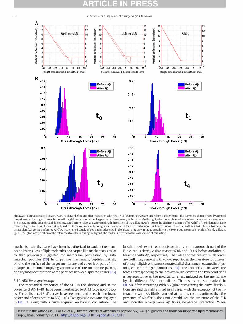
Fig. 5. A: F–d curves acquired on a POPC/POPS bilayer before and after interactionwith Aβ(1–40) (example curves are taken from t1 experiment). The curves are characterized by a typicaljump-to-contact; at higher forces the breakthrough force is recorded and appears as a discontinuity in the curve. On the right, a F–d curve obtained on a silicon dioxide surface is reported.B: Histograms of the breakthrough forces measured before (blue) and after (pink) administration of the different Aβ(1–40) to the SLB in phosphate buffer. A shift of the indentation forcetowards higher values is observed at t0, t1 and t2. On the contrary, at t4 no significant variation of the force distributions is detected upon interaction with Aβ(1–40) fibers. To verify sta-tistical significance, we performed ANOVA test on the 4 couple of populations depicted in the histograms: only in the t4 experiment the two group means are not significantly different(p b 0.05). (For interpretation of the references to color in this figure legend, the reader is referred to the web version of this article.)
6 C. Canale et al. / Biophysical Chemistry xxx (2013) xxx–xxx
mechanisms, in that case, have been hypothesized to explain the mem-brane lesions: loss of lipidmolecules or a carpet-likemechanism similarto that previously suggested for membrane permeation by anti-microbial peptides [26]. In carpet-like mechanism, peptides initiallybind to the surface of the target membrane and cover it or part of it ina carpet-like manner implying an increase of the membrane packingdensity by direct insertion of the peptides between lipidmolecules [26].
3.3.2. AFM force spectroscopyThe mechanical properties of the SLB in the absence and in the
presence of Aβ(1–40) have been investigated by AFM force spectrosco-py. Force–distance (F–d) curves have been recorded on eachmembranebefore and after exposure to Aβ(1–40). Two typical curves are displayedin Fig. 5A, along with a curve acquired on bare silicon nitride. The
Please cite this article as: C. Canale, et al., Different effects of Alzheimer's pBiophysical Chemistry (2013), http://dx.doi.org/10.1016/j.bpc.2013.07.01
breakthrough event i.e., the discontinuity in the approach part of theF–d curve, is clearly visible at about 6 nN and 10 nN, before and after in-teraction with Aβ, respectively. The values of the breakthrough forcesare well in agreement with values reported in the literature for bilayersof phospholipidswith anunsaturated alkyl chain andmeasured inphys-iological ion strength conditions [27]. The comparison between theforces corresponding to the breakthrough event in the two conditionsis representative of the mechanical effect induced on the membraneby the different Aβ intermediates. The results are summarized inFig. 5B. After interacting with Aβ (pink histograms) the curve distribu-tions are slightly right shifted in all cases, with the exception of the in-teraction with Aβ fibrils sampled at t4; this result confirms that thepresence of Aβ fibrils does not destabilizes the structure of the SLBand indicates a very weak Aβ fibrils/membrane interaction. When
eptide Aβ(1–40) oligomers and fibrils on supported lipid membranes,0
7C. Canale et al. / Biophysical Chemistry xxx (2013) xxx–xxx
smaller intermediates interact with the SLB, the removal of part of thebilayer evidenced byAFM imaging is unexpectedly accompanied by a ri-gidifying effect of the membrane on the silicon substrate. The effect israther small at t0, but it becomes significant at t1 and t2, where theforce distributions, before and after interaction with Aβ, overlap onlypartially. This result is very different from a previous result of ourgroup in which Aβ(1–42) was found to mechanically weaken SLB onmica and on charged polymer cushions [11]. The experiment describedin the current paper differs from the previous one for several aspects:first of all, the utilized substrates (and their surface properties)were dif-ferent (silicon vs. mica and polymer cushions), as well as the Aβ pep-tides (Aβ(1–42) vs. Aβ(1–40)) and the pretreatment used for each ofthem; additionally in the experiment here described we have exploitedthe slow kinetics of aggregation of Aβ(1–40) to separate the Aβ inter-mediates at different instants, reducing the variability of the system.
Moreover, it is worthy to mention that, in the current investigation,all the acquired force–distance curves are characterized by a significantjump to contact feature, which was never observed in the experimentdisplaying a decrease of the breakthrough force upon peptide action.This indicates once more that the interaction between membrane andAβ may follow different paths depending on many parameters, such asthe boundary conditions, the interactionwith the substrate, and especial-ly on the Aβ species present in solution. From our results it seems thatsmall protofibrils (i.e., the intermediates present in the range of the ki-netics between t2 and t3) could create complexes with the lipid matrixand solubilize it. On the other hand, the lipid region topographicallynot affected after Aβ(1–40) interaction, become mechanically more sta-ble, such as the lipid packing would become more dense in these areas.Interestingly, the β-amyloid peptide-25–35 has been found to decreasethefluidity ofmouse brain andhuman lymphocytemembranes,with im-portant consequences on the membrane-associated ionic channel func-tions [28]. When the disruption of the lipid membrane is heavy, such asin the case of Aβ(1–40) at t2 (Fig. 4B) and Aβ(1–42) on mica [11], theforce spectroscopy data measured after peptide action are taken fromlipid patches of very small size (tens to hundreds of nanometers), possi-bly loosely bound to the substrate, and are tremendously affected byboundary effect; it is therefore not surprising that the force distributionsindicate, in those cases, a complete mechanical destabilization of thesystem.
4. Conclusions
The results presented here confirm the toxic action exerted byAβ(1–40) prefibrillar oligomers, thus considered the species mainly re-sponsible for the amyloid pathogenicity. Moreover, they contribute toattribute to the membrane damage a crucial role in the onset of diseaseand neuronal cell degenerationwith the lipid fluidity a potential key pa-rameter indicative of the membrane state.
In the future, a detailed analysis of force–distance curve, by the samemethodological approach, performed in the presence of cholesterol,connected to AD, will further clear the role assumed by membrane ri-gidity in the general toxic framework. The comparison of these structur-al measurements to membrane–peptide interaction kinetics, by thestudy of leakage formation in lipid vesicles, will also be important inorder to significantly improve our understanding of protein–membraneinterface.
Supplementary data to this article can be found online at http://dx.doi.org/10.1016/j.bpc.2013.07.010.
Acknowledgments
This work has been partially supported by the following pro-jects: MERIT ‘Basi molecolari nelle sindromi degenerative corre-late con l'invecchiamento’; and FIRB RBFR12SIPT MIND: ‘Indaginemultidisciplinare per lo sviluppo di farmaci neuro-protettori’.
Please cite this article as: C. Canale, et al., Different effects of Alzheimer's pBiophysical Chemistry (2013), http://dx.doi.org/10.1016/j.bpc.2013.07.01
References
[1] D.J. Selkoe, The molecular pathology of Alzheimer's disease, Neuron 6 (1991)487–498.
[2] R. Kayed, A. Pensalfini, L. Margol, Y. Sokolov, F. Sarsoza, E. Head, J. Hall, C. Glabe,Annular protofibrils are a structurally and functionally distinct type of amyloidoligomer, Journal of Biological Chemistry 284 (2009) 4230–4237.
[3] R. Kayed, Y. Sokolov, B. Edmonds, T.M. McIntire, S.C. Milton, J.E. Hall, C.G. Glabe,Permeabilization of lipid bilayers is a common conformation-dependent activityof soluble amyloid oligomers in proteinmisfolding diseases, Journal of BiologicalChemistry 279 (2004) 46363–46366.
[4] Y.V. Sokolov, R. Kayed, A. Kozak, B. Edmonds, T.M. McIntire, S. Milton, M. Cahalan,C.G. Glabe, J.E. Hall, Soluble amyloid oligomers increase lipid bilayer conductanceby increasing the dielectric constant of the hydrocarbon core, Biophysical Journal86 (2004) 382A-382A.
[5] S.M. Butterfield, H.A. Lashuel, Amyloidogenic protein membrane interactions:mechanistic insight frommodel systems, Angewandte Chemie International Edition49 (2010) 5628–5654.
[6] D.M. Hartley, D.M.Walsh, C.P. Ye, T. Diehl, S. Vasquez, P.M. Vassilev, D.B. Teplow, D.J.Selkoe, Protofibrillar intermediates of amyloid β-protein induce acute electrophysi-ological changes and progressive neurotoxicity in cortical neurons, Journal of Neuro-science 19 (1999) 8876–8884.
[7] D.M. Walsh, I. Klyubin, J.V. Fadeeva, W.K. Cullen, R. Anwyl, M.S. Wolfe, M.J. Rowan,D.J. Selkoe, Naturally secreted oligomers of amyloid beta protein potently inhibithippocampal long-term potentiation in vivo, Nature 416 (2002) 535–539.
[8] J. McLaurin, A. Chakrabartty, Membrane disruption by Alzheimer β-amyloidpeptides mediated through specific binding to either phospholipids or ganglio-sides. Implications for neurotoxicity, Journal of Biological Chemistry 271 (1996)26482–26489.
[9] C.M. Yip, J.McLaurin, Amyloid-beta peptide assembly: a critical step infibrillogenesisand membrane disruption, Biophysical Journal 80 (2001) 1359–1371.
[10] A. Demuro, E. Mina, R. Kayed, S.C. Milton, I. Parker, C.G. Glabe, Calciumdysregulation and membrane disruption as a ubiquitous neurotoxic mechanismof soluble amyloid oligomers, Journal of Biological Chemistry 280 (2005)17294–17300.
[11] S. Dante, T. Hauss, R. Steitz, C. Canale, N.A.Dencher,Nanoscale structural andmechan-ical effects of beta-amyloid (1–42) on polymer cushioned membranes: a combinedstudy by neutron reflectometry and AFM force spectroscopy, BBA — Biomembranes1808 (2011) 2646–2655.
[12] A. Relini, O. Cavalleri, R. Rolandi, A. Gliozzi, The two-fold aspect of the interplay ofamyloidogenic proteins with lipid membranes, Chemistry and Physics of Lipids158 (2009) 1–9.
[13] R. Richter, A. Mukhopadhyay, A. Brisson, Pathways of lipid vesicle deposition onsolid surfaces: a combined QCM-D and AFM study, Biophysical Journal 85 (2003)3035–3047.
[14] C. Rossi, J. Chopineau, Biomimetic tethered lipid membranes designed for mem-brane–protein interaction studies, European Biophysics Journal 36 (2007) 955–965.
[15] C. Canale, M. Jacono, A. Diaspro, S. Dante, Force spectroscopy as a tool to investigatesolid supported lipid membranes, Microscopy Research and Technique 73 (2010)965–972.
[16] M.P. Mingeot-Leclercq, M. Deleu, R. Brasseur, Y.F. Dufrene, Atomic force microscopyof supported lipid bilayers, Nature Protocols 3 (2008) 1654–1659.
[17] S. Dante, T. Hauss, A. Brandt, N.A. Dencher, Membrane fusogenic activity of theAlzheimer's peptide A beta(1–42) demonstrated by small-angle neutron scattering,Journal of Molecular Biology 376 (2008) 393–404.
[18] S. Dante, T. Hauss, N.A. Dencher, Beta-amyloid 25 to 35 is intercalated in anionic andzwitterionic lipid membranes to different extents, Biophysical Journal 83 (2002)2610–2616.
[19] Y. Fezoui, D.M. Hartley, J.D. Harper, R. Khurana, D.M. Walsh, M.M. Condron, D.J.Selkoe, P.T. Lansbury, A.L. Fink, D.B. Teplow, An improved method of preparing theamyloid beta-protein for fibrillogenesis and neurotoxicity experiments, Amyloid 7(2000) 166–178.
[20] H. Edelhoch, Spectroscopic determination of tryptophan and tyrosine in proteins,Biochemistry 6 (1967) 1948–1954.
[21] J.L. Hutter, J. Bechhoefer, Calibration of atomic-force microscopy tips, The Review ofScientific Instruments 64 (1993) 1868–1873.
[22] R. Carrotta, C. Canale, A. Diaspro, A. Trapani, P.L. San BIagio, D. Bulone, Inhibiting ef-fect of alpha(s1)-casein on Abeta(1–40) fibrillogenesis, Biochimica et BiophysicaActa 1820 (2012) 124–132.
[23] C. Canale, B. Torre, D. Ricci, P.C. Braga, Recognizing and avoiding artifacts inatomic force microscopy imaging, Methods in Molecular Biology 736 (2011)31–43.
[24] J. McLaurin, C.M. Yip, Modulation of A beta fibrillogenesis on the bilayer surface,Amyloid - Journal of Protein Folding Disorders 8 (2001) 138–139.
[25] C.M. Pfefferkorn, Z. Jiang, J.C. Lee, Biophysics of alpha-synuclein membrane interac-tions, Biochimica et Biophysica Acta 1818 (2012) 162–171.
[26] J.D. Green, L. Kreplak, C. Goldsbury, X.L. Blatter, M. Stolz, G.S. Cooper, A. Seelig, J.Kist-Ler, U. Aebi, Atomic force microscopy reveals defects within mica supportedlipid bilayers induced by the amyloidogenic human amylin peptide, Journal of Mo-lecular Biology 342 (2004) 877–887.
[27] S. Garcia-Manyes, L. Redondo-Morata, G. Oncins, F. Sanz, Nanomechanics of lipid bi-layers: heads or tails? Journal of the American Chemical Society 132 (2010)12874–12886.
[28] W.E. Muller, S. Koch, A. Eckert, H. Hartmann, K. Scheuer, Beta-amyloid peptide de-creases membrane fluidity, Brain Research 674 (1995) 133–136.
eptide Aβ(1–40) oligomers and fibrils on supported lipid membranes,0
Recommended


