
Effect of liposome encapsulation of tea catechinson their accumulation in basal cell carcinomas
Jia-You Fang a,*, Woan-Ruoh Lee b,c, Shing-Chuan Shen d,e,Yen-Ling Huang a
Journal of Dermatological Science (2006) 42, 101—109
www.intl.elsevierhealth.com/journals/jods
a Pharmaceutics Laboratory, Graduate Institute of Natural Products,Chang Gung University, Kweishan, Taoyuan, TaiwanbDepartment of Dermatology, Taipei Medical University Hospital, Taipei, TaiwancGraduate Institute of Medical Sciences, Taipei Medical University, Taipei, TaiwandDepartment of Dermatology, College of Medicine, Taipei Medical University, Taipei, TaiwaneDepartment of Dermatology, Taipei Municipal Wan Fang Hospital, Taipei, Taiwan
Received 20 September 2005; received in revised form 21 November 2005; accepted 15 December 2005
KEYWORDS(�)-Epigallocatechingallate;Liposomes;Intratumor;Basal cell carcinoma
Summary
Background: (�)-Epigallocatechin gallate (EGCG), the main active polyphenol ingreen tea, is associated with antioxidant and anticancer activities.Objective: The aim of this study was to evaluate the feasibility of using liposomes forintratumor distribution of EGCG and its derivative, (+)-catechin.Method: Liposomes containing egg phosphatidylcholine, cholesterol, or anionicsurfactant in the presence of 15% ethanol were prepared. The physicochemicalcharacteristics including vesicle size, zeta potential, drug entrapment, and drugrelease of liposomal formulations were determined. The liposomes containing EGCGwere injected into basal cell carcinomas (BCCs), melanomas, and colon tumors toexamine the tumor uptake of the drug. Liposomes were also incubated with a givennumber of BCC cells, and the cell viability was estimated.Result: Almost no drug molecules were observed when free EGCG was administeredto BCCs. EGCG encapsulated in liposomes with deoxycholic acid (DA) and ethanolincreased drug deposition by 20-fold as compared to the free form. The larger vesiclesize of this formulation was suggested to be the predominant factor governing thisenhancement. The liposomes without ethanol showed low or negligible enhancementon EGCG uptake in BCCs. Liposomes protected EGCG from degradation, resulting inthe induction of greater BCC death compared to that by free EGCG at lowerconcentrations.
* Corresponding author at: Pharmaceutics Laboratory, Graduate Institute of Natural Products, Chang Gung University, 259 Wen-Hwa1st Road, Kweishan, Taoyuan 333, Taiwan. Tel.: +886 3 2118800x5521; fax: +886 3 2118236.
E-mail address: [email protected] (J.-Y. Fang).
0923-1811/$30.00 # 2005 Japanese Society for Investigative Dermatology. Published by Elsevier Ireland Ltd. All rights reserved.doi:10.1016/j.jdermsci.2005.12.010

102 J.-Y. Fang et al.
Conclusion: These results suggest that the intratumor injection of liposomes contain-ing EGCG with moderate modification is an effective approach for increasing EGCGdeposition in BCCs.# 2005 Japanese Society for Investigative Dermatology. Published by Elsevier IrelandLtd. All rights reserved.
1. Introduction
Green tea polyphenols are antioxidants and havebeen shown to exhibit chemopreventive and anti-tumor activities [1,2]. The major and most-activeconstituent in green tea responsible for these bio-logical effects is (�)-epigallocatechin gallate(EGCG). With respect to the skin, green tea extractsand EGCG itself have been reported to be beneficialin treating UV-induced photodamage, basal cellcarcinomas (BCCs), melanomas, and skin papillomas[3—6]. Hence, green tea catechins may be suitableto be the therapeutic agents or adjuvants for treat-ing skin disorders [2,7].
The oral bioavailability of tea catechins is knownto be low with a bioavailability of less than 2—5%[8,9]. The systemic clearance of these catechins isalso high [8,10]. Hence the direct delivery such as byan intratumor route to localized sites is highly desir-able since it would allow local pathologies to betreated without significant systemic side effects. Ofthe various options for administration, an intratu-mor injection of drugs is one of the most-promisingapproaches for solid tumors to minimize side effectsand maximize cytotoxicity at the tumor site [11].However, in most cases, local retention of drugsinjected intratumorally is very low because of thelarge diffusion capability due to their small mole-cular size. To overcome this problem, drug—carriersystems such as liposomes are suitable because oftheir favorable characteristics as a biodegradabledrug reservoir.
Liposomes are microscopic vesicles consisting ofmembrane-like phospholipid bilayers surroundingan aqueous medium. The aim of this study was toinvestigate the effect of liposomal systems on theintratumor accumulation of EGCG. (+)-Catechinand its isomer, (�)-epicatechin, were also usedas model drugs since it has shown moderate antic-arcinogenic activity toward skin cancer [12]. BCCwas used as the skin tumor model since it is themost-common type of skin cancer in the world [13].Both in vivo drug deposition within the tumor andin vitro cell viability were performed to evaluatethe efficacy of liposomes for intratumoral EGCGadministration.
2. Materials and methods
2.1. Materials
EGCG, (+)-catechin, (�)-epicatechin, cholesterol(CH), and deoxycholic acid (DA) were purchasedfrom Sigma Chemical (St. Louis, MO, USA). Tween80 was obtained from Kanto Chemical (Tokyo,Japan). Egg phosphatidylcholine (EPC, 99%) wassupplied by Nippon Oil (Tokyo, Japan). Dulbecco’smodified Eagle’s medium (DMEM), RPMI 1640, andfetal calf serum (FCS) were purchased from Biowest(Nuaille, France). The BCC cell line (BCC-1/KMC)was kindly provided by Prof. Hsin-Su Yu (Departmentof Dermatology, National Taiwan University, Taipei,Taiwan). BCC-1/KMC is a long-term culture of humanBCC derived from the undifferentiated type of facialBCC tumor on the thermal traumatic scar, which wasaneuploidy and subculture for more than 100 pas-sages [14,15]. This immortalized and tumorigeniccell line expresses epithelial markers of keratinfilaments and desmosomes. BCC-1/KMC has success-fully adapted to grow in RPMI 1640 and at a mod-erate concentration of calcium (0.4 mM). Themelanoma (B16-F0) and colon cancer (HT-29) celllines were supplied by American Type Culture Col-lection (Rockville, MD, USA). All other chemicals andsolvents were of analytical grade.
2.2. Preparation of liposomes
EPC (4%, w/v), CH (1%, w/v), and other additiveswere dissolved in a 10 ml volume of a chloroform:-methanol (2:1) solution. The organic solvent wasevaporated in a rotary evaporator at 40 8C, andsolvent traces were removed by maintainingthe lipid film under a vacuum overnight. The filmswere hydrated with double-distilled water or 15%ethanol in water containing 17.2 mM of the drugusing a probe-type sonicator (VCX 600, Sonics andMaterials, USA) at 25 W for 30 min. In some experi-ments, the liposomes were extruded through aLipoFast extruder (Avestin, Canada) using a poly-carbonate membrane with a pore size of 200 nm.The liposomal systems used in this study are listedin Table 1.

Effect of liposome encapsulation of tea catechins 103
Table 1 The composition and characterization of EGCG, (+)-catechin, and (�)-epicatechin liposomes by vesicle size,zeta potential, and drug encapsulation
Code Compositiona Size (nm) Zeta potential(mV)
Encapsulation (%)
EGCG (+)-Catechin (�)-EpicatechinF1 EPC + CH = 4:1b 131.1 � 0.3 �0.9 � 0.4 99.6 � 0.1 39.5 � 4.8 31.9 � 2.7F2 EPC + CH + DA
= 4:1:0.25378.2 � 10.9 �26.2 � 0.9 99.0 � 0.1 53.4 � 0.6 62.7 � 7.8
F3 EPC + CH + Tween 80= 4:1:1.64
104.6 � 2.3 �12.1 � 0.3 84.6 � 3.8 46.9 � 4.5 57.8 � 9.1
F4 F2 extrude 215.8 � 21.3 �36.1 � 1.7 98.1 � 2.5 57.0 � 6.6 64.7 � 7.9
EPC, egg phosphatidylcholine; CH, cholesterol; DA, deoxycholic acid. Each value represents the mean � S.D. (n = 3).a The ratio of liposome composition is weight ratio (%).b All formulations contained a 15% ethanol in the systems except F3.
2.3. Determination of vesicle size andzeta potential
The mean vesicle size and zeta potential of theliposomes were measured by a laser scatteringmethod (Nano ZS1 90, Malvern, UK). Liposomalsuspensions were diluted 100-fold with double-dis-tilled water before the measurement. The determi-nation was repeated three times per sample forthree samples.
2.4. Drug encapsulation percentage inliposomes
The liposomal suspension with drugs was centri-fuged at 48,000 � g and 4 8C for 30 min in a BeckmanOptima MAX1 ultracentrifuge (Beckman Coulter,USA) in order to separate the encapsulated drugfrom the free form. The supernatant and precipitatewere analyzed by HPLC to determine the encapsula-tion percentage [16]. A 25 cm long, 4 mm innerdiameter stainless RP-18 column (Merck, Darm-stadt, Germany) was used. The mobile phase forcatechins was 10:90 acetonitrile:2.7% acetic acid/water at a flow rate of 1.2 ml/min. The fluorescencedetector was set at 280 nm for excitation and320 nm for emission. The detection limit of (+)-catechin, (�)-epicatechin and EGCG was 10, 10,and 40 mg/ml, respectively.
2.5. In vitro release
Drug release from liposomes was measured using aFranz diffusion cell. A cellulose membrane (Cellu-Sep1 T2, with a molecular weight cutoff of 6000—8000, Membrane Filtration Products, USA) wasmounted between the donor and receptor compart-ments. The donor medium consisted of 1 ml of aliposomal formulation. The receptor medium con-sisted of 10 ml of pH 7.4 citrate—phosphate buffer.
The available diffusion area between cells was1.539 cm2. The stirring rate and temperature werekept at 600 rpm and 37 8C, respectively. At appro-priate intervals, 300 ml aliquots of the receptormedium were withdrawn and immediately replacedwith an equal volume of fresh buffer. The amount ofdrugs was determined by HPLC.
2.6. In vivo intratumor administration
The BCC and HT-29 cell lines were maintained at37 8C in RPMI 1640 medium containing 10% FCS andwere subcultured twice a week. The B16-F0 cell linewas cultured in DMEM and 10% FCS. The solid tumorswere obtained by a subcutaneous injection of 107
BCC and HT-29 cells or 106 B16-F0 cells into the backof female nude mice (Balb/c-nu strain, 6—8 weeksold). The nude mice were purchased from NationalLaboratory Animal Center, Taipei, Taiwan and werehoused under pathogen-free conditions according toChang Gung University animal care guidelines. Allanimal experiments were reviewed and approved bythe Institutional Animal Care Committee at ChangGung University. On day 7 as the size of BCCs,melanomas, and colon carcinoma grows to approxi-mately 150, 250, and 300 mm3, respectively, 50 mlof liposomes was intratumorally injected with a 29-gauge needle. Tumor volume was determined bydirect measurement with calipers. The tumorvolume was calculated using the formulaw2 � l � p/6, where the length (l) is the longestdimension and the width ðwÞ is the dimension per-pendicular to the length. At 24 h later, the tumorsites were cut away and weighed.
2.7. Extraction of drug from the tumors
After excising a tumor, it was weighed and mincedwith scissors, positioned in a glass homogenizercontaining 1 ml of 0.1 N HCl, and ground for 5 min

104 J.-Y. Fang et al.
with an electric stirrer. The resulting solution wascentrifuged for 10 min at 10,000 rpm and then fil-tered through a PVDF membrane (with a pore size of0.45 mm, Millipore, USA). The drug amount in thesupernatant was determined by HPLC.
2.8. Cytotoxicity assay
BCC cells were seeded at an initial concentrationof 3 � 104 cells/well in 24-well culture plates, andincubated in medium (RPMI 1640 supplemented with10% FCS). Twenty microliters of liposomes with orwithout drug diluted with medium was added at 24 hpost-inoculation, and plates were incubated in a 5%CO2 atmosphere at 37 8C for 24 h. After PBS wash,cells were incubated with 5 mg/ml 3-[4,5-dimethylthiazol-2-yl]-2,5-diphenyl-tetrazolium bro-mide (MTT) in RPMI 1640 for 2 h at 37 8C. Formozancrystals resulting from MTTreduction were dissolvedby adding 200 ml DMSO and gently agitated for30 min. The absorbance of the supernatant was thenmeasured spectrophotometrically in an ELISA readerat 550 nm. Cell viability was calculated as thepercentage of the control.
2.9. Statistical analysis
Statistical analysis of differences between differenttreatments was performed using unpaired Student’st-test. A 0.05 level of probability was taken as theminimal level of significance.
Fig. 1 Release of EGCG (A), (+)-catechin (B), and (�)-epicatechin (C) across a cellulose membrane from ahydroalcoholic solution and liposomes. Each value repre-sents the mean and S.D. (n = 4).
3. Results
3.1. Size, zeta potential, and drugentrapment of liposomes
The sizes and zeta potentials of the prepared lipo-somes are shown in Table 1. The liposomes madefrom EPC and CH in the presence of ethanol (F1) hada relatively small size of 133.1 nm. The furtheraddition of DA (F2) significantly increased( p < 0.05) the size of the vesicles. The conventionalliposomes without ethanol also produced relativelysmall vesicles. There were almost no surfacecharges on F1 because of the negligible charges ofphosphatidylcholine, the major component of EPC[17]. The addition of DA (F2), an anionic surfactant,resulted in an increase in the negative charges onthe vesicle surface. The absolute zeta potential ofF3 vesicles showed a value of �12.1 mV.
EGCG showed a high rate of encapsulation ofnearly 100% in liposomes incorporated with ethanol(F1 and F2, Table 1). However, this high trappingefficiency was not observed for conventional lipo-
somes (F3). EGCG always exhibited higher encapsu-lation than did (+)-catechin or (�)-epicatechin. Theencapsulation of both isomers with the same mole-cular weight in liposomes was similar for all formu-lations tested ( p > 0.05).
Effect of liposome encapsulation of tea catechins 105
Table 2 Tumor uptake of EGCG and (+)-catechin at24 h from a 15% ethanol solution and various liposomesystems after intratumor injection into a basal cellcarcinoma
Code Tumor uptake (nmol of drug/g of tumor)
EGCG (+)-Catechin
F0a 2.80 � 2.02 0.05 � 0.02F1 39.26 � 12.80 27.33 � 6.75F2 56.34 � 9.48 62.71 � 20.86F3 6.31 � 3.45 0.13 � 0.05F4 23.57 � 10.70 1.49 � 0.45
Each value represents the mean � S.D. (n = 6).a The formulation of code 0 means EGCG or (+)-catechin in a
15% ethanol solution (control group).
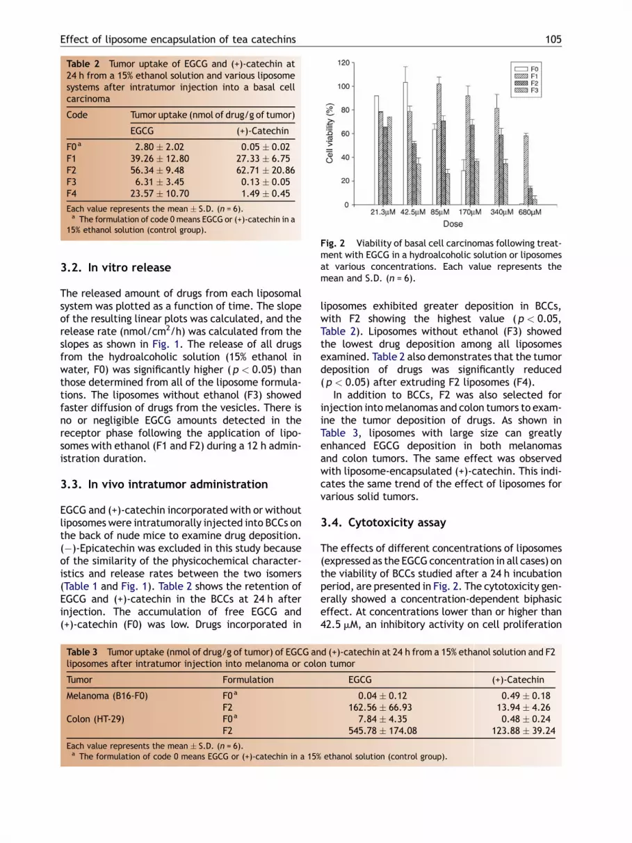
Fig. 2 Viability of basal cell carcinomas following treat-ment with EGCG in a hydroalcoholic solution or liposomesat various concentrations. Each value represents themean and S.D. (n = 6).
3.2. In vitro release
The released amount of drugs from each liposomalsystem was plotted as a function of time. The slopeof the resulting linear plots was calculated, and therelease rate (nmol/cm2/h) was calculated from theslopes as shown in Fig. 1. The release of all drugsfrom the hydroalcoholic solution (15% ethanol inwater, F0) was significantly higher (p < 0.05) thanthose determined from all of the liposome formula-tions. The liposomes without ethanol (F3) showedfaster diffusion of drugs from the vesicles. There isno or negligible EGCG amounts detected in thereceptor phase following the application of lipo-somes with ethanol (F1 and F2) during a 12 h admin-istration duration.
3.3. In vivo intratumor administration
EGCG and (+)-catechin incorporated with or withoutliposomes were intratumorally injected into BCCs onthe back of nude mice to examine drug deposition.(�)-Epicatechin was excluded in this study becauseof the similarity of the physicochemical character-istics and release rates between the two isomers(Table 1 and Fig. 1). Table 2 shows the retention ofEGCG and (+)-catechin in the BCCs at 24 h afterinjection. The accumulation of free EGCG and(+)-catechin (F0) was low. Drugs incorporated in
Table 3 Tumor uptake (nmol of drug/g of tumor) of EGCG anliposomes after intratumor injection into melanoma or colo
Tumor Formulation
Melanoma (B16-F0) F0a
F2Colon (HT-29) F0a
F2
Each value represents the mean � S.D. (n = 6).a The formulation of code 0 means EGCG or (+)-catechin in a 15%
liposomes exhibited greater deposition in BCCs,with F2 showing the highest value (p < 0.05,Table 2). Liposomes without ethanol (F3) showedthe lowest drug deposition among all liposomesexamined. Table 2 also demonstrates that the tumordeposition of drugs was significantly reduced(p < 0.05) after extruding F2 liposomes (F4).
In addition to BCCs, F2 was also selected forinjection intomelanomas and colon tumors to exam-ine the tumor deposition of drugs. As shown inTable 3, liposomes with large size can greatlyenhanced EGCG deposition in both melanomasand colon tumors. The same effect was observedwith liposome-encapsulated (+)-catechin. This indi-cates the same trend of the effect of liposomes forvarious solid tumors.
3.4. Cytotoxicity assay
The effects of different concentrations of liposomes(expressed as the EGCG concentration in all cases) onthe viability of BCCs studied after a 24 h incubationperiod, are presented in Fig. 2. The cytotoxicity gen-erally showed a concentration-dependent biphasiceffect. At concentrations lower than or higher than42.5 mM, an inhibitory activity on cell proliferation
d (+)-catechin at 24 h from a 15% ethanol solution and F2n tumor
EGCG (+)-Catechin
0.04 � 0.12 0.49 � 0.18162.56 � 66.93 13.94 � 4.26
7.84 � 4.35 0.48 � 0.24545.78 � 174.08 123.88 � 39.24
ethanol solution (control group).

106 J.-Y. Fang et al.
Fig. 3 Viability of basal cell carcinomas following treat-ment with a hydroalcoholic solution or liposomes withoutEGCG at various concentrations. Each value representsthe mean and S.D. (n = 6).
Fig. 4 EGCG remaining in a hydroalcoholic solution orliposomes following incubation for various times in thesame medium used for the MTTassay at 37 8C. Each valuerepresents the mean and S.D. (n = 3).
appeared for the free EGCG formulation (F0). Theantiproliferative activity was negligible for an EGCGdose of 42.5 mM. A similar trend was observed for F1and F2 liposomes, which showed significant cytotoxi-city at concentrations lower than 85 mM and higherthan 170 mM. Blank liposomes without EGCG werealso used to perform the MTTassay. When the effectsof different phospholipid concentrations tested onviability ofBCCswerecompared, it is clear fromFig. 3that in most cases except at the highest concentra-tion, the vesicles themselves had no effect on thecytotoxicity. The MTT assay of melanoma and colontumor was also performed by treating F0 and F2 withEGCG concentration at 42.5 mM as shown in Table 4.Similar to the results of BCCs, the liposomes contrib-uted tohigher suppressionof cell growthas comparedto free EGCG formulation.
EGCG in hydroalcoholic solution and in F2 lipo-somes was incubated in the medium the same aswith the MTT assay, and the residual amounts ofEGCG were determined by HPLC as a function oftime at 37 8C (Fig. 4). The EGCG remaining percen-tage is calculated as the residual EGCG amount inmedium after incubation divides by EGCG amount inmedium at time zero. F2 liposomes loaded withEGCG were quite stable, showing that �50% ofthe EGCG still remained at 2 h, while EGCG in thehydroalcoholic solution was unstable, with only�8%
Table 4 Viability (%) of melanoma or colon tumorfollowing treatment with hydroalcoholic solution (F0)or F2 liposomes with EGCG at 42.5 mM
Tumor F0 F2
Melanoma (B16-F0) 103.61 � 7.17 89.02 � 5.38Colon (HT-29) 92.36 � 4.41 76.23 � 6.83
Each value represents the mean � S.D. (n = 6).
of the EGCG remaining. Free EGCG had beencompletely degraded by 4 h after incubation.
4. Discussion
In contrast to conventional liposomes, vesicles pre-pared from an ethanolic solution of phospholipidswere shown to exhibit high encapsulation efficiencyfor both hydrophilic and lipophilic drugs [18,19].The so-called ethosomes contain vesicles with inter-digitated fluid bilayers. A 15% ethanol was incorpo-rated in the liposomal systems in this study. Theresults suggest that ethanol in these liposomal sys-tems can increase the entrapment of EGCG. Theencapsulation of (+)-catechin and (�)-epicatechinby liposomes was always lower than that of EGCG.EGCG contains a galloyl group which is absent fromthe other two catechins. The n-octanol/water par-titioning coefficient indicates greater lipophilicityof EGCG (16.0) compared to (�)-epicatechin (1.4)[20]. EGCG may strongly locate to the surface of thephospholipids bilayers, resulting in high entrapmentwithin liposomes. The interdigitated fluid mem-brane of ethanol-containing liposomes may be ben-eficial to this interaction between EGCG and thebilayers.
(+)-Catechin and (�)-epicatechin showed differ-ent manners of liposome entrapment as comparedto EGCG. The trapping efficiency of a water-solublemolecule may be reduced by the lack of interactionswith the bilayers. The probability of two isomersbeing incorporated into the aqueous cores inside thebilayers is similar. The incorporation of DA-contain-ing and Tween 80-containing liposomes resulted in adramatic increase in (+)-catechin and (�)-epicate-chin encapsulation (F1 versus F2, p < 0.05). Drug

Effect of liposome encapsulation of tea catechins 107
entrapment in conventional liposomes (F3) wascomparable to that of DA-containing liposomes(F2 versus F3, p > 0.05). Drug leakage from lipo-somes is often involved in the aggregation or fusionof liposomal membranes [21]. The high surfacepotential of liposomes (F2 and F3) tends to increasethe interbilayer distance owing to electrostaticrepulsive forces. This may reduce the leakage of(+)-catechin and (�)-epicatechin from the aqueouscores of liposomes.
For development of liposomes encapsulated withantitumor agents in an in vivo status, it is importantto optimize the ability of drug released from vesi-cles. The pore size of the permeated membraneused was below 10 nm, and free molecules wereable to permeate across the membrane. Drugrelease rates from liposomes were much lower thanthose determined from the hydroalcoholic solution(control). This may suggest that drugs do not diffusefreely when entrapped in the vesicles. No or negli-gible EGCG release from liposomes was detected.This may be due to the high entrapment and stronginteraction of EGCG within the liposomal bilayers.
Relatively low accumulation in BCCs wasachieved with EGCG and (+)-catechin in a hydro-alcoholic solution. It is difficult to obtain the desiredtherapeutic results without side effects by main-taining an effective drug concentration in tumorcells, because the free drug with a small molecularmass rapidly diffuses away from the site of injec-tion. Liposomes with DA and ethanol (F2) showedthe greatest ability to enhance local retention ofEGCG and (+)-catechin in BCCs. In tumors, thepermeability of the capillary endothelium isenhanced compared to that of normal tissues[22]. F2 had the largest structure with an averagediameter of 378.2 nm compared to the other for-mulations. Liposomes with a greater size may bemore-readily trapped by the fiber network, and theleakiness of capillary walls may be insufficient toallow their efflux from the interstitial into thevascular space. This result indicates that vesiclesize is an important determinant of the retentionof these liposomes in the tumor.
The smallest size of F3 may have allowed thoseliposomal vesicles quickly escape from the tumorinterstitium into the vascular space. Another possi-bility is that free EGCG and (+)-catechin are easilyreleased from liposomes without ethanol (F3,Fig. 1), leading to the invalidity of liposomes fordrug delivery in this case. In order to further explorethe influence of vesicle size on the tumor uptake ofdrugs, F2 was extruded from a polycarbonate mem-brane with a pore size of 200 nm (F4). As shown inTable 1, no difference (p > 0.05) was found inencapsulation before or after extrusion. The result
of tumor uptake by F4 confirms the importance ofvesicle size on drug deposition. The intratumordistribution of F4 was also lower than that of F1,although the vesicle size of F4 (215.8 nm) was largerthan that of F1 (131.1 nm). This finding indicatesthat liposome accumulation by tumors is not alwayscorrelated with liposome size. In addition to size,zeta potential and liposomal composition may alsoplay crucial roles in determining tumor uptake.Since the vesicle surface of F4 was highly ionizedto a negative charge, the negatively charged pro-teoglycans in the tumor interstitium may repel F4liposomes and increase the outflow of F4 from theinterstitium to the capillaries [11]. Another expla-nation is the effect of DA on the phospholipidbilayers. A previous report has suggested that lipo-somes with rigid bilayers show superior tumor accu-mulation [23]. The inclusion of DA in the liposomeswith ethanol further reduced the rigidity of thebilayers [24], resulting in lower tumor uptake com-pared to liposomes without DA.
Although F3 showed a rigid structure in thebilayers because of the lack of ethanol and DA, itsability to retain drugs within BCCs was low. Thissuggests that the size of and drug release fromliposomes, rather than bilayer rigidity, still predo-minate tumor accumulation. The presence of etha-nol may also play an important role. Theincorporation of ethanol allows the formation ofinterdigitated and malleable vesicles.
This characteristic of liposomes would acceleratethe fusion of vesicles with membranes of fibroblasts(3T3) and delivery of vesicular components togetherwith encapsulation of molecules inside the cell [18].
In addition to BCCs, EGCG has been reported to bebeneficial in treating melanomas and colon cancer[25,26]. F2 liposomes were selected to examine theintratumor EGCG amounts in melanomas and colontumors. The results indicated that liposomes withethanol and DA have a widespread utility in varioussolid tumors. The EGCG uptake in melanomas wasespecially low with injection of the free form (F0,Table 3). This may be due to that melanoma has avasculature that is characterized by the presence ofblood channels and which lacks a well-defined intra-tumor matrix [27]. The use of liposomes may sig-nificantly improve this easy escape of free drug frommelanomas. The EGCG deposition in melanoma andcolon adenocarcinoma was higher than that in BCCsin the liposomal form (Tables 2 and 3). Melanomaand colon tumor showed an abundant vasculaturearound the tumor [28,29], contributing to a higherpercentage of capillary volume to the whole tumor.The nodular BCCs always lack vasculature. Lipo-somes-encapsulated EGCG may locate in the inter-stitial part of the tumor, resulting in the more

108 J.-Y. Fang et al.
concentrated EGCG in the interstitial space of mel-anoma and colon tumor. Another possible reasonmay be that BCCs provided higher amounts of pro-tein or enzyme to attack the lipid bilayers of lipo-somes. The real mechanisms of this effect were notexactly known in the present study and need to befurther explored.
BCCs showed similar uptake to liposomes-encap-sulated EGCG and (+)-catechin (Table 2). However,the deposition of liposomes-encapsulated EGCG inmelanoma and colon tumor was much higher thanthat of (+)-catechin (Table 3). It can be explained bythe higher vasculature system in both tumors. Asobserved in Table 1, the entrapment percentage ofEGCG in F2 liposomes (nearly 100%) is higher thanthat of (+)-catechin by a two-fold. The drugs in freeform may easily escape from tumors by passingthrough the capillary endothelium, resulting inthe greater loss of (+)-catechin as compared toEGCG from tumors with an abundant vasculature.
The intermediate EGCG doses tested in this studydid not exhibit a crucial cytotoxic effect towardsBCCs. This phenomenon was shown by both the freeform and liposomal formulations. The presence ofethanol may have produced this biphasic effect. Aprevious study reported that ethanol exhibits anantioxidative capacity at low concentrations, whileat higher concentrations this effect is reversed [30].As a result, the cytotoxicity due to EGCG at someconcentrations may be partly offset by this parti-cular characteristic of ethanol. The vesicles them-selves generally had no effect on the cytotoxicitybased on the data in Fig. 3. This indicates that thecytotoxicity toward BCCs was mainly a consequenceof the EGCG molecules. It is noticeable that theliposomal systems showed higher antiproliferativeactivities than the free form ( p < 0.05) at lowerdoses (21.3 and 42.5 mM). Not only the BCCs, mel-anoma and colon cancer cell growth was signifi-cantly inhibited by F2 liposomes encapsulatedwith EGCG at 42.5 mM. It is noteworthy that freeEGCG (F0) at 42.5 mM could not induce cell viabilityof the three cell lines examined here. The stabilityof EGCG is susceptible to body fluids such as serum[10]. The encapsulation of EGCG by liposomes mayprotect the drug from oxidation and degradation. Asshown in Fig. 4, free EGCGwas quickly degraded in a10% FCS medium. Liposomes provided stable reten-tion of the drug within the vesicles, contributing tothe successful anticancer activity of EGCG at lowerconcentrations. EGCG in liposomesexhibited its cyto-toxicitybybothdirect fusionof vesicleswithcells andrelease from vesicles after a stable retention.
Free EGCG (F0) showed stronger cytotoxicitytoward BCCs compared to liposomes at higher con-centrations (>85 mM). Although EGCG was not
stable in the medium, a large amount of EGCGmolecules in the formulations with higher doses stillexisted after degradation. These intact moleculesmay have rapidly entered tumor cells to generatethe subsequent cell death. The slower leakage ofEGCG from liposomes limited the availability of freeEGCG. This result may explain the relatively lowerviability of BCCs treated by F3 than by the otherliposomes, since liposomes without ethanol quicklyreleased the encapsulated EGCG (Fig. 1).
5. Conclusions
The purpose of this study was attempted to assessthe feasibility of using liposomes to promote thetumor accumulation of EGCG and its derivatives.The incorporation of DA in the liposomes in thepresence of 15% ethanol greatly increased EGCGuptake by the tumor. The key to this enhancementmay be due to the vesicle size. Larger vesicles aremore-readily trapped in tumors by the fiber net-work, and the leakiness of capillary walls is insuffi-cient to allow vesicle efflux from the interstitialspace. The intratumor distribution of liposomes mayalso be mediated on the basis of surface charges andthe flexibility of the bilayers. Liposomes inducedcell death of BCCs more effectively than did thehydroalcoholic solution at lower EGCG doses. Thiswas due to the ability of liposomes tomaintain EGCGstability. Therefore the incorporation of liposomesmay reduce the drug dose required to avoid sideeffects evoked by the drugs. From the data pre-sented in this study, liposome delivery systemsshowed that they can serve as effective tea cate-chins carriers after moderate formulation design. Itwas also shown that liposomes were able to providehigher EGCG accumulation within BCCs, and toincrease the stability of EGCG inside the vesicles.
References
[1] Katiyar SK, Ahmad N, Mukhtar H. Green tea and skin. ArchDermatol 2000;136:989—94.
[2] Fujiki H, Suganuma M, Imai K, Nakachi K. Green tea: cancerpreventive beverage and/or drug. Cancer Lett 2002;188:9—13.
[3] Wang ZY, Huang MT, Ho CT, Chang R, Ma W, Ferraro T, et al.Inhibitory effect of green tea on the growth of establishedskin papillomas in mice. Cancer Res 1992;52:6657—65.
[4] Mukhtar H, Katiyar SK, Agarwal R. Green tea-anticarcino-genic effects. J Invest Dermatol 1994;102:3—7.
[5] Levin C, Maibach HI. Exploration of alternative and naturaldrugs in dermatology. Arch Dermatol 2002;138:207—11.
[6] Huang CC, Fang JY, Wu WB, Chiang HS, Wei YJ, Hung CF.Protective effects of (�)-epigallocatechin gallate on UVA-induced damage in HaCaT keratinocytes. Arch Dermatol Res2005;296:473—81.

Effect of liposome encapsulation of tea catechins 109
[7] Suganuma M, Okabe S, Kai Y, Sueoka N, Sueoka E, Fujiki H.Synergistic effects of (�)-epigallocatechin gallate with (�)-epicatechin, sulindac, or tamoxifen on cancer cell line PC-9.Cancer Res 1999;59:44—7.
[8] Dvorakova K, Dorr RT, Valcic S, Timmermann B, Alberts DS.Pharmacokinetics of the green tea derivatives, EGCG, by thetopical route of administration in mouse and human skin.Cancer Chemother Pharmacol 1999;43:331—5.
[9] Catterall F, King LJ, Clifford MN, Ioannides C. Bioavailabilityof dietary doses of 3H-labelled tea antioxidants (+)-catechinand (�)-epicatechin in rat. Xenobiotica 2003;33:743—53.
[10] Cai Y, Anavy ND, Chow HSS. Contribution of presystemichepatic extraction to the low oral bioavailability ofgreen tea catechins in rats. Drug Metab Dispos 2002;30:1246—9.
[11] Kawakami S, Yamashita F, Hashida M. Disposition character-istics of emulsions and incorporated drugs after systemic orlocal injection. Adv Drug Deliv Rev 2000;45:77—88.
[12] Soleas GJ, Grass L, Josephy PD, Goldberg DM, Diamandis EP.A comparison of the anticarcinogenic properties of four redwine polyphenols. Clin Biochem 2002;35:119—24.
[13] Thissen MRTM, Neumann MHA, Schouten LJ. A systematicreview of treatment modalities for primary basal cell carci-nomas. Arch Dermatol 1999;135:1177—83.
[14] Chiang LC, Chiang W, Yu HS, Sheu HM, Chen HY. Establish-ment and characterization of a continuous human basal cellcarcinoma cell line from facial skin (I) cytological behaviorof early passages. Kaohsiung J Med Sci 1994;10:170—6.
[15] Yen HT, Chiang LC, Wen KH, Tsai CC, Yu CL, Yu HS. Theexpression of cytokines by an established basal cell carci-noma cell line (BCC-1/KMC) compared with cultured normalkeratinocytes. Arch Dermatol Res 1996;288:157—61.
[16] Fang JY, Hung CF, Hwang TL, Huang YL. Physicochemicalcharacteristics and in vivo deposition of liposome-encapsu-lated tea catechins by topical and intratumor administra-tions. J Drug Target 2005;13:19—27.
[17] Buszello K, Harnisch S, Muller RH, Muller BW. The influenceof alkali fatty acids on the properties and the stability ofparenteral O/W emulsions modified with Solutol HS 151.Eur J Pharm Biopharm 2000;49:143—9.
[18] Dayan N, Touitou E. Carriers for skin delivery of trihexyphe-nidyl HCl: ethosomes vs. liposomes. Biomaterials 2000;21:1879—85.
[19] Godin B, Touitou E. Mechanism of bacitracin permeationenhancement through the skin and cellular membranes froman ethosomal carrier. J Contr Rel 2004;94:365—79.
[20] Hashimoto T, Kumazawa S, Nanjo F, Hara Y, Nakayama T.Interaction of tea catechins with lipid bilayers investigatedwith liposome systems. Biosci Biotechnol Biochem 1999;63:2252—5.
[21] Yamaguchi T. Lipid microspheres as drug carriers: apharmaceutical point of view. Adv Drug Deliv Rev 1996;20:117—30.
[22] Mizuguchi H, Nakanishi T, Nakanishi M, Nakagawa T, Naka-gawa S, Mayumi T. Intratumor administration of fusogenicliposomes containing fragment A of diphtheria toxin sup-presses tumor growth. Cancer Lett 1996;100:63—9.
[23] Nagayasu A, Uchiyama K, Kiwada H. The size of liposomes: afactor which affects their targeting efficiency to tumors andtherapeutic activity of liposomal antitumor drugs. Adv DrugDeliv Rev 1999;40:75—87.
[24] El Maghraby GMM, Williams AC, Barry BW. Skin delivery of 5-fluorouracil from ultradeformable and standard liposomes invitro. J Pharm Pharmacol 2001;53:1069—77.
[25] Suzuki Y, Isemura M. Inhibitory effect of epigallocatechingallate on adhesion of murine melanoma cells to laminin.Cancer Lett 2001;173:15—20.
[26] McLoughlin P, Roengvoraphoj M, Cornelia Q, Hescheler J,Certa U, Sachinidis A. Transcriptional responses to epigallo-catechin-3 gallate in HT 29 colon carcinoma spheroids.Genes Cells 2004;9:661—9.
[27] Harasym TO, Cullis PR, Bally MB. Intratumor distribution ofdoxorubicin following i.v. administration of drug encapsu-lated in egg phosphatidylcholine/cholesterol liposomes.Cancer Chemother Pharmacol 1997;40:309—17.
[28] Arashiro K. The tumor vasculature in cutaneous malignantmelanoma: scanning electron microscopy of corrosion casts.Plast Reconstr Surg 2002;110:717—8.
[29] Marzola P, Degrassi A, Calderan L, Farace P, Crescimanno C,Nicolato E, et al. In vivo assessment of antiangiogenicactivity of SU6668 in an experimental colon carcinomamodel. Clin Cancer Res 2004;10:739—50.
[30] Videla LA. Assessment of the scavenging action of reducedglutathione, (+)cyanidanol-3 and ethanol by the chemi-luminescent response of the xanthine oxidase reaction.Experientia 1983;39:500—2.
Recommended


