
Esophagogastric Junction Distensibility After FundoplicationAssessed with a Novel Functional Luminal Imaging Probe
Monika A. Kwiatek,Department of Medicine, Feinberg School of Medicine, Northwestern University, Chicago, IL, USA.Division of Gastroenterology, Department of Medicine, Feinberg School of Medicine, NorthwesternUniversity, 676 N. St. Clair Street, Suite 1400, Chicago, IL 60611-2951, USA
Peter J. Kahrilas,Department of Medicine, Feinberg School of Medicine, Northwestern University, Chicago, IL, USA
Nathaniel J. Soper,Department of Surgery, Feinberg School of Medicine, Northwestern University, Chicago, IL, USA
William J. Bulsiewicz,Department of Medicine, Feinberg School of Medicine, Northwestern University, Chicago, IL, USA
Barry P. McMahon,Medical Physics & Clinical Engineering, Department of Clinical Medicine, Trinity College, Dublin,Ireland
Hans Gregersen, andMech-Sense, Aalborg Hospital, Århus University Hospital, Århus, Denmark
John E. PandolfinoDepartment of Medicine, Feinberg School of Medicine, Northwestern University, Chicago, IL, USAMonika A. Kwiatek: [email protected]
AbstractObjective—The aim of the study was to compare the esophagogastric junction (EGJ) compliancein response to controlled distension in fundoplication (FP) patients and controls using the functionalluminal imaging probe (FLIP).
Background—FP aims to replicate normal EGJ distensibility. FLIP is a new technology that usesimpedance planimetry to measure intraluminal cross-sectional area (CSA) during controlleddistension.
Methods—Ten controls and ten FP patients were studied with high-resolution esophageal pressuretopography (HREPT) and then the FLIP placed across the EGJ. Deglutitive and interdeglutitive EGJdistensibility was assessed with volume-controlled distension. The FLIP measured eight CSAsspaced 4 mm apart within a cylindrical saline-filled bag along with the corresponding intrabagpressure.
Results—The EGJ formed an hourglass shape during distensions with the central constriction atthe diaphragmatic hiatus. The distensibility of the hiatus was significantly greater during deglutitiverelaxation in both subject groups, but FP patients exhibited reduced EGJ distensibility and
Correspondence to: Monika A. Kwiatek, [email protected] paper was presented at the Digestive Disease Week and the 109th Annual Meeting of the American Gastroenterological AssociationInstitute, May 17–22, 2008, San Diego, CA, USA.
NIH Public AccessAuthor ManuscriptJ Gastrointest Surg. Author manuscript; available in PMC 2011 February 1.
Published in final edited form as:J Gastrointest Surg. 2010 February ; 14(2): 268–276. doi:10.1007/s11605-009-1086-1.
NIH
-PA Author Manuscript
NIH
-PA Author Manuscript
NIH
-PA Author Manuscript

compliance compared to controls. During the interglutitive period, the corresponding increase inintrabag pressures at larger volumes were also greater in FP patients implying a longer segment ofEGJ constriction. The EGJ distensibility characteristics did not correlate with HREPT measures.
Conclusions—FLIP technology was used to compare EGJ distensibility in FP patients and controlsubjects. The least distensible locus within the EGJ was always at the hiatus. EGJ distensibility wassignificantly reduced, and the length of constriction increased in FP patients. Future FLIP studieswill compare patients with and without post-FP dysphagia and gas bloat, symptoms suggestive of anoverly restrictive FP.
KeywordsEsophagogastric junction; Fundoplication; Functional luminal imaging probe; Manometry
Patients with gastroesophageal reflux disease have an abnormally compliant esophagogastricjunction (EGJ)1–4 that inadequately impedes reflux of gastric contents and thus contributes toa greater likelihood of esophageal mucosa injury and reflux-related symptoms. Increased EGJcompliance is likely multifactorial with potential contributing defects of lower esophagealsphincter pressure, extrinsic compression by the crural diaphragm, and misalignment betweenthe two manifest as a sliding hiatal hernia.3,5,6 Potential deleterious mechanical consequencesof increased EGJ compliance include increased volumes of liquid reflux,7 a reduced thresholdfor eliciting transient LES relaxations,8 and allowing gastric juice to track within the closedsphincter.9–11 Surgical antireflux procedures aim to correct the defective EGJ by fashioninga mechanical antireflux barrier that allows adequate EGJ opening for passage of swallowedingesta into the stomach as well as gastric venting when required.12,13 Ideally, a normal healthyEGJ would be replicated.
Postoperative integrity of the EGJ junction is usually assessed by manometry. Such functionalassessments are often provoked by persistent or recurrent gastroesophageal reflux symptomssuggestive of a defective fundoplication (FP) or because of postoperative dysphagia. Althoughmanometric technology has evolved recently to high-resolution esophageal pressuretopography (HREPT),14–16 it still fundamentally measures intraluminal pressure. However,the surgical modification of the EGJ during fundoplication may not be best gauged bymeasurement of intraluminal pressure. Fundoplication entails tightening of the diaphragmatichiatus and construction of a loose floppy fundic wrap around the distal esophagus, neither ofwhich necessarily affects the intraluminal pressures. Alternatively, FP integrity may be betterassessed when challenged with intraluminal distension.17
Measurement of intraluminal distensibility at the EGJ is complex. The distending pressuremust be localized within the EGJ and dimensional measurements restricted to the area ofinterest. Although this can be achieved with a barostat (or hydrostat), this is somewhatcumbersome and requires concurrent fluoroscopic imaging.2–4 Nonetheless, barostatassessment of compliance at the narrowest locus within the EGJ after FP suggested it to besimilar in asymptomatic FP patients compared to control subjects.2 A potentially more robustmethod for measuring EGJ distensibility, capable of making measurements at multiple adjacentsegments without need for fluoroscopy, is by adaptation of the principle of impedanceplanimetry18,19 into a functional luminal imaging probe (FLIP). FLIP recordings allowdynamic imaging of EGJ distention as a three-dimensional structure based on instantaneousmeasurement of multiple intraluminal cross-sectional areas with concurrent pressuremeasurements, thereby facilitating measurement of EGJ distensibility.20,21 Hence, the aim ofthe current study was to compare the EGJ distensibility in FP patients during the interdeglutitiveperiod and during deglutitive EGJ relaxation to that of asymptomatic control subjects usingthe FLIP.
Kwiatek et al. Page 2
J Gastrointest Surg. Author manuscript; available in PMC 2011 February 1.
NIH
-PA Author Manuscript
NIH
-PA Author Manuscript
NIH
-PA Author Manuscript

Materials and MethodsSubjects
Ten asymptomatic control subjects (2M, 23–50 years) and ten patients who have hadlaparoscopic Nissen FP surgery (2M, 42–68 years) were studied. The control subjects wererecruited from a pool of volunteers who had neither gastrointestinal symptoms, any priorgastrointestinal surgery, nor were taking medications known to affect gastrointestinal function.FP patients were recruited successively from referrals to the Gastroenterology OutpatientClinic and Gastrointestinal Diagnostic Laboratory for follow-up assessment of mild tomoderate postoperative symptoms. All subjects gave written informed consent. The studyprotocol was approved by the Northwestern University Institutional Review Board.
Functional Luminal Imaging ProbeEsophagogastric junction distensibility was measured using a custom-made FLIP designed tomeasure intraluminal cross-sectional areas (CSAs) as a function of distention pressure aspreviously described.21 In brief, the probe assembly was 80 cm long, with the proximal 68 cmconstructed from a 4.5-mm outer diameter nine-lumen polyurethane tube and the distal 12 cmconstructed of a 1.6-mm outer diameter double-lumen polyethylene tube (Fig. 1; GMCMedical, Hornslet, Denmark). A noncompliant 35-μm-thick polyestherurethane bag wasmounted on the distal end. Within the bag was a 3.2-cm segment comprised of nine-ringelectrodes spaced 4 mm apart for impedance planimetry measurement. Excitation electrodesat either end emitted a constant low current of 100 μA at a frequency of 5 kHz making thevoltage measured across each of the eight adjacent pairs of ring electrodes proportional to theimpedance between them. As the bag was filled with 0.2% saline, the impedance across eachsegment was thus inversely proportional to the CSA of the bag at that locus. Maximal bagdiameter was 3.2 cm. The probe also contained two low compliance saline perfused channels(1 mm ID), connected to external pressure transducers (Edwards TruWave, EdwardsLifesciences, Irvine, CA, USA), providing pressure measurements within and 2.5 cm proximalto the bag.
Measurements from the eight electrode pairs and pressure transducers were sampled at 10 Hzwith the data acquisition system, transmitted serially to a personal computer, and displayed inreal-time using custom-made software programmed in Labview® version 6.1 (NationalInstruments, Austin, TX, USA). The probe was calibrated at body temperature prior to eachstudy by filling the bag with 0.2% saline within a calibration block containing a set ofcylindrical cutouts with CSAs ranging from 50 to 616 mm2. The pressure transducers werecalibrated at 0 and 75 mmHg.
High-Resolution ManometryHREPT data were obtained using a solid-state manometric assembly (4.2 mm outer diameter)with 36 circumferential sensors spaced at 1-cm intervals (Sierra Scientific Instruments, LosAngeles, CA, USA), the recording characteristics of which have been previously described.22,23 Pressure transducers were calibrated at 0 and 100 mmHg using externally applied pressureprior to the study.
Experimental ProtocolStudies were performed in a supine position after at least a 6-h fast. Patients underwenttransnasal placement of the manometry assembly, which was positioned to record from thehypopharynx to the stomach with about five intragastric sensors. The assembly was fixed inplace by taping it to the nose. The manometric protocol included at least a 30-s period ofbaseline recording in a supine position followed by a series of ten 5-ml and two 10-ml test
Kwiatek et al. Page 3
J Gastrointest Surg. Author manuscript; available in PMC 2011 February 1.
NIH
-PA Author Manuscript
NIH
-PA Author Manuscript
NIH
-PA Author Manuscript

water swallows. Once the manometric assembly was removed, the FLIP was placedtransnasally into the stomach and withdrawn until the bag was centered at the EGJ based onHREPT measurements.23,24 Bag position was also confirmed fluoroscopically by partiallyfilling the FLIP bag (20–30 ml) and observing transit of swallowed barium into the stomach(Fig. 2). The probe was then fixed in place by taping it to the nose. Interdeglutitive (30 s) anddeglutitive (dry swallow) FLIP measures of CSA and distention pressure were made with thebag filled to 30, 40, 50, and 60 ml. Each volume was tested in triplicate and repeated if thesubject inadvertently swallowed. Swallows were evident by a peristaltic contraction at theperfused channel 2.5 cm proximal to the bag. EGJ geometry was monitored in real time toassure proper bag placement, and instances of suspected migration were confirmedfluoroscopically before repositioning and repeating the measurement.20
Data AnalysisHigh-Resolution Manometry—The HREPT plots were analyzed to characterize EGJmorphology and deglutitive function in terms of end-expiratory EGJ pressure, inspiratoryaugmentation of EGJ pressure, length of the EGJ high-pressure zone (HPZ), abdominal lengthof the EGJ HPZ, and integrated relaxation pressure (IRP) during deglutitive relaxation aspreviously described.22–27 Distal esophageal peristalsis was considered normal when theperistaltic amplitude and velocity were ≥30 mmHg and <10 cm/s. Failed or hypotensiveperistalsis with 50–60% of test swallows constituted intermittent hypotensive peristalsis, 70–90% frequent hypotensive peristalsis, and 100% absent peristalsis. Distal esophagealcontractile vigor was measured by the distal contractile integral (DCI). Peristalsis-relatedintrabolus pressure (IBP) was measured 1 cm proximal to the EGJ and summarized as anaverage pressure during the 3 s of maximal IBP during esophageal emptying (IBPesoph).28
Functional Luminal Imaging Probe—Interdeglutitive EGJ CSAs and intrabag pressurewere assessed at each FLIP bag volume by quantifying the 50th percentile of each measureduring each test 30-s recording. The corresponding deglutitive EGJ measures were assessedduring the period between a dry swallow and the distal esophageal peristaltic or postdeglutitiveEGJ contraction. The deglutitive EGJ response was quantified by the 1-s nadir in the intrabagpressure and the corresponding CSAs. Measurements of CSA were made at each of the eightelectrode pairs covering a span of 3.2 cm.
EGJ compliance (volume vs. pressure) was calculated based on the intrabag pressure and anapproximation of EGJ volume across the range of FLIP bag volumes associated withmeasureable distention. EGJ volume was estimated by identifying the narrowest CSA(invariably at the diaphragmatic hiatus), extending distally for three additional CSAs andapplying the formula (CSAx + CSAx+4 mm + CSAx+8 mm + CSAx+12 mm) × 0.004 to convertthe 4-mm segment CSAs (square millimeters) to milliliters. A linear regression analysis wasthen applied with the slope of the line representing EGJ compliance (milliliters per millimetersof mercury).
Statistical AnalysisThe data from triplicate trials were averaged to describe the EGJ response at each FLIP bagvolume for each subject. Data from all the subjects was then expressed as median (5th–95thpercentile). Statistical comparisons were performed using Wilcoxon matched pairs test andKruskal–Wallis test. The relationships between measures provided by the FLIP and HREPTwere assessed with Spearman’s rank correlation coefficient (rs). A p value<0.05 wasconsidered significant.
Kwiatek et al. Page 4
J Gastrointest Surg. Author manuscript; available in PMC 2011 February 1.
NIH
-PA Author Manuscript
NIH
-PA Author Manuscript
NIH
-PA Author Manuscript

ResultsDemographic and HREPT Data
The FP patients were assessed with FLIP at 4 months to 7 years postoperative with eight ofthe ten having had their surgery at Northwestern Memorial Hospital (NMH). Those eightoperative reports uniformly described a laparoscopic “short floppy” Nissen fundoplication,2.0–2.5 cm in length, constructed with a 51–60-Fr Maloney dilator placed within the esophagusand mobilization of the fundus by dividing the short gastric vessels. Operative reports werenot available for the two patients who had their surgery elsewhere but who were certain thatthey had complete 360° fundoplication on the basis of preoperative consultation with theirsurgeons.
The symptoms prior to and following the surgery were recorded in seven of the eight FP patientswho had their surgery at NMH. Symptoms of heartburn and regurgitation were consistentlypresent in six of the seven patients prior to the surgery. The three other patients reported havingsevere heartburn and regurgitation before the surgery. At the time of the study, four of the tenpatients reported mild dysphagia, three of the ten patients reported bloating, three of the tenpatients reported chest pain, one of the ten patients reported nausea, one of the ten patientsreported abdominal pain, two of the ten patients reported heartburn, and one of the ten patientsreported heartburn and regurgitation. None of these problems were sufficiently severe for anyof these patients to undergo revision surgery.
HREPT data on EGJ parameters showing similar contractile function between control subjectsand FP patients are summarized in Table 1. One significant difference between groups wasthat the length of the EGJ HPZ, both total and intra-abdominal, was slightly shorter in FPpatients (p<0.02). None of the subjects had a HREPT signature of hiatal hernia defined as aseparation greater than 2 cm between the components of the EGJ HPZ (LES and cruraldiaphragm).24 The barium swallow used to confirm the position of the FLIP bag across theEGJ, verified the absence of hiatal hernia. With respect to peristaltic function, one of the normalcontrols had frequent hypotensive peristalsis while the remainder were normal. Among the FPpatients, two had frequent and one had intermittent hypotensive peristalsis. However, the distalesophageal contractile vigor, summarized as the DCI of the normal and hypotensive peristalticcontractions, was comparable between groups (control, 2,640 (1,297–3,429); FP, 2,193 (418–6,578) mmHg s cm, p=0.55). An abnormally high deglutitive IRP (>15 mmHg) was detectedin one control and two FP patients.
EGJ DistensibilityWhen straddling the EGJ, the FLIP bag assumed an hourglass shape with the centralconstriction at the diaphragmatic hiatus in both control subjects and FP patients. The hourglassshape was present during both the interdeglutitive period and deglutitive relaxation at all FLIPbag volumes (Fig. 3). In fact, evident in Table 2, many subjects in both groups had CSAmeasurements at the hiatus that were the minimum detectable (50 mm2) implying that all ofthe saline within the FLIP bag displaced proximal or distal to it. This suggested that the hiatuswas uniformly the least distensible locus within the EGJ. Only with the FLIP bag volume of60 ml was there nearly consistent hiatal distention above the minimum, at which point thehourglass opened and closed with respiration confirming this to be the diaphragmatic hiatus.Of note, the 60-ml bag volume resulted in pronounced hiatal opening in controls duringdeglutitive relaxation to a CSA significantly greater than that observed in FP patients (Table2; p<0.001).
In the EGJ dynamic described above, distensile pressure within the FLIP bag increased withincreasing bag volume in both subject groups and in both test conditions (Table 3; p<0.0001).
Kwiatek et al. Page 5
J Gastrointest Surg. Author manuscript; available in PMC 2011 February 1.
NIH
-PA Author Manuscript
NIH
-PA Author Manuscript
NIH
-PA Author Manuscript

Furthermore, the distending pressure within the FLIP bag was consistently greater in FPpatients than in control subjects particularly with FLIP bag volumes of 40, 50, and 60 ml(p<0.05; Table 3). Conceptually, pressure within the FLIP bag increased when the increasedvolume of saline within it could no longer disperse to highly compliant regions proximal ordistal to the EGJ presumably because the bag was filled to capacity in those regions. Hence,the observed difference in pressure between the control subjects and FP patients implies thatthere was a longer zone of measured constriction in the FP patients. This difference was furtherbrought out by the estimated EGJ volume, a measure that utilized the CSA of the hiatus andthree distal adjacent FLIP segments. Examining Fig. 4, both during the interdeglutitive periodand during deglutitive relaxation, the EGJ of control subjects was widely distended at distensilepressures insufficient to achieve any measureable opening during the same conditions in theFP patients.
The data in Fig. 4 can also be utilized to estimate EGJ compliance, defined as the slope of theEGJ volume vs. intrabag pressure relationship. Since the data points associated with the 40-ml FLIP bag volume did not achieve measureable EGJ distention, this could only be done withthe 50- and 60-ml data points. As evident in Fig. 5a, interdeglutitive EGJ compliance wascomparable between control subjects and FP patients (p=0.13). As expected, deglutition tendedto increase EGJ compliance in controls (p=0.08); the same change in compliance was not seenin FP patients (p=0.92; Fig. 5b).
HREPT vs. FLIP EGJ MeasuresThe data from controls and FP patients were pooled to test hypotheses on association betweenFLIP vs. HREPT measures of distal esophageal function. Hypothetically, a less compliant EGJmight exert greater closing pressure and distal resistance for the bolus traversing the esophagusresulting in greater intrabolus pressure. However, there were no significant correlationsbetween FLIP measure of interdeglutitive EGJ compliance and HREPT measures of expiratoryEGJ pressure (rs=−0.12, p=0.63) or inspiratory EGJ augmentation (rs=0.25, p =0.29).Likewise, deglutitive EGJ compliance did not correlate with IBPesoph (rs=0.14, p=0.56).However, IBPesoph was related to DCI (rs=0.56, p=0.01) suggesting that the contractile vigorof the distal esophagus increased with outflow resistance. An interesting contrast between thetwo technologies was that while HREPT estimates of sphincter length found the FP patientsto have a significantly shorter HPZ than control subjects, FLIP measures of EGJ volume, byinference length, found the FP patients to have a significantly longer zone of constriction.
DiscussionThe EGJ has two distinct dimensions of function—that during nondeglutitive periods to preventreflux by maintaining closure and that during periods of opening to facilitate trans-EGJ flow,be it esophagogastric or gastro-esophageal. Manometry, or more recently HREPT, directlymeasures closure forces. This investigation tested the ability of the FLIP, a novel device basedon impedance planimetry technology, to quantify EGJ opening CSA in response to controlledintraluminal distension variables. Studies were done on control subjects and patients withsatisfactory to good functional outcome from laparoscopic Nissen fundoplication. The majorfindings of the study were that (1) the FLIP isolated the hiatus as the least distensible locuswithin the EGJ in both subject groups, (2) the distensibility of the hiatus was significantlygreater during deglutitive relaxation in both subject groups, (3) fundoplication patientsexhibiting reduced EGJ distensibility and reduced EGJ compliance during deglutitiverelaxation compared to control subjects, (4) fundoplication patients exhibited a longer segmentof reduced distensibility than did controls, and (5) EGJ attributes demonstrated with FLIPmeasurements were not mirrored by HREPT findings.
Kwiatek et al. Page 6
J Gastrointest Surg. Author manuscript; available in PMC 2011 February 1.
NIH
-PA Author Manuscript
NIH
-PA Author Manuscript
NIH
-PA Author Manuscript

The finding that the least distensible locus within the EGJ is at the hiatus supports similarfindings made using barostat2,4 or hydrostat3 technology. This was found to be true irrespectiveof the presence of hiatus hernia or fundoplication. The significance of quantifying thismeasurement is that this variable dominates the equation for trans-EGJ flow (Flow rate = dP× D4/CVL) in which dP is the trans-EGJ pressure gradient, D is the opening diameter, C is aconstant, V is viscosity, and L is the length of constriction.29 Although the length of constrictionalso figures into the equation, note that D, the diameter of maximal constriction, is raised tothe fourth power causing it to be the dominant variable. It follows that this variable is a keydeterminant of both the efficacy of swallow-related esophageal emptying and the volume ofrefluxate during periods of sphincter relaxation.2 In postfundoplication patients, distensibilitywithin the hiatus is a direct consequence of the details of operative hiatal repair. Quite possibly,this variable, a generally underappreciated source of technical variability in fundoplicationsurgery, is a major determinant of postoperative outcome in terms of dysphagia and gas bloat.
The FLIP findings of a less distensible hiatus and a longer length of constriction post-FP relativeto control subjects, despite somewhat conflicting conclusions based on HREPT measures(Table 1), highlight the distinction between measuring resistance to physically opening theEGJ lumen (FLIP) and measuring contraction within a closed lumen (HREPT). To assume thatthese techniques are equivalent, assumes that a decrease in contractile pressure, mainlyattributable to LES and crural diaphragm contraction, parallels luminal opening dimensions inthe absence of that contractile activity. In fact, these two properties have no necessaryrelationship to each other as the latter is instead related to wall properties of the EGJ and theexternal constraint on the EGJ imposed by the diaphragmatic hiatus and fundoplication, ifpresent. Fundoplication surgery is clearly designed to modify these latter variables and for thatreason, the outcome is better measured with a technique such as FLIP. A “short floppy”fundoplication constructed with a larger caliber dilator within the esophageal lumen shouldhave no obvious effect on the contractility of the LES or crural diaphragm but should limitEGJ distensibility. In fact, evident in Fig. 4, this is what was observed. EGJ distensibility duringthe interdeglutitive period (with both the LES and crural diaphragm contracting) was similarbetween subject groups but distensibility during deglutitive relaxation was significantly greaterin the control subjects. Although beyond the scope of the current work, it would be of greatinterest to examine the profile of EGJ distensibility in postfundoplication patients withbothersome dysphagia or gas bloat to see if they are quantifiably different.
Although findings from the current study generally corroborate those obtained from a barostatdistention study of a similar population of fundoplication patients,2 there is an importantdifference. Both studies demonstrated increased length of the constricted segment afterfundoplication but only the current FLIP study demonstrated reduced compliance duringdeglutitive relaxation; the barostat study suggested distensibility similar to that of controlsubjects.2 The explanation for this discrepancy is likely methodological. In the barostat study,only a single two-dimensional plane was imaged leaving it vulnerable to error related toasymmetry of the EGJ. FLIP, on the other hand, calculates CSA bases on impedancecharacteristics irrespective of luminal shape and, thus, is inherently more accurate. FLIP alsohas the advantage of utilizing data from several adjacent segments within the EGJ, whereasthe barostat study analyzed only the single locus of greatest constriction. Together, theseadvantages, as well as the rapid sampling of the FLIP device, argue that the FLIP is likely themore accurate method for ascertaining intraluminal CSA.
The key data related to EGJ distensibility and compliance gleaned from the FLIP measurements(summarized in Figs. 3 and 4) depend on measurement of the pressure within the FLIP bagrather than the volume within the bag. Although the design of the device does allow formeasurement of intrabag pressure, the initial concept of its design was for volumetricdistension, which is less relevant when assessing the EGJ. The problem with volumetric
Kwiatek et al. Page 7
J Gastrointest Surg. Author manuscript; available in PMC 2011 February 1.
NIH
-PA Author Manuscript
NIH
-PA Author Manuscript
NIH
-PA Author Manuscript

distension is that a substantial portion of the measurement length of the FLIP resides outsideof the zone of interest (the EGJ and hiatus), instead residing in the far more compliant distalesophagus or the nearly infinitely compliant proximal stomach. Hence, the initial saline volumeinstilled into the FLIP bag disperses to these more compliant ends before challenging the areaof interest. EGJ distension occurs only when the more compliant ends are filled to capacityand intrabag pressure increases with added volume. In the current study, this occurred onlywith bag volumes of 50 and 60 ml (Fig. 3) making the data obtained with lesser distentionvolumes irrelevant to the EGJ. Given these considerations, improvements in FLIP designmaking it more applicable to the EGJ would reduce the overall bag capacity so that lesservolumes are required to achieve EGJ distension, make the pressure sensor more robust byincorporating solid state technology, and, hopefully, introduce an easier method to achievepressure controlled distension, akin to hydrostat technology.3
ConclusionThis experiment evaluated the utility of FLIP technology in a comparison of EGJ distensibilityin FP patients and control subjects. The FLIP found the least distensible locus to be at the hiatusin both subject groups. The other major finding was that EGJ distensibility was reduced andthe length of constriction increased post-FP. These features were not paralleled by manometricfindings emphasizing the difference between assessing contractility in a closed lumen anddistensibility (opening dimensions) in the setting of EGJ relaxation. Further study will beneeded to ascertain whether or not differences in FLIP measures of EGJ distensibility correlatewith significant postoperative symptoms of dysphagia or gas bloat.
AcknowledgmentsThe authors would like to thank Mr. Patrick N. Smith-Ray (Department of Surgery, Feinberg School of Medicine,Northwestern University) for providing patient symptomatology reports and Dr. Sudip K. Ghosh (Department ofMedicine, Feinberg School of Medicine, Northwestern University) for initial assistance with the study.
Funding This work was supported by R01 DC00646 (P.J.K. and J. E.P.) from the Public Health Service and the AGAJune and Donald O Castell Esophageal Clinical Research Award (J.E.P.).
References1. Jenkinson AD, Scott SM, Yazaki E, Fusai G, Walker SM, Kadirkamanathan SS, Evans DF. Compliance
measurement of lower esophageal sphincter and esophageal body in achalasia and gastroesophagealreflux disease. Dig Dis Sci 2001;46:1937–1942. [PubMed: 11575446]
2. Pandolfino JE, Curry J, Shi G, Joehl RJ, Brasseur JG, Kahrilas PJ. Restoration of normal distensivecharacteristics of the esophagogastric junction after fundoplication. Ann Surg 2005;242:43–48.[PubMed: 15973100]
3. Pandolfino JE, Shi G, Trueworthy B, Kahrilas PJ. Esophagogastric junction opening during relaxationdistinguishes nonhernia reflux patients, hernia patients, and normal subjects. Gastroenterology2003;125:1018–1024. [PubMed: 14517784]
4. Pandolfino JE, Shi G, Curry J, Joehl RJ, Brasseur JG, Kahrilas PJ. Esophagogastric junctiondistensibility: a factor contributing to sphincter incompetence. Am J Physiol Gastrointest Liver Physiol2002;282:G1052–G1058. [PubMed: 12016131]
5. Kahrilas PJ, Lin S, Manka M, Shi G, Joehl RJ. Esophagogastric junction pressure topography afterfundoplication. Surgery 2000;127:200–208. [PubMed: 10686986]
6. Lord RV, DeMeester SR, Peters JH, Hagen JA, Elyssnia D, Sheth CT, DeMeester TR. Hiatal hernia,lower esophageal sphincter incompetence, and effectiveness of Nissen fundoplication in the spectrumof gastroesophageal reflux disease. J Gastrointest Surg 2008;13:602–610. [PubMed: 19050984]
7. Ghosh SK, Kahrilas PJ, Brasseur JG. Liquid in the gastroesophageal segment promotes reflux, butcompliance does not: a mathematical modeling study. Am J Physiol Gastrointest Liver Physiol2008;295:G920–G933. [PubMed: 18718998]
Kwiatek et al. Page 8
J Gastrointest Surg. Author manuscript; available in PMC 2011 February 1.
NIH
-PA Author Manuscript
NIH
-PA Author Manuscript
NIH
-PA Author Manuscript
8. Orlando RC. Overview of the mechanisms of gastroesophageal reflux. Am J Med 2001;111(Suppl8A):174S–177S. [PubMed: 11749946]
9. Pandolfino JE, Zhang Q, Ghosh SK, Post J, Kwiatek M, Kahrilas PJ. Acidity surrounding thesquamocolumnar junction in GERD patients: “acid pocket” versus “acid film”. Am J Gastroenterol2007;102:2633–2641. [PubMed: 17714553]
10. Fletcher J, Wirz A, Young J, Vallance R, McColl KE. Unbuffered highly acidic gastric juice existsat the gastroesophageal junction after a meal. Gastroenterology 2001;121:775–783. [PubMed:11606490]
11. Fletcher J, Wirz A, Henry E, McColl KE. Studies of acid exposure immediately above the gastro-oesophageal squamocolumnar junction: evidence of short segment reflux. Gut 2004;53:168–173.[PubMed: 14724145]
12. DeMeester TR, Bonavina L, Albertucci M. Nissen fundoplication for gastroesophageal reflux disease.Evaluation of primary repair in 100 consecutive patients. Ann Surg 1986;204:9–20. [PubMed:3729589]
13. Watson DI, Mathew G, Pike GK, Jamieson GG. Comparison of anterior, posterior and totalfundoplication using a viscera model. Dis Esophagus 1997;10:110–114. [PubMed: 9179480]
14. Clouse RE, Prakash C. Topographic esophageal manometry: an emerging clinical and investigativeapproach. Dig Dis 2000;18:64–74. [PubMed: 11060469]
15. Kahrilas PJ, Sifrim D. High-resolution manometry and impedance-pH/manometry: valuable tools inclinical and investigational esophagology. Gastroenterology 2008;135:756–769. [PubMed:18639550]
16. Pandolfino JE, Fox MR, Bredenoord AJ, Kahrilas PJ. High-resolution manometry in clinical practice:utilizing pressure topography to classify oesophageal motility abnormalities. NeurogastroenterolMotil 2009;21:796–806. [PubMed: 19413684]
17. Harris LD, Pope CE 2nd. “Squeeze” vs. resistance: an evaluation of the mechanism of sphinctercompetence. J Clin Invest 1964;43:2272–2278. [PubMed: 14234823]
18. McMahon BP, Drewes AM, Gregersen H. Functional oesophago-gastric junction imaging. World JGastroenterol 2006;12:2818–2824. [PubMed: 16718804]
19. Gregersen, H. Biomechanics of the Gastrointestinal Tract: New Perspectives in Motility Researchand Diagnostics. Heidelberg: Springer; 2003. p. 268
20. McMahon BP, Frokjaer JB, Kunwald P, Liao D, Funch-Jensen P, Drewes AM, Gregersen H. Thefunctional lumen imaging probe (FLIP) for evaluation of the esophagogastric junction. Am J PhysiolGastrointest Liver Physiol 2007;292:G377–G384. [PubMed: 16950760]
21. McMahon BP, Frokjaer JB, Liao D, Kunwald P, Drewes AM, Gregersen H. A new technique forevaluating sphincter function in visceral organs: application of the functional lumen imaging probe(FLIP) for the evaluation of the oesophago-gastric junction. Physiol Meas 2005;26:823–836.[PubMed: 16088071]
22. Ghosh SK, Pandolfino JE, Zhang Q, Jarosz A, Shah N, Kahrilas PJ. Quantifying esophageal peristalsiswith high-resolution manometry: a study of 75 asymptomatic volunteers. Am J Physiol GastrointestLiver Physiol 2006;290:G988–G997. [PubMed: 16410365]
23. Pandolfino JE, Ghosh SK, Zhang Q, Jarosz A, Shah N, Kahrilas PJ. Quantifying EGJ morphologyand relaxation with high-resolution manometry: a study of 75 asymptomatic volunteers. Am J PhysiolGastrointest Liver Physiol 2006;290:G1033–G1040. [PubMed: 16455788]
24. Pandolfino JE, Kim H, Ghosh SK, Clarke JO, Zhang Q, Kahrilas PJ. High-resolution manometry ofthe EGJ: an analysis of crural diaphragm function in GERD. Am J Gastroenterol 2007;102:1056–1063. [PubMed: 17319930]
25. Ghosh SK, Pandolfino JE, Rice J, Clarke JO, Kwiatek M, Kahrilas PJ. Impaired deglutitive EGJrelaxation in clinical esophageal manometry: a quantitative analysis of 400 patients and 75 controls.Am J Physiol Gastrointest Liver Physiol 2007;293:G878–G885. [PubMed: 17690172]
26. Scheffer RC, Samsom M, Haverkamp A, Oors J, Hebbard GS, Gooszen HG. Impaired bolus transitacross the esophagogastric junction in postfundoplication dysphagia. Am J Gastroenterol2005;100:1677–1684. [PubMed: 16086702]
Kwiatek et al. Page 9
J Gastrointest Surg. Author manuscript; available in PMC 2011 February 1.
NIH
-PA Author Manuscript
NIH
-PA Author Manuscript
NIH
-PA Author Manuscript

27. Pandolfino JE, Ghosh SK, Rice J, Clarke JO, Kwiatek MA, Kahrilas PJ. Classifying esophagealmotility by pressure topography characteristics: a study of 400 patients and 75 controls. Am JGastroenterol 2008;103:27–37. [PubMed: 17900331]
28. Mitchell BM, Pandolfino JE, Leslie E, Parks TR, Kwiatek MA, Kahrilas PJ. Measurement ofintrabolus pressure (IBP) using high-resolution manometry: normative ranges in the upright andsupine position. Gastroenterology 2009;136:A528.
29. White, FM. Fluid Mechanics. New York: McGraw-Hill; 1999.
Kwiatek et al. Page 10
J Gastrointest Surg. Author manuscript; available in PMC 2011 February 1.
NIH
-PA Author Manuscript
NIH
-PA Author Manuscript
NIH
-PA Author Manuscript

Figure 1.Distal end of FLIP showing the electrode and pressure measurement loci within the bag(modified from McMahon et al.20).
Kwiatek et al. Page 11
J Gastrointest Surg. Author manuscript; available in PMC 2011 February 1.
NIH
-PA Author Manuscript
NIH
-PA Author Manuscript
NIH
-PA Author Manuscript

Figure 2.A fluoroscopic image with a distended FLIP bag in situ straddling the EGJ following a 5-mlbarium swallow.
Kwiatek et al. Page 12
J Gastrointest Surg. Author manuscript; available in PMC 2011 February 1.
NIH
-PA Author Manuscript
NIH
-PA Author Manuscript
NIH
-PA Author Manuscript
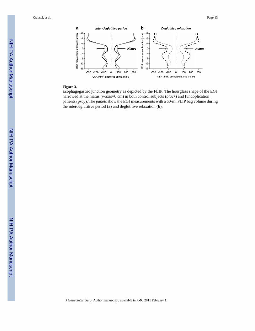
Figure 3.Esophagogastric junction geometry as depicted by the FLIP. The hourglass shape of the EGJnarrowed at the hiatus (y-axis=0 cm) in both control subjects (black) and fundoplicationpatients (gray). The panels show the EGJ measurements with a 60-ml FLIP bag volume duringthe interdeglutitive period (a) and deglutitive relaxation (b).
Kwiatek et al. Page 13
J Gastrointest Surg. Author manuscript; available in PMC 2011 February 1.
NIH
-PA Author Manuscript
NIH
-PA Author Manuscript
NIH
-PA Author Manuscript

Figure 4.Measured FLIP bag distensile pressure and estimated EGJ volume with the FLIP bag filled to40 ml (lower dots), 50 ml (middle dots), and 60 ml (upper dots). Both control subjects(black) and fundoplication patients (gray) exhibited measureable EGJ distention during theinterdeglutitive period (solid lines) and deglutitive relaxation (dashed lines) only with 50- and60-ml FLIP bag volumes. Both groups exhibited increased EGJ volume during deglutitiverelaxation. However, the distensile pressures associated with EGJ distention were consistently8–10 mmHg greater in the FP patients compared to the control subjects (see also Table 3).
Kwiatek et al. Page 14
J Gastrointest Surg. Author manuscript; available in PMC 2011 February 1.
NIH
-PA Author Manuscript
NIH
-PA Author Manuscript
NIH
-PA Author Manuscript

Figure 5.Esophagogastric junction compliance in controls and postfundoplication patients during theinterdeglutitive period (a) and deglutitive relaxation (b). Median (5th–95th percentile);*p<0.05 vs. controls.
Kwiatek et al. Page 15
J Gastrointest Surg. Author manuscript; available in PMC 2011 February 1.
NIH
-PA Author Manuscript
NIH
-PA Author Manuscript
NIH
-PA Author Manuscript

NIH
-PA Author Manuscript
NIH
-PA Author Manuscript
NIH
-PA Author Manuscript
Kwiatek et al. Page 16
Table 1
Esophagogastric Junction Pressure Morphology and Deglutitive Function Reported as Median (5th–95thPercentile)
Controls Fundoplication
Expiratory EGJ pressure (mmHg) 18 (10–36) 15 (6–22)
Inspiratory EGJ augmentation (mmHg) 17 (8–37) 13 (7–26)
Length of EGJ HPZ (mm) 45 (42–57) 37 (29–45)*
Abdominal length of EGJ HPZ (mm) 28 (24–44) 23 (19–30)*
Deglutitive IRP (mmHg) 12 (5–17) 13 (6–17)
IBPesoph 17 (12–21) 18 (11–24)
Deglutitive IRP was abnormal (>15 mmHg) in one control and two patients. Median (5th–95th percentile)
HPZ high-pressure zone, IRP integrated relaxation pressure
*p<0.05 vs. controls
J Gastrointest Surg. Author manuscript; available in PMC 2011 February 1.

NIH
-PA Author Manuscript
NIH
-PA Author Manuscript
NIH
-PA Author Manuscript
Kwiatek et al. Page 17
Table 2
CSA (Square Millimeters) of the Diaphragmatic Hiatus (Narrowest EGJ CSA Measured by the FLIP) DuringVolume Distensions Reported as Median (5th–95th Percentile)
FLIP bag volume (ml) Control subjects Fundoplication patients
Interdeglutitive Deglutitive Interdeglutitive Deglutitive
30 50 (50–68) 52 (50–166) 51 (50–60) 52 (50–70)
40 50 (50–50) 51 (50–55) 51 (50–56) 50 (50–54)
50 50 (50–52) 54 (50–106) 61 (52–88) 60 (50–109)
60 93 (50–182) 233 (55–429)** 159 (68–245) 102 (68–272)*
The minimal detectable CSA was 50 mm2
*p<0.001 vs. controls;
**p<0.05 vs. interdeglutitive period
J Gastrointest Surg. Author manuscript; available in PMC 2011 February 1.

NIH
-PA Author Manuscript
NIH
-PA Author Manuscript
NIH
-PA Author Manuscript
Kwiatek et al. Page 18
Table 3
Pressure (Millimeters of Mercury) Within the FLIP Bag During Distension Reported as Median (5th–95thPercentile)
FLIP bag volume (ml) Control subjects Fundoplication patients
Interdeglutitive Deglutitive Interdeglutitive Deglutitive
30 16 (13–19) 13 (10–16)** 19 (13–21) 13 (8–18)**
40 17 (11–21) 14 (8–18)** 22 (17–27)* 17 (12–20)**
50 20 (13–26) 15 (10–20)** 26 (19–31)* 21 (14–26)*, **
60 23 (15–30) 17 (11–25)** 30 (21–34)* 23 (16–28)*, **
*p<0.05 vs. controls;
**p<0.05 vs. interdeglutitive period
J Gastrointest Surg. Author manuscript; available in PMC 2011 February 1.
Recommended


